UNIVERSIDADE DE LISBOA
Faculdade de Medicina Veterinária
‘THE USE OF MANUKA HONEY AS A TREATMENT OF WOUNDS IN HORSES’
TERESA DIOGO MARQUES MAGALHÃES
2019 LISBOA CONSTITUIÇÃO DO JÚRI
Doutora Berta Maria Fernandes Ferreira São Braz Doutora Paula Alexandra Botelho Garcia de Andrade Pimenta Tilley
Doutor Luís Ressano Garcia Pardon Lamas
ORIENTADOR Doutor Andrew Dart CO-ORIENTADOR
UNIVERSIDADE DE LISBOA
Faculdade de Medicina Veterinária
‘THE USE OF MANUKA HONEY AS A TREATMENT OF WOUNDS IN HORSES’
TERESA DIOGO MARQUES MAGALHÃES
DISSERTAÇÃO DE MESTRADO INTEGRADO EM MEDICINA VETERINÁRIA
2019 LISBOA CONSTITUIÇÃO DO JÚRI
Doutora Berta Maria Fernandes Ferreira São Braz Doutora Paula Alexandra Botelho Garcia de Andrade Pimenta Tilley
Doutor Luís Ressano Garcia Pardon Lamas
ORIENTADOR Doutor Andrew Dart CO-ORIENTADOR
Acknowledgments
Em primeiro lugar gostaria de agradecer ao Professor Doutor Luís Lamas, por toda a orientação e tempo dispensado para me ajudar na realização desta tese.
To Professor Doctor Andrew Dart for all the support during the internship and during the realization of this thesis, he was a true help emotionally and professionally, and also for helping me collect the data needed.
Gostaria de agradecer aos meus pais, por toda a ajuda emocional que me deram ao longo destes 6 anos, ao meu pai por nunca me deixar desistir de nada, e à minha mãe por ter sempre o dom de me acalmar e saber sempre o que dizer nos meus momentos de fraqueza, são o meu maior pilar.
Aos meus tios, Carminho e Tozé pela ajuda fundamental nos primeiros 3 anos de vida académica, à minha prima Sara que foi sempre um suporte emocional e sempre pronta a ajudar. Aos meus avós por todo o carinho e orgulho que demonstraram por mim ao longo de toda a minha vida.
Aos meus amigos de Coimbra, o bando dos 7 por tanto companheirismo e cumplicidade, a minha Diana, o meu afilhado, que foram as pessoas mais importantes e especiais que este curso me deu, nunca vos vou esquecer e prometo estar presente sempre que puder, levo-vos comigo para a vida.
Os meus amigos de sempre e para sempre, que foram um constante apoio, a Salé pelas noites de estudo, por me dar um ombro para chorar, e por ser a minha maior confidente, a Janica por ser como uma irmã e demostrar sempre que se sente orgulhosa, a Inês por me dizer que vou conseguir, ao Diogo por uma amizade sempre incondicional e por não me deixar dramatizar, ao Migas pelas gargalhadas, ao Francisco pelo seu lado racional, a Carlota por me conseguir por sempre em cima, a Ana pela positividade e a Catarina por todo o apoio, foram importantes em todas as fases da minha vida e espero que continuem a ser sempre, vocês são insubstituíveis! Ao Ricardo, a melhor pessoa que eu conheço, por me ter acompanhado em todo o processo, por nunca me deixar desistir, pelo apoio incondicional a nível emocional e profissional, por me ter dito infinitas vezes que eu ia conseguir chegar até aqui, por acreditar mais em mim do que eu própria, queres sempre o melhor para mim e obrigada por me aturares nas fases de stress, que tantas foram (não sei como consegues), sou uma sortuda!
Aos amigos que apareceram mais no fim desta caminhada, a Cristina, o Serra, o Mendes, o Gonçalo, foram fulcrais na minha integração e desejo-vos todo o sucesso do Mundo.
Resumo
O uso de mel de Manuka no tratamento de feridas em cavalos:
Os antibióticos são extremamente importantes na redução das doenças infeciosas que atualmente existem a nível mundial. No entanto, à medida que emergem as bactérias resistentes, a eficácia dos antibióticos torna-se mais limitada. A resistência bacteriana aos compostos antimicrobianos, representa uma ameaça muito séria à saúde pública uma vez que continua a aumentar exponencialmente em todo o Mundo. Para além das propriedades gerais do mel, o mel de Manuka tem uma atividade antimicrobiana diferente, na qual a sua atividade não surge devido a ter na sua composição o peróxido de hidrogénio mas sim devido maioritariamente à presença do metilglioxal. A qualidade e quantidade de evidência de que o mel de Manuka também tem capacidade de modular a resposta inflamatória inicial, através da ativação do TRL-4 nos monócitos fazendo com que haja um aumento da produção de citoquinas importantes na reparação e regeneração tecidular, tem crescido.
As feridas do membro distal em cavalos cicatrizam geralmente por segunda intenção, pois a oclusão por primeira intenção não é possível devido a um variado leque de fatores. Na maioria dos casos, estas feridas apresentam um nível elevado de contaminação, uma perda de tecido avaliada de moderada a grave, uma grande taxa de retração do tecido, tempos de contração da pele mais lentos e interrupção antecipada da contração da ferida.
O objetivo de aplicar medicação tópica nas feridas é a manipulação do ambiente da mesma. O mel de Manuka quando utilizado topicamente demonstra que é capaz de modular as primeiras fases de cicatrização das feridas que fecham por segunda intenção em equinos, assim como é capaz de atuar no controlo da contaminação que caracteriza estas feridas.
Neste trabalho, foi realizada uma revisão sistemática da literatura, com o objetivo de demonstrar as evidências que existem, assim como a sua qualidade quando se fala no uso do mel de Manuka no processo de cicatrização de feridas em medicina veterinária, mais especificamente em equinos, apresentando critérios de inclusão e exclusão.
São também reportados três casos clínicos, onde o tratamento com mel de Manuka teve um papel importante e resultados benéficos de forma a validar e fornecer evidências de que é um tratamento alternativo que vale a pena ter em mente quando se lida com feridas em cavalos.
Abstract
The use of Manuka honey in wounds healing in horses:
Antimicrobial compounds are essentially important in reducing the global burden of infectious diseases. However, as resistant pathogens develop and spread, the effectiveness of the antibiotics is diminished. This type of bacterial resistance to the antimicrobial agents poses a very serious threat to public health, and for all kinds of antibiotics, including the major last-resort drugs, the frequencies of resistance are increasing worldwide. In addition to the generic properties of honey, Manuka honey has a non-peroxide antimicrobial activity largely attributed to methylglyoxal. Evidence that Manuka honey can also modulate the initial inflammatory response, through activation of toll-like receptor 4 on monocytes to enhance production of cytokines important in tissue repair and regeneration, have arisen.
Distal limb wounds in horses usually heal by second intention because primary or delayed primary closure cannot be accomplished. In most cases these wounds have gross contamination and moderate to severe tissue loss that would make closure difficult due to a greater wound retraction, slower rates and earlier cessation of wound contraction. They must heal completely through the process of contraction, granulation, and epithelialization. The aim of applying topical medication to wounds left to heal by second intention is to manipulate the wound environment. As a topical preparation, Manuka honey has been shown to modulate the early-period of second-intention wound healing in the horse, but this precise mechanism of action is still unclear, and has also been shown to control the contamination of the wound environment. The objective of this study is to show the actual evidences of the use of Manuka honey in the process of wound healing in Veterinary medicine, more specifically in horses by presenting a brief review of literature with inclusion and exclusion criteria, and by presenting three cases where Manuka honey treatment had a beneficial role and great outcomes, which provides evidence that it is an alternative treatment worth having in mind when dealing with wounds in horses.
Table of contents Acknowledgments ___________________________________________________________ i Resumo __________________________________________________________________ ii Abstract _________________________________________________________________ iii Table of contents ___________________________________________________________ iv List of figures _____________________________________________________________ vi List of tables _____________________________________________________________ vii List of Abbreviatures and Symbols __________________________________________ viii Internship short-report _____________________________________________________ 1 Introduction ______________________________________________________________ 3 CHAPTER I – Literature Review _____________________________________________ 5 1. Skin anatomy ______________________________________________________________ 5 2. Wound healing _____________________________________________________________ 5 2.1. Hemostasis ____________________________________________________________________ 6 2.2. Inflammatory phase ____________________________________________________________ 6 2.3. Proliferative phase ______________________________________________________________ 7 2.4. Remodeling phase ______________________________________________________________ 9 3. Mediators of inflammation __________________________________________________ 11 4. Bacterial resistance – the reality _____________________________________________ 12 5. Physical characteristics of honey in general ____________________________________ 12
5.1. pH __________________________________________________________________________ 12 5.2. Sugar/Osmose ________________________________________________________________ 13 5.3. Anti-oxidant __________________________________________________________________ 13 5.4. Methylglyoxal (MGO) vs Hydrogen Peroxide ______________________________________ 14 5.5. Others _______________________________________________________________________ 15
6. Honey in wound healing ____________________________________________________ 15 7. Manuka honey ____________________________________________________________ 15
7.1. UMF rating ___________________________________________________________________ 18 7.2. What’s the difference between MGO and UMF on the label of the commercial products? 18 7.3. Effect on biofilms ______________________________________________________________ 19 7.4. Effect in gram (–) and gram (+) __________________________________________________ 19
8. Effect in malodour _________________________________________________________ 20 9. Storage __________________________________________________________________ 20 10. Can we use General food honey as a wound dressing as well? ___________________ 20 11. Recommendations on using a Manuka honey wound dressing __________________ 21 CHAPTER II – Use of Manuka honey to treat wounds in horses – state of the art and case reports __________________________________________________________________ 23
2. State of the art about of Manuka honey in wound healing in Veterinary Medicine using inclusion and exclusion criteria __________________________________________________ 23
2.1. Search strategy and Data sources ________________________________________________ 23 2.2. Selection of articles ____________________________________________________________ 23 2.3. Screening process _____________________________________________________________ 24
3. Results __________________________________________________________________ 24
3.1. State of the arts – search results _________________________________________________ 24 3.2. Overview of the included studies _________________________________________________ 25 3.3. Reviewed studies ______________________________________________________________ 27 3.3.1. In vivo studies ______________________________________________________________ 27 3.3.2. In vitro studies ______________________________________________________________ 30 4. Case reports ____________________________________________________________________ 30 4.1. Case 1 _____________________________________________________________________ 30 4.2. Case 2 _____________________________________________________________________ 33 4.3. Case 3 _____________________________________________________________________ 37 5. Discussion ___________________________________________________________ 39 5.1. Advantages of Manuka honey _____________________________________________ 39 5.2. Manuka honey as an anti-inflammatory agent _______________________________ 39 5.3. Manuka honey as an antimicrobial agent ____________________________________ 40 5.4. Action of manuka honey against resistant bacteria ____________________________ 40 5.5. Other types of honey and its capacity as a possible wound healing dressing _______ 41 5.6. Short or long-term treatment _____________________________________________ 41 5.7. Bandaged or unbandaged wounds _________________________________________ 42 5.8. Benefits of conducting a literature review ___________________________________ 42 5.9. Benefits of clinical cases presentation _______________________________________ 43 5.10. Limitations _____________________________________________________________ 43 6. Conclusion ___________________________________________________________ 43 7. Future directions _____________________________________________________ 44 Bibliography _____________________________________________________________ 46
List of figures:
Figure 1 – Temporal explanation of the inflammatory phases, in order to fully understand this process, and to understand that some phases occur almost at the same time. Source: Modified by Marco Langlois (Faculté de médecine vétérinaire, Université de Montréal) from Stashak & Theoret, 2014.
Figure 2 and 3 – Photographs of a Standardbred horse with wounds along the side of the horse and down back leg caused by a clostridial myositis after injection with a dirty needle
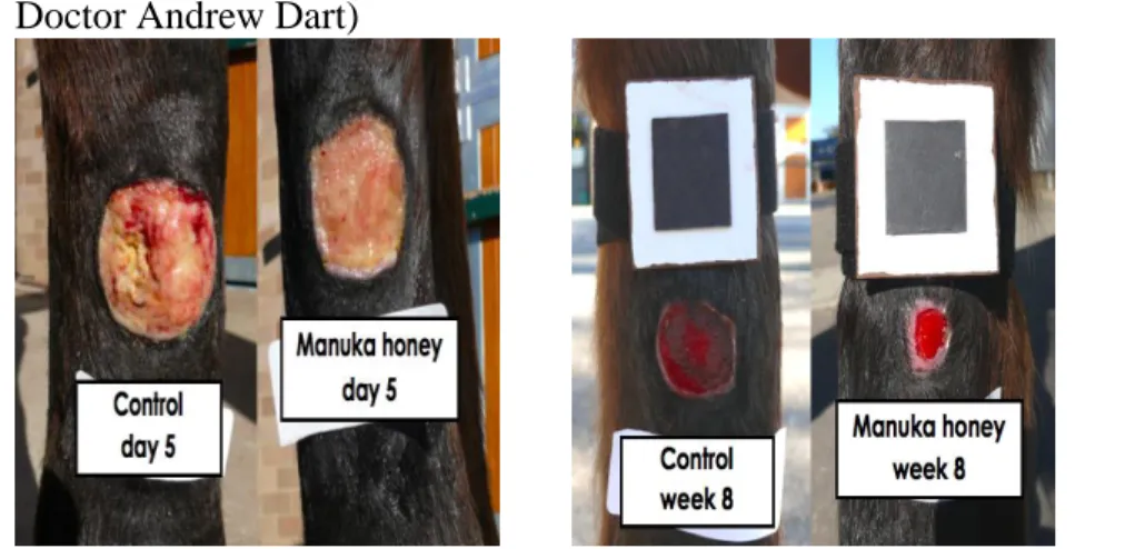
Figure 4 – Photographs showing the action Manuka honey had in the treatment of a surgically created wound compared to a control wound, at day 5 of treatment. (Courtesy of Professor Doctor Andrew Dart)
Figure 5 – Photograph showing the effect of Manuka honey on a surgically created wound compared to a surgically created control wound on week 8 of treatment. (Courtesy of Professor Doctor Andrew Dart)
Figure 6 – Flow diagram of the study selection process.
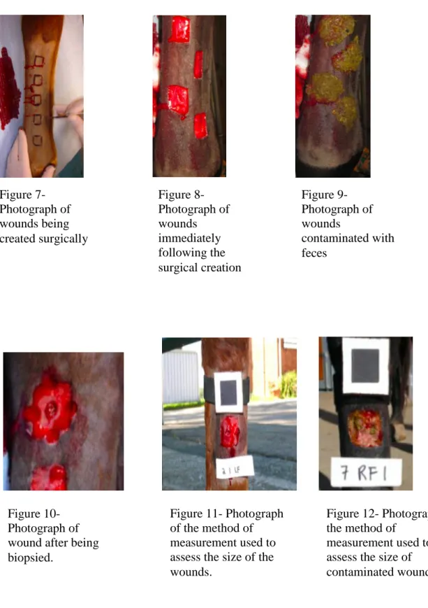
Figure 7 – Photograph of wounds being created surgically (Courtesy of Professor Doctor Andrew Dart)
Figure 8 – Photograph of created wounds after the surgery (Courtesy of Professor Doctor Andrew Dart)
Figure 9 – Photograph of wounds contaminated with feces ( Courtesy of Professor Doctor Andrew Dart)
Figure 10 – Photograph of wound after being biopsied (Courtesy of Professor Doctor Andrew Dart)
Figure 11 – Photograph of the method of measurement used to assess the size of the wounds (Courtesy of Professor Doctor Andrew Dart)
Figure 12 – Photograph of the method of measurement used to assess the size of contaminated wounds (Courtesy of Professor Doctor Andrew Dart)
Figure 13 – Photograph of wound of case 1 few days after wounding. (Courtesy of Professor Doctor Andrew Dart)
Figure 14 – Photograph of wound of case 1 one week after treatment with Manuka honey. (Courtesy of Professor Doctor Andrew Dart)
Figure 15 – Photograph of wound of case 1 two weeks of treatment with Manuka honey. (Courtesy of Professor Doctor Andrew Dart)
Figure 16 – Photograph of wound of case 1 three weeks after daily application of Manuka honey. (Courtesy of Professor Doctor Andrew Dart)
Figure 17 – Photograph of the wound of case 1 one month after treatment with Manuka honey started
Figure 18 – Photograph of wound of case 2 on day 1 of wounding. (Courtesy of Professor Doctor Andrew Dart)
Figure 19 – Photograph of wound of case 2 after two weeks of treatment with Manuka honey. (Courtesy of Professor Doctor Andrew Dart)
Figure 20 – Photograph of the wound of case 2, two months after treatment with Manuka honey started. (Courtesy of Professor Doctor Andrew Dart)
Figure 21 and 22 – Photograph of a miniature horse with a severe neck injurie one week after receiving a perivascular injection of flunixin meglumine, showing extensive sloughing of the soft tissues and thrombosis of the jugular vein. (Courtesy of Professor Doctor Andrew Dart) Figure 23 –– Photograph of the neck seven days after surgically debridement and treatment that consisted in a broad-spectrum antimicrobial, non-steroidal anti-inflammatory therapy and application of Manuka honey UMF 20 twice daily (Courtesy of Professor Doctor Andrew Dart) Figure 24 and 25 – Photographs of the outcome of the miniature horse with a complete regenerated and recovered skin, showing the scar in the end of the treatment. (Courtesy of Professor Doctor Andrew Dart)
List of tables:
Table 1 – Cytokines involved in wound repair. (Stashak & Theoret, 2008) Table 2 – Research collected data of the use of Manuka honey in vivo Table 3 – Research collected data of the use of Manuka honey in vitro
List of Abbreviatures and Symbols % - Percentage
spp. - species
MIC- minimum inhibitory concentration MBC- Minimum bactericidal concentration HBM- honey based membrane
MRSA- methicillin-resistance Staphylococcus aureus MSSA- methicillin-sensible Staphylococcus aureus UMF- Unique Manuka Factor
IL- Interleukin
TNF- Tumour necrosis factor TLR4- Toll-like receptor 4 TGF-Transforming growth factor pH- Power of hydrogen
ROS- Reactive oxygen species COX- Cyclooxygenase
LPS- Lipopolysaccharide MGO- Methylglyoxal
DNA- Deoxyribonucleic acid RNA- Ribonucleic acid DHA- Dihydroxyacetone GH- General honey
EGT- Excessive granulation tissue ECM – Extracellular matrix
Internship short-report
My internship took place at the Camden Equine Centre of Sydney University, Australia. It started at the 1st of October 2018 and ended 31st of January 2019, which makes a total of 850 hours of work.
Camden Equine Centre is a specialist equine hospital that is specialized in Sports medicine (performance problems, lameness and orthopaedics), Surgery, Diagnostic Imaging (with MRI, Scintigraphy, high speed treadmill with endoscopy), and Internal Medicine, as well as an Ambulatory Service.
My supervisor Professor Doctor Andrew Dart, alongside an amazing team of specialists, residents and interns guided me throughout the internship, helped me with all the theory questions and all the practice knowledge, and made these months really worth wide.
During the 4 months, my internship consisted in rotating between the sports medicine, surgery and internal medicine. I had the freedom to join the cases I had most interest on, and I was able to go on ambulatory calls when I could.
In the surgeries I participated, I was able to learn a lot about the anaesthetics, with Dr. Eduardo Uquillas, able to learn about and practice asepsis before and after the surgery, and I was also able to participate in some surgeries, which for me was always the highlight of my day. On Sports medicine, I learned a lot about MRI and Scintigraphy. Dr Robin Bell and Dr Nicole Symonds always explain everything and let students practice a lot when it comes to radiographs, ultrasound, nerve blocks, and try to help us understand all about the performance evaluation. In the internal medicine cases, we had a lot of mare and foal care. I learned more about ophthalmology, gastrointestinal diseases and also about contagious and infectious diseases. Dr Gaby Van Galen always prompted students to discuss our clinical assessment of each case which improved my clinical knowledge.
On ambulatory calls, Dr Sara Biasutti was an outstanding teacher. During those hours in the car Dr Biasutti made us think about every possible differential diagnosis, based on the information the owners had provided prior to the call. So by the time we got to the patient we had a clear mindset about what we were going to see, and it was so much easier to understand everything we were seeing without the need to always be asking questions.
With my supervisor, Professor Andrew Dart, I had a lot of discussions about wound healing, He has a specific interest in the use of Manuka honey in second intention wound healing in horses. He introduced me to the publications he had performed, the results he had accomplished, and always advised me with respect to this thesis. Dr Dart was a great help during the internship.
Since Camden Equine Centre is a teaching hospital, I was able to spend a lot of time with the students, and participated in treatments, rounds and case presentation as one of them, which was a great learning experience as well.
From March 1st until the end of the month, I did a second externship that took place in Faculty
of Veterinary Medicine of University of Lisbon in SCUE (Serviço de Cirurgia e Urgência de Equinos) with the orientation of Professor Doutor Luís Lamas, Dr Mariana Magalhães, Dr Teresa Rosa and Dr Gonçalo Silva. In this externship I got the opportunity to follow a variety of referred clinical cases, as well as improve my monitoring skills and inpatient care.
Introduction
Wounds in horses are very common, particularly wounds to the distal limb. In horses wounds usually present with tissue avulsion and contamination, which precludes primary closure so these wounds are left to heal by second intention (Bigbie, Schumacher, Swaim, Purohit, & Wright, 1991).
For many centuries, honey was used for medicinal purposes by the ancient Egyptians, Greeks and Chinese. Historical uses of honey include gastrointestinal aliments, pain relief and treatment of infections. However, it’s most common use was to promote wound healing and for this purpose it was often combined with other substances such as animal fats and lint (Rogalska, 2016). In the last century the use of honey for healing decreased with the development of antibiotics (Oryan, Alemzadeh, & Moshiri, 2016). However, more recently, the emerging problem of bacterial resistance to antibiotics has seen the re-emergence of honey and other natural products. There are several properties that make honey an attractive wound dressing. It provides a moist environment, has a low pH which is favourable to the antibacterial activity, it releases oxygen from haemoglobin acting as an anti-oxidant, it draws fluid out of the wound bed because of its osmotic properties initiating an autolytic debridement amongst other properties that will be detailed further ahead (Blair, Cokcetin, Harry, & Carter, 2009; Tsang et
al., 2018).
The antibacterial action of most honeys is related to hydrogen peroxide. Hydrogen peroxide is generated by the enzyme glucose oxidase which is produced by the worker bees, converting glucose into gluconic acid and hydrogen peroxide. Glucose oxidase is only activated when honey is diluted, which happens when in contact with the wound exudate. This is called the peroxide activity of the honey (Cooper, 2014).
There are many different honeys, some of which have non-peroxide activity. Manuka honey derived from the Leptospermum trees, more specifically Leptospermum scoparium is native to New Zealand and is the most well-known non-peroxide honey. Leptospermum honeys have advantages when compared to the others honeys. They are very stable during storage (light and temperature) and when in presence of body fluids and they show no toxicity towards mammalian cells. Manuka honey has superior antibacterial action that is graded against a standard antiseptic, phenol, to provide a measure of antimicrobial activity referred to as the unique Manuka factor (UMF). The higher the UMF, the greater the antimicrobial activity (Dart, Bischofberger, Dart, & Jeffcott, 2015). This antibiotic activity is largely attributed to the presence of Methylglyoxal.
The antimicrobial effect is not the only advantage that Manuka Honey has to offer when it comes to wound healing. It has also been shown that it modulates the initial inflammatory
response through activation of toll-like receptor 4 on monocytes to enhance production of cytokines that are important in tissue repair and regeneration. It has been shown to stimulate the production of cytokines such as TNF-alfa, IL-1beta and IL-6, it’s also believed to help enhance angiogenesis, promote autolytic debridement and accelerate re-epithelization and wound closure (Tonks et al., 2003).
Compared with other topical treatments such as hydro-fiber silver or silver sulfadiazine, honey has been shown to be more effective in elimination of microbial contamination, reduction of wound area, promotion of re-epithelialization and improves the outcome of the wound healing by reducing the incidence of excessive scar formation (Oryan et al., 2016). Resistance to honey has not yet been proven to exist under the conditions that rapidly induced resistance to antibiotics.
It is important to refer that there are similarities between wounds in the distal limb of horses and diabetic ulcers observed in people, including relatively poor blood supply and chronic inflammation. Reviewing the human literature may provide insight and direction into studies in the future (Tsang et al., 2018).
The purpose of this review is to critically analyse the current knowledge on Manuka Honey activity in second intention wound and the healing of burns.
CHAPTER I – Literature Review 1. Skin anatomy
The skin is the largest organ of the body and serves key functions including physical protection, sensation, temperature regulation, and insulation. It is composed of 2 compartments; the epidermis and dermis. The dermal compartment consists of two regions, the papillary dermis and the reticular dermis. This compartment is composed of dense, fibroelastic connective tissue and constitutes the bulk of the skin. The strength of the skin is provided by a network of collagen fibers. The resilience of the skin is provided by elastin and glycosaminoglycans (GAGs). Collagen type I is the major collagen of the dermis whereas collagen type III comprises about 15% of the dermis. The fibroblast is the principal type of cell found in the dermis along with perivascular mast cells and tissue macrophages. Only the dermal compartment is vascularized with nutrients that reach epidermis by diffusion (Stashak & Theoret, 2008).
In the horse, the epidermis consists of five layers of keratinocytes: the stratum basale, the
stratum spinosum, the stratum granulosum, stratum lucidum and stratum corneum.
Although 90-95% of the cell population of the epidermis consists of keratinocytes, this compartment also includes melanocytes, Langerhans cells and Merkel cells. The epidermis is attached to the dermis at the level of the basement membrane, a thin, glycoprotein-rich layer composed primarily of laminin and type IV collagen (Proksch, Brandner, & Jensen, 2008).
2. Wound healing
Wounds heal through regeneration and repair.
Regeneration is replacement of the damaged tissue with normal cells through mitosis. Repair is when the body tries to re-establish the continuity of interrupted tissues with undifferentiated scar tissue which is less biologically suitable than the tissue it replaced. The point of repair is to re-establish an epithelial cover, recover the integrity, strength, and function of the skin even when the adjacent tissues that were normal are affected by this process. Second intention repair of full-thickness cutaneous wounds is divided in 4 phases (Haemostasis, inflammatory phase, proliferative phase and remodelling phase). These phases consist in interactions between multiple cellular types, their surrounding matrix, and the soluble mediators that govern the numerous activities required to rebuild the tissue. This interaction is not static but rather in a state of constant flux, resulting in a microenvironment that is continually evolving as the wound heals (Stashak & Theoret, 2008).
2.1.Hemostasis
The first phase of second intention repair is Hemostasis/Coagulation. It begins immediately after the lesion occurs. It takes several hours and the goal is to establish hemostasis and the formation of a provisional wound matrix (De Groot, Urbanus, & Roest, 2012).
Phospholipids released by the endothelial membrane after injury are transform into arachidonic acid which mediates both vascular tone and permeability. As peripheral vasoconstriction occurs, it deprives the surrounding tissues of oxygen and nutrients that are usually carried by the blood. Hypoxia, the pH changes and increases glycose to activate the platelets and enhance their adhesion and aggregation, which together initiate the intrinsic coagulation cascade that will end up forming a blood clot that not only seals the vessel, but it also gives strength to the wound and creates a provisional matrix that fills the space made by the wound, serving as a scaffold for migrating cells.
Inflammatory and stromal cells have special surface receptors (integrins) that recognise binding sites on the proteins present in the scaffold, which leads to the ingrowth of cells involved in healing (Herter, Rossaint, & Zarbock, 2014).
Mediators liberated by platelets and mast cells modulate vascular tone and increase vascular permeability facilitating cellular migration and diffusion of nutrients and oxygen needed to support healing. Over time the surface clot desiccates to form a scab that protects the wound from infection. This scab is, in turn, lysed by plasmin and sloughs along with dead inflammatory cells and bacteria as healing proceeds (Stashak & Theoret, 2008).
2.2.Inflammatory phase
The second phase is inflammation, also referred to clinically as the debridement phase.
The intensity of the inflammatory response is strongly correlated to the gravity of trauma and determines the extent of scarring.
It can be divided it into an early phase, characterized by recruitment of neutrophils, and a late phase, characterized by the appearance and transformation of monocytes.
Leukocytes are recruited into the injured tissues by several vasoactive mediators and chemo-attractants (Wulff & Wilgus, 2013). These signals initiate the processes of rolling, activation, tight adhesion, and, finally, transmigration of inflammatory cells through the microvascular endothelium. Chemo-attractants stimulate the release of enzymes by the activated neutrophils, expediting their penetration through vascular basement membranes. The cellular influx begins within minutes of the injury and the concentration of neutrophils at the wound gradually increases to reach a peak 1–2 days after injury.
bacteria through phagocytosis and subsequent enzymatic and radical oxygen mechanisms. Neutrophil migration and phagocytosis stop when contaminating particles are cleared from the site of injury. Most cells then become imprisoned within the clot, which is sloughed during the upcoming phases of repair. The neutrophils that remain die in a few days and are phagocytized by the tissue macrophages or modified wound fibroblasts, indicating the end of the early inflammatory phase of repair. Although neutrophils help create a better environment within the wound and serve as a source of proinflammatory cytokines, they are not crucial for the repair of noninfected wounds (Stashak & Theoret, 2008).
Macrophages play an important role in all phases of wound healing and control the overall process. During the early inflammatory phase, macrophages have proinflammatory functions, such as antigen presentation, phagocytosis, and the production of inflammatory cytokines and growth factors that help the wound healing. Later, during the proliferative phase of healing, macrophages stimulate proliferation of dermal, endothelial, and epithelial tissue to complete formation of the extracellular matrix (ECM), angiogenesis, and epithelialization. Macrophages can then change the composition of the ECM during the remodeling phase by delivering degra-dative enzymes (Delavary, van der Veer, van Egmond, Niessen, & Beelen, 2011).
After injury, once infection has been countered and repair completed, all the inflammatory cells disperse from the wound. For inflammation to resolve, each of the events that took place must stop and sometimes even reversed. Apoptosis is the universal pathway for eliminating unneeded cells in a phagocytic process. This mechanism predominates in all phases of wound repair because each phase depends on rapid increases in specific cellular populations that prepare the wound for repair (inflammatory cells) or deposit new matrices and mature the wound (stromal cells), but then must be eliminated prior to progression to the next phase of repair (Greenhalgh, 1998).
2.3.Proliferative phase:
The third phase is the proliferative phase (clinically referred as repair phase), that builds protection for the wound’s surface by forming the granulation tissue, a new epithelial cover and the vascular properties for the new tissues. The proliferative phase is characterized by the appearance of red, fleshy granulation tissue, which occupies the defect. For the first 3–5 days following injury, fibroblasts, endothelial and epithelial cells rapidly invade the wound in preparation for synthesis and maturation of the matrix or for wound coverage. Granulation tissue is composed by three elements that simultaneously move into the defect created by the wound, macrophages, fibroblasts and new blood vessels. Macrophages debride and produce mediators, such as cytokines and growth factors, that stimulate angiogenesis and fibroplasia,
fibroblasts proliferate and synthesize new components of the ECM, and the new blood vessels will carry oxygen and nutrients necessary for the metabolism and growth of cells, and give the granulation tissue its red, granular appearance (Martins-Green, 2013).
This granulation bed is supposed to replace the fibrin clot with the purpose to serve as barrier to infection and at the same time works as a surface where cells are able to migrate. Fibroblasts from adjacent uninjured dermis and subcutaneous tissue are stimulated to proliferate and express integrin receptors to mediate interactions between the cells and their environment and assist them in migrating to the defect.
Migration of fibroblasts happens before advancing capillary endothelial buds but follows macrophages that have opened a path by phagocytizing debris. After the arrival of the fibroblasts in the defect, they begin to proliferate and change their function to protein synthesis. Gradually they start to replace the provisional matrix with one that is rich in collagen, under the influence of growth factors and cytokines, to increase the ratio of type I to type III (immature) collagen.
The greatest rate of accumulation of connective tissue within the wound occurs 7–14 days after injury.
The granulation tissue rich in fibroblasts is replaced by a relatively avascular and acellular scar tissue, as the capillaries in the wound regress and fibroblasts either undergo apoptosis or transform into myofibroblasts that participate in the wound contraction (Desmoulière, Redard, Darby, & Gabbiani, 1995). The wound will have a much slower gain in tensile strength as wound remodels also because the collagen content decreases. If the signal to downregulate fibroblast activity is overdue beyond a specific time point, apoptosis is permanently impaired and leads to an imbalance between collagen synthesis and degradation and the formation of excessive scar tissue. The creation of new capillaries from the ones that already exist (angiogenesis) is important to restore oxygenation and to provide the nutrients needed to the newly formed granulation tissue and that is why the microvascular endothelial cells play a key role in the proliferative phase of repair.
Angiogenesis, is a complex and dynamic process that occur in response to tissue damage and hypoxia and it’s mediated by various soluble factors, the serum and the surrounding ECM. These factors are usually released during the inflammatory phase and they are angiogenic inducers, including growth factors, chemokines, angiogenic enzymes, endothelial specific receptors, and adhesion molecules, such as integrins (Liekens, De Clercq, & Neyts, 2001). There is the need of an organized and sequential way to construct a vascular network, and that includes an higher microvascular permeability, the liberation of proteinases with local degradation of the basement membrane that surrounds the existing vessel after, then the
endothelial cells need to migrate into the interstitial space, endothelial cellular proliferation and differentiation into mature blood vessels that will be followed by the regression of the newly formed vasculature as the tissue remodels.
In the end this intense angiogenic response ends up in a density of vessel that exceed the normal capillaries in an uninjured tissue, and this is what gives the granulation tissue a red, granular appearance (Dipietro, 2013).
When the stroma has been reconstituted and this vascular supply is no longer needed, the pro-angiogenic stimuli is down-regulated and the recent capillary network involutes (Li, Zhang, & Kirsner, 2003). After this the wound will start to acquire a pale colour. Epithelialization is the process of covering denuded epithelial surfaces and is crucial for successful closure of the wound but it can only proceed once the bed of granulation tissue is formed.
While the hemostatic activities that have the purpose of creating a temporary barrier, the residual epithelium under the clot is moving centripetally in order to close of wound. The regenerative capacity of the epidermis relies on keratinocyte stem cells that reside within specific microenvironments referred to as stem cell niches. To close the defect in the epidermis, keratinocytes at the wound’s edge must first loosen their adhesions to each other and to the basal lamina and develop the flexibility required to migrate over the new matrix. Once the surface of the wound is covered by epithelial cells contacting one another, further migration is inhibited by the expression, within the ECM, of laminin, a factor responsible for adhesion of epithelial cells. Although initial cellular migration does not need an increase in cellular multiplication, basal keratinocytes at the wound’s margin do begin to proliferate 1–2 days after injury to restock the migratory front. In equine full-thickness wounds healing by second intention, the provisional matrix will end up being replaced by a mature basement membrane. This is a time-consuming part of the process that occurs long after the total coverage of the wound by the epithelium. This is why the neo-epidermis is fragile for so long after repair is macroscopically complete (Stashak & Theoret, 2008).
2.4.Remodeling phase:
The remodeling phase is characterized by synthesis of connective tissue, lysis, and remodeling creating scar tissue. The mature ECM is an acellular scaffold and it is made of proteins, glycosaminoglycans, polysaccharides, and water. It helps the bidirectional communication between cells and their biochemical/ biophysical environment (Wong, Gurtner, & Longaker, 2013). The remodeling phase takes place from 3 weeks to 1 year after injury, while the ECM and its components undergo changes to make sure the replacement tissue will have enough function, integrity and strength.
Successful closure of the wound by contraction is a process where both dermis and epidermis bordering a full-thickness wound are drawn centripetally over the exposed defect (Desmoulière & Gabbiani, 1988).
Wound contraction not only accelerates closure, it also provides a better cosmetic appearance and strength of the scar because proportionally less area of the wound must be covered by newly formed epithelium. Newly formed epithelium is of inferior quality. It is fragile and doesn’t have normal nervous, glandular, follicular, and vascular components (Madison & Gronwall, 1992). After an initial lag phase, the skin edges retract and for 1-2 weeks the area of the wound gets bigger. After a period of rapid contraction, contraction slows as the wound approaches complete closure. Wound contraction is greater in regions of the body with loose skin than in regions where skin is under tension, such as the distal extremity of the equine limb. The new tissue remodels and randomly orientated collagen fibers start to reform along lines of stress. Points of weakness exist where the new collagen weaves into pre-existing tissue and are subject to damage when under stress. This process can take up to 2 years, during which there will be a rearrangement of the collagen fibers into a more organized lattice-like structure, under the influence of local mechanical factors that increase the tensile strength of scar tissue. The majority of type III collagen fibers laid down early in healing are replaced by collagen type I, fibers become increasingly crosslinked, and the normal skin ratio of 4:1 type I to type III collagen is reestablished. Glycosaminoglycans are steadily degraded until they reach concentrations found in normal dermis. The duration of the maturation phase depends on a variety of factors, including the patient’s genetic makeup, age, location of the wound, type of injury, and duration of inflammation (Stashak & Theoret, 2008).
Figure 1- Temporal explanation of the inflammatory phases, in order to fully understand this process, and to understand that some phases occur almost at the same time. Source: Modified by Marco Langlois (Faculté de médecine vétérinaire, Université de Montréal) from Stashak & Theoret, 2014.
3. Mediators of inflammation
The process of wound repair is a set of interactive processes that involve various elements of blood, extracellular matrix, and mesenchymal cells. Recovery of structural functional properties and integrity mostly relies on soluble mediators, synthesized by cells present in the wound or within the surrounding tissue, that coordinate migration, proliferation, and synthesis of proteins by the various cellular properties involved in this process.
Cytokines are glycoproteins released by most nucleated cells and are among the most important soluble mediators regulating wound repair (Barrientos, Brem, Stojadinovic, & Tomic-Canic, 2014).
TGF-1 peaks in first 24 hours and is a profibrotic cytokine that orchestrates the healing process, and the peak in horses is often gradual and develops over several days leading to a depressed TGF-3 response and dysregulated fibroplasia. TGF-3 has a reciprocal peak 5-9 days after wounding and modulates the initial inflammatory process (Bischofberger et al., 2016; Tonks et al., 2007).
Toll-like receptor 4 (TLR4) is believed to stimulate monocyte and macrophage activity and thereby increasing production of IL-1, IL-6 and TNF-, that perform a major role in regeneration and repair of the skin and fastens the initiation of the inflammatory response (Tonks et al., 2003).
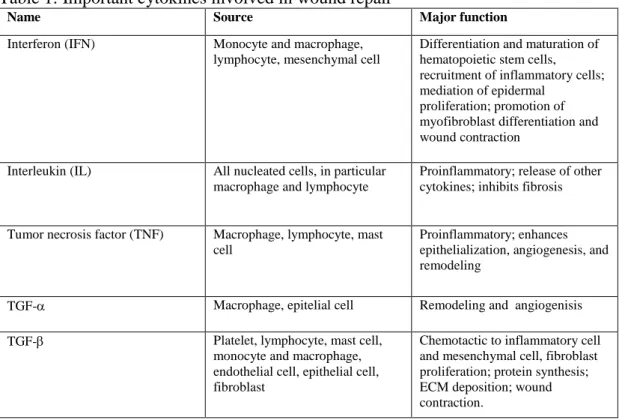
The cytokines that play an important role in wound repair are in table 1. Table 1: Important cytokines involved in wound repair
Name Source Major function
Interferon (IFN) Monocyte and macrophage, lymphocyte, mesenchymal cell
Differentiation and maturation of hematopoietic stem cells, recruitment of inflammatory cells; mediation of epidermal
proliferation; promotion of myofibroblast differentiation and wound contraction
Interleukin (IL) All nucleated cells, in particular macrophage and lymphocyte
Proinflammatory; release of other cytokines; inhibits fibrosis
Tumor necrosis factor (TNF) Macrophage, lymphocyte, mast cell
Proinflammatory; enhances epithelialization, angiogenesis, and remodeling
TGF- Macrophage, epitelial cell Remodeling and angiogenisis TGF- Platelet, lymphocyte, mast cell,
monocyte and macrophage, endothelial cell, epithelial cell, fibroblast
Chemotactic to inflammatory cell and mesenchymal cell, fibroblast proliferation; protein synthesis; ECM deposition; wound contraction.
4. Bacterial resistance – the reality
The development of antibiotics saw a decrease in the use of natural alternatives like honey. Nowadays most antimicrobials when tested have at least three different types of resistant bacteria. At the same time there is not enough research into producing new antimicrobials because the financial benefits to the pharmaceutic industry are not returned because resistance of new antibiotics is realized. Interest in ancient remedies, like plants and plant-based products, including honey are being re-evaluated (Mandal & Mandal, 2011).
It has been suggested that the risk of bacteria developing resistance to honey will be low if high concentrations are used clinically. High levels of antimicrobial agents can effectively inhibit viable bacteria before resistant strains emerge. Unlike some of the biocides, high amount of manuka honey is not cytotoxic (Cooper, Jenkins, Henriques, Duggan, & Burton, 2010). The fact that Manuka honey works has an antimicrobial due to more than one factor, and also affects the bacteria in different ways, is one of reasons believed to explain the non-resistance of honey (Oryan et al., 2016). Future research is needed in order to enhance the outcome of honey, there is still a gap of knowledge when it comes to know how all the properties work.
5. Physical characteristics of honey in general
5.1.pH
Honey is acidic and has a pH of around 3.2-4.5. This low pH is due to the formation of gluconic acid. During the dilution of honey, glucose oxidase catalyses glucose into gluconic acid and hydrogen peroxide where the gluconic acid results in acidity.
It is well known that topical acidification of wounds promotes healing by increasing the release of oxygen from haemoglobin in capillaries and because it is unfavourable for bacterial growth (Oryan et al., 2016).
In addition, this pH is less favourable for protease activity. Protease contributes to poor tissue healing by damaging the extracellular matrix and destroying cytokines and growth factors. So by reducing the destruction of the matrix needed for tissue repair, it contributes to wound contraction.
Acidification also assists in the antibacterial action of macrophages and prevents the ammonia produced by the bacterial metabolism from harming body tissues (Molan & Rhodes, 2015).
5.2. Sugar/Osmose
Honey is made of 80% sugars, 18% water and a complex mixture of amino acids, peptides, arabinogalactan proteins, organic acids, polyphenols, carotenoid-like substances, flavonoids, vitamins and minerals. The high sugar content produces a high osmotic gradient that leads to bacterial dehydration and shrinkage of the cell wall. This high osmolarity also initiates an influx of fluid, lymph and nutrients into the wound bed, creating a moist environment rich in nutrients. These properties promote an autolytic wound debridement and enhance tissue healing (Tsang et al., 2018).
High osmolarity solutions such as honey, tie up water molecules. Honey, as a viscous fluid, provides a protective barrier and prevents cross-infection of wounds. When honey is topically applied over a wound, due to its high osmolarity, bacteria have insufficient access to water for growth and development. Consequently, the microorganisms become dehydrated and eventually die. Also osmotic pressure from honey draws out lymphatic fluid from the subcutaneous tissue to wound surface, which aids in removal of debris, necrotic and devitalized tissues (Oryan et al., 2016).
The dilution of the honey by the wound exudates could be a problem, but it is believed that if the honey does not become too diluted it still inhibits the growth of bacteria (Minden-Birkenmaier & Bowlin, 2018).
5.3.Anti-oxidant
Chronic wounds are highly oxidizing environments because of release of reactive oxygen species (ROS) from persistent neutrophils and macrophages infiltration (Majtan, 2013). There are several components that can also contribute to the antioxidant activity of honey such as, flavonoids, phenolic acids, catalase, peroxidase, ascorbic acid, and carotenoids.
The presence and concentrations of these antioxidants differs between the many types of honey and therefore the antioxidant properties.
Woo et al. found that chrysin, a natural flavonoid found in many plant extracts, honey, and propolis acted to inhibit cyclooxygenase-2 (COX-2) gene expression in LPS- stimulated cultured macrophages. This effect was mediated through inhibition of the binding activity of nuclear factor IL-6 (Woo, Jeong, Inoue, Park, & Kwon, 2005).
A decrease in neutrophil superoxide production has been reported (Van den Berg et al., 2008). So, the antioxidant activity of honeys was mostly attributed to the inhibition of ROS formation either by inhibiting the respiratory burst of neutrophils or by direct ROS scavenging. A dose-dependent reduction in the production of superoxide from human neutrophils by some honeys
did not correlate with the level of known honey-based phenolic compounds, which are well-known free radical scavengers. This indicates that the anti-oxidant activity of honey is most probably caused by inhibition of the respiratory burst of neutrophils (Majtan, 2013).
5.4.Methylglyoxal (MGO) vs Hydrogen Peroxide
Hydrogen Peroxide is formed in the honey by an enzyme called glucose oxidase (Mavric, Wittmann, Barth, & Henle, 2008; Song, Salcido, & Erdman, 2011).
This enzyme is produced by worker bees and is found in all types of honey in varying concentrations and it is responsible for oxidizing glucose into gluconic acid and releasing hydrogen peroxide.
Hydrogen peroxide is only detected in diluted honey because dilution leads to activation of glucose oxidase. The generation of hydrogen peroxide is greatest when honey is diluted 30-50%, so in the presence of wound exudate the antimicrobial activity of hydrogen peroxide, which is one of the most used disinfectants, may contribute significantly to the antibacterial activity in some honey varietals (Cooper, 2014; Tsang et al., 2018).
Other factors that can contribute to the variability of the activity and concentration of hydrogen peroxide can be because of the plant species, environmental conditions and entomological factors, including age of the bee and foraging patterns (Tsang et al., 2018).
It is known that high concentrations of catalase present in body fluids, inhibits the activity of hydrogen peroxide. However it has been shown that honeys derived from some plant sources have superior antimicrobial activity even in the presence of this enzyme.
This has been identified has the non-peroxide activity. Manuka honey is a non-peroxide honey and this degree of activity is measured and reported as the Unique Manuka Factor (UMF) (Tsang et al., 2018).
MGO is an organic compound found in manuka honey and it has been shown to be responsible for the majority of the antimicrobial activity. The mode of action is still not fully elucidated but from what it’s known it acts through its capacity to involve a combination of enzymatic and non-enzymatic processes that involve the capacity to interact with macromolecules such as DNA and RNA (Irish, Blair, & Carter, 2011; Tsang et al., 2018).
MGO comes from a non-enzymatic conversion of a chemical found in high concentrations in the flower of the manuka tree, dihydroxyacetone (DHA). DHA has recently been shown to limit the production of hydrogen peroxide by inhibiting the enzyme glucose oxidase (Tsang et al., 2018), so hydrogen peroxide does not contribute to the antimicrobial activity of manuka honey (Oryan et al., 2016).
5.5.Others
It has been suggested that the antibacterial factors within honey are likely to interact together, thereby creating a synergistic effect and a regulating activity of other chemicals (Molan, 2008). Another characteristic of honey dressings is that it reduces pain. The pain in wounds results from the nerve endings being sensitized by prostaglandins produced in the process of inflammation, as well from the pressure on tissues resulting from oedema. Because of this high sugar content, honey also prevents pain on dressing changes as, by mobilizing the oedema from the surrounding tissues, it keeps the wound surface moist (Simon et al., 2009).
6. Honey in wound healing
The honey we use generally in wound healing is a Medical grade honey.
Honey has been considered a prized and exceptional remedy with a lot of different properties; it has a clinically proven effect in wound healing, it eliminates pathogens from wounds and it even provides an appropriate moist environment for effective healing (Majtan, 2013).
Variations in the type and level of antimicrobial activity in honey is associated with the floral source. However, while some floral sources appear to be associated with particular levels of hydrogen peroxide activity, variation in this activity among honeys from within the same floral species has also been observed. This may be due to the geographical location of the floral source and the prevailing environmental conditions, which affect the physiology of the floral species, or bee-related factors, such as age or colony health, which may affect the production or activity of glucose oxidase. Honeys with non-peroxide antimicrobial activity on the other hand, are more closely associated with floral source, being generally derived from Leptospermum species (Irish et al., 2011).
The honey this review studied in detail is the one that has its origin in the Leptospermum
scoparium commonly known as Manuka bush.
7. Manuka honey
Manuka honey is a non-peroxide type of honey, and it’s produced from the manuka tree (Leptospermum scoparium) found in New Zealand (Shah, 2017).
Honey derived from the manuka tree has been found to display superior antimicrobial activity compared to many other honey varietals. As a result, the increasing public awareness of its health benefits, has seen the cost of raw manuka honey increase more than 10-fold over the past 20 years (Stephens et al., 2010).
Manuka honey is not affected by catalase, and it has a unique antibacterial activity. This activity is due to the methylglyoxal, which forms by spontaneous conversion in ripened honey from its precursor substance dihydroxyacetone that is found in the manuka nectar. Methylglyoxal on its own is a cytotoxic substance, so it has been raised that manuka honey may contribute to delayed wound healing, however it seems that, the combination and ratio with other components in the manuka honey neutralizes any toxic effects (Majtan, 2011).
One important advantage about manuka honey and about the non-peroxide antimicrobial activity honeys in general, is that their activity is not destroyed by the enzyme catalase present in body fluids. This, together with the fact that it’s not affected by the gamma-radiation, allows the honey to be sterilized for medicinal use (Irish et al., 2011).
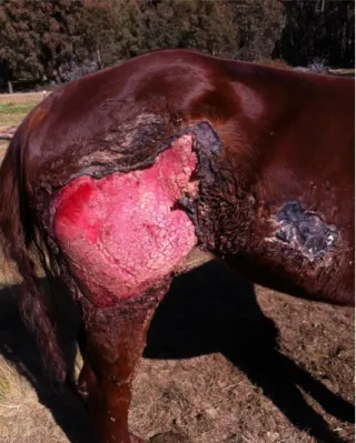
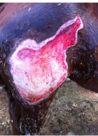
Figure 2 and 3: Photographs of a Standardbred horse with wounds along its flank and down the back leg caused by a clostridial myositis after injection with a contaminated needle. (Courtesy of Professor Doctor Andrew Dart).
Clinical case of a horse presented with extensive subcutaneous enphysema, febrile, depressed and urinating bloody stream of urine. Under local anaesthesia the affected area was sliced open with a scalpel in order to open the area to oxygen. Horse was placed on penicillin and fluids. Wounds were treated with Manuka honey daily. Photographs taken several weeks after horse being in, and treatment was continued throughout healing. Horse went back to racing.
7.1.UMF rating
The so-called Unique Manuka Factor is a trademarked grading system placed on the label of commercially available Manuka honey by licensed producers in New Zealand. The UMF rating assures the purity and quality of the product by representing a similar zone of growth inhibition in a radial diffusion assay with Staphylococcus aureus when compared with a known concentration of an antiseptic (phenol) solution. Each individual batch of manuka honey is tested for antimicrobial activity, however, it is worth noting this testing does not demonstrate precisely which components contribute to this property. In general, a UMF rating 10-15 is required for the batch to be considered therapeutically useful, whilst a UMF 16-30 has superior activity with high antimicrobial efficacy. A UMF rating 5-9 has minimal antimicrobial activity and is not recommended for therapeutic use as an antimicrobial agent, whilst a UMF 0-4 has no detectable antimicrobial activity (Tsang et al., 2018).
Tsang et al. in 2017 compared the effects of topical application of UMF20 and UMF5 manuka honey with a generic multi-floral honey. The results showed that when using the UMF20 the total healing time is reduced when compared to UMF5 and general honey (GH). Furthermore, healing times of wounds treated with GH and UMF5 were not significantly different. In this same study, it was concluded that application of UMF20 to wounds for 12 days showed a longer total healing time compared to using UMF20 until the wound is completely healed (A. S. Tsang et al., 2017).
7.2.What’s the difference between MGO and UMF on the label of the commercial products?
Commercial available manuka honey products are often labelled in different ways, so it is important to understand what it means. The UMF factor is the internationally registered and recognised trademark. It is produced by companies that measure the antibacterial activity of a batch of honey against phenol, a standard antiseptic. The higher the UMF, the greater the antibacterial efficacy. There are products labelled with an MGO concentration and others with a UMF. Products showing MGO concentration have measured the weight by volume of MGO in that batch of honey. While it is accepted that an increasing concentration of MGO is related to the antibacterial activity, the antibacterial activity has not been tested. Studies have shown that taken out of Manuka honey, the activity of MGO can vary, suggesting that other factors in Manuka honey can affect the antimicrobial activity of Manuka honey (Tsang et al., 2018). So, while concentration of MGO in any batch of Manuka honey broadly correlates with antibacterial activity only honey with the registered international trademark provides assurance of the medical grade of Manuka honey.
7.3.Effect on biofilms
Bacterial biofilms are known to cause difficult healing of wounds in both humans and horses due to the ability of biofilms to interfere with the activity of many antibiotics. Manuka honey has been shown to inhibit the formation of biofilms and cause detachment of established biofilms, although the exact mechanism of action is not fully known. Exposure of bacteria to manuka honey in vitro has demonstrated downregulation of genes coding for surface-binding proteins important in biofilm formation, virulence, and cell to cell communication. Low concentrations of manuka honey may prevent biofilm maturation by disrupting cellular communications, whilst higher concentrations of manuka honey are able to penetrate and detach established biofilms. The methylglyoxal in manuka honey appears to play a critical role in the burst of biofilms but it is not totally responsible for this property (Tsang et al., 2018).
The antibacterial component of manuka honey is a small water-soluble molecule that diffuses easily, which explains why manuka honey has also exhibited efficacy against bacteria contained in biofilms. Prolonged chronicity of wounds can be attributed to wound colonization that develops into a biofilm in which the bacteria stay protected by the matrix of the biofilm. These bacteria cannot be cleared by the host immune system and show resistance to both systemic and topical antimicrobial agents. This may explain why antibiotics are of limited use in treating chronic wounds. Manuka honey at a concentration of 40% has been found to give significantly reduced biofilm mass with in vitro testing of clinical isolates of P.aeruginosa that had developed into a biofilm (Cooper, 2009). Similar findings were seen in studies with
Streptococcus pyogenes (Maddocks, Lopez, Rowlands, & Cooper, 2012), methicillin-resistant Staphyloccus aureus (MRSA) (Alandejani, Marsan, Ferris, Slinger, & Chan, 2009), where it
was found biofilms were sensitive to manuka honey treatment (Molan & Rhodes, 2015).
7.4.Effect in gram (–) and gram (+)
The mode of action of Manuka honey differs with the different types of bacteria that it’s dealing with. Exposure of gram-positive organisms to MGO leads to the downregulation of autolysin, an enzyme involved in cell division, and the cleavage of bacterial cell wall components. On the other hand, when exposure of gram-negative bacteria to MGO occurs, it appears to lead to altered gene expression of proteins involved in the structural stability of the cell wall and cell lysis (Dart et al., 2015). A decrease in virulence factors of bacteria has also been observed following treatment with manuka honey, including downregulation of flagella-associated proteins, inhibition of siderophore formation, and reversal of antibiotic resistance (Tsang et al., 2018).
8. Effect in malodour
The deodorisation of offensive odour from wounds is an expected consequence of honey’s antibacterial action. The malodour is due to ammonia, amines and sulphur compounds, which are produced when infecting bacteria metabolise amino acids from proteins in the serum and necrotic tissue in a wound. The rapidity of honey’s deodorising action is probably due to the provision of a rich source of glucose, which would be used by the infecting bacteria in preference to amino acids, resulting in the production of lactic acid instead of malodours compounds (Molan, 2014).
9. Storage
Processing and storage conditions, including heat, light and catalases produced by damaged cells, pollen and some bacteria can inactivate hydrogen peroxide which leads to a variable antibacterial activity of some types of honey under different conditions (Dart et al., 2015). In contrast, the levels of DHA in stored Manuka honey slowly decreases over time as it is converted into MGO, which subsequently increases at a similar pace when stored at 37ºC. The concentration of MGO in stored honey plateaus after 3-4 months and maintains a consistent ratio of DHA to MGO of 2:1(Oryan et al., 2016; Tsang et al., 2018).
10. Can we use General food honey as a wound dressing as well?
Recent studies made at the University of Sydney, showed that generic honey from a supermarket was no more effective than isotonic saline to treat experimental contaminated wounds (Tsang et al., 2017).
Within the environment there are spores that pervade, existing in the soil, air, dust and raw agricultural products, including Clostridium spp. Clostridial spores are a real concern in the anaerobic environment of some open wounds. It is recommended that honey used in wound healing be sterilized. Heat can change the properties of some honeys to eliminate spores. Medical grade honey is often gamma-irradiated to inactivate spores such as those from Clostridium spp. This does not have a detrimental impact on the antibacterial activity of honey (Simon et al., 2009).
11. Recommendations on using a Manuka honey wound dressing
The recommendations and approaches on how to use honey as a dressing depends on the type of wound we’re dealing with, the site of the wound, the level of contamination that is present, and among other factors, the way the commercial product is made.
Contaminated or severely traumatized wounds should be surgically debrided to remove necrotic tissue and debris to optimize healing.
The application of Manuka should start as soon as possible in order to achieve the best outcome, preferably within the first 24 hours after wounding. The honey should be at a normal room temperature for an easier application.
When bacterial contamination and tissue trauma is present, manuka honey with a UMF ≥15 should be used (Bischofberger et al. 2013). However, it’s important to note that as the UMF of manuka honey increases so does the cost.
A study by Dart (2013) found that an effective way to decrease the costs, is to dilute the manuka honey to 66% together with 34% of sterile water-based gel. Wound healing was found to be unaffected by dilution (Bischofberger et al., 2015).
Bandages can have as many advantages as they can have disadvantages. Bandages will improve the contact time between the honey and wound and may be useful in the early stages of wound healing. However, ongoing bandaging may be associated with high incidence of extra granulation tissue (EGT) development, and in this case the EGT should be excised as it arises, and the application of honey continued (Carr, 2003). If a bandage is applied, the duration of application should be kept to a minimum, preferably under 12 days, to reduce the chances of EGT (Bischofberger, Dart, Perkins, & Dart, 2011).
The first application will have to be changed sooner than the ones to follow, because the amount of exudate will be greater due to the osmotic action of the honey happening at the same time as the peak activity of the inflammatory phase that causes a high level of exudate (Kennedy, 2018). On the following applications, as long as the dressing still has Manuka honey available and in contact with the wound and if there is no exudate soaking through the overlying bandage, it can be left in place for a maximum of 5 days, after that a change of bandage is recommended (Hollis, 2016). In open wounds, applying a thin film 2-3 times daily, may be the best approach, because the contact time and the efficacy are both optimized.
According to the duration of the treatment there are also different opinions. A study was undertaken to try to understand the outcomes between application of manuka for 21 days, and the application until the wound healing is complete. It showed that a wound heals faster if the treatment continues until the healing is complete (Dart et al., 2015).
Figure 4 and 5: Photographs showing the action Manuka honey had in the treatment of one wound compared to a wound without treatment in different time lines. (Courtesy of Professor Doctor Andrew Dart)
CHAPTER II – Use of Manuka honey to treat wounds in horses – state of the art and case reports
1. Aims
The aim of this literature review was to evaluate the quality of the available data on this subject, to identify gaps in current knowledge and ensure there is justification for future research on the subject and to add more information on the matter to this dissertation.
The aim of the clinical cases presentation is to report three real cases where Manuka honey was used as the major treatment, and to show the practical application and the outcome that I was able to follow part of the process during my internship, and observe all the theory I had studied put into actions.
2. State of the art about of Manuka honey in wound healing in Veterinary Medicine using inclusion and exclusion criteria
2.1.Search strategy and Data sources
This search was conducted in accordance with the evidence-based guidelines for systematic reviews set forth in the PRISMA statement. Published studies were identified from 2009 to 2019, that included information about the use of Manuka honey in wounds in Veterinary Medicine, more specifically in equine practice. The search was conducted in 2 relevant databases (PubMed and ScienceDirect). In each database, the following search terms were used: “wound”, “wound healing”; “veterinary”, “equine”, “horses”; “Manuka honey”. Each individual search consisted of a combination of the 3 terms connected by “AND”. Each individual search within a database was then combined using “OR” to account for duplication. The electronic search was complemented with a hand-search in references of review articles and book chapters
2.2.Selection of articles
The abstracts of articles were selected by applying the following inclusion criteria: (a) research studies and case reports; (b) published studies; (c) written in English language; (d) including research using Manuka honey in wounds; (e) studies related to Veterinary medicine; (f) studies with important information for this review with relevant results.
Because a large number of new studies have been conducted in the last few years, the information before 2009 was not included.
By analyzing the titles of the publications, I was able to determine that most studies did not meet the inclusion criteria because they did not focus specifically on the topic.