ACTA MEDICA PORTUGUESA 1985; 6: 137-141
ORIGINAL
RED CELL ABNORMALITIES IN A KINDRED WITH
AN UNCOMMON FORM OF HEREDITARY
SPHEROCYTOSIS
GABRIEL OLIM, SANDRA MARQUES, CARLOTA SALDANHA, DULCE SANTOS,
PAULO BARROCA, J. MARTINS E SILVA
Department of Biochemistry- Faculty of Medicine and Hematology Unit- Departmentof Medicine III. Santa Maria University Hospital.
Lisbon. Portugal.
SUMMARY
Red cell acetylcholinesterase(AChE) and Na,+ K ~- adenosinetriphosphatase (ATPase) activities, cell
2,3 -diphosphoglycerate (2,3 -DPG) and adenosinetriphosphate (ATP) content and filterability ratio were
studied in two children (with moderate hemolytic anemia and marked spherocytosis) and their parents. Patients’ parents have no medical problem but evidenced discrete spherocytosis on peripheral smear. Except some increased apparent red cell rigidity detected in the father, all the parameters studied in both parents were found to be normal, as compared to healthy controls. In contrast, red cell rigidity,
2,3-DPGand ATP levels and Na,+ K+ ATPase activity were increased in both children, whereas AChE
activity was similar to values of normal subjects. These observations suggest that both affected patients suffered from homozygous hereditary spherocytosis linked to an apparently recessively inherited red cell membrane defect.
RESUMO
Anomalias eritrocitãrias em criancas portadoras de uma variedade rara de esferocitose hereditãria
Em duas criancas (corn anemia hemolitica moderadae esferocitose acentuada) e seus pais foi determi
nada a actividade da acetilcolinesterase (AChE) e adenosinotrifosfatase (ATP-ase)-Na,+ K + --dependente da membrana eritrocitária, conteüdo globular em 2,3 -difosfoglicerato (2,3 -DPG) e adeno
sinotrifosfato (ATP) e indice de filtrabilidade eritrocitãria. Nenhum dos progenitores evidenciava altera cOes patolôgicas ou anornalias naqueles pararnetros laboratoriais, além da esferocitose ligeira em ambos e
aumento aparente da rigidez globular no pai. Pelo contrário, ambos os doentes, além de sinais clinicos
marcados, evidenciavam filtrabilidade eritrocitária nula a par do aumento da concentraçAo globular do 2,3 -DPG e ATP; aactividade de ATPase Na,~ K + -dependente era superior ao normal apenas em urn
dos doentes, sendo a actividade daAChE equivalentea dos controlos normais em ambos. Os resultados obtidos parecem evidenciar uma forma rara de esferocitose hereditária em ambos os doentes eventualmente
dependentede urn defeito da membrana hereditária corn caracteristicas recessivas.
INTRODUCTION
Hereditary spherocytosis (HS) is a heterogenous disor der, either clinically or in terms of the molecular defects, in apparent association with a primary abnormality of the red cell membrane.”2Although in the majority of the cases the
biochemical defect is still uncertain,3 in a small group of patients exhibiting marked spherocytosis skeletal protein changes involving the spectrin molecule have been describ ed.4’ These abnormalities might result in membrane insta bility,
red
cell fragmentation under circulatory stress and/or loss of erythrocyte deformability.The increased permeability to sodium (Na) ions observed in intact erythrocytes is another functional deficiency of HS
cells, perhaps due to structural changes in the membrane.6 In addition to the differences reported in the several classes of red cell membrane ATPase,7’8 there is also some eviden
ce that the profile of acetylcholinesterase (AChE) activity from HS patients is clearly distint from other hemolytic
anemias.9
In the present paper we focus attention on red cell mem brane Na, ~ K ~ - ATPase and acetylcholinesterase (AChE)
activities, in association to the index of erythrocyte filterabi lity (IF), adenosinetriphosphate (ATP) and 2,3 -- diphosphoglycerate (2,3 - DPG) concentrations, from two
siblings (the sons of related but normal parents) affected with inherited spherocytosis.
MATERIAL AND METHODS
Case reports and routine evaluation- A suggestive form
of hereditary spherocytosis was detected in a twelve-year-old white boy (patient 1) first seen by us for clear anemia, skele tal abnormalities and hepatosplenomegaly. A similar clinical situation was soon observed in patient 2, a five-year-old brother of patient 1. Both patients, still non-splenectomized at the moment, showed hemolytic anemia characterized by hemoglobin values ranging from 7 to 8 g/dl, elevated reticu locyte count, spherocytes on peripheral smear and mild ele vation of unconjugated bilirubin. The most important routi ne laboratory findings of both siblings and their parents (fourth degree cousins) are summarised in Table 1.
In contrast with the physical and hematologic abnormali ties found in both siblings, either parent has had any clinical problem and both had normal physical and routine labora tory tests. However, a small percentage of spherocytes as well as poiquilocytes and increased reticulocyte count (3-4, 5 Wo) were observed in blood smears of both parents.
In all members of this family there was no abnormal hemoglobin detected, HbA2 ande HbF levels were not rais ed, Coombs’ tests (direct and indirect) were negative and enzyme activities of red cell pyruvate-kinase and glucose 6-phosphate dehydrogenase were found to be comparable to those of normal subjects.
Procedures- Blood from each patient and both parents
was drawn into vacutainer tubes containing heparin or acid citrate dextrose as anticoagulants, and immediately subdivi ded in aliquots before being processed further.
Red cell morphology, blood cell counts, immediate osmotic fragility, autohemolysis in vitro, direct and indirect Coombs’ tests, hemoglobin electrophoresis and total and unconjugated bilirubin were performed by routine labora tory techniques.
Red cell 2,3 - diphosphoglycerate concentration was de
termined in 8 % trichloroacetic acid extracts from one blood aliquot, according to the method of Rose and Liebowitz.’° The erythrocyte adenosinetriphosphate content was measur ed in the 10% perchloric acid extract of each blood speci men by the method of Jaworek and Coworkers.”
Red cell acetylcholinesterase activity was measured cob rimetrically by the method of Kapland,’2 in a prepared 50% suspension three times saline-washed erythrocytes and using acetylthiocholine iodide as substracte.
Hemoglobin-free erythrocyte ghosts were prepared by hypotonic lysis, as described by Cha et al.’3 Protein content was measured by the method of Lowry.’4 Membrane Na,+ K,+ ATPase activity was determined as the release of inor ganic phosphate during incubation of ghosts with ATP, according to the technique of Tausshy et al.’5
The erythrocyte filterability index was carried out by a modification of the method of Reid et al,’6 using Nucleopo re polycarbonate sieves (Nucleopore Corp, Pleasanton, U.S.A.), with a pore diameter of 5 /L; erythrocytes suspend ed in Tris-albumin buffer (pH 7.4) at a hematocrit of 8 % were used for testing and compared to the filtration time of the solvent; the calculated index of filtration incorporated a correction for measured hematocrjt.’7
RESULTS
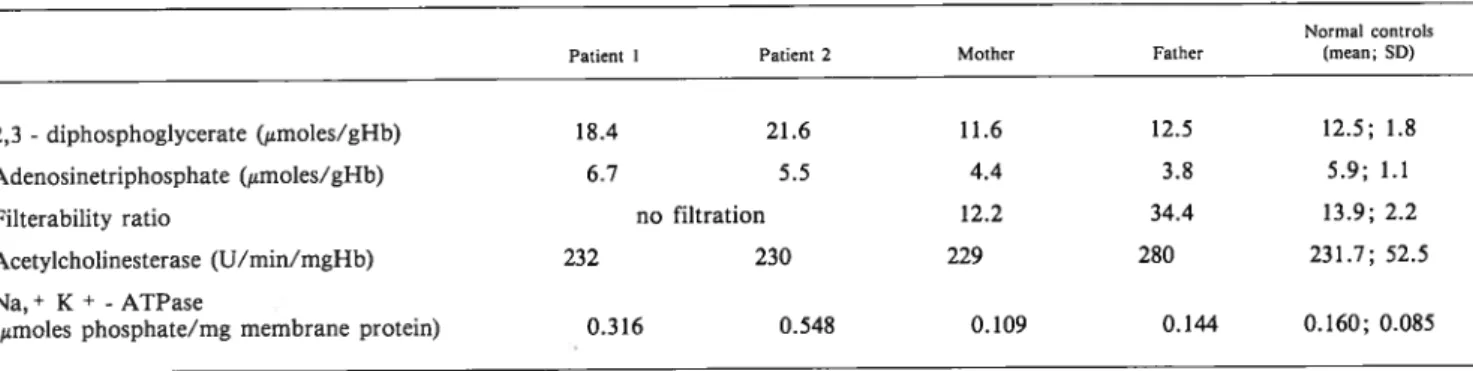
The results for the red cell studied parameters in all members of the family are shown in Table 2. Red cell AChE activity of patients and their parents failed to show any consistent variation from the normal pattern. Membrane Na,+ K+ - ATPase activity from the parents showed a
quite normal value, whereas raised activity levels were remarkable in their sons. A similar feature was observed with
TABLE 1 Standard hematologic data on members of the family studied
Patient Patient 2 Mother Father
Hemoglobin (g/dl) 7.6 7.1 13.7 16.6
Hematocrit (1/1) 0.22 0.22 0.45 0.52
Red cell count (x10 12/1) 2.7 2.6 5.0 5.6
Mean corpuscular volume (I’l) 81.5 84.6 90.0 93.6
Mean corpucular hemoglobin concentration (g/dl) 34.5 32.3 30.4 31.7
Reticulocytes (%) 7 18.5 3.5 4.2 Leucocyte count (x l0~ /1) 9.7 5.9 6.3 6.0 Platelet count (x 109/1) 300 360 300 280 Total bilirubin (mg/dl) 2.5 — 0.43 0.9 Unconjugated bilirubin (mg/dl) 1.9 — — 0.8 Serum iron (mg/dl) 87 128 141 121
Total iron binding capacity (mg/ dl) 187 — — —
Coombs tests (direct and indirect) negative negative — —
Hemoglobin F (°lo) 2.5 2.0 — —
Hemoglobin A2 (%) 3.5 3.0 — —
Osmotic fragility (g/l NaC1)
hemolysis begins 5.0 5.0 5.5 4.5
50% hemolysis (normal range: 4.0-4.45) 4.65 4.45 4.35 4.15 Autohemolysis 37°C (%)
48 h, without added glucose (normal range: 0.2-4.5) 6.0 4.9 5.1 2.8 48 h, with added glucose (normal range: 0-0.5) 1.21 0.95 0.73 0.80 Blood peripheral smear
spherocytic RBC + + + +
nucleated RBC + + — —
poikilocytosis and fragmented RBC + + + +
anisocytosis + + + + + ÷ — —
achantocytic RBC — — —
HEREDITARY SPHEROCYTOSIS
the 2,3- DPG concentration. No clear variation on ATP
levels was detected in all members of the family. As far as the index of red cell filtration is concerned, the increased values observed in both patients and, also in their father, might indicate a loss of deformability, in contrast to the normal value detected in the mother.
DISCUSSION
Hereditary spherocytosis seems to be the result of struc tural defect of erythrocyte membrane, yet to be defined in the most frequent form.1-3Usually this disorder occurs with
discreet clinical symptoms and mild anaemia, in apparent dependence of a mendelian autosomal trait, perhaps with variable penetrance; no cases of homozygoty have been described.’8’19
The presence of spherocytic cells in all members of the family we have studied appears as an uncommon event. Although no clinical manifestations were apparent in the (distantly related) patients’ parents, mild spherocytosis and other signs of presumed hemolytic compensation were detected on blood smear. In contrast, both siblings showed remarkable spherocytosis and other red cell abnormalities (Table 1), in association with clear clinical symptomatology. The obvious poikilocytosis and the normal values of mean corpuscular hemoglobin concentration, as observed on these cases, are unusual laboratory events in HS. Al though some confusion may arise with other spherocytic hemolytic anemias, it is unsafe to assume that one is not dealing with HS. In fact, the combination of anemia, increas ed reticulocytosis (particularly evident in patient 2), slight elevation of serum-indirect reacting bilirubin (in patient 1), spherocytosis, osmotic fragility (in patient 1), autohemolysis (in both), splenomegaly and skeleton abnormalities, as observed, support the diagnosis of HS.
Meanwhile, the reported negative Coombs’ test and cor rected autohemolysis by glucose excludes the diagnosis of immune spherocytosis; the differentiation from the various causes of splenomegaly and hypersplenism offered no doubt by the physical examination; the inexistence of red cell glucose 6-phosphate dehydrogenase and pyruvate-kinase deficiencies, or the absence of sickle cell trait and other he moglobinopathies also distinguished the presenting cases from congenital non-spherocytic hemolytic anemias.
Such appearance may suggest that the patients are ho mozygous for a recessive trait, responsible for a congenital hemolytic anemia with the main characteristics of hereditary spherocytosis.
In addition, both patients clearly exhibited decreased erythrocyte deformability, also observed in a slightest de gree in their father. The reduced deformability, docummen ted by various methods as a characteristic abnormality of HS erythrocytes,2° might be in the presenting cases a conse quence of diminished surface area/volume ratio21 and / or reduced mechanical stability of spherocytic membranes.22 As a consequence of a reduced deformability, progres sive membrane loss and cell fragmentation occurs in the circulation and might be related to the severity of hemolysis and resulting anemia in HS patients.’9’2° In fact, the abnor mal osmotic properties23 and decreased lifespan of HS
erythrocytes21 have been attributed to a diminished surface
area/volume ratio 23 also suggested as the primary factor
leading to decreased deformability. In addition, the reduced surface area/ volume ratio appears to be correlated ‘with the degree of spherocyte rigidity.24
Although our patients’ parents also presented spherocy tic cells, the percentage of their expected undeformable cells was not sufficient to produce the hematologic pattern of typical HS, but a discreet reduced erythrocyte deformability in the father.
For the majority of HS patients, the red cells have a nor mal discocytic shape and about normal osmotic and mem brane properties; only a variable subpopulation of truly spherocytic cells accounts for the difference in deformability and the degree of the anemia.24 So far, the moderate ane mia and osmotic fragility showed in both siblings might be explained by a remarkable percentage of underformable spherocytes, as observed.
As a likely consequence of the red cell fragmentation, the increased 2,3 - DPG content found in both patients
could account for reduced oxygen affinity of hemoglobin25 and hence more oxygen being released to the hypoxic tissues.26 Our data oppose former results of low 2,3 - DPG
levels in HS patients, also with intact spleen,27 but are in agreement with more recent results of increased red cell 2,3 -DPG content in HS children.28
The alteration in red cell 2,3 - DPG levels might be also
governed by some other metabolic factors, namely the ove rall rate of erythrocyte glycolytic pathway.29
Although the rate of glycolysis is decreased in human erythrocytes containing high concentrations of 2,3 - DPG
under in vitro conditions,29 the concentration of ATP were found to be slightly elevated in high DPG cells.30 Further more there is no evidence of a major decrease in the levels of ATP when 2,3 - DPG concentration increases in various
hypoxic conditions•31
The major cation flux abnormality in HS cells appears to be an excessive sodium permeability.6 HS cells oppose the
TABLE 2 Red cell 2,3-diphosphoglycerate and adenosinetriphosphate concentration, filterability ratio, membrane acetylcholinesterase and Na, ~ K - adenosinetriphosphate activities
Normal controls
Patient I Patient 2 Mother Father (mean; SD)
2,3 -diphosphoglycerate (1tmoles/gHb) 18.4 21.6 11.6 12.5 12.5; 1.8
Adenosinetriphosphate (~smoles/gHb) 6.7 5.5 4.4 3.8 5.9; 1.1 Filterability ratio no filtration 12.2 34.4 13.9; 2.2 Acetylcholinesterase (U/min/mgHb) 232 230 229 280 231.7; 52.5 Na,+ K+ -ATPase
abnormaly high Na~ influx by increased pumping, depen dent of higher ATP generation by glycolysis.32’ ~ The higher than normal Na,+ K~ ATPase activity observed in both
siblings, confirming former studies in HS patients,7’33 could be a reflection of an excessive sodium permeability in HS cells, being the Na~ pumping sustained by increase glycoly sis; thus an elevation in ATP levels (with parallel increase of 2,3 -DPG) might be required to maintain the ion pumping.
However, such gain in intracellular ATP was not evident in our study, and seems dissociated of increased 2,3 - DPG
levels.
In HS cells, the Na,~ K~ - ATPase was shown in corre
lation with the rate of sodium efflux;33 however, none of these parameters seems to influence the red cell survival.34 In addition, neither the HS cells became spherical due to an increase in total cation and water content23 nor the spheroi dal shape itself is responsible for increased glycolysis;35 similar abnormalities in cation flux and pumping are also found in some other hemolytic36 or nonhemolytic
disorders. ~
All these observations suggest that excessive sodium per meability and erythrocyte metabolic adaptation, as well as some other described characteristics of HS cells (e. g. sphe rodicity, increased osmotic fragility, changes in membrane lipids or abnormalities in membrane proteins and lipid phosphorylation) are secondary to basic structural changes in red cell membrane.1-3, 19, 20, 37
This view seems to be also partly supported in a recent study, where the profile for erythrocyte membrane acetyl cholinesterase in HS was clearly distint of normal and some other hemolytic anemias;9 the elevation of AChE activity was detected exclusively by using an ionic substracte and was inapparent with a lipophylic one, perhaps due to a more fully exposition of the enzyme molecules at the outer membrane side; however the AChE activity was not influenced by the spherocytic shape.
Conversely, we were unable to confirm by similar metho dology an elevation of erythrocyte AChE activity in our patients or their parents. This discrepancy between our results and those of Streichman et a19 may be due to changes that particular cytoskeletal membrane interactions may impose to the AChE molecules, in dependence of the transmembra ne control of integral membrane protein distribution.38
In spite of all spherocytosis is not the same,13, 18, 19 and
the underlying molecular abnormality is still undefinied for the majority of HS patients,39 most data point towards the red cell membrane cytoskeleton as being the site of the primary genetic defect in HS.
In this regard and associated to the heterogeneity in inheritance, the basic lesion in HS might reflect diverse ab normalities of the membrane cytoskeleton, perhaps as final consequence of many distinct biochemical defects, as re cently suggested.40
The clinical course and uncommon inheritance pattern of our patients may be related to a such still uncovered mem brane defect. Further investigations upon the red cell mem brane characteristics in this family are now in progress in our laboratory.
ACKNOWLEDGMENTS
This study was supported in part by a grant from INIC (MbL2). The technical assistance of Mr. Chim W. San is gratefully acknowledged. The authors also thank Miss Emi ha Alves for typing the manuscript.
REFERENCES
I. CONDREA, E.: Hemolytic disorders associated with a pri mary red cell membrane defect. Experientia. 1976; 32: 537--542.
2. VALENTINE, W. N.: The molecular lesion of hereditary spherocytosis (HS): a continuing enigma. Blood., 1977; 49: 241-245.
3. LUX, S. E.; WOLFE, L. C.: Inherited disorders of the red cell membrane skeleton. Pediatr C/in. Nth. Am., 1980; 27: 463--486.
4. AGRE, P.; ORRINGER, E. P.; BENNETT, V.: Deficient red cell spectrin in severe, recessively inherited spherocytosis. N. EngI. J. Med., 1982; 306: 1155-1 161.
5. GOODMAN, S. R.; SHIFFER, K. A.; CASORIA, L. A.; EYSTER, M. E.: Identification of the molecular defect in the erythrocyte membrane skeleton of some kindreds with heredi tary spherocytosis. Blood., 1982; 60: 722-784.
6. BERTLES. J. F.: Sodium transport across the surface mem brane of red blood cells in hereditary spherocytosis. J. C/in. Invest., 1957; 36: 816-824.
7. NAKAO, R.; KURASHINA, S.; NAKAO, M.: Adenosinetri phosphatase activity of erythrocyte membrane in hereditary spherocytosis. Life Sci., 1967; 6: 595-600.
8. KIRKPATRICK, F. H.; WOODS, 0. M.; LA CELLE, P. L.:
Absence of one component of spectrin adenosinetriphosphatase
in hereditary spherocytosis. Blood., 1975; 46: 945-954.
9. STREICHMAN, S.; KLIN, A.; TATARSKY, I.; LIVNE, A.: Unique profile for erythrocyte membrane acetylcholinesterase in hereditary spherocytosis. Biochim. Biophys. Acta., 1983; 757: 168-175.
10. ROSE, Z. B.; LIEBOWITZ, J.: Direct determination of 2,3 -- diphosphoglycerate. Anal. Biochem., 1970; 35: 177-180.
11. JAWOREK, D.; GRUBER, W.; BERGMEYER, H. U.: Adenosine-5’-triphosphate. Determination with 3 -phos
phoglycerate kinase. In: Methods in Enzymatic Analysis, vol. 3, 2 and ed. H. U. Bergmeyer (ed), Verlag chemie, Weinheim/ /Academic Press New York London., 1974; 2097-2101. 12. KAPLAN, E.; HERZ, F.; HSU, K. S.: Erythrocyte acetylcho
linesterase activity in ABO hemolytic diseaseof the newborn.
Pediat., 1964; 33: 205-211.
13. CHA, Y. S.; SHIN, B. S.; LEE, K. S.: Active uptake of Ca2+ and Ca2+ activated Mg2+ ATPase in red membrane frag
ments.J. Gen. Physiol., 1971; 87: 202-214.
14. LOWRY, 0. H.; ROSENBROUGH, N. J.; FARR, A. C.; RANDELL, R. J.: Protein measurement with the Folin phenol reagent. J. Biol. Chem., 1951; 193: 265-275.
15. TAUSSHY, H. H.; SHORR, E.: A microcolorimetric method for the determination of inorganic phosphate. J. Biol. Chem., 1953; 202: 675-679.
16. REID, H. C.; BARNES, A. J.; LOCK, P. J.; DORMANDY, J. A.; DORMANDY, T. L.: A simple method for measuring erythrocyte deformability. J. C/in. Pathol., 1976; 29: 855-858. 17. HANSS, M.: Erythrocyte filterability measurement by the ini
tial flow rate method. Biorheology., 1983; 20: 199-211. 18. YOUNG, L. E.: Hereditary spherocytosis. Ann. J. Med.,
1955; 18: 486-497.
19. WEED, R. S.: Hereditary spherocytosis: A review. Arch. Int. Med., 1975; 135: 1316-1323.
20. MOHANDAS, N.; PHILIPS, W. M.; BESSIS, M.: Red blood
cell deformability and hemolytic anemias. Semin. Hematol., 1979; 16: 95-1 14.
HEREDITARY SPHEROCYTOSIS
21. COOPER, R. A.; JANDL, J. H.: The role of membrane lipids in the survival of red cells in hereditary spherocytosis. J. Clin. Invest., 1969; 48: 736-744.
22. WAUGH, R. E.; LA CELLE, P. L.: Abnormalities in the membrane material properties of hereditary spherocytes. J. Biomech. Eng., 1980; 102: 240-246.
23. SELWYN, J. C.; DACIE, J. V.: Autohemolysis and other changes resulting from the incubation in vitro of red cells from patients with congenital hemolytic anemia. Blood., 1954; 9: 414-438.
24. LEBLOND, P. F.; BESSIS, M.; DE BOISFLUERY, A.: La forme des érythrocytes dans Ia spherocytose héréditaire; étude au microscope
a
balayage. Relation avec leur déformabilité. Nouv. Rev. Franc. Hématol., 1973; 13: 873-883.25. BENESCH, R.; BENESCH, R. E.: The effect of organic phos phate from the human erythrocyte on the allosteric properties of hemoglobin. Biochem. Biophys. Res. Commun., 1977; 26: 162-167.
26. HJELM, M.; WADMAN, B.: Clinical symptoms, haemoglo bin concentration and erythrocyte biochemistry. Clin. Haema tol., 1974; 3: 689-703.
27. FERNANDES, L. A.; ERSLEV, A. J.: Oxygen affinity and compensated hemolysis in hereditary spherocytosis. J. Lab. Clin. Med., 1972; 80: 780-785.
28. HAIDAS, S.; ZANNOS-MARIOLEA, L.; MATSANIOTES, N.: Red cell 2,3 -diphosphoglycerate levels in children with
hereditary haemolytic anemias. Br. J. Haematol., 1975; 31: 521-530.
29. DUHM, J.: Studies on 2,3 -diphosphoglycerate: effect on
hemoglobin glycolysis and on buffering properties of human erythrocytes. In: Erythrocyte Structure and Functions. Alan R. Liss Inc. New York., 1975; 167-192.
30. DUHM, J.: Inosine permeability and purine nucleoside phos phorylase activity as limiting factors for the synthesis of 2,3 --diphosphoglycerate from inosine, pyruvate and inorganic
phosphate in erythrocytes of various mammalian species. Bio chem. Biophys. Acta. 1974; 343: 89-100.
31. BREWER, 0. J.; EATON, J. W.: Erythrocyte metabolism: in teraction with oxygen transport. Science 1971; 171: 1205-1211. 32. MOHLER, D. N.: Adenosine triphosphate metabolism in here
ditary sherocytosis. J. Clin. Invest., 1965; 44: 1417-1421. 33. WILEY, J. S.: Co-ordinated increase of sodium leak and so
dium pump in hereditary spherocytosis. Br. Haematol., 1972; 22: 529-542.
34. WILEY, J. S.: Red cell survival studies in hereditary spherocy tosis. J. Clin. Invest., 1970; 49: 666-672.
35. COOPER, R. A.; JANDL, J. H.: The relative and conjoint loss of red cell lipids. J. Clin. Invest., 1969; 48: 909-914. 36. BERNARD, J. F.; BOURMIER, 0.; RENOUX, M.; CHAR
RON, D.; BOIVIN, P.: Unclassified haemolytic anemia with splenomegaly and erythrocyte cation abnormalities: a disease of the spleen? Scand. J. Haematol., 1976; 17: 231-239. 37. NATHAN, D. 0.; SHOHET, S. B.: Erythrocyte ion transport
defects and hemolytic anemia: <<hydrocytosis>> and <<desiccyto sis>> Semin. Hematol. 1970; 7: 381-408.
38. NICOLSON, G. L.; PAINTER, R. 0.: Anionic sites of human erythrocyte membrane. II Anti-spectrin-induced transmembra ne aggregation of the binding sites for positively charged coloi dal particles. J. Cell. Biol., 1973; 59: 395-406.
39. PALEK, J.; LUX, J. F.: Red cell membrane skeletal defects in hereditary and acquired hemolytic anemias. Semin. Hematol.
1983; 20: 189-224.
40. BURKE, B. E.; SHOTTON, D. M.: skeleton abnormalities in hereditary Haematol., 1983; 54: 173-187.
Erythrocyte membrane spherocytosis. Brit. J.
Address for reprints: J. Martins e Silva Biochemistry Department Faculty of Medicine
Santa Maria University Hospital 1600 Lisbon. Portugal.