Le uko cyte adhe sio n - a fundam e ntal
pro ce ss in le uko cyte physio lo gy
Department of Biosciences, Division of Biochemistry, University of Helsinki, Helsinki, Finland
C.G. Gahmberg, L. Valmu, L. Tian, P. Kotovuori, S. Fagerholm, A. Kotovuori, C. Kantor and T. Hilden
Abstract
Leukocyte adhesion is of pivotal functional importance. The adhesion involves several different adhesion molecules, the most important of which are the leukocyte ß2-integrins (CD11/CD18), the intercellular adhesion molecules, and the selectins. We and others have extensively studied the specificity and binding sites in the integrins and the intercellular adhesion molecules for their receptors and ligands. The integrins have to become activated to exert their functions but the possible mechanisms of activation remain poorly understood. Impor-tantly, a few novel intercellular adhesion molecules have been re-cently described, which seem to function only in specific tissues. Furthermore, it is becoming increasingly apparent that changes in integrins and intercellular adhesion molecules are associated with a number of acute and chronic diseases.
Co rre spo nde nce
C.G. Gahmberg Department of Biosciences Division of Biochemistry University of Helsinki Viikinkaari 5 00014 Helsinki Finland
Fax: + 358-9-708-59068 E-mail: carl.gahmberg@ helsinki.fi
Presented at the 5th Brazilian Symposium on Extracellular Matrix - SIMEC, Angra dos Reis, RJ, Brasil, September 7-10, 1998.
Research supported by the Academy of Finland, the Sigrid Jusélius Foundation, the Finnish Cancer Society and the Magnus Ehrnrooth Foundation.
Received O ctober 19, 1998 Accepted O ctober 29, 1998
Ke y wo rds
·Leukocyte
·Adhesion
·Integrin
·ICAM
·Membrane
·Glycoprotein
Intro ductio n
Most leukocyte functions depend on ad-hesion. These include cytotoxicity by T lym-phocytes and natural killer (NK) cells, im-munoglobulin synthesis by B lymphocytes, phagocytosis and chemotaxis by granulo-cytes and macrophages and homing to lym-phoid organs (1-7). It is furthermore essen-tial that adhesion-dependent functions are strictly regulated, otherwise chaos would develop in vivo.
A number of adhesion molecules are needed and they must often cooperate to function properly. The most important for leukocytes are the carbohydrate-binding
selectins, the CD11/CD18 or ß2-integrins,
and the intercellular adhesion molecules (ICAMs) (1,6,7). These molecular families
include several members and a certain re-dundancy is evidently important. Thus, an individual leukocyte always contains more than one adhesion molecule belonging to a certain family.
Absence or defective ß2-integrins are
known to result in the hereditary disease, leukocyte adhesion deficiency type 1 (LAD1). Patients affected by this disease suffer from life-threatening infections due to defective granulocytes and macrophages and the in-ability to produce immunoglobulins (8,9). Impaired synthesis of selectin ligands due to malfunction of specific glycosyl transferases results in leukocyte adhesion deficiency type 2. Although this is a milder disease, infec-tions are also common.
man-kind, adhesion is partially defective. There-fore, much effort is currently focused on the elucidation of how the adhesion molecules function at the molecular level. A detailed understanding may certainly facilitate the development of drugs, which would target adhesion.
In this short review we discuss the struc-tures of adhesion molecules, how they may be regulated and the potential applications of adhesion-related drugs.
Se le ctins are ne e de d fo r the initial inte ractio n be twe e n le uko cyte s and e ndo the lial ce lls
Currently three members of the selectin
family are known (10). These are E-selectin, P-selectin and L-selectin (Figure 1A). E-selectin is often induced on endothelial cells, P-selectin is found in platelets and endothe-lial cells and L-selectin is leukocyte-specif-ic. They are carbohydrate-binding lectins
which recognize sialyl Lex
, sialyl Lea and similar sugars (Figure 1B). L-selectin also binds to sulfated structures including sulfatide glycolipids (11). Their function is regulated in two major ways: 1) induced expression of the selectins themselves or 2) induced ex-pression (synthesis) of their ligands.
Endothelial cells contain low amounts of selectins in their resting state. When acti-vated by cytokines P-selectin is rapidly trans-located from intracellular Weibel-Palade bodies to the cell surface. This occurs in minutes and no protein synthesis is needed. The expression of E-selectin peaks later, approximately at 4-6 h and depends on new protein synthesis. L-selectin is present on different types of leukocytes and is easily shed from the cell surface upon activation, evidently due to cell surface proteolysis.
An increased expression of selectin ligands (sialyl Lex
, etc.) occurs in activated tissues, for example during transplantation rejection (12). In this way leukocyte adhe-sion may be increased.
The selectins induce rolling of leuko-cytes along the vessel walls. This means that due to relatively weak interactions, the leu-kocytes (mainly neutrophils) do not become firmly attached, but slowly move along the activated endothelial cell surfaces. This is, however, an important event during which integrins become activated resulting in firm adhesion.
The le uko cyte CD 11/CD 18 inte grins
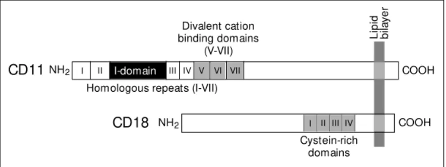
A schematic structure of a leukocyte
CD11/CD18 (ß2) integrin is shown in
Fig-ure 2. Four ß2-integrins are currently known.
They are composed of a common ß2-chain
(CD18) forming heterodimers with four
dif-Figure 1 - Selectins and selectin ligands. A, Schematic structures of selectins. The lectin domains are at the NH2-termini, follow ed by epidermal grow th factor domains (EGF) and complement consensus repeats. B, Structures of the sialyl Lex and sialyl Lea ligands.
NH2
L-selectin NH2
NH2
Lectin
Lectin
Lectin
EGFCR1 CR2 COOH
EGF
EGF
! ! ! ! !
! !!
! !
!! !
! !
!
! ! ! ! !
! !
! ! ! !
! ! ! ! ! !
í ì
î ï ï ï ï ï ï ï ï
í
ì ï ï ï ï ï ï ï ï ï ï ï ï ï ï î
í
ì ï ï ï ï ï ï ï ï î
Lipid bilayer
COOH
COOH
CR1 CR2 CR3 CR4 CR5 CR6
CR1 CR2 CR3 CR4 CR5 CR6 CR7 CR8 CR9
A
B
P-selectin E-selectin
Lex
Sialyl Lex
Neu-NAc a2 3 Gal ß1 4 GIcNAc ß1 3 Gal ß1
3
Fuc a1
Fuc a1 4 GIcNAc ß1 3 Gal ß1
3
a2
Neu-NAc ß1
3 Gal
ferent a-chains (CD11). CD11a/CD18 (LFA-1, aLß2) is primarily expressed on lympho-cytes, CD11b/CD18 (Mac-1, aMß2) on granu-locytes and monocytes, whereas CD11c/ CD18 and CD11d/CD18 are mainly ex-pressed on monocytes/macrophages.
The integrins are glycoproteins contain-ing a complex mixture of N-glycosidic oli-gosaccharides (13). There is no evidence for O-glycosylation. A large proportion (38%) of the oligosaccharides are high mannose-type oligosaccharides, which bind for ex-ample E. coli bacteria (14). Interestingly, the complex oligosaccharides contain large
a-mounts of the sialyl Lex
epitope, and the integrins bind in fact E-selectin in vitro (15). The a-chains contain an I (intervening) or A-domain, and this region is known to bind the ICAM-ligands. The I-domains from CD11b and CD11a have been crystallized and their structures determined (16,17). They form a metal ion-dependent adhesive site
(MIDAS). An Mg2+
-ion is bound to the I-domain with one coordination site left free, and this may be utilized in ligand binding. It has been proposed that the I-domain sits on top of a ß-propeller structure formed by seven feet (18). This structural prediction is based on the homology with structurally known G-proteins.
Divalent cations are needed for integrin activity. Thus, EDTA inhibits their function.
Mg2+
is probably essential, but can be
re-placed by Mn2+
, which shows a stronger activation ability (19). The role of Ca2+
is more controversial. In some systems it is inhibitory, but it may also be important in integrin clustering in the plane of the mem-brane and in this way increasing the avidity of integrin interactions.
One of the most important but also still poorly understood questions is how integrins are activated. This topic has recently been extensively discussed (6). Most probably there exist two major routes of activation, one from the outside of the membrane and one from the inside. Several monoclonal
antibodies are known, which react with the ß2-integrins and activate them. We have found a peptide, derived from the ligand ICAM-2, which binds to CD11a/CD18 and CD11b/ CD18 and strongly activates these integrins (20,21). How this happens is not exactly known, but most probably it involves a con-formational change in the integrins. Mono-clonal antibodies to several other cell sur-face glycoproteins such as CD3, CD43, CD44 and CD45 may also activate the integrins. In these cases, the activation most probably involves intracellular signals, with final acti-vation by inside-out actiacti-vation.
Phorbol esters have long been known to be potent activators of leukocyte integrins (22). Their cellular receptor is protein kinase C, a Ca2+
-dependent serine/threonine pro-tein kinase. Several groups have therefore studied the possible phosphorylation of integrins. By labeling with 32
P-phosphate it became obvious that the a-chains are consti-tutively phosphorylated, whereas the ß-chain is phosphorylated only upon activation (23-26). The major phosphorylated amino acid was found to be serine-756 (Figure 3). How-ever, when this amino acid was mutated, no effect on adhesion was observed (27). If, however, the threonine residues at positions 758-760 were mutated, all adhesion was ab-rogated. Although no clear phosphorylation of these residues had been observed, we then found that in the presence of the phosphatase inhibitor, okadaic acid, in fact a strong
threo-Li
pi
d
bi
la
ye
r
Figure 2 - Schematic structure of a leukocyte CD11/CD18 integrin. The a-chains (CD11) contain 7 homologous repeats and the important binding domain (I). The ß-chain (CD18) contains a cysteine-rich region, w hich may be important for the stabilization of the polypep-tide.
CD11NH2 COOH
Divalent cation binding domains
(V-VII)
Homologous repeats (I-VII)
NH2
CD18 COOH
Cystein-rich domains
I II III IV V VI VII
I II III IV
nine phosphorylation was seen (28). Evi-dently, in activated cells there is a continu-ous rapid threonine phosphorylation/dephos-phorylation cycle, which is not easily ob-served because of strong phosphatase activ-ity. Importantly, threonine phosphorylation was also seen after stimulation of the T cell receptor using anti-CD3 antibodies (28).
Our recent experiments now show that phosphorylation increases the binding of integrins to the cytoskeleton (Valmu L,
Fagerholm S, Suila H and Gahmberg CG, unpublished results). An attractive hypoth-esis is that the lateral mobility of membrane proteins is increased by phosphorylation (29), resulting in clustering of the integrins and increased avidity for their ligands.
The inte rce llular adhe sio n m o le cule s
The cellular ligands for the leukocyte integrins are the ICAMs. They are members of the immunoglobulin superfamily, which contain characteristic Ig-domains composed of two ß-sheets connected by conserved cys-teines. Presently, five ICAMs have been de-scribed, ICAM-1-ICAM-5 (1,6,7,30-32). ICAM-1 and ICAM-3 contain five Ig-do-mains, whereas ICAM-2 and ICAM-4 con-tain only two. ICAM-5 (telencephalin) is unusually complex with its nine Ig-domains (Figure 4).
ICAM-1 (CD54) was the first to be de-scribed. It is present on leukocytes and endo-thelial cells, but it is also expressed in sev-eral other tissues. Characteristic is its easy induction by cytokines such as tumor necro-sis factor-a, interferons, etc. It is probably the major ligand for integrins in most organs. ICAM-2 (CD102) is also present on leu-kocytes and endothelial cells. It shows a more stable expression and is not easily in-duced (33). It can, however, be inin-duced as seen in lymphomas (34). A major function of ICAM-2 may actually be its stimulatory ac-tion on leukocytes. Evidence for this has come from studies on a peptide from the first domain of ICAM-2. This peptide inhibits the binding between endothelial cells and integrins (20), but also shows a strong stimu-latory activity on various leukocytes includ-ing NK cells. The cytotoxicity and migration of NK cells strongly increased after treat-ment with the peptide (21,35). A similar activity has also been observed with a soluble construct of the external portion of ICAM-2 (Kotovuori A, unpublished results).
ICAM-3 (CD50) was actually found
Lipid bilayer
KA LIHLS DLREY RRFEK EKLKS QWNND NPLFK
S
AT T T
VM NPK FAES 769 760759 758 756 724
Figure 3 - The cytoplasmic region of CD18. The major phosphorylation site is serine-756, but the three consecutive threonines are functionally important and partially phosphorylated.
NH2
SS II SS
S I S
S I S
S I S
SS II SS
COOH
NH2
SS II SS
S I S
S I S
S I S
S I S
SS II SS SS II SS NH2
NH2
SS II SS
S I S
S I S
S I S
SS II SS
SS II SS
SS II SS
SS II SS
SI S
COOH
COOH COOH
COOH ICAM -1
(CD54)
ICAM -2 (CD102)
ICAM -3 (CD50)
ICAM -4 (LW)
NH2
SS II SS
S I S
ICAM -5 (TLN)
Figure 4 - The ICAM s schematically show n. The CD-names and previous names (LW = Landsteiner-Wiener and TLN = telencephalin) are indicated.
rather early, but only when it was cloned and sequenced did it become obvious that it could be an ICAM. It is strongly expressed on leukocytes, and again it may be that it is not most important as an adhesion molecule, but more as a signaling component. Interest-ingly, it binds well to CD11d/CD18 (36).
ICAM-4 has been known for a long time as the Landsteiner-Wiener (LW) blood group antigen. Initially LW was thought to be the same as the Rh-antigen but subsequent ge-netic and serological studies showed that this was not the case. It is red-cell specific. When it was cloned and sequenced it turned out that it had a clear homology with the then known ICAMs (37). Subsequently, it was shown to be able to bind to leukocyte integrins and an ICAM-4 antibody blocked the adhe-sion (30). Its physiological function(s) is still not known.
ICAM-5 is brain-specific. It was first characterized and named telencephalin, re-flecting its distribution in brain. Also here, sequence analysis showed its homology to the ICAMs and later work showed that it is an ICAM (31,32). Thus, it is able to bind
leukocytes through the ß2-integrins, but
whether it solely acts as a leukocyte-binding protein in brain is not known. In some of the ICAMs the integrin-binding domain and the adhesion sites have been mapped. CD11a/ CD18 binds to all studied ICAMs and the first Ig-domain seems most important in in-tegrin binding (6,7,38,39). CD11b/CD18, however, binds to the third Ig-domain in ICAMs and is thus different in this respect (40). It also binds to 2 and the ICAM-2-derived adhesion peptide, but the binding may be weaker than the binding to ICAM-1 (41).
Most ICAM genes are clustered on chro-mosome 19 p13-2, with the exception of the ICAM-2 gene, which is located on chromo-some 17 q23-25 (6). This indicates that they have arisen from gene duplication.
The recent discoveries of ICAM-4 and ICAM-5 show that organ-specific ICAMs
exist. It is anticipated that in the future more such ICAMs will be found.
Adhesion molecules evidently possess some general stickiness, which makes them suitable to act as receptors for various mi-crobes. ICAM-1 was found to act as a major rhinovirus receptor (38,42) and as receptor for Plasmodium falciparum-infected red cells (43). Interestingly, the binding site of rhi-noviruses, which is in the first domain of ICAM-1, is different from that used by CD11a/CD18, although it is also located in the first Ig-domain. This fact is important when drugs are developed, which specifical-ly could inhibit microbe attachment but not leukocyte adhesion. Otherwise, side effects would be a major problem.
D e ve lo pme nts in the future
neutro-phils in the damaged cardiac tissue, which may result in further destruction of the tis-sue. Preliminary results using monoclonal antibodies to leukocyte integrins and ICAMs look promising (6,44).
In some instances it could be important to develop drugs which would enhance inte-grin/ICAM adhesion. The results with the ICAM-2 peptide P1 already show that such an approach could be useful (21,35). During infections it could be advantageous to in-crease integrin activity in order to inin-crease the accumulation of leukocytes in infected tissues. An especially important field of ap-plication could be to enhance the activity of cytotoxic T lymphocytes and NK cells in patients with malignant diseases. Reagents
are now becoming available which increase integrin activity, and certainly some of them could be developed to become clinically use-ful. But also here the necessary basic knowl-edge of the mechanisms of activation of leukocyte adhesion is very limited, which makes drug development difficult.
We think that during the years to come we will witness a rapid development in the clinical application of adhesion research, and it will be especially rewarding to be part of that effort.
Ackno wle dgm e nts
We thank Yvonne Heinilä for secretarial assistance.
Re fe re nce s
1. Springer TA (1990). Adhesion receptors of the immune system. Nature, 346:
425-434.
2. Arnaout M A (1990). Structure and func-tion of the leukocyte adhesion molecules CD11/CD18. Blood, 75: 1037-1050.
3. Patarroyo M , Prieto J, Rincon J, Timonen T, Lundberg C, Lindbom L, Åsjö B & Gahmberg CG (1990). Leukocyte-cell ad-hesion: A molecular process fundamental in leukocyte physiology. Immunological Review s, 114: 67-108.
4. Hogg N (1989). The leukocyte integrins.
Immunology Today, 10: 111-114. 5. Carlos TM & Harlan JM (1994).
Leuko-cyte-endothelial adhesion molecules.
Blood,84: 2068-2101.
6. Gahmberg CG, Tolvanen M & Kotovuori P (1997). Leukocyte adhesion. Structure and function of human leukocyte ß2-integrins and their cellular ligands. Euro-pean Journal of Biochemistry, 245: 215-232.
7. Gahmberg CG (1997). Leukocyte adhe-sion. CD11/CD18 integrins and intercellu-lar adhesion molecules. Current Opinion in Cell Biology,9: 643-650.
8. Arnaout M A (1990). Leukocyte adhesion molecules deficiency: Its structural basis, pathophysiology and implications for mod-ulating the inflammatory response. Immu-nological Review s,114: 145-180.
9. Springer TA (1985). The LFA-1, M ac-1
gly-coprotein family and its deficiency in an inherited disease. Federation Proceed-ings, 44: 2660-2663.
10. Bevilacqua M , Butcher E, Furie B, Furie B, Gallat in M , Gim brone M , Harlan J, Kishimoto K, Lasky L, M cEver R, Paulson J, Rosen S, Seed B, Siegelman M , Springer T, Stoolman L, Tedder T, Varki A, Wagner D, Weissman I & Zimmerman G (1991). Selectins: a family of adhesion re-ceptors. Cell, 67: 233.
11. Aruffo A, Kolanus W, Walz G, Fredman P & Seed B (1991). CD62/P-selectin recog-nition of myeloid and tumor cell sulfatides.
Cell, 67: 35-44.
12. Turunen JP, M ajuri M -L, Seppo A, Tiisala S, Paavonen T, M iyasaka M , Lemström K, Penttilä L, Renkonen O & Renkonen R (1995). De novo expression of endothelial sialyl Lew is a and sialyl Lew is x during cardiac transplant rejection: superior ca-pacity of a tetravalent sialyl Lew is x oli-gosaccharide in inhibiting L-selectin-de-pendent lymphocyte adhesion. Journal of Experimental M edicine, 182: 1133-1142. 13. Asada M , Furukaw a K, Kant or C,
Gahmberg CG & Kobata A (1991). Struc-tural study of the sugar chains of human leukocyte cell adhesion molecules CD11/ CD18. Biochemistry,30: 1561-1571. 14. Gbarah A, Gahmberg CG, Ofek I, Jacobi U
& Sharon N (1991). Identification of the leukocyte adhesion molecules CD11 and
CD18 as receptors for type 1 fimbriated (mannose specific) Escherichia coli. Infec-tion and Immunity, 59: 4524-4530.
15. Kotovuori P, Tontti E, Pigott R, Shepherd M , Kiso M , Hasegaw a A, Renkonen R, Nortamo P, Altieri DC & Gahmberg CG (1993). The vascular E-selectin binds to the leukocyte integrins CD11/CD18.
Glycobiology, 3: 131-136.
16. Lee J-O, Rieu P, Arnaout M A & Liddington R (1995). Crystal structure of the A do-main from the a subunit of integrin CR3 (CD11b/CD18). Cell, 80: 631-638. 17. Qu A & Leahy DJ (1995). Crystal structure
of the I-domain from the CD11a/CD18 (LFA-1,aLß2) integrin. Proceedings of the National Academy of Sciences, USA, 92: 10277-10281.
18. Springer TA (1997). Folding of the N-ter-minal, ligand-binding region of integrin a -subunits into a ß-propeller domain. Pro-ceedings of the National Academy of Sci-ences, USA, 94: 65-72.
19. Altieri DC (1991). Occupancy of CD11b/ CD18 (M ac-1) divalent ion binding site(s) induces leukocyte adhesion. Journal of Immunology, 147: 1891-1898.
21. Li R, Nortamo P, Kantor C, Kovanen P, Timonen T & Gahmberg CG (1993). A leu-kocyte integrin binding peptide from in-tercellular adhesion molecule-2 stimu-lates T cell adhesion and natural killer cell activity. Journal of Biological Chemistry, 268: 21474-21477.
22. Patarroyo M , Beatty PG, Fabre JW & Gahmberg CG (1985). Identification of a cell surface protein complex mediating phorbol ester-induced adhesion (binding) among human mononuclear leukocytes.
Scandinavian Journal of Immunology, 22: 171-182.
23. Hara T & Fu SM (1986). Phosphorylation of a, ß subunits of 180/100-Kd polypep-tides (LFA-1) and related antigens. In: Reinherz EL, Haynes BF, Nadler LM & Bernstein ID (Editors), Leukocyte Typing II. Vol. 3. Springer-Verlag, New York,
77-84.
24. Chatila TA, Geha RS & Arnaout M A (1989). Constitutive and stimulus-induced phos-phorylation of CD11/CD18 leukocyte ad-hesion molecules. Journal of Cell Biology,
109: 3435-3444.
25. Buyon JP, Slade SG, Reibm an J, Abramson SB, Philips M R, Weissmann G & Winchester R (1990). Constitutive and induced phosphorylation of the a- and ß-chains of the CD11/CD18 leukocyte inte-grin family. Journal of Immunology, 144: 191-197.
26. Valmu L, Autero M , Siljander P, Patarroyo M & Gahmberg CG (1991). Phosphoryla-tion of the ß-subunit of CD11/CD18 integrins by protein kinase C correlates w ith leukocyte adhesion. European Jour-nal of Immunology, 21: 2857-2862. 27. Hibbs M L, Jakes S, Stacker SA, Wallace
RW & Springer TA (1991). The cytoplas-mic domain of the integrin lymphocyte function-associated antigen 1 ß subunit: Sites required for binding to intercellular adhesion molecule 1 and the phorbol es-ter-stimulated phosphorylation site. Jour-nal of Experimental M edicine, 174: 1227-1238.
28. Valmu L & Gahmberg C (1995). Treatment w ith okadaic acid reveals strong
threo-nine phosphorylation of CD18 after acti-vation of CD11/CD18 leukocyte integrins w ith phorbol esters or CD3 antibodies.
Journal of Immunology,155: 1175-1183. 29. Patarroyo M & Gahmberg CG (1984). Phorbol 12,13 dibutyrate enhances lateral redistribution of membrane glycoproteins in human blood lymphocytes. European Journal of Immunology, 14: 781-787. 30. Bailly P, Tontti E, Hermand P, Cartron J-P
& Gahmberg CG (1995). The red cell LW blood group protein is an intercellular ad-hesion molecule w hich binds to CD11/ CD18 leukocyte integrins. European Jour-nal of Immunology, 25: 3316-3320.
31. Tian L, Yoshihara Y, M izuno T, M ori K & Gahmberg CG (1997). The neuronal gly-coprotein telencephalin is a cellular ligand for the CD11a/CD18 leukocyte integrin.
Journal of Immunology, 158: 928-936.
32. M izuno T, Yoshihara Y, Inazaw a J, Kagamiyama H & M ori K (1997). cDNA cloning and chromosomal localization of the human telencephalin and its distinc-tive interaction w ith lymphocyte function-associated antigen-1. Journal of Biologi-cal Chemistry, 272: 1156-1163.
33. Nortamo P, Li R, Renkonen R, Timonen T, Prieto J, Patarroyo M & Gahmberg CG (1991). The expression of human intercel-lular adhesion molecule-2 is refractory to inflammatory cytokines. European Jour-nal of Immunology, 21: 2629-2632. 34. Renkonen R, Paavonen T, Nortamo P &
Gahmberg CG (1992). Regulation of the expression of endothelial adhesion mol-ecules in vivo. Increased expression of ICAM -2 in lymphoid malignancies. Ameri-can Journal of Pathology, 140: 763-767. 35. Som ersalo K, Carpén O, Saksela E,
Gahmberg CG, Nortamo P & Timonen T (1995). Activation of natural killer cell mi-gration by leukocyte integrin-binding pep-tide from intercellular adhesion molecule-2 (ICAM -molecule-2). Journal of Biological Chemis-try, 270: 8629-8636.
36. Van der Vieren M , Le Trong H, Wood CL, M oore PF, St John T, Staunton DE & Gallatin WM (1995). A novel leukointegrin, adß2, binds preferentially to ICAM -3.
Im-munity, 3: 683-690.
37. Bailly P, Herm and P, Callebaut I, Sonneborn HH, Khamlichi S, M ornon J-P & Cartron J-P (1994). The LW blood group glycoprotein is homologous to intercellu-lar adhesion molecules. Proceedings of the National Academy of Sciences, USA, 91: 5306-5310.
38. Staunton DE, Dustin M L, Erickson HP & Springer TA (1990). The arrangement of the im m unoglobulin-like dom ains of ICAM -1 and the binding sites for LFA-1 and rhinovirus. Cell, 61: 243-254.
39. Berendt AR, M cDow all A, Craig AG, Bates PA, Sternberg M JE, M arsh K, New bold CI & Hogg N (1992). The binding site on ICAM -1 for Plasmodium falciparum
-in-fected erythrocytes overlaps, but is dis-tinct from, the LFA-1-binding site. Cell,
68: 71-81.
40. Diamond M S, Staunton DE, M arlin SD & Springer TA (1991). Binding of the inte-grin M ac-1 (CD11b/CD18) to the third im-munoglobulin-like domain of ICAM -1 (CD54) and its regulation by glycosylation.
Cell, 65: 961-971.
41. Xie J, Li R, Kot ovuori P, Verm ot -Desroches C, Wijdenes J, Arnaout M A, Nortamo P & Gahmberg CG (1995). Inter-cellular adhesion molecule-2 (CD102) binds to the leukocyte integrin CD11b/ CD18 through the A domain. Journal of Immunology, 155: 3619-3628.
42. Greve JM , Davis G, M eyer AM , Forte CP, Connolly Yost S, M arlor CW, Kamarck M E & M cClelland A (1989). The major human rhinovirus receptor is ICAM -1. Cell, 56: 839-847.
43. Berendt AR, Simmons DL, Tansey J, New bold CI & M arsh K (1989). Intercellu-lar adhesion molecule-1 is an endothelial cell adhesion receptor for Plasmodium fal-ciparum. Nature, 341: 57-59.
44. Gahmberg CG, Tolvanen M , Nortamo P, Li R, Kantor C, Xie J, Kotovuori P, Puustinen A, Valmu L, Ylänne J & Tontti E (1995). The intercellular adhesion mol-ecules (ICAM s). In: Horton M A (Editor),