Universidade de Lisboa
Faculdade de Farmácia
NOVEL STRATEGY TO DETECT AND LOCALIZE
LATENT HIV RESERVOIRS
Miguel Gomes Cardoso
Dissertação orientada pelo Professor Doutor João Gonçalves
Mestrado em Ciências Biofarmacêuticas
Faculdade de Farmácia
NOVEL STRATEGY TO DETECT AND LOCALIZE
LATENT HIV RESERVOIRS
Miguel Gomes Cardoso
Dissertação orientada pelo Professor Doutor João Gonçalves
Mestrado em Ciências Biofarmacêuticas
Novel Strategy to Detect and Localize Latent HIV Reservoirs
iii
Agradecimentos
Em primeiro lugar, gostaria de agradecer ao Professor Doutor João Gonçalves a oportunidade dada para realizar o meu projecto de mestrado no seu laboratório, pela confiança e incentivo dados ao longo do ano e por todas as sugestões para me manter na direcção certa.
Quero agradecer às minhas colegas de laboratório, Inês, Luciana, Rafaela, Tatiana e Vera, por todos os momentos engraçados, frustrações partilhadas por ensaios falhados e pelas conversas, por vezes complicadas, quando eu tentava explicar o que tinha corrido mal. À Ana Catarina, Catarina e Pedro por todas as dicas e ensinamentos passados que foram extremamente importantes para me conseguir orientar no início da tese. À Paula por todos os pormenores transmitidos que fazem toda a diferença.
Agradeço à Professora Doutora Madalena Pimentel por todos os momentos de incentivo dados quando nos cruzávamos no laboratório.
Aos meus colegas do iMed.ULisboa, Ana, Mariana e Pedro, um obrigado por todos os cafés, conversas e desabafos que iniciaram praticamente todos os dias de trabalho. Sem eles, não teria sido tão divertido acordar cedo para trabalhar.
Aos meus amigos, que me deram grandes momentos para guardar ao longo deste ano, que conseguiram muitas vezes fazer-me abstrair do trabalho de forma a divertir-me, que me ouviram desabafar vezes sem conta sobre a tese (apesar de se cansarem depressa), que foram verdadeiros amigos. Sem precisar dizer nomes, agradeço profundamente todo o apoio e presença.
Quero agradecer à minha família, toda a preocupação e apoio dados ao longo da minha. Agradeço especialmente à minha Mãe e ao meu Pai por tudo o que me ensinaram e continuarão a ensinar, pelo incentivo e apoio nos bons e maus momentos, pelas oportunidades que me deram para ser uma pessoa melhor, por serem os meus pais.
Novel Strategy to Detect and Localize Latent HIV Reservoirs
v
Abbreviations
AIDS Acquired immunodeficiency syndrome
Amp Ampicillin
ATCC American Type Culture Collection
BSA Bovine serum albumin
CA Capsid
CAR Chimeric antigen receptor
cART Combinational antiretroviral therapy
CCR5 C-C chemokine receptor type 5
CD32a Cluster of differentiation 32a
CD4 Cluster of differentiation 4
CD44 Cluster of differentiation 44
CMV Cytomegalovirus
CXCR4 C-X-C chemokine receptor type 4
DMEM Dulbecco's modified Eagle's medium
DNA Deoxyribonucleic acid
EGFP Enhanced green fluorescent protein
Env Envelope
FBS Fetal bovine serum
FITC Fluorescein isothiocyanate
FP Fusion peptide
Gag Group-specific antigen
Gal4DBDVP64 Gal4 DNA binding domain VP64
GFP Green fluorescent protein
HA Hemagglutinin
HER2 Human Epidermal growth factor Receptor 2
HIV Human immunodeficiency virus
HIV-1 Human immunodeficiency virus type 1
Novel Strategy to Detect and Localize Latent HIV Reservoirs
vi IκB Inhibitor of kappa B
IN Integrase
INSTI Integrase strand transfer inhibitor
JKT Jurkat
Kan Kanamycin A
LDL Low-density lipoprotein
LNR Lin12-Notch repeat
LRA Latency-reversing agent
LTR Long terminal repeats
MA Matrix
MyD88 Myeloid differentiation primary response 88
NC Nucleocapsid
Nef Negative regulatory factor
NF-κB Nuclear factor kappa B
NNRTI Non-nucleoside reverse transcriptase inhibitor
NRTI Nucleoside reverse transcriptase inhibitor
NtRTI Nucleotide reverse transcriptase inhibitor
Ø Diameter
ORF Open Reading Frame
PAMP Pathogen-associated molecular pattern
PBS Phosphate-buffered saline PGK Phosphoglycerate kinase PI Propidium iodide Pol Polymerase PR Protease PSA Penicillin-Streptomycin-Amphotericin B
RNA Ribonucleic acid
RPMI Roswell Park Memorial Institute
RT Reverse transcriptase
vii scFv single-chain variable fragment
SFFV Spleen focus-forming virus
SV40 Simian virus 40
tBFP tag Blue florescent protein
TCR T-cell receptor
TLR Toll-like receptor
TU (Lentiviral) Transducing units UAS Upstream activating sequence
Vif Viral infectivity factor
Vpr Viral protein R
Novel Strategy to Detect and Localize Latent HIV Reservoirs
ix
Abstract
Over the years, the scientific eagerness to find a cure for human immunodeficiency virus (HIV) led us to, try after try, be closer to finding it. For the first time, in 1996, appears a new therapeutic strategy that consists in combinations of different antiretroviral drugs. Over the last years, the most used therapy against HIV has been combinational antiretroviral therapy (cART) although, it is not able to eradicate HIV from the host due to HIV latency. The cART works by stopping the replication cycle of HIV, but, when the virus is latent, its replication cycle is interrupted, thus making impossible for cART to fully eliminate the provirus from the host. Nowadays, the HIV/acquired immunodeficiency syndrome (AIDS) research has the goal to discover the answer to the question of the last years: what is the best method to reactivate the latent HIV? There are records of a wide range of compounds that can stimulate the HIV reactivation (latency-reversing agents – LRAs), but everyone has a disadvantage or a low efficacy in vivo. The search for a more efficient and specific non-toxic LRA has greatly increased. New strategies have emerged that consist in the combination of more potent LRAs with specific vehicles to delivery. Cell receptors have the ability to generate a response after the binding of an antigen. These receptors can be customized allowing to choose the sensing and response behaviors. With this new concept, we developed a strategy that comprehends the reactivation of latent reservoirs upon receptor recognition and the induction of T cell activation which leads to the death of the infected cell. Here, our results shown that the developed receptor is capable of sensing surface-expressed gp120 and inducing the expression of a reporter vector allowing us to detect gp120+ cells. Thus, this customized cell response is a promising method to be employed as a therapeutic strategy after future optimizations.
Novel Strategy to Detect and Localize Latent HIV Reservoirs
xi
Resumo
O vírus da imunodeficiência humana (do inglês, HIV) é responsável pela síndrome da imunodeficiência adquirida (SIDA), uma doença que debilita o sistema imunitário ficando sem capacidade para combater infecções oportunistas. O HIV-1 reconhece diferentes receptores presentes na superfície dos linfócitos T e de outras células, como monócitos, células dendríticas e macrófagos, levando à fusão do virião com a membrana celular. Inicialmente, a proteína gp120 liga-se ao receptor CD4 e, de seguida, a um dos co-receptores, CCR5 ou CXCR4, aproximando a proteína gp41 da superfície celular e resultando na fusão com a célula. O RNA viral, após a entrada do vírus, é convertido em DNA pela transcriptase reversa e logo transportado para o núcleo onde será integrado no genoma da célula hospedeira. Finalmente, a célula inicia a transcrição e tradução das proteínas virais para formar novas partículas virais que, depois de secretadas, irão infectar novas células após sofrerem maturação.
As terapias actualmente utilizadas para o tratamento desta infecção, dependem da replicação activa do vírus para serem eficazes. Estas consistem na combinação de diferentes fármacos antiretrovirais que têm como alvo diferentes passos do ciclo replicativo do HIV-1. Assim, os reservatórios latentes existentes no organismo do hospedeiro tornam-se o maior obstáculo à erradicação do vírus. Estes são compostos por células infectadas num estado de latência, que é definido pela ausência de transcrição viral, e por isso, a terapia combinatória antiretroviral (cART) é incapaz de eliminar estas células. As principais células que formam os reservatórios são linfócitos T CD4+ de memória que têm um tempo de meia-vida aproximadamente igual a 44 meses, estimando-se que demoraria cerca de 73 anos para eliminar totalmente o HIV-1 do hospedeiro apenas com cART.
Dada a incapacidade das terapias actuais erradicarem o HIV-1 do organismo, têm vindo a emergir vários estudos que procuram desenvolver estratégias inovadoras para ultrapassar o obstáculo que são os reservatórios latentes. As novas abordagens terapêuticas têm de ser capazes de eliminar as células latentes de forma específica, como acontece com a terapia génica e a terapia celular. Também têm sido estudadas outras abordagens que dependem da reactivação das células latentes, pois mostraram ser um método que tanto possibilita o sistema imunitário de reconhecer essas células como infectadas e então induzir a sua morte, como também possibilita que as terapias correntes sejam eficazes visto que, as células passam a ter o ciclo de replicação do HIV-1 activo. O mecanismo mais utilizado para reactivar as células consiste na indução de cascatas de sinalização que leva à activação da transcrição do genoma do HIV-1 e consequentemente, à produção viral. Estudos revelam que a
Novel Strategy to Detect and Localize Latent HIV Reservoirs
xii estimulação do receptor TLR5 pela flagelina remove as células do seu estado de latência. Este receptor tem um papel importante na imunidade inata e tem como mecanismo de transdução de sinal a activação do NF-κB que, por sua vez, activa a transcrição.
Na terapia celular, células são modificadas para expressar receptores sintéticos específicos que reconhecem antigénios associados a doenças. As células expressam naturalmente receptores que têm papeis importantes no desenvolvimento e manutenção celular em tecidos. Estudos actuais estão focados no estudo de estratégias promissoras que têm como base os mecanismos de activação e de transdução de sinal destes receptores. Um receptor que se mostrou bastante promissor foi o receptor Notch, pois não necessita de mensageiros secundários para a transdução de sinal e porque tanto o domínio de reconhecimento (extracelular) como o domínio intracelular são facilmente personalizados. Os receptores synNotch providenciam flexibilidade na modificação dos seus domínios concedendo a capacidade de especificar o alvo molecular e a resposta do receptor ao estímulo. Através da transdução de células, são criadas linhas celulares que, ao reconhecerem células específicas, têm uma resposta pré-definida.
Baseando-nos nestas estratégias, propusemo-nos a desenvolver um receptor (VRC01_synNotch) capaz de detectar a presença de gp120 na superfície celular e, quando estimulado activa a expressão de um vector repórter fluorescente. Este receptor é o primeiro passo para o desenvolvimento de uma estratégia que consiste na detecção de linfócitos T CD4+ de memória induzindo a expressão de flagelina que activará a célula de memória e do receptor VCR01_synNotch. Caso esta célula esteja infectada, passará a expressar as proteínas virais, nomeadamente a gp120, que, depois do receptor VRC01_synNotch a reconhecer, será desencadeada a activação do linfócito T eliminando a célula infectada.
Para avaliar o funcionamento deste receptor também criámos células que expressam os antigénios alvo. No caso do VRC01_synNotch, as células alvo expressam gp120 à superfície e, no caso do controlo positivo (LaG17_synNotch) que reconhece a proteína EGFP, as células alvo expressam uma versão modificada da EGFP à superfície da célula. Depois de várias optimizações da expressão do receptor LaG17_synNotch, as células com o receptor tiveram uma activação de 15% após a co-cultura com células EGFP+. De forma a tentar aumentar o nível de expressão de tBFP, decidimos usar apenas células Jurkat como células receptoras e apresentadoras.
Depois de criadas, as células foram outra vez incubadas em co-cultura e, após a análise por citometria de fluxo, as células LaG17_synNotch+ apresentaram, no máximo, 5.3% de células positivas para tBFP e as VRC01_synNotch+ apresentaram, no máximo, 2.29% de células tBFP+. Apesar das células terem mais facilidade na interacção célula-a-célula, o
xiii número de células transduzidas com êxito foi baixo, com excepção das células EGFP+, havendo pouca estimulação dos receptores e consequente activação.
As células infectadas quando estão no estado de latência são indistinguíveis das outras células não infectadas. Ao reactivar estas células, como o genoma do HIV-1 começa a ser expresso, proteínas específicas da infecção, como a gp120, são transportadas para a superfície da célula. A presença destes antigénios nas células facilita o desenvolvimento de abordagens específicas. Para isso, foi estudado o efeito da flagelina na reactivação de células latentes (J-Lat 10.6) e também na expressão do receptor (Fas) e do ligando (FasL) que estão associados à sinalização da apoptose. Os resultados que obtivemos demonstraram que a estimulação da flagelina não afecta a expressão de Fas/FasL, mas é capaz de reactivar 80% das células latentes.
No futuro, as células transduzidas têm de ser separadas e purificadas (cell sorting) para optimizar os ensaios de activação e assim estudar detalhadamente as interacções entre as células receptoras e as células apresentadoras. Têm de ser desenvolvidos novos receptores que reconheçam os marcadores de células de memória (CD44 ou CD32a) e levem à expressão da flagelina para reactivar a célula latente. Um estudo pioneiro identificou a proteína CD32a como um antigénio com elevada expressão em células de memória latentes, que poderá ser utilizada numa abordagem independente da reactivação. Por fim, o receptor VRC01_synNotch tem de ser modificado para que, após estimulação com gp120, a célula receptora seja activada induzindo a morte da célula infectada.
Os resultados apresentados neste trabalho demostraram que o receptor desenvolvido é capaz de induzir uma resposta específica após o reconhecimento de gp120 expresso na superfície da célula. Através de modificações dos domínios do receptor, podemos obter respostas personalizadas a específicos estímulos. Para obter respostas mais complexas, podem-se combinar diferentes receptores de forma a obter uma reposta apenas quando dois ou mais antigénios são reconhecidos. Concluindo, estes receptores mostraram ser uma promissora abordagem para erradicação do HIV-1.
Novel Strategy to Detect and Localize Latent HIV Reservoirs
xv
Table of Contents
Agradecimentos ...iii Abbreviations ... v Abstract ...ix Resumo ...xi Table of Contents ...xv 1. General Introduction ... 11.1. Human Immunodeficiency Virus ... 1
1.1.1. Structure and Genome ... 1
1.1.2. Replication Cycle ... 2
1.1.3. Latency ... 4
1.1.4. Reactivation ... 4
1.2. Notch Receptors ... 6
2. Objective ... 9
3. Material and Methods ...11
3.1. Bacterial Strains and Media...11
3.2. Plasmids ...11
3.3. Cell Lines and Culture Conditions ...12
3.4. Cell Transfection ...12 3.5. Viral Production ...12 3.6. Cell Transduction ...13 3.7. Co-Culture of Cells ...13 3.8. Flagellin Stimulation ...13 3.9. Flow Cytometry ...13 4. Results ...15
4.1. Syn-Notch Receptors Transduction ...15
4.2. Sender Ligands Transfection ...18
4.3. Receiver and Sender Cells Co-Culture ...20
4.4. Flagellin Stimulation ...23
5. Discussion ...25
6. Concluding Remarks and Future Perspectives ...29
7. References ...31 8. Annexes ... I
1
1. General Introduction
1.1. HUMAN IMMUNODEFICIENCY VIRUS
The human immunodeficiency virus (HIV), which belongs to a subgroup of Retrovirae family named lentivirus, is responsible, over time, for acquired immunodeficiency syndrome (AIDS) (Nye & Parkin, 1994 - Chapter 1). There are two types of HIV: HIV-1 and HIV-2, where HIV-1 virulence and infectivity are higher than HIV-2 and HIV-1 has a global prevalence while HIV-2 is confined to West Africa (Sharp & Hahn, 2011). The major differences between the two types of HIV are viral replication, the rate of evolution, viral load in semen and the co-receptor usage. Viral replication and viral load in semen of HIV-1 are greater than in HIV-2, although the rate of evolution and co-receptor usage are higher in HIV-2. There are some similarities, for example, the cytopathicity1 (Nyamweya et al., 2013).
1.1.1. STRUCTURE AND GENOME
HIV is composed of a matrix layer of p17 proteins (MA) surrounded by a host-sourced lipid bilayer scattered with external (gp120) and transmembrane (gp41) glycoproteins which are bound by non-covalent interactions to form the gp160 complex and also other proteins present in the host cell, including major histocompatibility antigens, actin and ubiquitin (Arthur et al., 1992). The viral matrix proteins involve a core capsid, composed of p24 proteins (CA), that contains two identical copies of single-stranded RNA, of approximately 9.7 kb (Foley et al., 2017), that are organized in a ribonucleoprotein complex. This complex comprises several copies of p7 proteins (NC), forming a nucleocapsid, and essential viral enzymes: reverse transcriptase (p51/p66 or RT), protease
(p15 or PR) and integrase (p31 or IN). HIV-1 virions also package accessory proteins, negative regulatory factor (Nef), viral protein R (Vpr) and viral infectivity factor (Vif). The other accessory proteins, Rev, Tat and Vpu are only expressed in the host cell (Bukovsky et al., 1997; Cohen et al., 1990; Liu et al., 1995; Nye & Parkin, 1994 - Chapter 1; Turner & Summers, 1999).
1 Changes in cell morphology caused by infecting virus (Cytopathic effect).
Figure 1| HIV-1 structure. Adapted from Splettstoesser,
Novel Strategy to Detect and Localize Latent HIV Reservoirs
2 HIV-1 genome (Figure 2) holds all the information needed to produce new virions after the infection. The genome is flanked by two long terminal repeats (LTR) and between them are the three genes encoding structural polyproteins, gag (structural proteins – MA, CA and NC), pol (viral enzymes – RT, IN and PR) and env (surface glycoproteins – gp120 and gp41), four genes encoding the accessory proteins, vif, vpr, vpu and nef, and finally two genes encoding the regulatory proteins, tat and rev. LTR regions are composed of three different sequences, U5, R and U3, and it contains the important regulatory regions for transcription initiation and polyadenylation.
1.1.2. REPLICATION CYCLE
The replication cycle (Figure 3) begins when the gp120 protein recognizes the primary receptor CD4, present in the membrane of T lymphocytes, monocytes, macrophages and dendritic cells; and the co-receptor, C-C chemokine receptor type 5 (CCR5) or C-X-C chemokine receptor type 4 (CXCR4). This interaction results in a conformational change in gp41 inducing the exposure of the fusion peptide, and then HIV proceeds with the fusion of the viral envelope with the host cell membrane and delivers the core capsid into the cytoplasm. The capsid is stripped and the viral RNA is released into the cytosol as the reverse transcription complex (RTC). Afterwards, the viral RNA is converted by the RT into viral double-stranded DNA and then imported into the nucleus where will be integrated into the host genome. Now, the host enzymes can transcribe the viral DNA generating new viral proteins which will develop new virions, and after the externalization, these virions become mature and ready to infect new cells (Deeks et al., 2015).
Throughout the last years, HIV treatments and therapeutic strategies have increased, making HIV infection a controllable condition rather than a fatal outcome. These treatments aim distinct stages and targets in the HIV replication cycle (Figure 3), and they are divided into different classes of drugs according to their mechanism of action. The most commonly applied therapeutic strategy consists in a combination of drugs from different classes and is designated as combinational antiretroviral therapy (cART) (Spivak & Planelles, 2016). The drugs usually used in this therapy belong to the following classes: nucleoside reverse transcriptase inhibitors
3 (NRTI), nucleotide reverse transcriptase inhibitors (NtRTI), integrase strand transfer inhibitors (INSTI), non-nucleoside reverse transcriptase inhibitors (NNRTI), protease inhibitors, fusion inhibitors and entry inhibitors (Lucas & Nelson, 2015). Although cART is currently the most effective therapy against HIV, the patients on cART must not stop their therapy due to the existence of latently infected cells that are not affected by cART. Therefore, if the patients cease their therapy, it is most likely to occur a viral rebound (Spivak & Planelles, 2016). This limitation makes cART a long-term treatment and not a cure; thereby it is imperative to find an effective cure.
Figure 3| Schematic overview of the HIV-1 replication cycle. HIV enters its target cells via CD4 and either CCR5 or CXCR4 through interaction with Env glycoprotein (step 1). After fusion and uncoating, the viral RNA is then reverse transcribed into DNA (step 2). The ensuing pre-integration complex is imported into the nucleus, and the viral DNA is then integrated into the host genome (step 3). Mediated by host enzymes, HIV DNA is transcribed to viral mRNAs (step 4). These mRNAs are then exported to the cytoplasm where translation occurs (step 5) to make viral proteins and eventually mature virions (step 6). Each step — HIV entry, reverse transcription, integration and protein maturation — in the HIV life cycle is a potential target for antiretroviral drugs. INSTI, integrase strand transfer inhibitor; NNRTI, non-nucleoside reverse transcriptase inhibitor; NRTI, nucleoside reverse transcriptase inhibitor. Adapted from Deeks, 2015
Novel Strategy to Detect and Localize Latent HIV Reservoirs
4 1.1.3. LATENCY
Viral latency hinders the success of cART as a cure for HIV since, to be effective, the cells must be activated and productively infected. The HIV-1 latency is a “state transcriptionally silent but potentially inducible genomic integration” (Spivak & Planelles, 2016) that occurs in resting memory CD4+ T cells. This state is established within days of initial infection when some activated CD4+ T cells survive after the viral DNA is integrated, and revert to a resting state indirectly forming the reservoirs, or when the virus directly infects resting memory CD4+ T cells (Siliciano & Greene, 2011). Latent cells, upon activation, are again capable of producing infectious virions, and as these cells persist and duplicate, latent reservoirs are formed (van der Sluis et al., 2013). Latent reservoirs, as Siliciano et al. estimated, would take 73 years to eradicate (Siliciano et al., 2003), owing to the fact that the half-life of resting memory cells is 43.9 months (Ruelas & Greene, 2013), thus making cART futile as a cure.
1.1.4. REACTIVATION
HIV-1 enters in a state of latency to achieve a long-term persistence in the host, as well as during this stage, there is no production of new virions. Moreover, latently infected resting CD4+ T cells cannot be differentiated from the uninfected resting cells, therefore making antiviral therapies inefficient in HIV eradication. Removing HIV from his state of latency allows the immune system to recognize the infected cells and a higher efficiency of cART, moving towards to the eradication of HIV (Figure 4).
The principal mechanism of HIV reactivation resorts to the cell signaling. The HIV-1 transcription activation can be induced by diverse T cell stimuli, like cytokines, T-cell receptor (TCR) and Toll-like receptor (TLR) ligands, and mitogens2 (Williams & Greene, 2007). There is not only one signaling pathway responsible for HIV reactivation, for different receptors stimulated there are different cell pathways activated. One of the possible pathways is through TLR5 stimulation which was already demonstrated that can reactivate HIV-1 latent cells leading to the expression of the viral genome (Thibault et al., 2009).
Toll-like receptors are a class of proteins (pattern recognition receptor - PRR) that enable cells to recognize conserved molecular patterns of pathogens (Pathogen-associated molecular patterns - PAMPs) having a key role in the innate immune system (Vasselon & Detmers, 2002). TLR5 is activated due to the binding of flagellin to the receptor which initiates a signaling cascade known as MyD88-dependent pathway which results, in the end, in the activation of Nuclear factor kappa B (NF-κB) through the degradation of the inhibitor of kappa B (IκB). The HIV-1 LTR comprises binding sites for several inducible transcription factors, where NF-κB is
5 one of those. In that way, the expression of HIV-1 genome is initiated once the TLR5 is activated (Thierry et al., 2016; Van Lint et al., 2013).
Figure 4| Sources of viral replication in patients infected with HIV-1 and strategies to control residual
viraemia. (Left panel) Plasma viraemia in non-treated patients infected with HIV-1 is mainly generated by repeated
cycles of infection–replication in activated CD4+ T cells; robust replication occurs in these cells, and they are highly susceptible to de novo infection. Reactivation of latently infected T cells contribute in lower proportions to viraemia. (Right panel) CD4+ T cells carrying latent proviruses can become activated and contribute to residual viraemia in patients treated with highly active antiretroviral therapy (HAART). The reactivation (REACT) should always be combined with HAART to block the spread of infection by new viral particles that are produced from the activated reservoirs. The use of immunosuppressive drugs (IM SUPP) can contribute to the maintenance of T cells in a resting state. HAART intensification (HAART INT) could achieve complete suppression of the residual viraemia that is caused by ongoing replication in T cells that escape from HAART control. Finally, the use of antibodies coupled to drugs or treatment with immunotoxins (TOXIN) are potential strategies for selective killing of the remaining unidentified cellular reservoirs. Adapted from Coiras et al., 2009
Novel Strategy to Detect and Localize Latent HIV Reservoirs
6
1.2. NOTCH RECEPTORS
During the development and maintenance of self-renewing adult tissues, cells require mechanisms to regulate their core activities and respond to the microenvironment conditions. For that, there is a vast array of signaling networks, where some are simple and some are more complex, which control various responses of the cell, enabling them to perform specific physiological functions and adapt their behavior (Pryciak, 2009). These signaling circuits lead to, depending on the signal, different outputs, and for that reason, scientists started to exploit these mechanisms to get customized responses. A new approach has emerged in science, the design and build of custom synthetic signaling circuits which are based on different natural signaling pathways (Lim, 2010). The goal was to modify the preexisting intra- and extracellular domains so that in the end, we can control the sensing (input) and response (output) behaviors. One of the many possibilities is the Notch pathway (Figure 5) that, since it does not depend on secondary messengers, is easier to customize the extracellular domain to a specific antigen and the intracellular domain to a specific transcription factor (Kopan, 2002).
Notch receptors are single-pass transmembrane proteins that rely on the binding of its ligand, Delta or Serrate, to induce a conformational change exposing the cleavage site (S2). A metalloprotease is recruited to S2 to mediate its cleavage, whereafter another protease complex, γ-secretase, cleaves multiple times within the transmembrane domain, releasing the intracellular domain to the cytoplasm. Once released, the endodomain translocates to the nucleus where assembles into a transcriptional activation complex (Chillakuri et al., 2012). This pathway depends on physical contact between cells to successfully activate the Notch receptor. Many studies suggest that the Notch-ligand interaction forces a conformational change when the ligand bound to the ectodomain undergo endocytosis, pulling the LNR repeats that shield the S2 cleavage site (mechanotransduction model) (DuFort et al., 2011; Wendy R. Gordon et al., 2007, 2015; Mumm & Kopan, 2000). These observations explain how cis-interacting and soluble ligands have an inhibitory function in the Notch signaling pathway. These types of ligands do not have the ability to exert a pulling force in the receptor therefore not activating and preventing the interaction with other trans-presenting ligands (Kopan & Ilagan, 2009). In the first attempt to customize the Notch signal, the intracellular domain was replaced with an artificial transcription factor in order to study the Notch activation (Struhl & Adachi, 1998). Morsut et al. explored the Notch receptor modularity aiming to engineer fully customized receptors where the sensing and the responses are user-specified creating the synNotch receptors (Morsut et al., 2016).
7
Figure 5| Modular configuration of synNotch receptors. (A) Left: in wild-type Notch receptor, the extracellular
domain recognizes the Delta ligand and then, through cleavage, the intracellular domain travels to the nucleus where regulates gene expression. Center: the first attempt to customize the Notch receptor, the intracellular domain was modified to a user-defined transcription activator, which induces the expression of a reporter vector. Right: finally, to have a completely customized and user-specified Notch-based receptor, the extracellular domain was changed to recognize a target molecule. (B) The wild-type Notch receptor structure is modified by swapping the extra- and intracellular domains. Different extracellular domains can be used to recognize varied targets (e.g., antibody based, peptide tags) and other intracellular domains to activate or repress specific genes. Adapted from Morsut et al., 2016
Novel Strategy to Detect and Localize Latent HIV Reservoirs
9
2. Objective
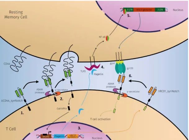
The general aim of this thesis is to develop a strategy, based on cell engineering, to detect and eliminate HIV-1 latent reservoirs that, due to its state of latency, cannot be eradicated with cART therapy. For this purpose, T cells (Jurkat cell line) will be designed to identify resting memory CD4+ T cells, since they are the primary reservoir of persistent HIV-1 infection, and upon the recognition, the cells will express a latency-reversing agent and another surface receptor. If the resting memory cells harbor the HIV-1 provirus, after the reactivation, gp120 glycoproteins will start to be expressed on the cell surface. At this point, the new receptor will detect the expression of gp120 that leads to the T cell activation, and finally destroying the infected cell (Figure 6).
The goal for this work is to build a Notch-based receptor capable of inducing the expression of reporter gene when stimulated with a target ligand – gp120.
Figure 6| Schematic overview of the presented strategy to detect and eliminate HIV-1 reservoirs.
(1.) T cells express on their surface a receptor capable of recognize resting memory CD4+ T cells by the CD44 marker. (2.) Once the receptor detects a memory cell, a conformational change leads to the cleavage of the intracellular domain (Gal4VP64) by ADAM protease and γ-secretase. (3.) Then, the Gal4VP64 travels to the nucleus binding with the response element (5xUAS), and activates the expression of flagellin and VRC01_synNotch receptor. (4.) The flagellin is secreted from the T cell and stimulates the TLR5 from the resting memory cell, which induces the activation of the NF-κB-dependent pathway. (5.) NF-κB moves into the nucleus where it activates the HIV replication resulting in the expression of gp120 on the cell surface. (6.) Finally, the VRC01_synNotch receptor detects the presence of gp120 and, after the cleavage, the T cells is activated.
Novel Strategy to Detect and Localize Latent HIV Reservoirs
11
3. Material and Methods
3.1. BACTERIAL STRAINS AND MEDIA
XL-1 Blue, XL-10 Gold and Stellar. The bacteria were grown in LB medium (10 g tryptone; 5 g yeast extract and 10 g NaCl in 1 L ddH2O) supplemented with 100 μg/mL of Ampicillin or 50 μg/mL of Kanamycin depending on the plasmid’s selectable marker. The incubations were carried out at 37°C or, in case of lentiviral plasmid propagation and cloning, 30°C to reduce recombination activity. All the bacterial strains used for DNA propagation and cloning were chemically competent, and they were transformed following the manufacturer's protocol. Genotypes are shown in Annexes.
3.2. PLASMIDS
pMDLg/pRRE, pCMV-VSV-G and pRSV-REV, encoding the packaging proteins Gag-Pol, VSV-G and Rev respectively, were used to produce lentiviral particles for cell transduction. VSV-G is an envelope protein, that is responsible for the viral packaging, which enables viral entry by mediating the binding with an LDL receptor. pHR_Gal4UAS_tBFP_PGK_mCherry is a lentiviral vector with a tBFP reporter for Gal4VP64 synNotch receptors with a constitutive mCherry (PGK promoter) where the tBFP reporter is regulated by the Gal4DBDVP64 target, 5xUAS, cloned 5’ to a minimal CMV promoter. pHR_PGK_LaG17_synNotch_Gal4VP64 (pHR_LaG17_synNotch) is a lentiviral vector for constitutive expression of the LaG17 anti-GFP nanobody synNotch Gal4VP64 receptor regulated by PGK promoter. The receptor was built by combining the LaG17 anti-GFP nanobody with the mouse Notch-1 minimal regulatory region (NM_008714 – I1427 to R1752) and the transcription activator Gal4VP64. The receptor also contains, for membrane targeting, an N-terminal CD8α signal peptide (MALPVTALLLPLALLLHAARP) and, for surface expression detection, a myc-tag (EQKLISEEDL). In the end, the receptor was cloned into a modified pHR’SIN:CSW vector. pHR_EGFPligand is a lentiviral vector for surface expressed EGFP regulated by SFFV promoter. All the plasmids above were obtained from Addgene repository.
SR2ENV is a vector for expression of gp120 at the surface, kindly given by Dr. Fabrizio Mammano. pHR_PGK_VRC01_synNotch_Gal4VP64 (pHR_VRC01_synNotch) and pHR_PGK_anti-CD44_synNotch_Gal4VP64 (pHR_anti-CD44_synNotch) were constructed by digesting the plasmid pHR_LaG17_synNotch with BstEII and MluI to remove the LaG17 nanobody cassette and the cloning VRC01 and anti-CD44-scFv fragments, respectively, with T4 ligase (Thermo Fisher). Since the myc-tag was incorporated in the LaG17
Novel Strategy to Detect and Localize Latent HIV Reservoirs
12 cassette, the VRC01 fragment has a HA-tag (YPYDVPDYA) and the anti-CD44 scFv fragment preserves the myc-tag. All plasmid maps are shown in Annexes (Figure A3 – A9).
3.3. CELL LINES AND CULTURE CONDITIONS
Human embryonic kidney cells 293T (HEK 293T) (ATCC, VA, USA) were cultured DMEM (Lonza, Basel, Switzerland) with 10% (v/v) FBS (Biowest, France), 2 mM L-Glutamine (Lonza) and PSA (100 U Penicillin, 100 µg Streptomycin, 0.25 µg Amphotericin B) (Lonza) [DMEM +/+]. These cells have a high propensity for transfection, their growth is reliable, and they express a mutant version of the SV40 large T antigen. With the expression of this allele of SV40 large T antigen, DNA that contains the SV40 origin of replication can replicate with a high copy number improving the quantity of recombinant protein or retrovirus produced by the cell.
Jurkat E6-1 cells (ATCC) are human CD4+ T lymphocytes derived from a leukemia patient. J-Lat 10.6 is a cell line widely used in the latency studies that is derived from Jurkat T cells which have a latent HIV provirus where the GFP ORF replaces Nef coding sequence and a frameshift mutation in env. These cell lines were cultured in RPMI-16 (Lonza) with 10% (v/v) FBS (Biowest), 2 mM L-Glutamine (Lonza) and PSA (Lonza) [RPMI +/+]. All the cell lines were cultured at 37°C with 5% CO2.
3.4. CELL TRANSFECTION
HEK 293T cells were transfected using Lipofectamine 3000 (Thermo Fisher Scientific, MA, USA). 1 × 10⁵ or 5 × 10⁵ cells were seeded in 24-well (15.6 mm Ø) or 6-well plates (34.7 mm Ø) (Orange Scientific, Belgium), respectively, for the day after. Following the manufacturer's protocol, 500 ng (24-well) or 2500 ng (6-well) of DNA per well were transfected. The cells were analyzed 48 hours post-transfection.
3.5. VIRAL PRODUCTION
HEK 293T were seeded in 6-well plates at a density of 1.2 × 10⁶ cells per well. Cells were transfected using Lipofectamine 3000 with 1.5 μg of transfer plasmid, 1.2 μg of pMDLg/pRRE, 0.6 μg of pRSV-REV and 0.3 μg of pCMV-VSV-G. The cells were left to incubate for 6 hours before the medium was replaced with fresh DMEM +/+ and then incubated overnight. At 24 hours post-transfection, the supernatant was collected and replaced with 2 mL of pre-warmed medium, and at 56 hours the supernatant was again collected. In the end, the supernatant was aliquoted and placed at -80°C. The viruses were quantified by the HIV-1 p24CA Antigen Capture Assay Kit (AIDS and Cancer Virus Program, Frederick National
13 Laboratory, Frederick, MD, USA) following the manufacturer’s instructions. To determine the lentiviral transduction units (TU) the ratio used was 1 pg of p24 protein for 10-100 TU.
3.6. CELL TRANSDUCTION
1 × 10⁵ Jurkat cells were transduced using different quantities of lentivirus in RPMI +/+ supplemented with polybrene (Hexadimethrine bromide – Sigma-Aldrich, MO, USA) at 8 μg/mL. The cell cultures were spinoculated at 1 200 × g for 90 minutes at 22°C and then incubated for 2 hours (O’Doherty et al., 2000). After that, the medium was replaced with fresh RPMI +/+ and left to incubate overnight.
3.7. CO-CULTURE OF CELLS
2 × 10⁵ receiver and sender cells were plated in a round bottom 96-well plate at a 1:1 ratio and centrifugated at 400 × g for 1 minute to promote the interaction between cells. After 24 hours of incubation, the cells were analyzed for tBFP expression by flow cytometry.
3.8. FLAGELLIN STIMULATION
In a 6-well plate, different concentrations (0, 5 and 10 ng/mL) of flagellin were used to stimulate 1 × 10⁵ Jurkat E6-1 or J-Lat 10.6 cells. The cells were incubated for 24h and then, the Jurkat E6-1 cells were prepared as explained in Flow Cytometry section, where the primary antibodies used were mouse anti-human Fas and rabbit anti-FasL (Santa Cruz Biotechnology, TX, USA) and the secondary were goat anti-mouse IgG–FITC (Sigma-Aldrich) and anti-rabbit IgG-FITC (Santa Cruz Biotechnology). The stimulated J-Lat 10.6 cells were directly analyzed for GFP expression.
3.9. FLOW CYTOMETRY
The cells were collected and washed thrice with cold PBS + 0.5% BSA and then were incubated with a primary antibody (1 µg per 1 × 10⁶ cells) at room temperature for 30 minutes. For the synNotch receptors, pHR_LaG17_synNotch has a myc tag and pHR_VRC01_synNotch has an HA tag, both in the extracellular domain, the primary antibodies used are c-myc Tag Monoclonal Antibody (Thermo Fisher) and HA Tag Monoclonal Antibody (2-2.2.14), HRP (Thermo Fisher). Cells transduced with pHR_EGFPligand vector were directly analyzed since they already express GFP, and with SR2ENV vector, the cells were stained with VRC01 (NIH AIDS Research and Reference Reagent Program, USA). The cells were washed three times again with cold PBS + 0.5% BSA and subsequently incubated with the
Novel Strategy to Detect and Localize Latent HIV Reservoirs
14 secondary antibody conjugated to FITC for 30 minutes in the dark. The primary antibodies used, apart from VRC01, were derived from mouse; VRC01 was derived from a human host. In these cases, the mouse primary antibodies were labeled with goat anti-mouse IgG–FITC (Sigma-Aldrich) and VRC01 with goat anti-Human IgG–FITC (Thermo Fisher). Finally, the cells were washed thrice with cold PBS + 0.5% BSA where, after the last wash, 10 µL of propidium iodide (10 µg/mL) was added to stain the dead cells. After the staining, all the cells were analyzed in Guava easyCyte™ 5HT (Merck Millipore, MA, USA). To evaluate the transcription activation, expression of tBFP, mCherry and GFP were analyzed directly in BD LSRFortessa™ X-20 (BD Biosciences, CA, USA). All flow cytometry data were analyzed with FlowJo software (TreeStar).
15
4. Results
4.1. SYN-NOTCH RECEPTORS TRANSDUCTION
SynNotch receptors (Figure 5B) maintain the core regulatory domain from the wild-type Notch and the intra- and extracellular domains are replaced by synthetic transcriptional activation domains and diverse recognition domains (for example, single-chain antibodies - scFv), respectively (Roybal, Williams, et al., 2016). In cell-based therapies, instead of triggering T cell activation like CARs do, which is unspecific, the synNotch receptors, with their customized transcriptional activator domain, only activates the expression of specific target genes. This control over the inputs and outputs of the synNotch pathway allows the use of more than one receptor without having interactions between them because they share no common signaling intermediates.
Jurkat E6-1 cells were firstly transduced with a reporter vector for the synNotch receptors (pHR_Gal4UAS_tBFP_PGK_mCherry), creating a new cell line (JKT tBFP_mCherry) in collaboration with Joana Ministro (Technophage). Then, these cells were again transduced with vectors for the expression of the synNotch receptors (pHR_LaG17_synNotch and pHR_VRC01_synNotch). After 48 hours, the cells were prepared and stained with antibodies that target protein tags present in the extracellular domain of the receptors, myc tag for the LaG17_synNotch receptor and HA tag for the VRC01_synNotch receptor. Following that, we analyzed the cells by flow cytometry (Figure 7). Increasing quantities of virus were used to
LaG17_synNotch 0 pg/mL 36.5 pg/mL 73 pg/mL 182.5 pg/mL 365 pg/mL 730 pg/mL p24 c on c ent rat io n (pg /m L) VRC01_synNotch 0 pg/mL 88 pg/mL 176 pg/mL 880 pg/mL 1320 pg/mL 1760 pg/mL p24 c on c ent rat io n (pg /m L)
Figure 7| Assessment of synNotch receptors expression. Different concentrations of lentiviruses were used to
transduce JKT tBFP_mCherry cells. After 48 hours of incubation, the cells were immunostained and analyzed by flow cytometry.
Novel Strategy to Detect and Localize Latent HIV Reservoirs
16 transduce the cells, however, as we can observe in Figure 7, none of them resulted in receptor-expressing cells.
With the intention to understand the absence of receptor expression, the pHR_LaG17_synNotch lentiviruses were produced with different lentiviral plasmids (pMDLg/pRRE, pCMV-VSV-G and pRSV-REV) kindly provided by Technophage. Subsequently, the JKT tBFP_mCherry cells were transduced with the fresh viruses, and after the immunostaining, the cells were analyzed by flow cytometry (Figure 8). Higher viral concentrations were used than the previous assay which resulted in an increased LaG17_synNotch receptor expression. With lentivirus at a p24 concentration of 39.5 ng/mL, 50.4% of the cells were expressing the receptor on their surface.
After we observed expression of the synNotch receptor, we transduced JKT tBFP_mCherry cells with LaG17_synNotch and VRC01_synNotch lentiviruses, and Jurkat E6-1 with EGFP ligand and gp120 ligand lentiviruses. To determine the optimal quantity of lentivirus we used different amounts of viral supernatant. As represented in Figure 9, the expression of the synNotch receptors and gp120 ligand increased when using 250 μL of viral supernatant and decreased with 500 μL, whereas the expression of EGFP ligand increased with higher volumes of viral supernatant.
0 n g /m L 3 ,9 n g /m L 7 ,8 9 n g /m L 1 9 ,7 n g /m L 3 9 ,5 n g /m L 0 1 0 2 0 3 0 4 0 5 0 6 0 7 0 8 0 p 2 4 c o n c e n t r a t i o n ( n g / m L ) % of La G 1 7 _ s y nN ot c h + c e lls 4 . 7 6 1 1 . 1 1 6 . 2 3 0 . 1 5 0 . 4
Figure 8| Assessment of LaG17_synNotch receptor expression. Different amounts of pHR_LaG17_synNotch
lentivirus were used to transduce JKT tBFP_mCherry cells. After 48 hours of incubation, the cells were immunostained for the myc tag and followed with the analysis by flow cytometry.
p2 4 c onc ent ra tio n (ng /m L) 39.5 ng/mL 19.7 ng/mL 7.89 ng/mL 3.9 ng/mL 0 ng/mL
17 L a G 1 7 _ s y n N o tc h E G F P _ lig a n d V R C 0 1 _ s y n N o tc h g p 1 2 0 _ lig a n d 0 1 0 2 0 3 0 4 0 5 0 6 0 7 0 8 0 % o f p o si ti ve c el ls 0 µ L 1 0 0 µ L 2 5 0 µ L 5 0 0 µ L 19.5 28.727.4 35.4 50.2 15.8 21.6 13.4 65.1 20.8 13.3 14.1
Figure 9| Assessment of synNotch receptors and respective ligands expression. For each transduction, were
used different quantities of viral supernatant. The p24 concentration of each lentivirus was 22.6 ng/mL for LaG17_synNotch receptor, 15.9 ng/mL for EGFP ligand, 34.0 ng/mL for VRC01_synNotch receptor and 45.6 ng/mL for gp120 ligand. After 48 hours of incubation, the cells were analyzed by flow cytometry. All except the EGFP ligand were immunostained against the tags present on the extracellular domain.
Novel Strategy to Detect and Localize Latent HIV Reservoirs
18
4.2. SENDER LIGANDS TRANSFECTION
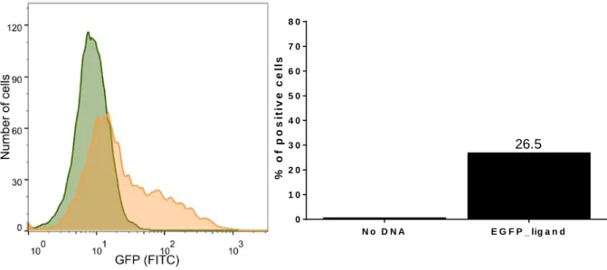
In order to subsequently evaluate the proper functioning of the LaG17_synNotch receptor, we transfected cells with the surface-expressed antigen, EGFP ligand, generating cells that will present the antigen to receptor cells (sender cells). HEK 293T cells were transfected with pHR_EGFPligand, and after 48 hours they were analyzed by flow cytometry to acquire the number of EGFP+ cells. As shown in Figure 10, 26.5% of the transfected cells were expressing EGFP.
Aiming to increase the expression of EGFP ligand we tested different conditions where we varied the number of cells (0.5 × 10⁵, 1 × 10⁵ and 2 × 10⁵ cells) and the volume of Lipofectamine 3000 (0.75 and 1.5 μL). As demonstrated in Figure 11, using 0.5 × 10⁵ cells and 1.5 μL of Lipofectamine 3000 had the highest EFGP ligand expression. When we transfected 2 × 10⁵ cells with both volumes of the reagent, the percentage of dead cells (PI+ cells) was more than 50%, although, when a lesser number of cells was used, the percentage of dead cells was smaller. It is important to note that there is not a significant difference in ligand expression between the two volumes of Lipofectamine 3000.
N o D N A E G F P _ lig a n d 0 1 0 2 0 3 0 4 0 5 0 6 0 7 0 8 0 % o f p o s it iv e c e ll s 26.5
Figure 10| Assessment of EGFP ligand expression in HEK 293T. 𝟐𝟐 × 𝟏𝟏𝟏𝟏𝟓𝟓 HEK 293T cells were plated in a
24-well plate, 24 hours before the transfection. 500 ng of DNA and 1.5 μL of Lipofectamine 3000 were added to unsupplemented DMEM medium which, thereafter, was added to the cells. 6 hours post-transfection, the medium was replaced with fresh DMEM +/+. After 48 hours of incubation, the cells were analyzed by flow cytometry.
19 0 .7 5 1 .5 0 1 2 3 4 5 6 7 8 9 1 0 1 1 1 2 L ip o f e c t a m in e 3 0 0 0 v o lu m e ( µ L ) Fol d C ha ng e 0 .5 x 1 05c e lls 1 x 1 05c e lls 2 x 1 05c e lls N o D N A 10.6 10.3 5.4 11.0 9.9 4.5 1.0 1.0
GFP
PI
Figure 11| Optimization of EGFP ligand transfection. HEK 293T cells were plated in a 24-well plate, 24 hours
prior to transfection. For every well, 500 ng of DNA were added with 0.75 or 1.5 μL of Lipofectamine 3000 to unsupplemented DMEM medium and then added to the cells. After 6 hours, the medium was replaced by fresh DMEM +/+ medium, and after 48 hours, propidium iodide was added to the cells in PBS + 0.5% BSA at a final concentration of 0.5 μg/mL and the cells were directly analyzed by flow cytometry.
No DNA
0.5 × 10⁵ cells 1 × 10⁵ cells 2 × 10⁵ cells
0.5 × 10⁵ cells 1 × 10⁵ cells 2 × 10⁵ cells
1. 5 μL of Li p of ec tam ine 30 00 0. 75 μL of Li pof ec tam ine 30 00
Novel Strategy to Detect and Localize Latent HIV Reservoirs
20
4.3. RECEIVER AND SENDER CELLS CO-CULTURE
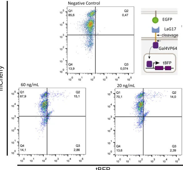
After generating the receiver (LaG17_synNotch+) and sender (EGFP+) cells, to assess the functionality of the synNotch receptor, both cells were co-cultured in a 1:1 ratio for 24 hours. First, we tested the interaction between Jurkat and HEK 293T cells. Jurkat cells expressed the LaG17_synNotch receptor and the HEK 293T cells expressed the EGFP ligand. In the first attempt, the cells appeared to be dead after the 24 hours of incubation. As we can observe in
Figure 12, the presence of EGFP+ cells did not activate the receiver cells since the expression
level of tBFP of the cultures, with and without sender cells, are the same. Nevertheless, the percentage of EGFP+ cells was very low in the LaG17_synNotch+ + EGFP+ co-culture.
In the second attempt, new JKT tBFP_mCherry cells were transduced with lentivirus with two different p24 concentrations (20 and 60 ng/mL) to express LaG17_synNotch receptors, and HEK 293T cells were transfected to express EGFP ligand (See Figure A1 and Figure A2 in Annexes). The cells transduced with lentivirus with 60 and 20 ng/mL of p24 presented 37.5% and 57.2% of positive cells for LaG17_synNotch receptors, respectively. Likewise, the receiver and sender cells were co-cultured in RPMI +/+ medium with a 1:1 ratio of the receiver to sender cells for 24 hours. As depicted in Figure 13, the stimulation with EFGP+ cells resulted in the activation of the receiver cells which led to the expression of tBFP, 15.1% for the cells transduced with lentivirus at 60 ng/mL of p24 and 14.0% for the cells transduced with lentivirus at 20 ng/mL of p24.
Figure 12| Assessment of LaG17_synNotch receptor activation. 𝟏𝟏 × 𝟏𝟏𝟏𝟏𝟓𝟓 of receiver and sender cells were co-cultured for 24 hours in a 1:1 ratio. Jurkat LaG17_synNotch+ were combined with sender cells EGFP positive and negative. After the incubation period, the cells were analyzed by flow cytometry. In the right plot, the orange color represents the LaG17_synNotch+ + EGFP- co-culture and the blue color represents the LaG17_synNotch+ + EGFP+ co-culture. LaG17_synNotch+ LaG17_synNotch+ + EGFP+ LaG17_synNotch+ + EGFP- 0.08% 7.30% 0.33% EGFP LaG17 Gal4VP64 tBFP
21 Previously, we studied the interaction of receiver and sender cells which had different systems of culture, receiver cells were in suspension and sender cells were adherent to the plate. In order to inquiry a more realistic scenario, following that we decided to study the interaction between receiver and sender cells in a suspension culture and the other synNotch receptor against gp120 glycoprotein (VRC01_synNotch). With the cells referred in Figure 9, the receiver cells were co-cultured with the respective sender cells in a 1:1 ratio and then a spin down at 400 x g for 1 minute was done to ensure the contact between cells. When the synNotch cells were stimulated with its specific ligand, the expression of tBFP increased (Figure 14) as larger volumes of viral supernatant were used. When co-cultured with negative sender cells, the LaG17_synNotch+ cells presented a residual expression of tBFP, however, the VRC01_synNotch+ cells had a similar but smaller expression of tBFP when compared to VRC01_synNotch+ + gp120+ co-culture. As expected, cells that did not express the synNotch receptors had no tBFP expression when stimulated with EGFP+ or gp120+ sender cells.
Negative Control 20 ng/mL 60 ng/mL
m
C
he
rr
y
tBFP
Figure 13| Assessment of LaG17_synNotch activation by EGFP+ cells. 𝟐𝟐 × 𝟏𝟏𝟏𝟏𝟓𝟓 of receiver and sender cells
were co-cultured for 24 hours in a 1:1 ratio. JKT tBFP_mCherry cells were transduced with lentivirus with 20 and 60 ng/mL of p24. Jurkat LaG17_synNotch+ were combined with sender cells EGFP positive and negative. After the incubation period, the cells were analyzed by flow cytometry. See also Figure A1 and Figure A2.
EGFP LaG17 Gal4VP64
Novel Strategy to Detect and Localize Latent HIV Reservoirs 22 E G F P+ g p 1 2 0+ E G F P+ E G F P- g p 1 2 0+ g p 1 2 0 -0 .-0 0 .5 1 .0 1 .5 2 .0 2 .5 3 .0 3 .5 4 .0 4 .5 5 .0 5 .5 6 .0 tB FP e x pr e s s ion ( % ) 1 0 0 µ L 2 5 0 µ L 5 0 0 µ L 0.03 0.07 0.06 0.04 0.03 0.03 2.23 3.81 5.30 0.29 0.590.52 0.74 1.93 2.29 0.68 1.72 1.99
Figure 14| Assessment of tBFP expression after the activation of the receiver cells, LaG17_synNotch
and VRC01_synNotch. The viral supernatant volume indicated was used to transduce each cell type. The
p24 concentration of each lentivirus was 22.6 ng/mL for LaG17_synNotch receptor, 15.9 ng/mL for EGFP ligand, 34.0 ng/mL for VRC01_synNotch receptor and 45.6 ng/mL for gp120 ligand.
LaG17_synNotch VRC01_synNotch No synNotch receptor EGFP LaG17 Gal4VP64 tBFP gp120 VRC01 Gal4VP64 tBFP
23
4.4. FLAGELLIN STIMULATION
Lastly, we wanted to study the influence of flagellin on the expression of Fas receptor and its ligand FasL, which are associated with the apoptosis pathway. To understand this, Jurkat E6-1 cells were stimulated with two concentrations of flagellin, 5 and 10 ng/mL, for 24 hours. After that time, the cells were immunostained to detect the expression of the Fas receptor and its ligand, FasL, and were analyzed by flow cytometry. In Figure 15A we can observe that the stimulation with different concentrations of flagellin did not make any difference in the expression of these proteins. However, when the flagellin stimulated J-Lat 10.6 cells, more than 80% were activated and expressed GFP (Figure 15B).
Fl age lli n co nc en tr at io n (n g/ m L) 10 ng/mL 5 ng/mL 0 ng/mL No staining Fl age lli n co nc en tr at io n (n g/ m L) 10 ng/mL 5 ng/mL 0 ng/mL No staining Fl age lli n co nc en tr at io n (n g/ m L) No staining 5 ng/mL 10 ng/mL B) A) FasL Fas 82.0% 80.8% 0.98%
Figure 15| Assessment of FasL and Fas expression, and J-Lat 10.6 cells reactivation after flagellin
stimulation. A) 𝟏𝟏 × 𝟏𝟏𝟏𝟏𝟓𝟓 Jurkat E6-1 cells were stimulated with flagellin at concentration of 0, 5 and 10 ng/mL. The
cells were incubated with flagellin for a period of 24 hours and afterward immunostained against Fas and FasL to be analyzed by flow cytometry. B) 𝟏𝟏 × 𝟏𝟏𝟏𝟏𝟓𝟓 J-Lat 10.6 cells were stimulated with 5 and 10 ng/mL of flagellin for
Novel Strategy to Detect and Localize Latent HIV Reservoirs
25
5. Discussion
Latent viral reservoirs have been the major obstacle to achieve eradication of HIV-1 infection. These reservoirs can be found primarily in resting memory CD4+ T cells which have the HIV-1 DNA integrated into their genome. The current antiretroviral therapies available depend on active viral replication to be efficient, thus they are incapable of eliminating the latent cells from the host. For that reason, it is crucial to develop a strategy that aims to destroy these latent reservoirs. Here, we developed a receptor capable of inducing expression of a reporter gene upon stimulation with infected cells expressing gp120. This receptor was based on Notch receptors for their need of cell-to-cell contact to be activated and the specific signal transduction mechanism. In this pathway, the signal is transduced via an intracellular domain that, after the stimulation of the extracellular domain, is cleaved and travels to the nucleus where it regulates gene expression. The intracellular domain can be customized to activate or repress the expression of specific genes, and the extracellular domain can be modified to a specific receptor, making possible the design of signal networks that are user-specified. The synNotch receptor we developed (VRC01_synNotch), when activated, cleaves and releases the Gal4VP64 domain, which moves to the nucleus where activates the expression of a fluorescent reporter, tBFP. The extracellular domain is a VRC01_scFv which recognizes gp120 glycoprotein. For the positive control, we used a synNotch receptor developed by Morsut et al., LaG17_synNotch, which, upon EGFP ligand stimulation, also releases a Gal4VP64 domain (Morsut et al., 2016).
The first attempt to express LaG17_synNotch receptors in T cells (Jurkat cells) was unsuccessful with no receptor expression. Moreover, after other attempts, we started to troubleshoot to identify what was hindering the expression of the synNotch receptor. In the end, we identified the purity of plasmids as the problem, caused by a phage contamination in the laboratory. New lentiviral vectors (pMDLg/pRRE, pCMV-VSV-G and pRSV-REV) and a DsRed reporter vector were used to test the production of lentiviruses. After transducing cells with the lentiviruses produced with these new vectors, they were analyzed and proved to be successfully transduced (data not shown). With these lentiviral vectors, we produced new LaG17_synNotch lentiviruses and then transduced cells that, after the evaluation, showed to express the synNotch receptor on the cell surface.
To assess the functionality of the synNotch receptor, sender cells had to be generated by transfecting HEK 293T cells with a vector for EGFP surface ligand expression and then co-cultured with LaG17_synNotch+ cells. The co-culture did not demonstrate any expression of tBFP, meaning that the receptors did not activate or the intracellular domain was incapable
Novel Strategy to Detect and Localize Latent HIV Reservoirs
26 of activating the gene expression. For that reason, we attempted again with new transduced cells with two viral concentrations (20 and 60 ng/mL of p24), and new transfected EGFP+ cells which we mixed them in a co-culture. Our results manifested a tBFP expression of 14% and 15.1% for 20 and 60 ng/mL of p24, respectively, therefore proving the proper functioning and activation of the LaG17_synNotch receptors. The low level of tBFP expression can be explained by the low levels of LaG17_synNotch+ (20 ng/mL: 37.5%; 60 ng/mL: 57.2%) and EGFP+ (33.5%) cells. Approximately 50% of cells could be activated to express tBFP, and just 30% could stimulate the receiver cells, and since the receptor needs cell-to-cell interaction, as the receiver cells were in suspension and the sender cells were adherent, the contact between cells was very limited. Knowing that, after the receptor cleavage upon interaction with the antigen, the extracellular domain bound to the antigen is endocytosed, meaning that the EGFP ligand can only activate a synNotch receptor once. Therefore, the tBFP expression similarity between the two concentrations used to transduce the receptor cells could be justified by the shortage of EGFP+ cells. To optimize the receptor activation, we decided to use Jurkat cells for both receiver and sender cells and promote the cell-to-cell contact with a spin down before incubation.
Since the LaG17_synNotch receptor is functional, we included VRC01_synNotch receptor in the activation assay. New cells were generated by transduction to express LaG17_synNotch, VRC01_synNotch, EGFP ligand or gp120 ligand, but when examined, the cells did not have a high expression level. Even so, we prepared the co-cultures and analyzed them to find that, while the percentage of tBFP+ cells was small in both co-cultures, it increased with the increment of synNotch+ cells and positive sender cells. In parallel with the negative control co-cultures, the LaG17_synNotch+/EFGP+ co-culture had a clear receptor activation unlike the LaG17_synNotch+/EFGP- co-culture, however, the VCR01_synNotch+/gp120+ co-culture had similar activation levels to VCR01_synNotch+/gp120- co-culture nevertheless, should be noted that the first had a higher expression of tBFP. Our results, as expected, demonstrated the absence of tBFP expression in synNotch- cells attesting the requirement of synNotch receptors activation to induce the expression of tBFP. Despite the fact that the cells had more cell-to-cell interaction than the Jurkat and HEK 293T cells co-culture, the activation levels were inferior. This outcome can be justified by the poor transduction of the receiver and sender cells. Every transduction was done with 100, 250 or 500 μL of viral supernatant and, with the increment of viral concentration, the expression of the receptors and ligands improved until the highest volume was used, leading to a small decline, with the exception of EGFP+ cells that greatly improved with all viral supernatant volumes. It is important to note that the expression of gp120 did not vary significantly with the different viral concentrations, for what can be the reason for the small discrepancy between the VRC01_synNotch co-cultures with
27 positive or negative sender cells. In order to improve the expression of tBFP in both co-cultures, the receiver and sender cells must be sorted to increase the number of cells expressing the synNotch receptors and ligands, generating new cell lines. Consequently, higher levels of tBFP+ cells will be obtained after the stimulation with target cells.
Following the strategic plan, to study a potential cytotoxic and latency-reversing agent, we stimulated Jurkat E6-1 and J-Lat 10.6 cells with flagellin. The J-Lat 10.6 cells are a HIV-1 latency model that expresses GFP, instead of viral particles, when reactivated. We evaluated if flagellin could induce the expression of Fas and FasL, which would result in cell apoptosis by the activation of Fas/FasL pathway (Waring & Müllbacher, 1999), and verified that the stimulation with flagellin did not induce the expression. Therefore, flagellin cannot be used to induce the death of the infected cell. However, when the J-Lat 10.6 cells were stimulated with flagellin, more than 80% of cells expressed GFP meaning they were reactivated from their latency state. For these reasons, flagellin can be potentially used as an LRA to promote, if the cell is latently infected, the reactivation and hence the expression of gp120.
We also developed a synNotch receptor against CD44, which is a resting memory T cell marker (Baaten et al., 2010; Rosenblum et al., 2016), but we did not test this receptor in this work. The idea is to use this receptor in the future to detect resting memory T cells, and upon detection, the expression of flagellin and VCR01_synNotch is induced. The flagellin will activate the latent cells, resulting in the expression of gp120. Then, the VRC01_synNotch recognizes the surface-expressed gp120 and induces T cell activation to kill the infected cell. Here we developed a receptor capable of regulating gene expression when stimulated with gp120+ cells. A recent study stated that CD32a is a T cell maker that is highly induced in HIV-1-infected resting CD4+ T cells, which can be a promising target to identify latent reservoirs and efficiently destroy them (Descours et al., 2017).
Novel Strategy to Detect and Localize Latent HIV Reservoirs
29
6. Concluding Remarks and Future Perspectives
cART therapy has been, for most patients, the only treatment that allows them to have a normal life. However, they must always live dependent on their therapy medicines. In order to increase the life quality for these people, it is crucial to develop a cure for the HIV-1 infection.
Here, we develop a receptor that is competent to detect the presence of a specific antigen, gp120 in this case, and when that happens, the intracellular domain is released and regulates a specific gene expression (tBFP expression). We verified that synNotch receptors are very dependent on cell-to-cell interactions to be activated. VRC01_synNotch it is part of a strategic plan that aims to destroy the latently infected T cells. However, the optimization of this strategy is critical for further understand and improve efficiency.
In the future, the developed cells expressing the synNotch receptors and its ligands must be sorted to create cell lines that will improve the interaction between these receptors and respective ligands. With new studies, novel markers have been demonstrated to identify latent reservoirs, which is the case of CD32a. This marker can be used to construct a new synNotch receptor to increase the specificity of this therapeutic approach, which would be anti-CD32a and would induce the expression of flagellin and the VCR01_synNotch receptor. The last receptor, instead of inducing the expression of a reporter gene once activated, would result in the activation of the T cell becoming cytotoxic and promoting the death of the infected cell. In the end, we will have a T cell with a combination of two customized-sensing circuits that will be able to have a ‘shock and kill’ approach to eradicate the latent HIV-1 reservoirs.
Novel Strategy to Detect and Localize Latent HIV Reservoirs
31