T
HE EFFECTS OF LEUCINE METABOLITES ON PERFORMANCE,
BODY COMPOSITION AND BIOCHEMICAL MARKERS OF MUSCLE DAMAGE ANDINFLAMMATION
FILIPE JOSÉ NOIA TEIXEIRA
Orientador: Prof.ª Doutora Cristina Paula Fidalgo de Negreiros Monteiro
Bento
Coorientador: Prof.ª Doutora Ana Catarina Francisco Nunes Matias
Tese especialmente elaborada para obtenção do grau de Doutor em
Motricidade Humana na especialidade de Fisiologia do Exercício
T
HE EFFECTS OF LEUCINE METABOLITES ON PERFORMANCE,
BODY COMPOSITION AND BIOCHEMICAL MARKERS OF MUSCLE DAMAGE ANDINFLAMMATION
F
ILIPEJ
OSÉN
OIAT
EIXEIRAOrientador: Prof.ª Doutora Cristina Paula Fidalgo de Negreiros Monteiro Bento Coorientador: Prof.ª Doutora Ana Catarina Francisco Nunes Matias
Tese especialmente elaborada para obtenção do grau de Doutor em Motricidade Humana na especialidade de Fisiologia do Exercício
Júri:
Presidente: Doutor Francisco José Bessone Ferreira Alves, Professor Catedrático e Presidente
do Conselho Científico da Faculdade de Motricidade Humana da Universidade de Lisboa
Vogais:
-Doutor Stuart Martin Phillips, Canada Research Chair (Tier 1), McMaster University, McMaster Department of Kinesiology, Hamilton, Ontario, Canada
-Doutora Analiza Mónica Lopes Almeida Silva, Professora Auxiliar com Agregação da Faculdade de Motricidade Humana da Universidade de Lisboa
-Doutor Vítor Hugo da Costa Gomes Moreira Teixeira, Professor Auxiliar da Faculdade de Ciências da Nutrição e Alimentação da Universidade do Porto
-Doutor Paulo Alexandre Silva Armada da Silva, Professor Auxiliar da Faculdade de Motricidade Humana da Universidade de Lisboa
-Doutora Cristina Paula Fidalgo Negreiros Monteiro Bento, Professora Auxiliar da Faculdade de Motricidade Humana da Universidade de Lisboa
A realização deste trabalho não teria sido possível sem o incansável apoio e orientação da Prof.ª Doutora Cristina Monteiro e da Prof.ª Doutora Catarina Matias. As mesmas foram exemplares desenvolvendo todos os esforços para que os trabalhos chegassem a “bom porto”. A Prof.ª Doutora Cristina Monteiro sempre me tentou ajudar com bons conselhos e suporte, moderando em muito a minha abordagem menos ponderada em relação a certos assuntos. Também agradeço todo o seu expertise em análises clínicas, inflamação e imunologia. É uma referência para mim. A Prof.ª Doutora Catarina Matias é aquilo que posso chamar uma autentica “força de trabalho e capacidade”. Foi o meu SOS em inúmeros momentos deste projecto, uma verdadeira ferramenta de soluções. Não poderia ter melhor aconselhamento em relação à composição corporal (DXA, BIS etc.), estatística e até à simples criação de gráficos! Às duas o meu sincero muito obrigado, por tudo. Impossível chegar até aqui sem o vosso apoio e conhecimento.
Ao meu mentor e supervisor externo Professor Doutor Catedrático Stuart M. Phillips. Quando há uns anos atrás comecei a interessar-me pela ingestão proteica havia um nome que admirava e que surgia como pioneiro no campo. Nunca pensei poder trabalhar com esse nome um dia ou ver artigos partilhados com este nome, nas melhores revistas de Medicina Desportiva do mundo. O Professor Stuart Phillips (que sempre me pediu para o tratar por Stu, desde a nossa primeira troca de e-mails) mais do que toda a ciência que me ensinou deixa-me duas grandes lições: 1) podemos ser o maior investigador do mundo no nosso campo, sendo humildes e simpáticos; 2) a integridade como cientista não tem preço. Podia elogiar as suas capacidades científicas como a
indexados no PUBMED/MEDLINE, mas vou focar-me no que pouca gente conhece: O Stu é uma excelente pessoa. Cada vez que escrevia algo com menos qualidade, nunca me criticou. Sempre disse: “sabes… eu acho que aqui consegues fazer melhor”. Sempre me incentivou a melhorar e sempre me ensinou a ciência pela positiva. Dizem que não devemos conhecer os nosso heróis, discordo totalmente. Thanks for everything Stu!
À Profª Doutora Maria João Valamatos por todo o seu apoio e conhecimento. Se hoje sou menos ignorante em relação a plataformas de salto vertical, ultrassom, testes de força isométrica e Wingate devo-lhe a si. Muito obrigado por tudo.
À Prof. Doutora Joana Reis, muito obrigado por todos os conselhos e apoio. Se hoje sou menos ignorante em relação aos conceitos de VO2max e cinética do O2 devo-lhe
a si. Outra parte importante do apoio da Joana é o seu conhecimento geral de Fisiologia do Exercício, impressionante (ou usando uma sua expressão: fortíssimo). Para quem não conhece a Joana, a sua boa disposição e sentido de humor são contagiantes.
Ao meu amigo Francisco Tavares, o expert no treino da força. Foi um prazer amigo, obrigado por todo o apoio e conhecimento nesta vertente. És uma referência para mim. Obrigado pela disponibilidade, conhecimento e amizade.
Às minhas colegas Ana Batista e Ana Oliveira, obrigado pela disponibilidade e responsabilidade no apoio dos inúmeros scans no DXA, BIS e dinamometria. Desejo-vos as maiores felicidades e mais uma vez muito obrigado.
Aos meus colegas de laboratório Christophe Domingos e Paulo Pires. O Christophe Domingos incentivou-me desde os primeiros seminários e foi quem não me deixou desistir na fase inicial do doutoramento. É um amigo e um investigador com grande potencial e a quem agradeço por todo o apoio no meu projecto de doutoramento.
gargalhadas.
Ao Professor Doutor Catedrático Francisco Alves, pelos seus ensinamentos ao longo do doutoramento na área da Fisiologia. Um verdadeiro expert no campo! Agradeço também pela oportunidade de ter desenvolvido este projecto sob a sua coordenação.
Ao Professor Doutor Catedrático Luís Bettencourt Sardinha, obrigado pela colaboração do Laboratório de Exercício e Saúde. Foi absolutamente fundamental. Agradeço também a sua revisão crítica aos manuscritos submetidos.
Aos diversos Professores que nos inspiraram e ensinaram ao longos dos diversos seminários do Doutoramento. Muito obrigado.
Aos colegas Scott Howell e Richard Kones que ofereceram o seu expertise médico no nosso estudo de caso. Foi um prazer colaborar convosco. Muitas mais colaborações virão!
Ao Heitor Oliveira Santos, um estimado colega com o qual tive o prazer de colaborar em vários artigos de revisão durante este doutoramento. Muito obrigado cara, valeu!
Last but not least…!
Às minhas famílias (o plural é propositado), aos meus pais, ao meu irmão Sandro, aos meus sobrinhos. Hoje, mais do que nunca, reconheço e valorizo a importância da família. Agradeço também à mulher da minha vida, a minha verdadeira companheira Vanessa. És a melhor pessoa que conheço, fonte de inspiração e motivo de orgulho para
mim. Sem o teu amor incondicional nada disto seria possível. Obrigado por seres quem és e por teres a inesgotável paciência para aturar o meu mau feitio.
À Rita Figueiredo pelos anos de colaboração à frente da Body Temple, Lda e da Proviant Labs. Teria sido impossível efectuar este doutoramento sem o teu apoio e o natural apoio destas empresas. Obrigado por terem permitido que mantivesse a isenção durante este doutoramento. Rita, sei que tens uma batalha dura pela frente, mas não tenho qualquer dúvida que terás sucesso. Coragem!
Aos meus amigos científicos e não científicos. Uma palavra de agradecimento ao meu amigo Vítor Hugo Teixeira pelos seus conselhos, apoio e amizade. Uma fonte de conhecimento e alguém que me incentiva a ser melhor. Aos meus restantes amigos “científicos” pelo constante debate e troca produtiva de ideias: Pedro Carvalho, António Pedro Mendes, Mário Simões, Gabriel Martins, César Leão e Mónica Sousa. Agradeço também a todas as instituições que confiam no meu trabalho e me desafiam constantemente a ensinar mais e melhor: Bwizer, Manz e Clínica das Conchas.
Aos meus amigos “não científicos” (embora gostem também de ciência), Pablo Vigarinho e Rodrigo Ribeiro. Obrigado pela vossa amizade e ajuda.
Por fim ao meu irmão Joaquim, quis o infortúnio que não estivesses cá neste momento. Se hoje me interesso por ciência devo-te a ti. Como químico foste a maior e melhor influencia científica que podia ter. Obrigado por tudo e obrigado por teres tomado conta de mim. Este trabalho de doutoramento é dedicado à tua memória!
Table of Contents
Abbreviations ... XIII Abstract ... XIX Resumo ... XXI CHAPTER 1 ... 23 General Introduction ... 23 1.1 Dissertation Structure ... 251.2 List of articles and conference abstracts as first author ... 26
CHAPTER 2 ... 29
Literature Review ... 29
2.1 Leucine ... 31
Metabolism ... 32
Action on muscle protein synthesis ... 35
Action on muscle protein degradation ... 36
Energy metabolism ... 37
Glucose homeostasis ... 37
Immunomodulatory role ... 38
Role in inflammation ... 39
Human research on performance and body composition ... 40
2.2.1 β-hydroxy-β-methylbutyrate (HMB) ... 43
Metabolism ... 44
Bioavailability between different commercial forms of HMB ... 45
Safety ... 47
Duration of supplementation, dose and timing ... 48
Proposed mechanisms of action ... 49
Improvements in body composition, strength, power and blood markers .. 54
Older and diseased populations ... 66
2.2.2 Leucic acid (α-HICA) ... 70
Overview... 70
Human research studies ... 71
Other leucine metabolites... 72
2.3 Aim of the investigation ... 73
2.4 References ... 74
CHAPTER 3 ... 91
Methodology... 91
3.1 Study design and sampling ... 93
Ethics and general study design ... 93
Study 1, 2 and 3 (chapters 4, 5 and 6)... 93
Study 4 (chapter 7) ... 95
Participants ... 95
Study 1, 2 and 3 (chapter 4, 5 and 6) ... 95
Study 4 (chapter 7) ... 97
3.4 Muscle strength and power assessments ... 101
Maximal isometric forearm strength ... 101
Maximal isometric leg Strength ... 101
Countermovement jump, 1 repetition maximum and Wingate test ... 102
3.5 Body composition measurements ... 104
Anthropometry ... 104
Bioelectrical impedance spectroscopy ... 104
Dual-Energy X-Ray Absorptiometry ... 105
Muscle Thickness ... 106
3.6 Blood markers ... 106
Hormones and proxy markers of muscle damage ... 106
Inflammatory markers ... 107
Blood markers requested under physician care ... 108
3.7 Statistical analysis ... 108
3.8 References ... 109
CHAPTER 4 ... 113
Leucine Metabolites Do Not Enhance Training-induced Performance or Muscle Thickness1 ... 113
Abstract ... 115
4.1 Introduction ... 116
Participants ... 119
Body Composition ... 119
Muscle Thickness... 120
Blood Markers ... 120
Muscle Strength and Power ... 121
Supplementation and Diet Control ... 121
Training and Exercise Protocols... 122
Statistics ... 123
4.3 Results ... 123
Body Composition ... 124
Blood Markers ... 124
Muscle Strength and Power ... 124
4.4 Discussion ... 126
4.5 References ... 131
CHAPTER 5 ... 135
No Effect Of HMB Or α-HICA Supplementation On Training-induced Changes In Body Composition2... 135
Abstract ... 137
5.1 Introduction ... 138
5.2 Methods ... 139
Ethics ... 139
General study design ... 139
Training and exercise protocols ... 142 Statistics ... 142 5.3 Results ... 143 5.4 Discussion ... 144 5.5 References ... 147 CHAPTER 6 ... 151
Leucine Metabolites Do Not Attenuate Training Induced Inflammation in Young Resistance Trained Men3 ... 151
Abstract ... 153 6.1 Introduction ... 154 6.2 Methods ... 155 Ethics ... 155 Study Design ... 155 Sample selection ... 156 Body composition ... 156 Muscle strength ... 157
Supplementation and diet control ... 157
Training protocol ... 157
Blood markers ... 158
Statistical analysis ... 158
6.4 Discussion ... 161
6.5 References ... 164
CHAPTER 7 ... 167
Effects of Alpha-hydroxy-isocaproic Acid Upon Body Composition in a Type I Diabetic Patient With Muscle Atrophy – A Case Study4 ... 167
Abstract ... 169
7.1 Introduction ... 170
7.2 Case Presentation ... 171
7.3 Measurements ... 172
Anthropometry ... 173
Dual X ray absorptiometry (DXA) ... 173
Strength ... 174 Diet ... 175 Supplementation protocol ... 175 Blood Analysis ... 175 7.4 Results ... 175 7.5 Discussion ... 177 Body Composition ... 177
Bone Mineral Density... 177
Strength ... 178
Blood Markers ... 178
Diet ... 179
7.7 Conclusion ... 183
7.8 References ... 183
CHAPTER 8 ... 189
General Discussion ... 189
8.1 Main research findings and discussion ... 192
Performance ... 193
Body Composition ... 195
Muscle damage, hormones and inflammation ... 197
8.2 Limitations and future prospects ... 198
8.3 Conclusions ... 199
Tables List
Table 1. Experimental studies with HMB-Ca in young healthy individuals ... 60
Table 2. Experimental studies with HMB-FA in young healthy individuals ... 64
Table 3. Experimental studies with HMB-Ca and HMB-FA in older and diseased populations ... 68
Table 4. Baseline characteristics of the participants (n = 40) ... 97
Table 5. Characteristics of the training protocol ... 100
Table 6. Baseline characteristics of the participants... 119
Table 7. Baseline participants’ reported dietary intakes. ... 122
Table 8. Power and strength measures throughout the protocol... 127
Table 9. Baseline characteristics of the participants... 140
Table 10. Characteristics of the training protocol during the study. ... 140
Table 11. Backwards elimination regression final output between resting inflammatory markers and % changes in strength and MT. ... 161
Table 12. Backwards elimination regression final output between initial, late and total changes in inflammatory markers in strength and MT. ... 161
Table 13. Body composition characteristics: Baseline vs. 120 days. ... 173
Table 14. BMD T and Z scores: Baseline vs. 120 days... 174
Table 15. Strength measures: Baseline vs. 120 days. ... 174
Table 16. Blood composition changes baseline vs. 120 days. ... 176
Figure 1. Simplified main steps of BCAAs catabolism adapted from (18). In the first step a conversion into their keto acids occurs in extrahepatic tissues (muscle), while in the liver they are irreversibly decarboxylated (second step). In the third and last step their downstream metabolites will enter into the adenosine triphosphate synthesis process. ... 33 Figure 2. Leucine metabolism in animals adapted from (38). ... 34 Figure 3. Leucine pathways that may influence MPS (65, 66). ... 36 Figure 4. Proposed mechanisms for a beneficial effect of leucine on obesity/metabolic syndrome (122). ... 42
Figure 5. HMB metabolism adapted from Nissen and Abumrad (31) and Zanchi et al. (43). ... 45
Figure 6. Different absorption kinetics after 1 g of either HMB-Ca or HMB-FA (151). ... 47
Figure 7. Proposed mechanisms of action for HMB adapted from (166). ... 50 Figure 8. Possible mechanisms regarding HMB and increased protein synthesis adapted from (166) ... 51
Figure 9. Positive actions of HMB by inhibiting PKR on protein degradation and synthesis adapted from (166) ... 52
Figure 10. Changes in plasma concentrations after ingesting a high protein supplement and either 3 g of leucine of 1.5 g of HMB-Ca (157) ... 67
Figure 11. Simplified catabolism of leucine during dairy fermentation by various microorganisms (287) ... 71
Figure 12. Research design for studies 1 (chapter 4), 2 (chapter 5) and 3 (chapter 6). ... 94
Figure 14. CONSORT diagram of the randomization and flow of participants through the study. ... 118
Figure 15. Changes in MT during the 8 week training protocol. Panel A, Δ baseline-week 8 for MT (RF); Panel B, Δ baseline-week 8 MT (VL). Data are shown as box and whisker plots were whiskers are the maximum and minimum and the box represents the interquartile range, the line the group median. Dots represent outliers. *Significantly different (P < 0.05) from baseline . ... 125
Figure 16. Serum hormone concentrations and creatine kinase activity during the protocol. (A) Cortisol concentration; (B) CK activity; (C) GH concentration; and (D) IGF-1 concentration. *Significantly different (P < 0.05) from baseline. ... 126
Figure 17. Changes in whole-body FFM and FM during the 8-week training protocol. Panel A: Δ Baseline-week 8 for FFM; Panel B: Δ Baseline-week 8 for FM. Data are shown as box and whisker plots where whiskers are the maximum and minimum and the box represents the interquartile range, the line the group median and the dashed line the group mean. Dots represent high and low responders. *Different from baseline (p < 0.05) ... 143
Figure 18. Changes in trunk FFM and FM during the 8-week training protocol. Panel A: Δ Baseline-week 8 for trunk FFM; Panel B: Δ Baseline-week 8 for trunk FM. Data are shown as box and whisker plots where whiskers are the maximum and minimum and the box represents the interquartile range, the line the group median and the dashed line the group mean. Dots represent high and low responders. *Different from baseline (p
< 0.05) ... 144
Figure 19. Study design. ... 156 Figure 20. Inflammatory markers assessments during the 8-week training protocol. Panel A: IL-6; Panel B: hsCRP; Panel C: TNF-α. Data are shown as box and whisker plots where whiskers are the maximum and minimum and the box represents the interquartile range, the line the group median and the dashed line the group mean. Dots represent high and low responders. *significantly different from baseline. ... 160
µL Microliter
1RM 1 repetition maximum 3-MH 3-methylhistidine 4E-BP1 4E binding protein 1
ACTH Adrenocorticotrophic hormone ALP Alkaline phosphatase
ALT Alanine aminotransferase AMPK AMP-activated protein kinase
ASCT2 Alanine-serine-cysteine-preferring transporter 2 AST Aspartate aminotransferase
ATP Adenosine triphosphate
B Baseline
BCAAs Branched-chain amino acids
BCAT Branched-chain amino acid aminotransferases
BCKDC Branched-chain α-keto acid dehydrogenase enzyme complex BIA Bioelectrical impedance analysis
BIS Bioelectrical impedance spectroscopy
BM Body mass
BMC Bone mineral content BMD Bone mineral density BMI Body mass index
CD4+ Cluster of differentiation 4
CK Creatine kinase
CONSORT Consolidated standards of reporting trials COX-2 Cyclooxygenase 2
CRP C-reactive protein CSA Cross-sectional area CV Coefficient of variation
dL Deciliter
DLD Daily living disability DM Diabetes mellitus
DOMS Delayed onset muscle soreness DXA Dual-energy X-ray absorptiometry EAAs Essential amino acids
ECoAH Enol-CoA hydrase ECW Extracellular water
EDTA Ethylenediaminetetraacetic acid eEF2K Eukarotic elongation factor-2 kinase eIF2α Eukaryotic initiation factor 2 α EIMD Exercise-induced muscle damage FFM Fat free mass
fL Femtoliter
FM Fat mass
FSR Muscle protein fractional synthetic rate GDH Glutamate dehydrogenase
GH Growth hormone
HDL High-density lipoprotein HMB β-hydroxy-β-methylbutyrate
HsCRP High-sensitivity C-reactive protein HSP Heat shock proteins
ICW Intracellular water IFN-γ Interferon gamma
IGF-1 Insulin-like growth factor 1 IL-1 Interleukin 1
IL-10 Interleukin 10 IL-1β Interleukin 1 beta IL-6 Interleukin 6 IU International unit IVA-CoA Isovaleryl CoA kcal kilocalorie
kg Kilogram
kHz Kilohertz
KICD α-KIC dioxygenase
LAT L-type amino acid transporter LDH Lactate dehydrogenase LDL Low-density lipoprotein LPS Lipopolysaccharide LST Lean soft tissue MC-CoA β-methyl-crotonyl-CoA
mg Miligram
mL Milliliter mmol Millimole
MPB Muscle protein breakdown MPS Muscle protein synthesis MT Muscle thickness
mTORC Mammalian target of rapamycin complex mTORC1 Mammalian target of rapamycin complex 1 MVC Maximal voluntary contraction
MyoD Myogenic differentiation factor
N Newton
NAD+ Nicotinamide adenine dinucleotide NF-κB Nuclear factor kappa B
ng Nanogram
P38 MAPK P38 mitogen-activated protein kinases p70S6K Ribosomal protein S6 kinase beta-1
pg Picogram
PI3K Phosphoinositide 3-kinase PKC Protein kinase C
PKR RNA-dependent protein kinase
PLA Placebo
R Whole body resistance RCT Randomized controlled trial RDA Recommended dietary allowances RET Resistance exercise training
RF Rectus femoris
RHEB Ras homolog enriched in brain ROS Reactive oxygen species
STAT3 Signal transducer and activator 3 TBW Total body water
TCA Tricarboxylic acid cycle
TDEE Total daily energy expenditure TNF-α Tumor necrosis factor α UPP Ubiquitin proteasome pathway
US Ultrasound
UWW Underwater weighting
VEGF Vascular endothelial growth factor VL Vastus lateralis
W Watt
WBPB Whole-body protein breakdown WBPS Whole-body protein synthesis Xc Whole body reactance α-HICA α-hydroxy-isocaproic acid α-KIC α-ketoisocaproate
Abstract
Leucine metabolites β-hydroxy-β-methylbutyrate (calcium, HMB-Ca and free acid, HMB-FA) and α-hydroxyisocaproic acid (α-HICA) have been proposed to enhance performance (muscle power and strength), body composition (muscle thickness, fat-free mass [FFM], fat mass [FM] and bone mineral content [BMC]) and to modulate training-induced hormonal (testosterone, cortisol, insulin-like growth factor-1 [IGF-1] and growth hormone [GH]), inflammatory (tumor necrosis factor α [TNF-α], interleukine 6 [IL6] and high-sensitivity C-reactive protein [hsCRP]) and muscle damage responses (creatine kinase [CK]), in healthy young resistance trained individuals. Additionally, some leucine metabolites have also been proposed to improve functionality and body composition in elderly populations and/or under clinical settings. The present dissertation is comprised of four research studies conducted to further elucidate the effects of these compounds in both young resistance trained men and an older type 1 diabetic individual. Three studies were conducted in 40 young resistance trained men, directly comparing these leucine metabolites with placebo, over 8 weeks of a supervised resistance exercise training, regarding changes in muscle thickness, body composition, several hormones, inflammation and proxy markers of muscle damage. One clinical case study was also conducted in a type 1 diabetic patient to assess the effects of α-HICA on body composition, isometric strength and full hematologic measures, over 120 days without any exercise. No leucine metabolite resulted in any ergogenic effects on any outcome variable in young resistance-trained men, during the 8 weeks of the resistance training, while in the clinical case study a body mass increase was detected due to an increase in trunk FFM. Small increases regarding handgrip strength and bone mineral density were also noted in this clinical case study, albeit of unknown clinical significance. Leucine metabolites are not an effective strategy to improve muscle strength or body composition in young resistance-trained men. More research is warranted regarding α-HICA in diseased populations, particularly to compare different leucine metabolites.
Key-words: β-hydroxy-β-methylbutyrate free acid, β-hydroxy-β-methylbutyrate calcium, α-hydroxyisocaproic acid, body composition, performance.
Resumo
Os metabolitos da leucina (LM) β-hidroxi-β-metilbutirato (cálcico, HMB-Ca e livre, HMB-FA) e ácido hidroxiisocapróico (α-HICA) têm sido propostos na melhoria do desempenho (potência e força muscular), composição corporal (espessuras musculares, massa isenta de gordura [FFM], massa gorda [FM], conteúdo mineral ósseo [BMC]) e modulação hormonal induzida pelo exercício (testosterona, cortisol, factor de crescimento semelhante à insulina [IGF-1] e hormona do crescimento [GH], inflamação (factor de necrose tumoral α [TNF-α], interleucina 6 [IL-6] e proteína C-reactiva de alta sensibilidade [hsCRP]) e marcadores de dano muscular (creatina cinase [CK]), em indivíduos jovens, saudáveis e com experiência no treino da força. Adicionalmente, alguns LM também foram propostos na melhoria da mobilidade e composição corporal de populações idosas e/ou em contextos clínicos. A presente dissertação é composta por quatro estudos, conduzidos de forma a fornecer mais evidência em relação aos efeitos destes compostos em homens jovens, treinados e num indivíduo diabético tipo 1. Três estudos foram conduzidos em 40 jovens treinados, comparando directamente estes LM com um placebo, durante 8 semanas de treino da força supervisionado, em relação a alterações na espessura muscular, composição corporal, hormonas, inflamação e dano muscular. Um estudo de caso foi realizado num diabético tipo 1 para avaliar os efeitos do α-HICA na composição corporal, força e parâmetros hematológicos, durante 120 dias, na ausência de exercício. Nenhum LM resultou em quaisquer efeitos ergogénicos, no que diz respeito a qualquer variável estudada, em homens treinados, durante as 8 semanas do protocolo de treino da força, enquanto no estudo de caso, foi detectado um aumento na FFM do tronco. Pequenos incrementos em relação à força de preensão manual e à densidade mineral óssea também foram observados neste estudo de caso, embora de significado clínico desconhecido. Suplementar com metabolitos da leucina não é uma estratégia eficaz para melhorar a força muscular ou a composição corporal em homens jovens treinados. São necessários mais estudos em relação ao α-HICA, em populações clínicas, que comparem de forma directa os diversos metabolitos da leucina.
Palavras-chave: β-hidroxi-β-metilbutirato livre, β-hidroxi-β-metilbutirato cálcico, ácido hidroxiisocapróico, composição corporal, desempenho.
CHAPTER 1
1.1 Dissertation Structure
The study of leucine metabolites on performance, body composition and several biochemical markers has been thoroughly investigated for over two decades. β-hydroxy-β-methylbutyrate (HMB), the deaminated and decarboxylated form of leucine, has been the most studied compound with over 50 peer reviewed published publications. The evidence pertaining leucic acid or α-hydroxy-isocaproic acid (α-HICA) is scant, with only one research study being published to date. The present dissertation, entitled “The effects of leucine metabolites on performance, body composition and biochemical markers of muscle damage and inflamamation”, aimed to clarify whether supplementing with these leucine metabolites may, in fact, be an effective strategy to enhance performance, improve body composition and ameliorate biochemical markers of muscle damage and inflammation.
In order to contextualize this investigation, that led to the publication of four research studies in peer-review journals with an established ISI Impact Factor or SCImago journal rank, a literature review was performed (Chapter 2), and a general discussion (Chapter 8), providing a summary and some insights regarding the main findings from these studies (Chapters 4-7). This dissertation is organized as follows:
Chapter 2 includes an extensive literature review regarding leucine and its
metabolism: action on muscle and energy metabolism, glucose homeostasis, immunomodulation and inflammation. A brief review on its effects in humans was also performed. Additionally HMB was also reviewed pertaining its metabolism, differences in bioavailability between different forms of HMB (HMB-Ca, HMB-FA), safety, duration of supplementation, dose and timing. Proposed mechanisms of action were also reviewed, as well as HMB’s actions in humans regarding improvements in body composition, performance and biochemical markers (both in young and elderly subjects). Bearing this in mind, we reviewed the current literature regarding leucine metabolites (HMB and α-HICA), along with their main limitations and recommended future prospects.
The four studies included a generalized description of the methods used in each particular investigation, however a detailed and more specific description of all methodologies used is described in Chapter 3.
Chapters 4 to 7 correspond to the four studies that were conducted to answer the
research goals that were described in Chapter 2.
Chapter 8 corresponds to a general discussion, further discussing the main
findings, limitations and futures prospects from the research from these four studies (chapters 4-7). General conclusions, bearing in mind the main findings of this investigation, were crafted at the end of this section.
The bibliographic references were presented in the end of each section, adopting a number format.
1.2 List of articles and conference abstracts as first author
As a result of the complementary work that occurred as a significant part of the doctoral research program, publications in international journals and communications (oral/poster) in international congresses were made as first author:P
EER-
REVIEWED ARTICLES PUBLISHED,
IN PRESS OR SUBMITTED FROM THEDISSERTATION
:
Teixeira FJ, Matias CN, Monteiro CP, Valamatos MJ, Reis JF, Tavares F, Batista A,
Domingos C, Alves F, Sardinha LB, Phillips SM. Leucine Metabolites Do Not Enhance
Training-induced Performance or Muscle Thickness. Medicine & Science in Sports
& Exercise. 2019;51(1):56-64.
Teixeira FJ, Matias CN, Monteiro CP, Valamatos MJ, Reis JF, Batista A, Oliveira AC,
Alves F, Sardinha LB, Phillips SM. No effect of HMB or α-HICA supplementation on
training-induced changes in body composition. European Journal of Sport Science.
2018:1-9.
Teixeira FJ, Matias CN, Monteiro CP, Valamatos MJ, Reis JF, Morton RW, Alves F,
inflammation in young resistance trained men. Journal of Sport Sciences (under
review).
Teixeira FJ, Matias CN, Monteiro CP, Kones R. Effects of Alpha-hydroxy-isocaproic acid upon Body Composition in a Type I Diabetic Patient with Muscle Atrophy - A Case Study. The Yale journal of biology and medicine. 2018;91(2):161-71.
A
BSTRACTS AND POSTERS THAT ARE RELATED WITH THE DISSERTATION:
Teixeira FJ, Matias CN, Monteiro CP, Valamatos MJ, Reis JF, Tavares F, Batista A,
Domingos C, Alves F, Sardinha LB, Phillips SM. No effect of HMB or α-HICA on
training-induced changes in performance or body composition. World Congress on
the Basic Science of Muscle Hypertrophy and Atrophy - American College of Sports Medicine; Minneapolis 2018.
Teixeira FJ, Matias CN, Monteiro CP, Valamatos MJ, Reis JF, Tavares F, Batista A,
Domingos C, Alves F, Sardinha LB, Phillips SM. No Effect Of Hmb Or α-hica On
Training-induced Changes In Performance Or Body Composition: 503 Board #1.
Medicine & Science in Sports & Exercise. 2018;50(5S).
O
THER PEER-
REVIEWED ARTICLES PUBLISHED DURING THE COMPLETION OF THEDISSERTATION
:
Batista A, Monteiro CP, Borrego R, Matias CN, Teixeira FJ, Valamatos MJ, Oliveira AC, Reis JF, Mendes L, Sardinha LB. Association between whey protein, regional fat
mass, and strength in resistance-trained men: a cross-sectional study. Applied
Physiology Nutrition, and Metabolism. 2019;44(1):7-12.
Santos HO, Teixeira FJ. Use of medicinal doses of zinc as a safe and efficient
Journal of The International Society for the Study of Aging Male. 2019;15:1.10. doi: 10.1080/13685538.2019.1573220
Santos HO, Howell S, Teixeira FJ. Beyond tribulus (Tribulus terrestris L.): The
effects of phytotherapics on testosterone, sperm and prostate parameters. Journal of
CHAPTER 2
Amino acids are more than precursors for de novo protein synthesis, in fact they also exert important roles in molecular signalling and regulation of several metabolic processes such as: energy regulation, insulin homeostasis and immunomodulation (1). Moreover, leucine has been proposed as a potent activator of protein synthesis and simultaneously as an inhibitor of muscle proteolysis, which may lead to an increased expression of the anabolic phenotype (2). Leucine seems to regulate an important evolutionary pool of kinases, referred as mechanistic target of rapamycin complex or mTORC1, with the former purportedly regulating protein synthesis (3). How exactly leucine may influence muscle protein synthesis and proteolysis is presently unclear, although Dodd & Tee (2) have proposed several mechanisms.
It should however be noted, that research in vivo regarding body composition and performance with leucine has exerted equivocal results (2). Conversely, the supplementation with leucine metabolites, namely β-hydroxy-β-methylbutyrate (HMB) and leucic acid (α-HICA) has displayed noteworthy results (4-7). Bearing this in mind, the evidence regarding leucine and its metabolites: HMB and α-HICA, will be reviewed during this chapter.
2.1 Leucine
Leucine (C6H13NO2), also known as 2-amino-4-methylpentanoic acid is an
indispensable amino acid with a primary role in haemoglobin formation (8). Due to its arrangement of carbon atoms, leucine is one of the three branched-chain amino acids (BCAAs), with the other two being valine and isoleucine. This amino acid is present in all proteins (9) specially in high-quality foods (10) displaying a singular signalling role in several types of cells (11). Main dietary sources of leucine are animal foods, specially dairy, with vegetable sources as maize displaying also significant concentrations of this amino acid (12).
Leucine is important in several biochemical pathways, that mediate both protein synthesis and glucose homeostasis, activating molecules like phosphoinositide 3-kinase/protein kinase B and mTOR (13, 14). Unlike other indispensable amino acids, BCAAs are initially metabolized in extrahepatic tissues by branched-chain amino acid aminotransferases (BCAT) and branched-chain α-keto acid dehydrogenase enzyme
complex (BCKDC) (15). Additionally, leucine may improve lipid flux into the muscle tissue, further supporting the high demand of energy required for protein synthesis (16).
Metabolism
Leucine is transported by the system L transporters (LAT) which include several sodium independent isoforms (1 to 4). Leucine transport is dependent on glutamine which is carried into the cells by the Na+-dependent alanine-serine-cysteine-preferring transporter 2 (ASCT2) and then is transported outside the cells by LAT1. The former uses intracellular glutamine as an efflux substrate to uptake extracellular leucine into the cells (17, 18). Since leucine may also increase gene expression of some amino acid transporters (i.e. ASCT2) it is reasonable that it may also regulate the transport of other neutral and cationic amino acids (19).
Once inside the cells, leucine is metabolized by reversible transamination to form α-ketoisocaproate (α-KIC) resulting in the production of glutamate by the enzyme BCAT (20). The later enzyme is highly expressed in the myocytes (21) but not in the liver (22, 23). This biochemical feature explains, at least partially, why leucine is primarily oxidized in the muscle and not in the liver.
The oxidation of leucine in the skeletal muscle depends on BCKDC, which presents a low level of activity at rest (5-8%) but may be further stimulated by exercise up to 20-25% (24, 25). This complex is activated at the onset of exercise by dephosphorylation, in response to high concentrations BCAAs inside the muscle fiber, low levels of glycogen, low pH and also a decreased ATP/ADP ratio (26). Thus, an increase in leucine oxidation is only detected when either abundant exogenous amounts are provided, muscle glycogen stores significantly decrease or the levels of energy in the muscle fibre drop (27). A decrease in glycogen degradation in both the muscle and the liver are noted, after leucine supplementation (28, 29).
One of the resulting products of leucine transamination is glutamate, which may either be amidinated with ammonia to form glutamine or react with pyruvate forming alanine and oxoglutarate (30). Eventually, all BCAAs may be transaminated and decarboxylated into end products that enter the tricarboxylic acid cycle (TCA) (figure 1).
Figure 1. Simplified main steps of BCAAs catabolism adapted from (18). In the first step a conversion into their keto acids occurs in extrahepatic tissues (muscle), while in the liver they are irreversibly decarboxylated (second step). In the third and last step their downstream metabolites will enter into the adenosine triphosphate synthesis process.
The tissue supply of leucine depends on both exogenous (dietary) or endogenous (protein breakdown) supply (31). Since leucine can surpass transamination processes in the liver, feeding provides a sharp rise in plasma concentrations (32). After leucine is converted into α-KIC, this metabolite is further oxidized or re-synthesised back to leucine by extrahepatic tissues. At the muscle site, leucine is mainly used for protein synthesis or interconverted to alanine and glutamine (33).
After being formed from leucine by reversible transamination, α-KIC undergoes a second step of irreversible oxidative decarboxylation by BCKDC in the liver mitochondrion to form isovaleryl CoA (IVA-CoA). Approximately 90% of all α-KIC is oxidized to IVA-CoA and downstream metabolites acetoacetate and acetyl-CoA. At the liver cytosol, the remaining α-KIC is converted to β-hydroxy-β-methylbutyrate (HMB) by α-KIC dioxygenase (KICD) (figure 2) (31). Thus, conversion into HMB is
simultaneously a route of elimination by the kidney (34-36) and a pathway for cholesterol synthesis – via β-Hydroxy-β-methylglutaryl-CoA (HMG-CoA) (37).
Figure 2. Leucine metabolism in animals adapted from (38).
Abbreviations: α-KG: α-ketoglutarate; α-KIC:α-ketoisocaproate; BCAT: Branched-chain amino acid aminotransferases; BCKD: Branched-chain α-keto acid dehydrogenase; Glu: glutamine; HMG-CoA: β-Hydroxy-β-methylglutaryl-CoA; IVA-CoA: Isovaleryl CoA; KICD: α-KIC dioxygenase.
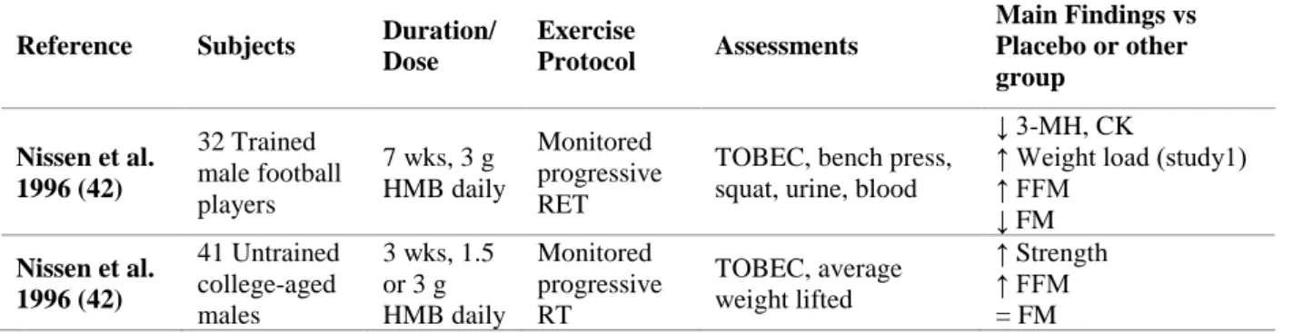
Many of the anticatabolic and anabolic actions of leucine have been attributed to the leucine metabolites α-KIC, HMB and α-HICA (6, 39). However the amount required to induce an effect cannot be consumed within a normal diet (40). Approximately 80% of leucine is used for protein synthesis with the remainder being converted to α-KIC and only 5% to HMB (38). Thus, within a normal diet, only 0.2-0.4 g HMB.day-1 is synthesized from leucine in a 70 kg individual (36, 41). Since the lower amount that exerted performance and body composition enhancement was 1.5 g.day-1 (42), this amount is deemed insufficient. To attain such a high intake of HMB, ≈60 g of leucine would have to be consumed daily, which is beyond the intake of a regular diet (43) and above the current recommended dietary allowances ([RDA] - 42 mg/kg.day-1) (39).
Although other BCAAs (isoleucine and valine) have also been proposed to induce similar effects, the current body of evidence does not support a similar metabolic action for these amino acids (37).
Action on muscle protein synthesis
Leucine actions upon muscle protein synthesis (MPS) have been researched for over four decades (44-46), yet the involved mechanisms remain unclear to date. It has been proposed that its downstream metabolites are, at least in part, responsible for this anabolic action (47-49). However, earlier studies proposed that leucine could influence MPS (50) without being converted to α-KIC (51). Consequently, leucine was thoroughly investigated regarding MPS (52-54).
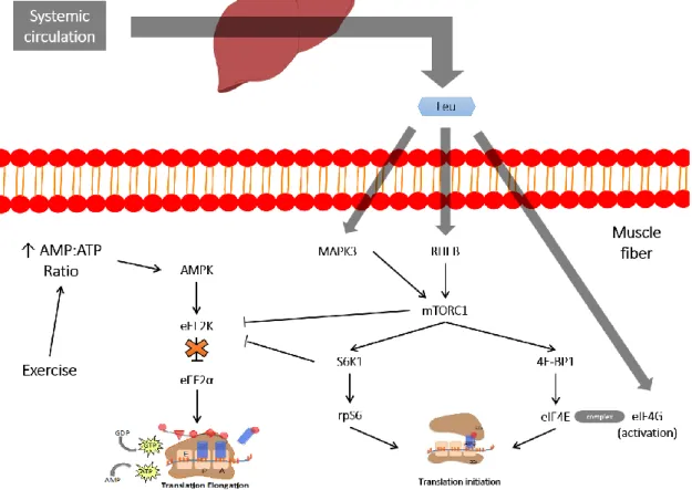
Leucine exerts a double action, both as substrate and as a signalling molecule for the initiation of MPS (55, 56). Several reviews have proposed numerous possible mechanisms, albeit a consensus has not been reached (10, 38, 57-60). As proposed by Duan et al (38), it is likely that after an increase in intracellular concentrations of leucine, Ras-related guanosine triphosphatases (61), human vacuolar sorting protein-34 (62) and mitogen activated protein kinase 3 (61) are activated causing the translocation of mTORC1 to the surface of late endosomes and lysosomes, where it is directly activated by Ras homolog enriched in brain (RHEB) at the lysosome’s surface. Further mTORC1 activation will lead to the downstream phosphorylation and activation of proteins that will regulate both translation initiation and elongation steps (figure 3). However, although leucine might increase insulin secretion via phosphoinositide 3-kinase (PI3K) activity, this does not seem important regarding mTORC1 stimulation (63, 64). Any insulin release by leucine, or its metabolites, is only permissive towards the muscle protein synthetic response and not the main driving factor for MPS to occur (38).
More recently this concept has been challenged, with some leucine metabolites – HMB and α-KIC – being touted has possessing direct anabolic properties and being responsible for the anabolic actions of leucine (20). Recent research, has further proposed a novel form of HMB (HMB free acid) and α-HICA to exert both anabolic and anti-catabolic properties (5-7). Please refer to chapter 2.2 for a more detailed discussion on these leucine metabolites.
Figure 3. Leucine pathways that may influence MPS (65, 66).
Action on muscle protein degradation
When low levels of amino acids or energy are sensed by the cells, MPS is hindered while the muscle protein breakdown (MPB) response is enhanced (degradation pathway) (67). Several mechanisms have been proposed, mainly from animal or in vitro studies, regarding the leucine effect on proteolysis although no consensus seems to exist so far. Some authors have proposed mTOR signalling, excluding an upstream kinase PI3K activation (68); while others have suggested the involvement of both PI3K and protein kinase C (PKC) (69).
It should also be noted, that the concentration of leucine used in some of these in
vitro studies (10 mM) is ≈ 50 fold the plasma concentration found in vivo, therefore it is
highly questionable that these levels may be attainable through dietary intake (38). Albeit some studies have suggested that protein inhibition with leucine might be mediated by
some of its metabolites (22,84,85), in particular HMB (70, 71), more recent research has displayed equivocal evidence (72). Definitely more research is commended to provide further mechanistical insights, regarding the suppression of proteolysis by leucine and also pertaining its downstream metabolites.
Energy metabolism
Leucine may also influence energy metabolism, since protein synthesis and energy are closely related through mTORC1 and AMP-activated protein kinase (AMPK) (57). In fact, leucine goes far beyond protein synthesis, since downstream regulatory proteins from the mTORC pool of kinases also promote mitochondrial biogenesis, further enhancing cellular respiration and energy partitioning (16, 73). It has been proposed, from observations in animal research, that by ameliorating cellular metabolism both fatty acid oxidation and lean tissue gains may be enhanced (57).
Some research has suggested that some positive effects regarding mitochondrial biogenesis and fatty acid oxidation, leading to an increase in energy expenditure, are the result of AMPK and silent information regulator transcript 1 (SIRT1) activation by leucine (73). Whether leucine mediates energy metabolism directly or through its metabolites, is unclear to date (38). Several proteins, that are under the influence of leucine or its metabolites, are involved in the regulation of both oxidative phosphorylation, glycolysis activation and fatty acid β-oxidation (74), with also the nitric oxide pathway probably being involved (75, 76).
Glucose homeostasis
Insulin may translocate the glucose transporter 4 to the plasma membrane of both muscle cells and adipocytes promoting glucose uptake (77). This process is mediated by intracellular signaling involving mTOR and PI3K. It has been shown that leucine may improve satiety and glucose metabolism using complex peripheral and central pathways (32, 53). Unlike insulin, leucine may activate protein kinase C and not B, thus promoting glucose uptake by a different pathway (78). Moreover, leucine may induce glucose uptake, for short periods of time, in a dose-dependent manner via mTORC1 and 2 (79).
It seems that this short-term glucose uptake (up to 45 min) (78), is likely due to the negative feedback resulting from the continuous mTOR stimulation (80). This small effect, in glucose uptake, is likely irrelevant, since research measuring in vivo net glucose transport, has failed to detect any effect of this amino acid (81). Albeit the stimulation of mTOR is deemed important in the insulin signaling process, an excessive activation may conversely lead to impaired insulin sensitivity (79).
Leucine may also stimulate insulin secretion from pancreatic β cells via two different mechanisms (82). It may serve as metabolic fuel for β cells or exert its action as an allosteric activator of glutamate dehydrogenase (GDH) (83), which may in turn promote insulin release from the pancreatic islets (84). It seems that both leucine or its transaminated product α-KIC, may act upon insulin secretion by direct inhibition of the ATP-regulated potassium channels (85). Both compounds regulate these channels, by increasing Ca2+ in the β cell’s cytosol, which would ultimately lead to the exocytosis of insulin secretory granules, probably via both protein phosphorylation and acylation mechanisms (86, 87). Other described mechanisms that may regulate β cell metabolism and insulin secretion, with leucine, are related with an increase in protein synthesis and gene expression (82).
Summarizing, leucine is able to acutely stimulate insulin secretion in β-cells, via ATP production and ATP-regulated potassium channels and also GDH activation. Moreover, acute effects and chronic effects have been described via increased protein synthesis and gene expression in β cells. It should however be noted, that most of the aforementioned mechanisms are the result of in vitro studies, with in vivo research failing to detect meaningful effects of leucine in glucose transport (81).
Immunomodulatory role
Few studies have investigated the role of leucine pertaining inflammation and immune cells (18). Immune cells have indeed high BCAT and BCKD activity levels, using also leucine among other BCAAs throughout the S phase of the cell cycle (88). During enhanced mitogenesis, it has been estimated that the BCAAs transport may be increased by ≈ 300%, while transamination and oxidation are increased by ≈ 200% and ≈ 130%, respectively (89).
When T cells (cluster of differentiation 4 [CD4+]) are activated, their requirements for amino acids increase. This is likely related with augmented protein synthesis requirements, which are mandatory for proper T cell function (18) and proliferation. The activated cells decrease the decarboxylation of pyruvate, yielding higher lactate levels that may supply nicotinamide adenine dinucleotide (NAD+). Indeed, in vitro studies have
shown, that BCAAs may activate T cells (90). Furthermore, a BCAT increase in immune cells, is positively correlated with both leucine transamination and mTOR activation, with the former being a critical regulator of T cell activation, differentiation, and metabolism (90). Albeit leucine has been implicated as an important amino acid pertaining lymphocyte activation, other BCAAs may also play an important role in this regard (91).
Although animal studies have shown promising results regarding leucine and its immunomodulatory role (92-95), few studies have been performed in humans (96). It seems indisputable, that leucine is both oxidized by immune cells and incorporated as a precursor for the synthesis of new cells (97). However, albeit promising, the effect of leucine or BCAAs regarding a possible positive role in immunity, should be further investigated in humans, definitely requiring further confirmatory research.
Role in inflammation
Inflammation plays an important role in tissue regeneration and protection (98) with the translocation of nuclear factor kappa B (NF-κB) to the cell nucleus long being implicated in the cell’s inflammatory response. Mechanisms that allow for this
translocation to occur seem related with a plethora of stimuli, i.e. increases in inflammatory markers (Tumor necrosis factor α [TNF-α] and interleukin 1 [IL-1]), lipopolysaccharides (LPS), heat shock proteins (HSP) and several T-cell activators (99). After being translocated, NF-κB will upregulate the expression of several immune-mediated factors at the cell nucleus, namely cyclooxygenase 2 (COX-2) and several inflammatory cytokines, that will ultimately aid on the eradication of several infectious agents (100, 101). Albeit inflammation is necessary for tissue regeneration and protection, overstimulating NF-κB may lead to life-threatening outcomes (18). More recently, it has
been suggested that NF-κB inhibition may inhibit apoptosis and reduce vascular endothelial growth factor (VEGF) (102-104).
Moreover, mTOR and NF-κB seem related (105). Indeed, the stimulus of NF-κB under certain oncological contexts, seems to contribute to cell proliferation, growth and angiogenesis (albeit this seems to be dependent on other factors like TNF-α) (106). Although inflammation may increase the mTOR response, it may paradoxically impair protein synthesis (107) and thus hinder the anabolic response to amino acids, especially leucine (108). Albeit a theoretical advantage could exist by supplementing with leucine in inflammatory diseases to hinder protein degradation, animal studies do not show advantages regarding supplementation, even with high doses of leucine or other BCAAs (107). This is likely due to the reduction of intramuscular glutamine from inflammatory diseases that may impair leucine transport to the muscle, since the transport of leucine is dependent on the intracellular concentration of glutamine (109, 110).
Few studies have investigated BCAAs or leucine regarding inflammation in humans. Recently, Nicastro et al. (111) evaluated both the supplementation with BCAAs, leucine or placebo (PLA) 15, 30, 60, 90 and 120 minutes after intake, in the absence of any exercise protocol. The authors reported no differences between groups insofar as TNF-α is concerned, however serum interleukin 6 (IL-6) decreased, while interleukin 10 (IL-10) significantly increased 60 min after, in the leucine group only. However, in this crossover controlled trial (n=8 participants) volunteers were refrained to perform resistance exercise 24 h prior to the supplementation protocol and blood collection, which precludes any extrapolation to a post-exercise context. Although statistically significant, these actions from leucine in the absence of an inflammatory stimulus, seem of minor importance regarding diseased populations or athletes.
Human research on performance and body composition
Leucine ingestion positively correlates with MPS in middle aged men, with a strong direct correlation being reported between peak plasma leucine concentrations and the postprandial muscle leucine accretion as evaluated by the postprandial muscle protein fractional synthetic rate (FSR) (112). Bearing this in mind, when aiming to induce maximal hypertrophic outcomes, some protein rich foodstuffs have been purported assuperior due to a higher leucine content and a faster digestion rate (113). Albeit leucine has been deemed important to initiate MPS (114), other essential amino acids are also necessary (115). Some research has suggested that administering leucine alone might rescue the FSR, when lower amounts of essential amino acids (EAAs) are present (116). Conversely, administering leucine with isoleucine and valine (other BCAAs), with limited amounts of other EAAs, might reduce the MPS response likely due to competition for the same gut transporter, which may decrease plasma leucine concentrations (115). In fact, a recent review suggests an absolute intake of 20-40 g of protein/meal (10-12 g of EAAs, 1-3 g of leucine) or 0.25 g/kgin young individuals, with higher doses being recommended for elderly individuals (≈40 g/meal), to maximize the MPS (117).
Few studies have investigated the effects of leucine in young healthy individuals. Some research has shown that leucine supplementation may increase strength, not improving body composition (i.e. not decreasing fat mass [FM] nor increasing fat free mass [FFM]) (118), with other research studies further confirming this evidence while failing to show improvements in body composition or muscle strength gains (119). A study by Crowe et al. (120), in young competitive canoeists, investigated the effects of 45 mg.kg-1.d-1 of leucine or placebo for six weeks. Although leucine concentrations increased in the blood and some improvements in endurance performance were reported, no effects on body mass (BM) or percentage body fat were found between groups. This was in agreement with a review by Balage et al. (121), stating that leucine-rich amino acid mixtures or proteins (providing sufficient EAAs) appear more efficient than leucine alone to improve muscle mass and performance, suggesting that the efficacy of leucine depends on the presence of other EAAs. Additionally, a review by Yao et al. (122) has proposed leucine as a promising nutritional tool to fight obesity, with several mechanisms being proposed (figure 4). However, most of the described pathways in this review are the result of animal studies, with few research studies being performed in humans (only 2 out of 14 studies).
Figure 4. Proposed mechanisms for a beneficial effect of leucine on obesity/metabolic syndrome (122).
Insofar as maintaining FFM, while undergoing food restriction, few studies have been performed in humans (younger or older individuals) with the majority being performed in animal models (123-127). Albeit some reviews have suggested an important role of leucine in preventing FFM losses, most seem unanimous on claiming that this amino acid requires other essential amino acids to be effective in preventing lean mass losses (1, 121). The current body of evidence pertaining leucine supplementation (alone), to prevent FFM while in caloric restriction is scant, with more human research being mandatory at this point.
Studies regarding leucine alone in elderly populations are also limited. Interestingly Ispoglou et al. (128) showed benefits with 6 g of leucine daily in addition to other essential amino acids. Elderly participants (n=36, 65-75 years old) displayed improvements regarding functional status and FFM, after 3 months of supplementation with a mixture of essential amino acids enriched with leucine. The authors proposed that leucine and BCAAs might be interesting to improve functionality and lean mass retention in older individuals, albeit reinforcing that leucine alone would not offer any additional benefits and that longer term studies are required. Some reviews have also suggested that leucine supplementation might be a promising strategy to prevent muscle mass loses with aging and to improve postprandial glycemic control in patients with type 2 diabetes (129). Still, authors state that there is insufficient evidence to recommend dietary supplementation with leucine to augment muscle mass, strength or improve glycemic
control. In fact, not all research supports a positive effect of leucine supplementation on muscle mass or strength, at least in healthy elderly men (130).
Although safe up to 1250 mg.kg-1.d-1 (131), studies performed in elderly populations have similar outcomes to their younger counterparts. Moreover, adding leucine to lower amounts of essential amino acids may pose as an effective strategy to stimulate muscle anabolism both at rest and after exercise (132). Murphy et al. (133) have further confirmed, that leucine co-ingestion with lower daily protein intakes (0.8 g.kg-1.d -1) will augment muscle protein synthesis. Since older individuals do not typically ingest
sufficient protein to optimally stimulate MPS (0.4 g.kg-1.meal-1) due to age-related issues
(134-136) or as imposed to slow the progression of certain chronic diseases (137), leucine supplementation along with meals may be an effective strategy to attenuate muscle loss. Additionally, combining leucine with other dietary supplements (i.e. vitamin D or whey), might also be promising to prevent sarcopenia in older persons and has been proposed in a recent review (138).
Altogether, it is plausible that leucine rich protein supplements might exert beneficial effects on body weight, body mass index and FFM in older persons prone to sarcopenia, but not muscle strength (albeit an elevated heterogenicity was detected between trials in the analysis performed by Komar et al.) (139). A more recent meta-analysis by Xu et al. (140) confirmed that leucine significantly increases MPS, albeit failing to show a significant effect on FFM. Increases in MPS may be important to prevent skeletal muscle losses with aging, however supplementing with leucine alone might not be the best strategy, since to maximize MPS all essential amino acids are required (1).
2.2 Leucine metabolites
2.2.1 β-hydroxy-β-methylbutyrate (HMB)
For decades, leucine metabolites have been thoroughly studied regarding their anabolic or anticatabolic potential (141). Several studies have been performed towards one particular metabolite, HMB (the deaminated and decarboxylated metabolite of
leucine). This was the result of an early study by Nissen et al. (42) that displayed interesting results regarding both body composition and performance outcomes and shifted the focus towards the inhibition of leucine transamination to spare muscle protein, via HMB (48). Since metabolites from other BCAAs lacked the same effect, the focus was placed towards leucine and its derivatives (37). As previously stated, nearly 60 g of leucine is required to produce 3 g of HMB (49). Such high amount, definitely requires a supplementation strategy with HMB, to elicit higher protein synthesis rates and minimize protein breakdown (142).
Several research studies have been performed to date with HMB (both HMB-Ca and HMB-FA), additionally several systematic reviews/meta-analysis (41, 143-150) and narrative reviews (43, 151-164) were also performed. Currently, it has been estimated that ≈2% of college athletes consume HMB (165). Although a considerable amount of evidence is available, some aspects of HMB remain controversial, with more research in some areas being commended.
METABOLISM
HMB is a natural biologically active compound present in many foods such as: catfish, alfalfa, asparagus, avocado, cauliflower and grapefruit (166). In animals and humans, HMB is mostly produced from leucine (167) in the liver from an extrahepatically derived metabolite (168). After leucine is converted to α-KIC in the muscle by BCAT, this α-ketoacid undergoes two pathways in the liver: a) the conversion to HMB in the cytosol by KICD (169) or b) the conversion to IVA-CoA by BCKDC in the mitochondria (31, 166). The main route for HMB synthesis is via KICD, albeit the enzyme enol-CoA hydrase (ECoAH) might also convert β-methyl-crotonyl-CoA (MC-CoA) produced from IVA-CoA to HMB when biotin is deficient. HMB undergoes conversion to HMG-CoA via a CO2 dependent pathway, being ultimately converted to either cholesterol via
HMG-CoA reductase (170) or acetoacetyl-HMG-CoA via HMG-HMG-CoA synthase (figure 5) (43). It seems undisputable, at this point, that HMB is in fact a precursor of cholesterol (171, 172).
When following the biochemical pathway of α-KIC to IVA-CoA via BCKDC, HMG-CoA will also be formed (figure 5). Under normal conditions most α-KIC is converted to IVA-CoA, with only 5% being converted to HMB (36, 49). Nissen and
Abumrad (173) have previously described that the main purpose of HMB is the conversion to HMG-CoA in the liver, to provide substrate for cholesterol synthesis. Bearing this in mind, it has been proposed that HMB supplementation might be important to assure proper cellular function, working as a substrate for HMG-CoA synthesis (43). Since the muscle seems to rely on de novo synthesis of cholesterol and the former is important for proper cell function, the role of HMB has been highlighted in this regard, albeit dependent on the degree of cell damage (43).
Figure 5. HMB metabolism adapted from Nissen and Abumrad (31) and Zanchi et al. (43).
BIOAVAILABILITY BETWEEN DIFFERENT COMMERCIAL FORMS OF HMB
Since it has been commercially available, HMB has been available in the mono-hydrated calcium salt form (HMB-Ca) with the following empirical formula: Ca (HMB)2
-H2O (151). Several factors have been described to influence its rate and magnitude of
appearance in the blood. The amount of HMB consumed and the co-ingestion with other nutrients seems to influence the HMB kinetics (174). For example it has been well documented that one g of HMB-Ca will peak HMB blood levels 120 min after ingestion,
while 3 g may speed the absorption kinetics to only 60 min, leading to higher plasma concentrations (487 vs 120 nmol/mL) (174).
It should also be noted that this faster and higher increase in the blood with the 3 g dose, will lead inevitably to higher HMB losses in urine (+14%) (174). Furthermore, adding glucose to HMB may also influence the rate and magnitude of its appearance, with 75 g of glucose delaying peak HMB levels in plasma by one hour and significantly lowering peak levels in the blood (352 vs 487 nmol/mL) (151). Thus, it was hypothesized that glucose might have both slowed gastric emptying and increased HMB clearance from circulation (151).
More recently a new form of HMB has been made commercially available, the free acid form of HMB (HMB-FA) (175). This type of HMB was associated with a gel containing a buffer (potassium carbonate – K2CO3) to raise pH to 4.5, which was though
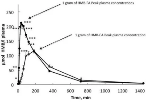
to stabilize HMB not hindering the absorption kinetics. In the initial commercially available form, the addition of Ca to HMB was thought to dissociate swiftly (15 min) in the gut and increase its solubility (176). However Fuller et al. (175, 177) showed that when comparing equivalent amounts of HMB from both HMB-Ca and HMB-FA, the former resulted in a two fold increase in peak plasma levels in just one fourth of the time (30 min vs. 120 min – figure 6) (175).
Additionally, the area under the curve analysis over three hours of ingestion was ≈97% higher with HMB-FA. The half-life time of both forms was similar, with a slightly higher time being reported for HMB-FA (3 h vs. 2,5 h) (175). Moreover, albeit HMB-FA leads to greater plasma concentrations, urinary losses were not different between both forms. Interestingly, the tissue uptake and utilization was also 25% higher with the free acid form, which was seen as promising towards amplifying HMB’s action within the cell. Albeit, some issues were raised towards the HMB-FA bioavailability in rats (178), more recent research in humans (177) confirmed the previous findings (175), towards a superior bioavailability of HMB-FA in humans, with better bioavailability through liquid-filled caps when comparing with gel format.
Figure 6. Different absorption kinetics after 1 g of either HMB-Ca or HMB-FA (151).
SAFETY
HMB has been thoroughly studied in humans (31, 172, 179, 180) and deemed to be safe. Human research has showed that 6 g of HMB-Ca daily for one month had no effect on cholesterol, haemoglobin, white blood cells, blood glucose, and liver and kidney function (181). Meta-analysis have also confirmed the high safety profile of HMB in humans (both young, old, diseased or healthy individuals) even when supplemented with amino acids (41, 172, 180). Furthermore, Baier et al. also confirmed the safety profile of HMB (2-3 g daily) for a longer period of time (one year) in elderly individuals, with no adverse effects being reported in blood or urine markers of hepatic or renal function (179). Also no detrimental effects were found towards blood lipids. The longer study performed to date with HMB-Ca, showed no adverse effects in an elderly population, consuming 3 g daily for 24 weeks (182).
Pertaining HMB-FA, data confirms its safety in humans, at doses of 402 and 459 mg/kg bodyweight.day-1 for men and women, respectively (183). Human research up to
12 weeks has found no adverse effects with HMB-FA (5, 7, 184). Some issues have been raised towards the safety of HMB in rats, due to a possible negative effect on plasma insulin, which was found increased after 320 mg/ kg body weight.day-1 for one month