• Nenhum resultado encontrado

Res. Biomed. Eng. vol.31 número3

N/A
N/A
Protected

Academic year: 2018

Share "Res. Biomed. Eng. vol.31 número3"

Copied!
8
0
0

Texto

(1)

DOI: http://dx.doi.org/10.1590/2446-4740.0736

*e-mail: rani@univap.br

Received: 23 February 2015 / Accepted: 01 September 2015

Effect of low-level laser therapy (GaAlAs - λ660 nm) on muscle

function

Vania Maria de Araújo Giaretta, Luiz Prudêncio Santos, Ana Maria Barbosa, Stephen Hyslop, Alexandre Pinto Corrado, Renata Amadei Nicolau*, José Carlos Cogo

Abstract Introduction: Low-level laser therapy (LLLT) is effective in preventing fatigue and in stimulating the microcirculation and cellular activity. In this study, we examined the effect of LLLT on injured tibial muscle in vivo by assessing muscle function during fatigue. Methods: Twenty-four male mice were used. Each mouse received an injection of sterile 0.9% saline solution (50 µL) in the right tibialis anterior muscle, after which the tendon of the muscle was exposed, connected to an isometric transducer and subjected to a resting tension of 1 g. A bipolar electrode was attached to the tibial nerve for electrical stimulation. The mice were randomly allocated to one of two groups: G1 (control: 3 h – n=8 and 9 h – n=5) and G2 (treated with GaAlAs laser,

λ660 nm, 35 mW, 0.6 J, 17 s: 3 h – n=6 and 9 h – n=5). Results: In G1 mice, the amplitude of the tetanic contracture in response to induced fatigue remained unchanged during six consecutive tetani. The amplitude of the tetanic contractions in response to electrical stimulation (4-8 mV) was also unchanged. These results indicated muscle intactness in response to the load imposed by tetanus. In G2 mice, there was an increase in the amplitude of contraction after 3 h and 9 h when compared to G1 at 83% tetanus. Conclusion: These results indicate that exposure of muscle to LLLT enhanced the contractile force and increased the resistance to muscle fatigue without causing morphological damage to cellular structures.

Keywords: Low-level laser therapy, Muscle activity, Muscle fatigue, Tetanus.

Introduction

Most studies of the effects of low-level laser therapy (LLLT) in laboratory animals have been done in rats and have shown that LLLT can reduce the weakness of local fatigue on muscle force and possibly reduce muscle damage after strenuous exercises (Albuquerque-Pontes et al., 2015; Caetano and Zanuto et al., 2013;

Lopes-Martins et al., 2006; Santos et al., 2014). LLLT

signiicantly enhances skeletal muscle performance and

protects skeletal muscle tissue against damage when applied immediately (Santos et al., 2014). LLLT also causes a dose- and wavelength-dependent increase in cytochrome c oxidase expression in intact skeletal muscle tissue, indicating that muscular metabolism can be enhanced by phototherapy (Albuquerque-Pontes et al., 2015).

Compared to rats, little is known of the responses to LLLT in other species, although various reports have

demonstrated the beneits of LLLT in improving muscle

function in dystrophic (mdx) mice (Leal-Junior et al., 2014; Oron et al., 2014; Silva et al., 2015). Mice are extensively used in toxinology to study the local effects (pain, edema, hemorrhage and necrosis) of a variety of venoms. Myonecrosis caused by Bothrops snake venoms is a well-known phenomenon experimentally

(Gutiérrez and Ownby, 2003) and clinically (França and Málaque, 2009; Warrell, 2004) and, in the latter case, can result in extensive tissue loss and permanent functional damage to the bitten limb. Experimental studies have shown that laser therapy can attenuate the local damage caused by snake venoms, as assessed

histologically and based on the quantiication of

marker proteins or enzymes (Barbosa et al., 2008; 2009; Doin-Silva et al., 2009; Dourado et al., 2003; Nadur-Andrade et al., 2012; 2014). However, none of these investigations has provided a functional assessment of the potential improvement in the contractile activity of muscle injected with venom and treated with LLLT.

In view of the lack of detailed studies on the

beneicial effects of LLLT on muscle function in

normal mice, and the potential usefulness of such a model for studying venom-induced and other lesions, in this work we examined the effect of LLLT on

normal (non-inlamed and non-lesioned) tibial muscle

by assessing muscle function in response to tetanic stimulation. The standardization of this model should provide a useful basis for assessing the effects of LLLT on muscle function in a variety of situations in mice.

(2)

Giaretta VMA, Santos LP, Barbosa AM, Hyslop S, Corrado AP, Nicolau RA, Cogo JC

Methods

Twenty-four Swiss mice (18-22 g; Animais de Laboratório - Anilab, Campinas, SP) were housed (six/cage) in the Laboratory of Physiology and Pharmacodynamics of the Institute of Research and Development at the Universidade do Vale do Paraíba (UNIVAP). The mice were maintained on a 12 light/dark cycle at 22-28 ºC and controlled humidity, with access to food and water ad libitum. The project was approved by the institutional Committee for Ethics in Animal Use at UNIVAP (protocol no. A001/CEUA/2012). The experiments were done within the ethical guidelines of the Brazilian Society for Laboratory Animal Science (SBCAL).

For use, the mice were randomly allocated to one of two experimental groups: G1 (control: 3 h and 9 h, n=8 and 5, respectively) and G2 (treated with

laser: 3 h and 9 h, n=6 and 5, respectively). For the

experiments, the right tibialis muscle of each mouse was injected with 50 µl of 0.9% NaCl and 30 min later the muscle of G2 mice was irradiated with a InGaAlP laser equipment (Twin Flex Evolution®,

MMOptics), operated in indirect contact mode (parameters described in Table 1).

Three and nine hours after saline injection the mice were anesthetized with a mixture of xylazine hydrochloride (20 mg/kg, i.p.), ketamine chloride (100 mg/kg, i.p.), atropine (0.25 mg/kg, i.p.) and

diazepam (5 mg/kg, i.p.) and ixed on a surgical table,

after which the tendon of the tibialis anterior muscle and the right tibial nerve were exposed. In the insert region, near the metatarsal plantar region, the muscle was connected through the tendon to an isometric transducer and the nerve was connected to a bipolar electrode. The muscle was subjected to a constant tension of 1 g and indirectly stimulated by individual pulses from a stimulator GRASS Technologies S48 square pulse stimulator. Pulses of 4-8 mV, 0.2 Hz and 2 ms duration were applied for 3 min; these parameters were chosen based on the minimum and maximum voltages needed to induce muscle contraction with

recruitment of all ibers (Doin-Silva et al., 2009;

Lopes-Martins et al., 2006). Tetanic stimulation was

done by increasing the frequency to 50 Hz for 10 s

and the tetanic proile was recorded. Next, six tetani

were applied at 3-min intervals while the stimuli for muscle contractions were maintained at the previously set parameters (4-8 mV, 0.2 Hz, 2 ms duration) to

conirm the intactness of the muscle ibers. Muscle

contractions and tetanic contractures were recorded

using a Gemini 7070 physiograph (Ugo Basile, Varese, Italy) and an Ugo Basile model 7003 isometric force transducer. Tetani were analyzed by measuring the

amplitude of the irst movement of the physiograph

pen up to the peak of each tetanus.

After completion of the records, the anesthetized mice were euthanized with an overdose of KCl (10%, i.c.) (Barbosa et al., 2009; 2010; Barbosa-Souza et al., 2011; Santos et al., 2010). The muscles were ixed in 4% formaldehyde for 24 h, followed by routine

procedures for dehydration and embedding in parafin

or paraplast. Sections 5 µm thick were cut with a microtome, mounted on microscope slides and stained with hematoxylin-eosin (HE). The slides were examined and photographed to facilitate examination of the cell membrane intactness, the position of the nuclei and the intercellular spaces. In addition, tibial muscles were analyzed histomorphometrically 3 h and 9 h after treatment with saline or LLLT. Muscle

iber diameter was analyzed in longitudinal sections

(n = 10 sections/animal) using ImageJ 1.49t software. The results were expressed as the mean ± standard

error of the mean (SEM). The signiicance of differences

between the control group and laser-treated groups was determined by ANOVA followed by the Tukey-Kramer post-test for multiple comparisons. Values of p<0.05

were considered signiicant.

Results

Tetanus was induced by increasing the frequency of electrical impulses from 0.2 Hz to 50 Hz for 10 s. Figure 1a and 1b show the myographic proile of the six tetanic records 3 h and 9 h after the injection of 0.9% saline. Between each tetanus, the muscle was stimulated at 4-8 mV for 2 ms and 0.2 Hz for 3 min to

assess the intactness of the ibers. In tetanus induction,

muscle contraction was initially high but decreased slightly and persisted for 10 s until the stimulus was turned off (Figure 1a and 1b).

The control records showed that the amplitude of the electrically-induced contractions for 3 min between the tetani remained unchanged, indicating that the muscles were functionally intact and not damaged by the repeated tetani. The records of six tetani in the G2 muscles that received saline or LLLT at 3 h showed

a signiicant increase in the amplitude of contraction

Table 1. Laser parameters.

Parameters Values

Energy density (J/cm2) 3

Energy (J) 0.6

Power (mW) 35

Irradiation time (s) 17

Spot (cm2) 0.19

Wavelength (nm) 660±5

(3)

in the control group, indicating an increase in muscle contractile force (Figures 1c and 1d). In the control

group, between the irst and the sixth tetanus, the mean decrease in tetanus amplitude was 17.2 ± 6%.

In muscles treated with LLLT, the decrease in tetanus amplitude was 34.0 ± 9.1%. Although the decrease in tetanus amplitude was greater in muscles treated with LLLT, these muscles showed a greater baseline amplitude and feedback when compared to control muscles. This was noted 3 h after irradiation. Figure 2

shows the development proile of the six tetani with

and without LLLT. Three hours after laser application there was potentiation of the contractions and an increase in muscle strength. The contractile force

developed in the irst three tetani was larger than

in subsequent tetani, indicating the onset of muscle fatigue, but was still above control values. Figure 3 shows the same situation 9 h after LLLT.

Histological analysis of the muscle ibers showed

that the cell membranes of control and LLLT-treated muscles were intact after 3 h and 9 h. The nuclei also showed no alterations in either group. These results indicated that even if G2 had a greater muscle activity, this did not cause morphological changes (Figures 4 and 5).

Histomorphometric analysis showed a signiicant increase in iber diameter in mice treated with LLLT

(3 h) compared with saline; no change in diameter was seen after 9 h (Figure 6).

Figure 1. Muscle contractions (a) and tetanic contractures (b) were recorded using a physiograph. Myographic proiles of tetanic trains in tibial muscle stimulated via the nerve. A – Control 3 h (n = 8), B – Control 9 h (n = 5), C – Laser 3 h (n = 6), D – Laser 9 h (n = 5). Note the

(4)

Giaretta VMA, Santos LP, Barbosa AM, Hyslop S, Corrado AP, Nicolau RA, Cogo JC

Discussion

In humans and rats, LLLT enhances the recovery of muscle contracture during tetanus. However, to date there has been no similar assessment in mice, a species widely used for testing drugs and toxins. In this study, we therefore sought to reproduce in mice the experimental technique previously used in rats. Thus, for example, the dose of LLLT used here has be extensively documented and standardized for rats, as described by Andrade et al. (2011), Caetano and Zanuto (2013), Lopes-Martins et al. (2006) and Santos et al. (2014). As shown in the present study, LLLT enhanced muscle contractility to tetanic stimuli

in the irst 3 h after laser application, but there was

no enhancement after 9 h.

The beneits of LLLT in skeletal muscle include

enhanced strength for everyday activities and sports,

without damage to the ibers or reduced muscle

functionality (Baroni et al., 2010; Caetano and Zanuto, 2013; Kelencz et al., 2010; Leal et al., 2009a; 2009b; Liu et al., 2009; Lopes-Martins et al., 2006; Maciel et al., 2013). The dose and form of LLLT application were selected to primarily enhance cellular activity (Andrade et al., 2011; Lopes-Martins et al.,

2007; Caetano and Zanuto et al., 2013; Santos et al., 2014). The 660 nm wavelength laser used here corresponded to the electromagnetic spectrum absorbed in the respiratory cycle (Zhang et al., 2008). This wavelength stimulates the respiratory chain and increases mitochondrial ATP production to provide energy to the cell via the chemical bonds of cytochrome c (Lopes-Martins et al., 2006). The data reported here suggest that cell activity was increased, probably because of an improved blood supply coupled with enhanced ATP formation. This ATP is used by troponins in phosphorylation to promote the necessary cross-bridges required for muscle contraction (Sussai et al., 2010).

The inference of an enhanced post-irradiation energy intake was based on the increase seen in the amplitude of contractile force in the myographic recordings. Figure 1 shows that the peak of tetanus was greater in irradiated mice, with tetanus lasting for ~10 s followed by a decrease at the third tetanus that was maintained for the remainder of the series. A similar relationship between exposure to LLLT and the duration of tetanus was also observed by

Lopes-Martins et al. (2006) and Santos et al. (2014) for rats.

The percentage decrease in amplitude between

the irst and sixth tetanic series in LLLT-treated mice

was similar to that reported by Lopes-Martins et al.

(2006), despite the different experimental conditions of the latter study. After three tetanic series, the degree of muscle fatigue was 34.0 ± 9.1% and probably

relected the restoration of muscle homeostasis after

use of the energy provided by LLLT-induced ATP formation (Sussai et al., 2010).

Studies such as those of Doin-Silva et al. (2009) and Lopes-Martins et al. (2006), originally done to evaluate neuromuscular activity, demonstrated an increase in muscle contractile force after application of low intensity laser in rats. LLLT has been described as an adjunctive treatment for athletes in aerobic performance, adaptation and recovery of skeletal muscle (Sussai et al., 2010). The longer time required for the onset of muscle fatigue and the gain in the

amplitude of the irst tetanic series agreed with clinical

studies describing greater torque in athletes treated

Figure 2. Muscle contractile force in the absence (control) and presence of LLLT (3 h post-LLLT). Note the greater contractile force in muscle treated with LLLT. Each point represents the mean of the

measurements of the irst muscle burst as explained in Figure 1. The

data are expressed as the mean ± SEM (control n=8; LLLT n=6).

*p<0.05 compared to control preparations.

Figure 3. Muscle contractile force in the absence and presence of LLLT (9 h post-LLLT). Note the greater contractile force in muscle treated with LLLT. Each point represents the mean of the

measurements of the irst muscle burst, as explained in Figure 1. The data are expressed as the mean ± SEM (control n=5; LLLT n=5).

There were no signiicant differences in the responses between the

two groups of animals.

(5)

with radiation different wavelengths (Andrade et al., 2011; Leal et al., 2010c; Lopes-Martins et al., 2007; Maciel et al., 2013; Place et al., 2010; Rocha and

Barbanti, 2007).

Histological analysis of muscle ibers exposed

to LLLT was done to examine whether LLLT could protect tissue from damage by subsequent tetanus. According to Sussai et al. (2010), stressful exercise

can cause muscle injury, iber breakdown, a decrease

in performance, depletion of ATP and calcium imbalance. As shown here, muscle structure and organization were unaffected by LLLT, indicating that the gain in energy input involved intracellular

biochemical alterations. Exposure to LLLT made the muscle more resistant to fatigue induced by indirect

electrical stimulation, a inding in agreement with

that of Leal et al. (2010a). Lopes-Martins et al. (2006) also observed a decrease in fatigue after application of LLLT in rat electrically-stimulated muscle. Leal et al. (2008; 2010b; 2010c) reported that LLLT increased the number and duration of contractions in high intensity, short duration exercises designed for volleyball players. Although LLLT apparently increased

the muscle iber diameter at 3 h, this enhancement probably relected the lower iber diameter in control

muscles rather than an increase caused by LLLT; this

(6)

Giaretta VMA, Santos LP, Barbosa AM, Hyslop S, Corrado AP, Nicolau RA, Cogo JC

conclusion is supported by the fact that the diameter of LLLT-treated muscles at 3 h was similar to the diameters of control and LLLT-treated muscles at

9 h. The reason for the lower iber diameter in the

3 h control group is unclear.

In conclusion, the results of this study indicate that LLLT enhances the contractile force of mouse tibialis muscle without causing morphological damage and increases the resistance of this muscle to fatigue. This model should be useful for studying a variety of pathological conditions in mice.

References

Albuquerque-Pontes GM, Vieira RP, Tomazoni SS, Caires CO, Nemeth V, Vanin AA, Santos LA, Pinto HD, Marcos RL, Bjordal JM, Carvalho PTC, Leal-Junior ECP. Effect

Figure 5. Histological sections of tibial muscle without LLLT (a) and (b) and 9 h after treatment with LLLT (c) and (d). (a) and (c) – Cross-sections and (b) and (d) – Longitudinal sections. Note the normal appearance of the muscles, e.g., intact membranes and peripherally located nuclei. Scale bar = 50 µm.

Figure 6. Histomorphometric analysis of control and LLLT-treated

tibial muscles 3 h and 9 h after LLLT. Muscle iber diameters were quantiied in longitudinal sections (n=10 sections/mouse) and the

columns represent the mean ± SEM.

(7)

of pre-irradiation with different doses, wavelengths, and application intervals of low-level laser therapy on cytochrome c oxidase activity in intact skeletal muscle of

rats. Lasers in Medical Science. 2015; 30(1):59-66. http://

dx.doi.org/10.1007/s10103-014-1616-2. PMid:24957189. Andrade LCG, Hauck LA, Maciel TS, Nicolau RA. Prevenção da fadiga em atletas de alta performance: revisão de literatura. In: Anais do 15° Encontro Latino Americano de Iniciação Científica e 11° Encontro Latino Americano de Pós-Graduação; 2011; São José dos Campos, Brasil. São José dos Campos: UNIVAP; 2011. p. 1-5

Barbosa AM, Villaverde AB, Guimarães-Sousa L, Soares AM, Zamuner SF, Cogo JC, Zamuner SR. Low-level laser therapy decreases local effects induced by myotoxins isolated from Bothrops jararacussu snake venom. The Journal of Venomous Animals and Toxins Including Tropical

Diseases. 2010; 16(3):470-9. http://dx.doi.org/10.1590/

S1678-91992010000300014.

Barbosa AM, Villaverde AB, Guimaraes-Souza L, Munin E, Fernandes CM, Cogo JC, Zamuner SR. Effect of the low level laser therapy in the myonecrosis induced by Bothrops jararacussu snake venom. Photomedicine and

Laser Surgery. 2009; 27(4):591-7. http://dx.doi.org/10.1089/

pho.2008.2296. PMid:19530909.

Barbosa AM, Villaverde AB, Guimarães-Souza L, Ribeiro W,

Cogo JC, Zamuner SR. Effect of low-level laser therapy in the inflammatory response induced by Bothrops jararacussu

snake venom. Toxicon. 2008; 51(7):1236-44. http://dx.doi.

org/10.1016/j.toxicon.2008.02.007. PMid:18439641.

Barbosa-Souza V, Contin DK, Bonventi W Fo, Araújo AB,

Irazusta SP, Cruz-Höfling MA. Osteopontin, a chemotactic protein with cytokine-like properties, is up-regulated in muscle injury caused by Bothrops lanceolatus (fer-de-lance) snake venom. Toxicon. 2011; 58(5):398-409. http://dx.doi.

org/10.1016/j.toxicon.2011.07.011. PMid:21839764. Baroni BM, Leal EC Jr, De Marchi T, Lopes AL, Salvador M, Vaz MA. Low level laser therapy before eccentric exercise reduces muscle damage markers in humans. European

Journal of Applied Physiology. 2010; 110(4):789-96. http://

dx.doi.org/10.1007/s00421-010-1562-z. PMid:20602109. Caetano HRS, Zanuto EAC. Ação da laserterapia de baixa intensidade sobre parâmetros bioquímicos. Uma revisão de

literatura. Colloquium Vitae. 2013; 5(Spec No):63-9. http:// dx.doi.org/10.5747/cv.2013.v05.nesp.000202.

Doin-Silva R, Baranauskas V, Rodrigues-Simioni L, Cruz-Höfling MA. The ability of low level laser therapy to prevent muscle tissue damage induced by snake venom.

Photochemistry and Photobiology. 2009; 85(1):63-9. http://

dx.doi.org/10.1111/j.1751-1097.2008.00397.x. PMid:18643907. Dourado DM, Favero S, Baranauskas V, Cruz-Höfling MA. Effects of the Ga-As laser irradiation on myonecrosis caused by Bothrops moojeni snake venom. Lasers in Surgery and

Medicine. 2003; 33(5):352-7. http://dx.doi.org/10.1002/

lsm.10237. PMid:14677163.

França FOS, Málaque CMS. Acidente botrópico. In: Cardoso

JLC, França FOS, Wen FH, Málaque CMS, Haddad V Jr,

editors. Animais peçonhentos no Brasil: biologia, clínica

e terapêutica dos acidentes. São Paulo: Sarvier/FAPESP; 2009. p. 81-95.

Gutiérrez JM, Ownby CL. Skeletal muscle degeneration induced by venom phospholipases A2: insights into the mechanisms of local and systemic myotoxicity. Toxicon. 2003; 42(8):915-31. http://dx.doi.org/10.1016/j.toxicon.2003.11.005. PMid:15019491.

Kelencz CA, Muñoz ISS, Amorim CF, Nicolau RA. Effect of low-power gallium-aluminum-arsenium noncoherent light

(640 nm) on muscle activity: a clinical study. Photomedicine and Laser Surgery. 2010; 28(5):647-52. http://dx.doi.

org/10.1089/pho.2008.2467. PMid:20961231.

Leal ECP Jr, Lopes-Martins RÁB, Dalan F, Ferrari M, Sbabo FM, Generosi RA, Baroni BM, Penna SC, Iversen

VV, Bjordal JM. Effect of 655-nm low-level 49 laser therapy

on exercise-induced skeletal muscle fatigue in humans.

Photomedicine and Laser Surgery. 2008; 26(5):419-24. http://dx.doi.org/10.1089/pho.2007.2160. PMid:18817474. Leal ECP Jr, Lopes-Martins RÁB, Baroni BM, Marchi T, Taufer D, Manfro DS, Rech M, Danna V, Grosselli D, Generosi RA, Marcos RL, Ramos L, Bjordal JM. Effect of 830 nm low-level laser therapy applied before high-intensity exercises on skeletal muscle recovery in athletes. Lasers

in Medical Science. 2009a; 24(6):857-63. http://dx.doi.

org/10.1007/s10103-008-0633-4. PMid:19057981. Leal ECP Jr, Lopes-Martins RÁB, Rossi RP, Marchi T, Baroni BM, Godoi V, Marcos RL, Ramos L, Bjordal JM. Effect of cluster multi-diode light emitting diode therapy (LEDT) on exercise-induced skeletal muscle fatigue and skeletal muscle recovery in humans. Lasers in Surgery and

Medicine. 2009b; 41(8):572-7. http://dx.Doi.org/10.1002/

lsm.20810.

Leal ECP Jr, Lopes-Martins RÁB, Almeida P, Ramos L, Iversen VV, Bjordal JM. Effect of low-level laser therapy (GaAs 904 nm) in skeletal muscle fatigue and biochemical markers of muscle damage in rats. European Journal of

Applied Physiology. 2010a; 108(6):1083-8. http://dx.doi.

org/10.1007/s00421-009-1321-1. PMid:20024577. Leal ECP Jr, Lopes-Martins RÁB, Frigo L, Marchi T, Rossi RP, Godoi V, Tomazoni SS, Silva DP, Basso M, Lotti P Fo, Corsetti FV, Iversen VV, Bjordal JM. Effects of low-level laser therapy (LLLT) in the development of exercise-induced skeletal muscle fatigue and changes in biochemical markers related to post-exercise recovery. The Journal of Orthopaedic and Sports Physical Therapy. 2010b; 40(8):524-32. http:// dx.doi.org/10.2519/jospt.2010.3294. PMid:20436237.

Leal ECP Jr, Nassar FR, Tomazoni SS, Bjordal J, Lopes-Martins RÁB. Laserterapia de baixa potência melhora o desempenho muscular mensurado por dinamometria isocinética

(8)

Giaretta VMA, Santos LP, Barbosa AM, Hyslop S, Corrado AP, Nicolau RA, Cogo JC

Liu XG, Zhou YJ, Liu TC, Yuan JQ. Effects of low-level laser irradiation on rat skeletal muscle injury after eccentric

exercise. Photomedicine and Laser Surgery. 2009; 27(6):863-9.

http://dx.doi.org/10.1089/pho.2008.2443. PMid:19697999.

Lopes-Martins RÁB, Labat RL, Ramos L, Pallota RC, Penn SC, Bjordal JM. Novos paradigmas no processo inflamatório: possíveis mecanismos de ação da laserterapia de baixa intensidade e considerações sobre dosimetria.

Jornal Brasileiro de Laser. 2007; 1(2):12-9.

Lopes-Martins RÁB, Marcos RL, Leonardo PS, Prianti AC Jr, Muscará MN, Aimbire F, Frigo L, Iversen VV, Bjordal

JM. Effect of low-level laser (Ga-Al-As 655 nm) on skeletal

muscle fatigue induced by electrical stimulation in rats. Journal

of Applied Physiology. 2006; 101(1):283-8. http://dx.doi. org/10.1152/japplphysiol.01318.2005. PMid:16627677.

Maciel TS, Silva J, Jorge FS, Nicolau RA. A influência do laser 830 nm no desempenho do salto de atletas de voleibol feminino. Revista Brasileira de Engenharia Biomédica. 2013; 29(2):199-205. http://dx.doi.org/10.4322/rbeb.2013.020.

Nadur-Andrade N, Barbosa AM, Carlos FP, Lima CJ, Cogo JC, Zamuner SR. Effects of photobiostimulation on edema and hemorrhage induced by Bothrops moojeni venom.

Lasers in Medical Science. 2012; 27(1):65-70. http://dx.doi.

org/10.1007/s10103-011-0914-1. PMid:21484453. Nadur-Andrade N, Dale CS, Santos AS, Soares AM, Lima CJ, Zamuner SR. Photobiostimulation reduces edema formation induced in mice by Lys-49 phospholipases A2 isolated from Bothrops moojeni venom. Photochemical

& Photobiological Sciences. 2014; 13(11):1561-7. http:// dx.doi.org/10.1039/C4PP00111G. PMid:25232894.

Oron A, Oron U, Sadeh M. Low-level laser therapy during postnatal development modulates degeneration and enhances regeneration processes in the hindlimb muscles of dystrophic

mice. Photomedicine and Laser Surgery. 2014; 32(11):606-11. http://dx.doi.org/10.1089/pho.2014.3757. PMid:25329504.

Place N, Yamada T, Bruton JD, Westerblad H. Muscle fatigue:

from observations in humans to underlying mechanisms studied in intact single muscle fibres. European Journal of Applied Physiology. 2010; 110(1):1-15. http://dx.doi.

org/10.1007/s00421-010-1480-0. PMid:20419312.

Rocha MA, Barbanti VJ. Análise das ações de saltos de ataque, bloqueio e levantamento no voleibol feminino. Revista Brasileira de Cineantropometria e Desempenho

Humano. 2007; 9(3):284-90.

Santos CM, Pereira ES, Rabelo NS, Zamuner SR, Barbosa AM, Cogo JC. Efeito do laser de baixa intensidade e LED na atividade hemorrágica induzida pelo veneno da serpente Bothrops jararaca. In: Anais do 14° Encontro Latino Americano de Iniciação Científica e 10° Encontro Latino Americano de Pós-Graduação; 2010; São José dos Campos, Brasil. São José dos Campos: UNIVAP; 2010. p. 1-4.

Santos LA, Marcos RL, Tomazoni SS, Vanin AA, Antonialli FC, Grandinetti VS, Albuquerque-Pontes GM, Paiva PRV, Lopes-Martins RAB, Carvalho PTC, Bjordal JM, Leal-Junior ECP. Effects of pre-irradiation of low-level laser therapy with different doses and wavelengths in skeletal muscle performance, fatigue, and skeletal muscle damage induced by tetanic contractions in rats. Lasers in Medical

Science. 2014; 29(5):1617-26. http://dx.doi.org/10.1007/ s10103-014-1560-1. PMid:24651950.

Silva AA, Leal-Junior ECP, D’Avila KA, Serra AJ, Albertini R, França CM, Nishida JA, Carvalho PT. Pre-exercise low-level laser therapy improves performance and low-levels of oxidative stress markers in mdx mice subjected to muscle fatigue by high-intensity exercise. Lasers in Medical

Science. 2015; 30(6):1719-27. http://dx.doi.org/10.1007/ s10103-015-1777-7. PMid:26076829.

Sussai DA, Carvalho PTC, Dourado DM, Belchior ACG, Reis FA, Pereira DM. Low-level laser therapy attenuates creatine kinase levels and apoptosis during forced swimming in rats. Lasers in Medical Science. 2010; 25(1):115-20. http://

dx.doi.org/10.1007/s10103-009-0697-9. PMid:19554361.

Warrell DA. Snakebites in Central and South America:

epidemiology, clinical features and clinical management.

In: Campbell JA, Lamar WW, editors. Venomous reptiles of the Western Hemisphere. Ithaca: Comstock Publishers/ Cornell University Press; 2004. Vol. 2, p. 709-61.

Zhang J, Xing D, Xuejuan G. Low-power laser irradiation activates Src tyrosine kinase through reactive oxygen species-mediated signaling pathway. Journal of Cellular

Physiology. 2008; 217(2):518-28. http://dx.doi.org/10.1002/ jcp.21529. PMid:18615581.

Authors

Vania Maria de Araújo Giaretta1, Luiz Prudêncio Santos2, Ana Maria Barbosa3, Stephen Hyslop4,

Alexandre Pinto Corrado5, Renata Amadei Nicolau6*, José Carlos Cogo1

1 Laboratório de Fisiologia e Farmacodinâmica, Instituto de Pesquisa e Desenvolvimento – IP&D, Universidade do Vale do Paraíba – UNIVAP, São José dos Campos, SP, Brazil.

2 Escola de Enfermagem Wenceslau Braz – EEWB, Itajubá, MG, Brazil.

3 Faculdade Anhanguera Educacional, Unidade I, Taubaté, SP, Brazil.

4 Departamento de Farmacologia, Faculdade de Ciências Médicas, Universidade Estadual de Campinas – UNICAMP, Campinas, SP, Brazil.

5 Departamento de Farmacologia, Faculdade de Medicina de Ribeirão Preto, Universidade de São Paulo – USP, Ribeirão Preto, SP, Brazil.

6 Centro de Laserterapia e Fotobiologia - CELAFO, Instituto de Pesquisa e Desenvolvimento - IP&D, Universidade do

Vale do Paraíba – UNIVAP, Avenida Shishima Hifumi, 2911, Bairro Urbanova, CEP 12244-000, São José dos Campos, SP, Brazil.

Imagem

Figure 1a and 1b show the myographic proile of the  six tetanic records 3 h and 9 h after the injection of  0.9% saline
Figure 1. Muscle contractions (a) and tetanic contractures (b) were recorded using a physiograph
Figure 3.  Muscle contractile force in the absence and presence  of LLLT (9 h post-LLLT)
Figure 4. Histological sections of tibial muscle without LLLT (a) and (b) and 3 h after treatment with LLLT (c) and (d)
+2

Referências

Documentos relacionados

Quando utilizar este documento em outro contexto, você deve dar crédito ao autor (ou autores), à Brasiliana Digital e ao acervo original, da forma como aparece na ficha

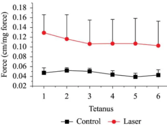
For a given sub- maximal stimulation, the combined effects of potentiation and fatigue could result in an increase, no change or a decrease in active force, depending on the

Creatine kinase (CK) content of mouse extensor digitorum longus muscle after perimuscular injection of Bothrops jararacussu venom (V; 1.0 mg/kg) alone and treatment with

Animals were randomly distributed into three groups: injured tibialis anterior muscle without treatment (IC: injury control); chronic ingestion of alcohol plus

A este método Eduardo Coutinho (1983, p.203) chamou de re vitalização da lin gua gem, ressaltan do, além da singu laridade de Rosa no manejo com a lín gua, o fato de o autor e

The Effects of Low-Level Laser Therapy on Bone in Diabetic and Nondiabetic Rats Photomedicine and Laser Surgery. The effects of infrared low-level laser therapy on

In the present study, bDNA was employed to improve our understanding of the interactions between low-intensity laser therapy and molecular and cellular mechanisms in skeletal