IN ST IT U T O D E C IÊ N C IA S B IO M ÉD IC A S A B EL S A LA Z A R FA C U LD A D E D E CI ÊN CI A S
R
ica
rdo
A
lv
es. C
yto
tox
icit
y i
nd
uce
d b
y e
xtr
act
s/co
m
po
un
ds o
f
Piso
lithus
tinc
tor
ius
sp
ore
s o
n h
um
an c
anc
er a
nd n
orm
al c
ell
lines
- e
valu
atio
n o
f a
nt
ica
nc
er p
ote
nt
ial
C
yto
to
xici
ty i
nd
uc
ed b
y e
xt
ra
cts
/com
po
un
ds
of
P
isol
ith
us
t
inc
to
riu
s s
po
re
s o
n h
um
an c
an
ce
r
an
d n
or
m
al c
ell l
in
es - e
va
lu
ati
on o
f a
nt
ica
nc
er
po
te
nt
ial
Ric
ard
o M
anu
el G
onç
alv
es
A
lves
Cytotoxicity induced by extracts/
compounds of Pisolithus tinctorius
spores on human cancer and normal
cell lines - evaluation of anticancer
potential
Ricardo Manuel Gonçalves Alves
M
2017M
.ICB
AS
2017
SE D E A D MIN IST RA TIVRicardo Manuel Gonçalves Alves
CYTOTOXICITY INDUCED BY EXTRACTS/COMPOUNDS OF
PISOLITHUS TINCTORIUS SPORES ON HUMAN CANCER AND
NORMAL CELL LINES - EVALUATION OF ANTICANCER
POTENTIAL
Dissertação de Candidatura ao grau de
Mestre em Toxicologia e Contaminação
Ambientais submetida ao Instituto de
Ciências Biomédicas de Abel Salazar
da Universidade do Porto.
Orientador
– Doutor Marco Aurélio
Correia Preto
Categoria – Investigador Auxiliar
Afiliação
– Blue Biotecnology and
Ecotoxicology
‐ Centro Interdisciplinar
de Investigação Marinha e Ambiental
(CIIMAR
)Co-orientador
– Doutor Rui Sérgio
Viana Sodré de Oliveira
Categoria – Assistente Convidado
Afiliação
– Escola Superior de Saúde,
Instituto Politécnico do Porto
(ESS-IPP); Centro de Ecologia Funcional;
Departamento de Ciências de Vida,
Universidade de Coimbra (CFE‐UC)
Acknowledgements
I would never be able to reach this point without the support of my supervisors. I am deeply grateful to Prof. Rui Oliveira for this dedication to this project, which were translated for me in motivation and inspiration in science. His commitment will sure a true example to follow in my personal and professional development.
Secondly, my greatly thanks to my co-supervisor, Dr. Marco Preto who helped me greatly in the chemical process, transmitting his huge knowledge on this field sciences and with a positive thinking that surely helped me through this dissertation.
My deepest thanks to Dra. Rosario Martins, for all knowledge related with the cell cultures and with practical issues with the cytotoxic assays. Her availability to help was crucial to the success of this thesis.
I am also grateful to BBE team, for the excellent guidance, caring and for providing good atmosphere for doing this research.
To my friends, João, Bruna, Filipe, Rui and Fabio, for the support each other by deliberating over our problems and findings, but also happily share true moments of friendship and shaping the person I am today. I am deeply indebted to them for their help.
I would like to acknowledge CIIMAR - Interdisciplinary Centre of Marine and Environmental Research and Health Superior School for permitting the use of all the equipment, installations, and facilities, proved to be essential to this project.
To my dearest Mother, for all the sacrifices she made, worthy of a true warrior. Without her effort, would not be possible to me, reach this stage.
This work was performed in the laboratories facilities of the Escola Superior de Saúde – Polytechnic of Porto (ESS-IPP) and in the laboratory facilities of the Interdisciplinary Centre of Marine and Environmental Research (CIIMAR-UP).
This research was partially supported by PAPRE-IPP (Program of Support to the Publication in High Quality Scientific Journals- Polytechnic of Porto) attributed to Maria do Rosário Martins.
This research was partially supported by the Strategic Funding UID/Multi/04423/2013 through national funds provided by FCT – Foundation for Science and Technology and European Regional Development Fund (ERDF), in the framework of the programme PT2020
The work hereby presented was financed by the project INNOVMAR - Innovation and Sustainability in the Management and Exploitation of Marine Resources (reference NORTE-01-0145-FEDER-000035, within Research Line NOVELMAR), supported by North Portugal Regional Operational Programme (NORTE 2020), under the PORTUGAL 2020 Partnership Agreement, through the European Regional Development Fund (ERDF).
Abstract
Cancer is classified as the greatest threat to human health in modern times. Currently conventional medicine uses chemotherapy as preferential treatment. However, this therapy entails acute side effects in patients, largely due to their lack of selectivity. Basidiomycetes are well known in the literature for the ability to produce secondary metabolites capable of being useful in different biomedical fields, including anticancer compounds. In a previous work, Pisolithus tinctorius, a globally distributed fungus capable of forming ectomycorrhizas in association with several trees, has already demonstrated a potential bioactivity with spores extracts and cancer cell lines. Following this work, the main objective of this dissertation was to optimize the extraction procedure of P. tinctorius spores, and following a bioassay guided fractionation to isolate compounds involved in the cytotoxicity. Initially two extractions were carried out only with solvent and sonication, resulting in a low extraction efficiency. With this, four different experimental methodologies were carried out to find the best extraction technique: extraction using only solvent (1), solvent and glass beads (2), solvent and sonication (3) and solvent, glass beads and sonication (4). Considering the results obtained, method 2 was selected for use in large-scale extraction. By vacuum liquid chromatography, using an increasing polarity gradient, it was possible to obtain 9 different fractions per extract. These fractions were tested for cytotoxicity by the 3-(4.5-dimethylthiazol-2-yl)-2.5-diphenyltetrazolium bromide (MTT) assay and using the cancer cell lines T47D, RKO, HEPG2 and the normal cell line HCMEC/D3. Fractions were tested at concentrations of 100, 10 and 1 µg/ ml over the 24 h, 48 h and 72 h. Due to the low mass recovered, the fractions that caused acute cytotoxic were grouped into a single pool, and then subjected to a gravity chromatography process (performed in parallel with thin layer chromatography), and 22 fractions were collected. Nuclear Magnetic Resonance analysis and cell viability assay of these fractions was performed to pooling the fractions that displayed the highest cytotoxic activity. This pool was subjected to high pressure liquid chromatography (HPLC), revealing a complexity, requiring further fractionation to identify and isolate the compound(s) responsible for this bioactivity. This study confirms the existence of
bioactive compounds with anticancer potential. The optimisation of the extraction process using glass beads is a first step in establishing new extraction techniques. However, further refinements are necessary to obtain sufficient mass, to be able to isolate and identify the compounds responsible for the bioactivity.
Resumo
O cancro é considerado como a maior ameaça para saúde humana nos tempos atuais. Atualmente a medicina convencional utiliza a quimioterapia como tratamento preferencial. Contudo esta terapia acarreta efeitos secundários nos pacientes, derivado em muito a sua falta de seletividade. Os basidiomicetes, são conhecidos na literatura pela sua capacidade de produzirem metabolitos secundários com potencial farmacológico nomeadamente potencial anticancerígeno. Num trabalho anterior, extratos de esporos de Pisolithus tinctorius, um fungo distribuído mundialmente e com capacidade de formar ectomicorizas em associações com diversas arvores, revelou potencial bioativo em linhagens celulares cancerígenas. Tendo em conta este trabalho o principalmente objetivo desta dissertação de mestrado foi otimizar o processo de extração com esporos de P. tinctorius e seguindo um fracionamento guiado por bioensaio isolar os compostos envolvidos na citotoxicidade. Inicialmente duas extrações forma efetuadas apenas com solvente e com a ação de um sonicador, resultando numa baixa eficiência de extração. Com isto quatro diferentes metodologias experimentais foram efetuadas: Extração com recurso apenas a solvente (1), uso de esferas de vidro com 3mm de diâmetro (2), solvente com auxilio de sonicação (3) e finalmente a extração com o recurso a esferas de vidro com diâmetro de 3mm, juntamente com sonicação (4). Tendo em conta os resultados obtidos, o método 2 foi selecionado para ser usado na extração em larga escala. Através de uma cromatografia liquida com vácuo, usando um gradiente crescente de polaridade, foi possível obter 9 frações distintas por extrato. Estas frações foram sujeitadas a um teste de citotoxicidade através de um ensaio com 3-(4.5-dimetilthiazol-2-il)-2.5-difeniltetrazolium bromedo (MTT), com as linhas celulares cancerígenas T47D, RKO, HEPG2 e uma linhagem celular normal HCMEC/D3, em concentrações de 100, 10 e 1µg/ml durante o período de 24h, 48h e 72h. Devida a baixa massa recuperada, as frações que despertavam resultados citotoxicologicos agudos, foram agrupadas numa única pool, sendo posteriormente sujeitas a um processo de cromatografia por gravidade, (realizado paralelamente uma cromatografia de camada fina, sendo recolhidos 22 frações e observando o ensaio de viabilidade celular estas frações, agrupou-se as que apresentavam maior atividade citotóxica. Esta pool foi submetida a uma cromatografia líquida de alta pressão (HPLC), revelando uma complexidade, sendo necessário mais um fracionamento de maneira a poder identificar e isolar o
composto(s) responsáveis por esta bioactividade. Este estudo vem confirmar a existência de compostos bioativos com potencial anticancerígeno. A otimização do processo de extração com o recurso a esferas de vidro, representa um primeiro passo em estabelecer novas técnicas de extração. No entanto, novos aperfeiçoamentos são necessários, de maneira a obter massa suficiente, para assim ser possível identificar e isolar os compostos responsáveis por esta bioatividade anticancerígena.
Index
LIST OF ABBREVIATIONS I
LIST OF FIGURES III
LIST OF TABLES IV
1. INTRODUCTION 1
1.1. BASIDIOMYCOTA AND CHEMICAL PROPERTIES; 1
1.2. PISOLITHUS TINCTORIUS; 5
1.3. BIOACTIVE POTENTIAL OF PISOLITHUS 6
1.3.1. Anti-cancer potential 6 1.3.3. Antimicrobial Potential 9 1.3.5. Other bioactivity potentials: 10 2. AIM AND OUTLINE OF THIS DISSERTATION 14
3. MATERIALS AND METHODS 15
3.1. COLLECTION OF PISOLITHUS TINCTORIUS SPORES 15
3.2. CRUDE EXTRACTION AND FRACTIONATION 15
3.3. IMPROVING THE EXTRACTION TECHNIQUE 15
3.4. FULL SCALE EXTRACTION WITH GLASS BEADS METHOD 16 3.5. FRACTIONATION OF THE E15094,PT01 AND E16152 CRUDE EXTRACT. 16
3.6. SUB-FRACTIONATION OF PTAFCES 17
3.7. PREPARATION OF THE TEST SAMPLES 19
3.8. PREPARATION OF THE PTAFCES FOR NUCLEAR MAGNETIC RESONANCE SPECTROSCOPY (NMR) 19
3.9. CHROMATOGRAPHIC SCAN USING HIGH PRESSURE LIQUID CHROMATOGRAPHY (HPLC) OF
PTAFCES SAMPLES 20
3.10. CELL CULTURES 20
3.11. CELL VIABILITY ASSAY 21
3.12. STATISTICAL ANALYSIS 21
4. RESULTS 22
4.1. IMPROVING THE EXTRACTION TECHNIQUE 22
4.2. FRACTIONATION OF THE E15094,PT01 AND E16152 CRUDE EXTRACT 23
4.3. THE NMR SPECTRA FROM THE PTAFCES. 26
4.4. RESULTS FROM THE CHROMATOGRAPH SCAN 28
4.5. CELL VIABILITY RESULTS 29
4.5.1. Cell viability results from E15094 29 4.5.2. Cell viability results from PT01 31 4.5.3. Cell viability results from E16151 36 4.5.4. Cell viability results from PTAFCES 41
5. DISCUSSION 42
6. CONCLUSION 46
7. APPENDIXES 48
7.1 APPENDIX A- 1H NMR DATA FOR SUB-FRACTIONS RESULTING FROM PTAFCES.
49
7.2 APPENDIX B - EFFECTS OF FRACTIONS FROM E16151 IN 20µG/ML IN THE
I
List of abbreviations
ATCC-American Type Culture Collection-
CDCl3 – Deuterochloroform
DCM- Dichloromethane DMSO- Dimethyl sulfoxide DNA- Deoxyribonucleic acid ECM- Ectomycorrhizal
hCMEC/D3- Human brain capillary endothelial cells
HIV/AIDS- Human immunodeficiency virus infection and acquired immune deficiency syndrome
HL-60- Leukemia cells
HPLC- High Pressure Liquid Chromatography LDH- Lactate dehydrogenase
MeCN- Acetonitrile MeOH- Methanol
Methicillin-Resistant Staphylococcus aureus MTT- Dimethyl thiazolyl diphenyl tetrazolium N- Nitrogen
NF‐κB- Nuclear factor kappa‐light‐chain‐enhancer of activated B cells NMR- Nuclear magnetic resonance spectroscopy
PBMC- Peripheral blood mononuclear cells PSK- Polysaccharide-K
RKO- Human colon adenocarcinoma cell ROS- Reactive oxygen species
SD-Standard Deviation
II TLC- Thin Layer Chromatography
USA-United States of America VLC- Vacuum liquid chromatography WHO- World Health Organization
III
List of figures
Figure 1 Mechanisms of action from the anticancer compounds from fungi 3
Figure 2 The bioassay-guided fractionation process 4
Figure 3 Chemical structure of pisosterol 8
Figure 4 NMR spectrum from the 22 fractions of PTAFCES fractions. 27
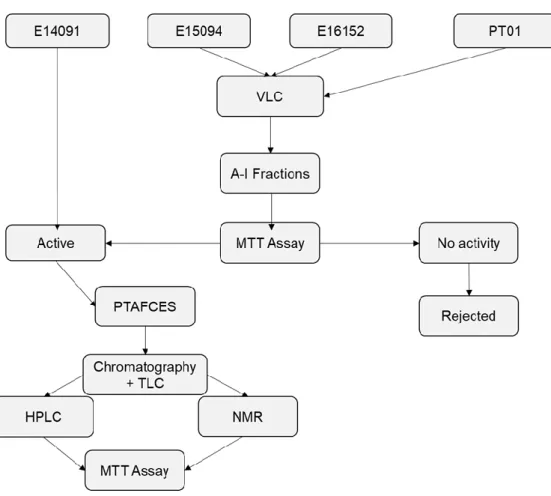
Figure 5 Bioassay guided fractionation design used in this study 29
Figure 6 Effects of fractions from E15094 in 10 µg/ml in the viability of
HCMEC/D3, RKO and T47D 30
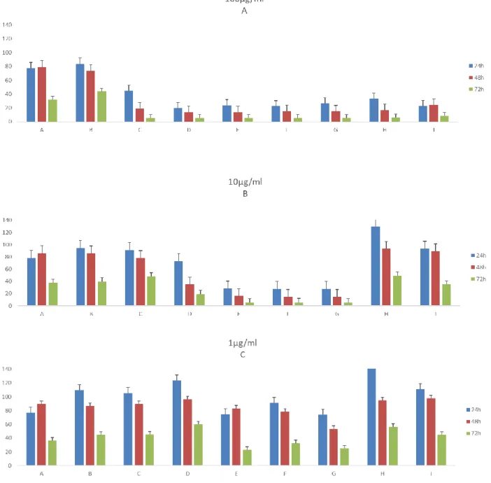
Figure 7 Effects of fractions from PT01 in 100 µg/ml (A), 10 µg/ml (B) and 1
µg/ml (C) in the viability of T47D. 31
Figure 8 Effects of fractions from PT01 in 100 µg/ml (A), 10µg/ml (B) and 1
µg/ml (C) in the viability of RKO 33
Figure 9 Effects of fractions from PT01 in 100 µg/ml (A), 10µg/ml and 1 µg/ml
(C) in the viability of HEPG2 34
Figure 10
Effects of fractions from PT01 in 100 µg/ml (A), 10µg/ml and 1 µg/ml
(C) in the viability of HCMEC/D3 35
Figure 11
Effects of fractions from E16151 in 100 µg/ml (A), 10µg/ml and 1
µg/ml (C) in the viability of T47D 37
Figure 12
Effects of fractions from E16151 in 100 µg/ml (A), 10µg/ml and 1
µg/ml (C) in the viability of RKO 38
Figure 13
Effects of fractions from E16151 in 100 µg/ml (A), 10µg/ml and 1
µg/ml (C) in the viability of HEPG2 39
Figure 14
Effects of fractions from E16151 in 100 µg/ml (A), 10µg/ml and 1
µg/ml (C) in the viability of HCMEC/D3 40
Figure 15
Effects of fractions from PTAFCES in 20 µg/ml in the viability of
HEPG2 (A) and HCMEC/D3 (B). 41
Figure 16
Chromatographic profile of the PTAFCES 12-19 through the
IV
List of tables
Table 1 Pisolithus compounds known for having bioactive properties 11 Table 2 Composing fractions of PTAFCES and total mass obtained 17 Table 3 Mixing ratio, volume and type of solvents used to perform the
gravitational chromatography from PTAFCES
18 Table 4 Fraction obtained with each tube and total mass recovered from
PTAFCES chromotography
18 Table 5 The four-different conditions of experimental extractions and
respective recovered mass and yield efficiency
22 Table 6 Data about E15094 fractionation through VLC and extraction
procedure
24 Table 7 Data about PT01 fractionation through VLC and extraction procedure 25 Table 8 Data about E16152 fractionation through VLC and extraction
procedure
1
1.
Introduction
1.1. Basidiomycota and chemical properties;
Basidiomycota are one of the big divisions of the higher kingdom of Fungi (Moore, 1998). In this group, we can include a vast number of types of Basidiomycota like mushrooms, puffballs, earthstars and other diverse organisms. Their ability to produce countless number of secondary metabolites aroused attention of the scientific community to discover the of these new compounds properties. The rise of synthetic substances and chemical syntheses did not stop the exploration of natural compounds. Furthermore, today, more than ever, the necessity to give response a wide range of problems, the search for secondary metabolites with biological activity happens at an explosive rate. Therefore, most of the new drugs created by pharmaceutical industries are from natural products, with 80% of this medicine having this origin (Wasser, 2014) (Chihara, Maeda, Hamuro, Sasaki, & Fukuoka, 1969). To reinforce the importance of these compounds, drugs like antibiotics (penicillin and erythromycin), anti-obesity pharmaceuticals (lovastatin) and immunosuppressants (cyclosporin) have their origin in secondary metabolites of Basidiomycota
Distinctive societies use these organisms in their diet, given their marked taste and the ease of growth in very extreme environments. Furthermore, they can be composed up to 40% of protein of its dry weight, being an excellent source of amino acids, possessing a good nutritional value (Money, 2016). Due to these facts, the ancient civilizations use the basidiomycotas to treat health problems. Oriental societies were pioneers in use of the Shiitake (Lentinus edodes) and Reishi (Ganoderma lucidum) in the treatment of different cancers and immunodeficiency disorders. The are other reports in the use of the psychadelic mushroom Amanita muscaria in the Mesoamerican, Celtic and Russian cultures with purpose of spiritualism and to reach a higher level of conscience (Chang & Miles, 1989). However, only with the advent of the chemical syntheses and the development of the biochemistry, the researchers started to get insights of the mechanisms of actions of these compounds. In 1969, it was isolated a polysaccharide, named Lentinan from Lentinus edodes, showing a strong anticancer activity in mice sarcoma 180. The isolation of this compound led researchers to look to the basidiomycota as new potential source of anticancer drugs.
2 As one of the deadliest disease, cancer is still draw the scientific community to find new approaches to fight this illness. In fact, the World Health Organization (WHO) reports that cancer is the main cause of mortality in 1 in 6 dead’s and is expected the number of oncological patients rise by 70% until 2030.(McGuire, 2016)
Traditionally, the western medicine relies on chemotherapy as a way to treat cancer. However, most of the drugs used in this technic are cytoxic substances, with a lack of selectiveness, targeting the normal and the oncological cells to induce the apoptosis. As consequence of this aggressive therapy, most patients suffer from side effects with immunosuppression, opening a door to opportunistic infections(Patel & Goyal, 2012). Nausea and acute pains are usually common, along with an impact in the patient self-esteem. Most of the drugs with origin in mushrooms are use as complementary of chemotherapy and radiotherapy, mostly to stimulate immune system to attack the cancer cells and preventing the infections from other diseases. Following this problematic, the modern pharmaceutical industry is trying to discover new compounds with different mechanisms of actions of the mainstream drugs. The lucrative market of the cancer drugs has reallocated most of the funds to the research of the innovative therapy’s with experience of the oriental cultures of medical mushrooms (Wasser, 2014). To reveal the importance, several drugs with origin in mushrooms are covered by the Japanese Health System. In 1993 25 % of anti-cancer drugs in the Nippon market had origin in mushrooms (Mizuno, 1999). Other study reports that workers from edible mushroom farm in Nagamo District (Japan), and consequently their diet were based in this Fungi, had a much lower rate of cancer incidence than the general population. (Ikekawa, 2001).
The most studied are the β-Glucans, a type of β-D-glucose polysaccharides, present in the cell wall in different organisms, including fungi. Most of this β-Glucans are chains
glucose polysaccharides connected by β-type glycosidic bonds (Lemieszek & Rzeski,
2012; Ren, Perera, & Hemar, 2012). Proteins can be connected in the side chains creating molecules like polysaccharide-K (PSK). This polysaccharide is extracted from a basidiomycota called Trametes versicolor and some studies show the potential of PSK to be used as adjuvant in the treatment of several cancers like leukaemia, breast and liver cancer. This compound is able to stimulate the instigation of natural killer cells, T-cells, dendritic cells and induce the production of cytokines. (Fritz et al., 2015). Although β-Glucans are well known in literature, secondary metabolites produce by the fungi can have a beneficial effect rising the interest from the therapeutic view. Compounds like terpenoids, triterpenoids, lactones and alkaloids have been identified
3 has potential anti-cancer drugs. (Petronelli, Pannitteri, & Testa, 2009). Some of this compounds are capable to interfere with intracellular pathways, like panepoxydone, Extracted from Lentinus crinitus, panepoxydone can inhibit the DNA binding of nuclear factor kappa-light-chain-enhancer of activated B cells (NF‐κB) (Arora et al., 2014; Erkel, Anke, & Sterner, 1996). The deregulation of this molecular mechanism is a characteristic of many cancers and compounds inhibitors of NF‐κB can be used to mediate inflammatory process and hold the cancer cells proliferation.(Sethi, Sung, & Aggarwal, 2008). Moreover, enzymes with antioxidant potential has been documented, such as superoxide dismutase, glucose oxidase and peroxidase. The presence of
these enzymes can help the cells to deal with reactive oxygen species (ROS’s),
highlighting the importance of the fungi to prevent damages from oxidative stress. (Wei, Helsper, & Van Griensven, 2008; Wei & Van Griensven, 2008).
Figure 1-Mechanisms of action from the anticancer compounds from fungi (Wasser, 2014)
These compounds can be obtain from different parts from the fungi with diverse extraction processes, being available to the pharmaceutical market: (Wasser, 2014).
1. Extracts from freeze dried fruit body powders. 2. Extracts from freeze dried mycelium;
4 The constant demanding for new compounds with bioactive response put some pressure on pharmaceutical industry. In this perspective, Wasser, 2014 established a protocol called Drug Discovery Procedure, to put a standard process to discover new therapeutic drugs.
Figure 2. The bioassay-guided fractionation process (Wasser, 2010; Weller, 2012)
Although these results are accepted in Eastern world, the scepticism in the Western Medicine can be explain by the rigid regulation enforced in these countries (Wasser, 2014) The lack of standardization and safety protocols can jeopardize the safe use of these compounds. The absence of information, leads Wasser, 2014 to also question if the bioactive effects are caused by an isolated compound or a group of them with a synergistic action. Other highlighted problem is the possibility of the compounds extracted from fungi interact with common used drugs, leading to unknown side effects.
Biomass
Production
Biomass
extraction
Chemical
Fractions
Screening for
toxicity
Chemical
Elucidation
Clinical drug
development
3. Biomass or extracts from mycelium from liquid culture grown in a bioreactor; 4. Spores and their extracts;
5
1.2. Pisolithus tinctorius;
Pisolithus tinctorius (Pers.) Coker & Couch 1928 belong to basidiomycota phylum and
to the Sclerodermataceae family. They have the ability to establish a symbiotic relationship with host trees, boosting their grow and development (Marx, 1977). The structure of P. tinctorius can be divided in two main regions: sporocarp, the zone connected to the soil, with a hard and rigid texture; and an upper zone with soft tissue called pseudoperidioles, containing a brownish pigment and the spores of this fungus. This organism can reach 20 cm of diameter,(Coker & Couch, 1928) weight up to 1 kg, with the spores size range between 7 to 11 μm (De Zubiria, Horner, & Lehrer, 1990). Due to large spectrum of heterogeneity in the morphology and with the help of DNA sequence data, some authors question the actual classification (Cairney, 2002). Furthermore, the results from (Martin, Diez, Dell, & Delaruelle, 2002), state that P.
tinctorius is not a so called “pan-global super fungus”, but a complex of multiple
species with more restrict geographical presence and limited host.
The ectomycorrhizal relationship is also essential to invasive tree species to colonise new habitats, with the prominent cases being the introduction of eucalypts in the
Iberian Peninsula (Díez, 2005) or the invasion of Pinus species in
Australia.(Richardson, 2000). In both cases, Pisolithus played a big role in contributing to the success of the invasion.
Highly used in reforestation, ectomycorrhizal fungi, such as P. tinctorius are key to the success of silviculture programs. The formation of fungal hyphae permits a deeper penetration in the soil allowing more efficient uptakes of N and transport to the host (Oliveira, Franco, & Castro, 2012). (Peay, Bruns, Kennedy, Bergemann, & Garbelotto, 2007) stated that the fungi can provide 86% of the N host need’s and receive 15% of carbohydrates. Nutrients are not the only benefit the host can receive. Since the ectomycorrhizas have a tolerance to high concentration of heavy metals, P. tinctorius showed a potential to be use for bioremediation of contaminated soils (Tam, 1995). The formation of the hyphal net around the host roots can serve of physical barrier preventing predators and pathogens to attack the tree. P. tinctorius have an important role in producing compounds with anti-fungal and anti-bacterial activity providing an extra protection. (Tsantrizos, Kope, Fortin, & Ogilvie, 1991).
6 This result, highlighted the possibility of use the secondary metabolites in diverse fields of the biomedical sciences, boosting the research on understanding all bioactive potential of P. tinctorius.
1.3. Bioactive potential of Pisolithus
As was stated before, nowadays the main potential of P. tinctorius is centrally in the economic value due to the ability to boost reforestation programs. This interaction host-fungi increases the concentration of certain compounds, as terpenes, in the host whose function is to protect it against fungal infections (Abreu, 1987). For this motive, studies were done to verify the presence of allelochemicals, in other words, secondary metabolites that can influence the growth of other organisms. The presence and the understanding of these compounds in P. tinctorius can open doors to new approaches in biomedical fields as the constant resistance to antibiotics and the search for new compounds, to be used as fungicides, explore the possible potential as antioxidant or even inhibit the grow of cancer cells as was demonstrated by Montenegro et al., 2004
1.3.1. Anti-cancer potential
As was describe before, cancer is considered of the most problematic disease that we face off. The actual therapies had demonstrated low efficiency with high collateral effects for the patients. Consequently, there has been a constant focus and growing interest from researchers to find new useful compounds to fight this infirmity. To verify the anticancer potential of P. tinctorius, some studies were made, especially with pisosterol, a compound primarily isolated by Gill, Kiefel, Skelton, & White, 1989, which is a triterpene present in the sporecarp of the fungus.
Montenegro et al. in 2004 conducted the first study concerning the anticarcinogenic action of pisosterol. This triterpene was tested in vitro, in erythrocytes of rats, sea urchin’s eggs and in the human cancer cell lines, such SF-268, B16, PC-3, MCF-7, HCT-8, HL-60 and CEM. By recurring to MTT assay, the authors were able to verify the mitochondrial activity of the cancer cell lines. Data revealed, membrane disruption in mouse erythrocytes was not observed and pisosterol has not interrupted the development of the sea urchin eggs. However, there was a strong growth inhibition in
7 all cancer cell lines, more notably in leukemia and melanoma cells. The same authors suggests that the selectivity have a different mechanism of action than the common cytotoxic drugs.
With this data, Montenegro and his team in 2007, conducted a study to determine if pisosterol was able to induce differentiation using HL-60 cell lines (leukemia cells) and normal cells peripheral blood mononuclear cells (PBMC). The data showed that HL-60 cells treated with pisosterol tended to differentiate into monocytic cells. Apoptosis was detected in HL-60 cells exposed to pisosterol, despite no cytotoxicity was observed in PBMC cells, even in the highest concentration (5 μg/ml), suggesting that pisosterol can be selective to cancer cells lines and revealing the potential of this compound to be used in the cancer treatment. This study was determinant since it is essential to evaluate the damages in the dividing proliferating lymphocytes, to prevent collateral damages. (Anazetti, Melo, Durán, & Haun, 2003)
The cancer cell HL-60 genome has a proto-oncogene called C-MYC, located in the homogeneously staining region (HSR) 8q24 aberration and has an important function on regulating the cell life cycle. It is known that, cancer cells with low degree of expression of C-MYC are less aggressive and so, more susceptible to chemotherapy treatment. (Eckhardt et al., 1994; Fegan, White, & Sweeney, 1995). It was observed that 90% of the analysed cells lacked this HSR region when threated with pisosterol. Consequently, these results opened the possibility of the use of pisosterol combined with chemotherapy, since this compound has the ability to block aggressive mechanisms of certain cancers.
Pisosterol was also tested in human glioblastoma cell lines.(Pereira et al., 2011) These type of cancer cells are known for their acute aggressiveness with an unfortunately, average rate survival of one year after being detected (Pinto et al., 2009). The study used two different cell lines, U343 and AHOL1, and before the treatment, 72% of U343 and 65% of AHOL1 cells registered more than two alleles of C-MYC. By being exposed to 1.8 µl/ml of pisosterol, only 33% of U343 cells and 15% of AHOL1 cells showed signs of containing C-MYC, signifying that pisosterol also blocks the expression of this proto-oncogene.
Since there was no data regarding the anticancer activity of pisosterol in in vivo assays, Montenegro et al., 2008 conducted a study where Sarcoma 180 tumours were transplanted to Swiss female mice and treated with 50 mg/m2 and 100 mg/m2 of pisosterol. The results showed that this compound was able to inhibit tumour growth by 43%. Morphological exams were made to evaluate the influence of pisosterol on the
8 mice organs. The treatment had an impact in the liver, showing kupffer cells hyperplasia, focal infiltrate of inflammatory cells, centriolobular vennus congestion, demonstrating that the liver is a target organ of pisosterol. Although, the authors concluded that the damages can be reversible since no stromal fibrosis was detected and conjunctive tissue was preserved.
Figure 3. Chemical structure of Pisosterol
Most of the studies that were done, have use the mycelium or the sporecarp to perform extraction, ignoring other structures of P. tinctorius such as the spores. The inoculation of the spores, through an allergy skin test, had provoked an allergy symptoms in 9% of the subjects (De Zubiria et al., 1990). Workers from silviculture industry and college students were subjected to the skin prick test with spores extracts. The workers group was the most affected, with 28.2% showing positive reactions, and 82% of individuals from these positive tests developed symptoms of respiratory allergy. The authors stated that, since this basidiomycete is used to boost reforestation, the exposure to spores can be considered a possible occupational hazard by provoking respiratory allergic asthmas.
Considering the absence of data in the literature, regarding the bioactivity potential of the P. tinctorius spores, (Alves, Preto, Vasconcelos, Oliveira, & Martins, 2015) conducted a work to assess the presence of anticancer substances. Firstly, it was evaluated which solvent had the major yield extraction and less mitochondrial activity. The result showed that the mixture of DCM:MeoH (2:1) had the best yield considering the mitochondrial activity. Starting from the crude extract, through successive bioassay-guided fractionation, 11 fractions were screened for cytotoxicity in MG63 (bone osteosarcoma),T47D (human breast tumour), RKO (rectal carcinoma) cell lines and simultaneity with normal endothelial cells. The data revealed a decrease in cancer cell
9 viability with a low impact in the normal cells. n other hand, considering the very low yield of extraction, it was not possible to purify, identify and isolated the compound(s), responsible for the anticancer response. So Alves et al., 2015 suggested in further studies, improving the extraction technique to help isolate the active compound.
1.3.3. Antimicrobial Potential
The discovery of penicillin has opened a new era in modern medicine, giving us the possibility to fight a large spectrum of diseases caused by bacteria (Golkar, Bagasra, & Pace, 2014). The massive use of antibiotics has lead us to an increasing resistance of the bacteria to these compounds. Some authors state this is probably the biggest threat to the human public health in the XXI century (Ventola, 2015). The statistics shows that more people die in USA of diseases provoked by methicillin-resistant
Staphylococcus aureus (MRSA) than HIV/AIDS, Parkinson’s disease, emphysema, and
homicide together. (Golkar et al., 2014; Rossolini, Arena, Pecile, & Pollini, 2014). Therefore, it has been an intense research to find new compounds that demonstrate efficiency to fight the so called super bugs, and fungi are a promising source for this type of drugs.
The first report of antimicrobial activity from P.tinctorius came from Kope and Fortin in 1989, when antifungal activity in cell free cultures was observed, provoking hyphal lysis to a different organisms. To understand the mechanism behind this activity, Tsantrizos et al., in 1991 isolated the pisolithin A and pisolithin B from P.tinctorius mycelium. . Although these metabolites were know in the literature, there was no data about their antifungal potential. These compounds showed a great capacity to reduce the mycelial grow of several phytopathogenic fungi such as Rhizoctonia praticola, Truncatella
hartigii, Sphaerosporella brunnea, Fusarium solani, Brunchorstia pinea and
dermatopathogenic like Microsporum gypseum and Trchophyton equinum. The authors noticed also that the inhibition was similar, and in some cases higher than antifungal chemical agents such as nystatin and polyoxin D.
Tackling the problematic of the resistance of the bacteria’s to the antibiotics, Ameri, Chanchala, Vaidya, & Deokule in 2011, conducted a study with crude extract and isolated fractions of P. albus against strains of MRSA. The collected fungi where associated with Acacia auriculiformis and Eucalyptus globulus trees. The crude was extracted with ethyl acetate, methanol and water and the fracts were from zones of
10 diterpenoids, triterpenoids, sesquiterpenoids, and polysaccharides I, II, IIIA and IIIB. The results showed that the crude extract with ethyl acetate and methanol had a strong antibacterial activity. Likewise, the fraction containing sesquiterpenoids displayed a robust action with the minimal inhibitory concentration determined to be in a range of 0.62 to 1.2 mg/ml. The author declared that with these results there are potential to the secondary metabolites of P. albus be use as antimicrobial drugs against certain strains of resistant bacteria. It also interesting to highlight the fact of P. albus collected from
Acacia auriculiformis, did not show any pronounced activity against the majority of
MRSA strains, proposing the possibility of that different hosts can induce the production of diverse compounds. More recently, an assay was conducted with extracts of eight species of ECM fungi (including P. albus) for tested anti-fungi potential against nine types of plant pathogenic fungi. It was observed that Pisolithus had an inhibitory effect in average 33.27% of those plant pathogenic fungi.(Mohan, Nivea, & Menon, 2015a)
1.3.5. Other bioactivity potential s:
Regarding the basis that supports the possibility of an immunosuppressive potential in fungal compounds, (Fujimoto, Nakayama, Nakayama, & Yamazaki, 1994) was responsible for isolating and characterising the components, using three different species of mushrooms, including P. tinctorius. With this fungus, he extracted compounds such as pisolactin and ergosterol peroxide, where the IC50 was evaluated in relation to the proliferation of lymphocytes in rat spleens stimulated with concanavalin A and lipopolysaccharides.
Additionally, in 2011, the antioxidant potential was also unveiled. In aerobic organisms, free radicals are constantly being produced during natural cell metabolism, and there are compounds that can counteract undesirable forms of active oxygen in the body, detoxifying it. For that effect, (Reis, Ferreira, Barros, & Martins, 2011), did a comparative study between tocopherol composition and the antioxidant properties of the fungus in vivo and in vitro, using fruiting bodies and mycelia respectively, obtaining as a result a high antioxidant capacity resulting from P. tinctorius, mainly when using
11
Extract/Compound Fungal structure
Fungal
species Assay Biological activity Reference
Pisosterol
Mycelium;
Sporecarp P. tinctorius
MTT (Montenegro et al. 2004; Montenegro et al. 2007) NBT reducing activity (Montenegro et
al. 2007)
Naphthyl acetate esterase activity (Montenegro et al. 2007) Antiproliferative effect (inhibition of DNA synthesis) Montenegro et al.
2007)
Mitotic index (Burbano et al. 2007;Pereira et al. 2011) In vivo assay (Montenegro et al. 2008)
Anticancer
(Montenegro et al., 2004) (Montenegro et al., 2007) (Burbano et al., 2009); (Carvalho Montenegro et al.,
2008); (Pereira et al., 2011)
12
Pisolithin A & Pisolithin B Mycelium P. tinctorius Spore Inhibition Antifungi (Tsantrizos et al., 1991)
Pisolactona Sporecarp P. tinctorius
Proliferation of mouse spleen lymphocytes stimulation with
concanavalin A Lipopolysaccharide
Immunosuppressive (Fujimoto et al., 1994)
Crude extracts Sporocarp P. albus Well agar diffusion method Antibacterial (Ameri et al., 2011)
Crude extracts Mycelium P.albus Well agar diffusion method
Chitinase activity Antibacterial
(Mohan, Nivea, & Menon, 2015)
Crude and Fraction
extracts from spores Spores P. tinctorius MTT Assay Anticancer (Alves et al., 2015)
Mycelium and Spores extracts
Mycelium;
13
Tocopherols Mycelium;
Sporocarp P. arhizus
DPPH radical-scavenging Activity
Reducing power and inhibition of b-carotene bleaching
(Reis et al., 2011)
14
2.
Aim and outline of this dissertation
All data collected until now supports the possibility of P. tinctorius to be used has a source of compounds with interesting bioactivity properties. Most of the studies were done with sporocarps as the source of fungal material, leaving the bioactive potential spores unexplored. The work performed by Alves et al. (2015) opened a door to explore this part of the fungus, more precisely its anticancer potential. Considering this, and the study executed by the main objective of this master thesis was to improve the extraction method from P. tinctorius spores, to obtain higher yield efficiency and to obtain more data about the compounds responsible for the cytotoxicity.
15
3.
Materials and Methods
3.1. Collection of Pisolithus tinctorius spores
In order to obtain samples of P. tinctorius, the method used by Alves et al. (2015) was followed. Fungi were collected from a forest ecosystem located in the north of Portugal, well known for the presence of this species. The P. tinctorius specimens were cut into small pieces, and the pseudoperidiole was lyophilised for 48 h, to dry the samples. After lyophilisation the spores were isolated with a help of a sieve with a pore of 1 mm.
3.2. Crude extraction and fractionation
Two extractions were performed following the method used by Alves et al., 2015. The spores were extracted with Dichloromethane-Methanol (2:1), through a sonication process. They were given the code names of PT01 and E15094. The conditions of the crude and fractionation are described in the table 5.
3.3. Improving the extraction technique
The low extraction yield with P.tinctorius spores obtained by the procedure already described, led to the necessary improvement of the method. In this sense, a step that uses glass beads was included. The use of glass beads to lyse cell walls and membranes is extensively used, as in the DNA extraction from cyanobacteria (Dederich et al., 2002; Mehta, Evitt, & Swartz, 2015). (Dederich et al., 2002; Mehta et al., 2015). Using this principle, a protocol was established to verify if the glass beads were useful to improve the extractions from spores, with four different experimental conditions. Sample 1 was extracted only with the solvent, dichloromethane-methanol in the proportion of 2 to 1. (DCM:MeOH (2:1)). Secondly, in sample 2 the same solvent mixed with 40 glass beads with a diameter of 3 mm. Next, with sample 3, the spores were extracted with DCM:MeOH (2:1) with the help of sonicator (Q55 Sonicator). Finally, the sample 4 was submitted an extraction the solvent DCM:MeOH (2:1) with the glass beads plus sonication. All the samples were extracted in 3 cycles with a duration of 5 minutes of sonication each with 10 minutes of rest (in part to avoid the over-heating, and to give time to the solvent penetrate
16 in spores) in a plastic tube of 50ml. Particularly, the sample 2 and 4 were subjected to agitation with vortex, to force the mechanical lysis through the glass beads. At the final of each cycle, the supernatant was decanted to 4 plastics tubes. After all the cycles were completed, the samples were left to rest during 24h, and the supernatants were transfer to pre-weighted vials with the assistance of a Pasteur pipette. These vials were evaporated under reduced pressure, with a rotary evaporator (Buchi Rotavapor® R-100) and the recovered weight was calculated. The vials were weighed before and after placement of each crude extract in order to calculate the extraction yield. The weight of spores used in each condition and volume of solvent used are describe in the Tables 6 and 7. Giving the results obtained using only the glass beads, this procedure was repeated for the subsequent preparation of extracts.
3.4. Full scale extraction with glass beads method
Given the obtained higher performance of extraction with glass beads, they were chosen to perform mechanical lyses. Instead of performing a full extraction, it was decided to extract the spores with small quantities and volumes of solvent, in order to optimise the solid/solvent ratio. With an average of 800 mg per plastic tube, the spores were extracted with 30 ml of DCM:MeOH (2:1) in 2 cycles with 40 to 50 glass beads with 3 mm of diameter. During each cycle, the tubes were agitated with a vortex for 5 min, and then allowed 5 min to rest. At the end of the sequence, the supernatant was decanted to a glass flask. Totally, 61.29g of spores were extracted with a total volume of 2250 ml of DCM:MeOH (2:1) mixture. After 24h, the supernatant was retired to a glass balloon, carefully with the help of Pasteur pipettes not to perturb the deposit in the bottom of the flask. Next, the solvent was evaporated under reduced pressure, with a rotary evaporator and transferred with chloroform into pre-weight vial and evaporated again. This crude extract, with the code name E16152, was stored in a pre-weight vial, and the recovered mass and the respective yield calculated.
3.5. Fractionation of the E15094, PT01 and E16152 crude extract.
All crude extracts were fractioned through vacuum liquid chromatography method (VLC) (Targett, Kilcoyne, & Green, 1979). The 5g of silica gel (Merkel 0.015-0.040) were packed hard with support of vacuum and the crude extracted was transferred into the top of the column and 400ml of hexane were continually being added until the column absolved all
17 extract compounds. Continuing, a series of different mixtures with increasing polarity was charged in the column, until obtaining 9 distinctive fractions. (A with lowest polarity and I with highest). The mixtures of solvents and their volumes, along with each mass recovered from the fraction are described in the Tables 6, 7 and 8. The fractions were collected in round bottom flask and all the solvents were dried under reduced pressure, in a rotary evaporator. The dried fractions were dissolved in chloroform and transferred to pre-weight vials and evaporated again to determine the weight of the dried mass.
3.6. Sub-Fractionation of PTAFCES
The fractions with higher anticancer activity had such low masses, that isolating and testing the compound(s) responsible for that response would prove very difficult. Therefore, it was decided to collect all the fractions with higher activity from E15094, E16152, PT01 and E14030 (this extraction was performed by (Alves et al., 2015)) in a single collection called PTAFCES. Table 2 describes which fraction were pooled together and their mass.
Table 2. Composing fractions of PTAFCS and total mass obtained Extraction code Fractions Total mass (mg)
E14030 G, H
133.43
E15094 F, G
PT01 E, F, G
E16152 E, F, G
All the mass of the PTAFCES were sub-fractionated through a chromatography column. Here it was used 20g of silica gel (Merkel 0.015-0.040) as stationary phase and a variety of solvents mixtures with increasing polarity as mobile phase. In total 187 test tubs were collected in seven different elution mixtures (Table 3). Previously to this, Thin Layer Chromatography (TLC) was performed to establish the mobile phase that result in better separations of the compounds present in the extract. Later TLC was used to identify the tubes to mixture in the same pool. Under this technique, a small sample (10 to 20 µL) of the fraction was collected from every 5 tubes with a capillary, and put in a TLC plate. The same plate was placed in a TLC chamber with the mobile phase with increasing polarity,
18 to drag the most polar substances. All the spots present were verified under a UV light and marked with a pencil.
Table 3. Mixing ratio, volume and type of solvents used to perform the gravitational chromatography from
PTAFCES
Tubes Solvent Mixing ratio (%) Volume (mL) 1-46 Hex- EtOAc 60-40 300 47-62 Hex- EtOAc 50-50 100 63-106 Hex- EtOAc 40-60 300 107-127 Hex- EtOAc 30-70 100 123-138 Hex- EtOAc 20-80 100 139-155 Hex- EtOAc 10-80 100 156-171 EtOAc 100 100 172-187 EtOAc-MeOH 50-50 100
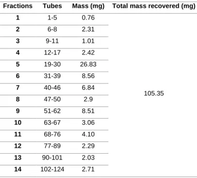
It was analysed the spots marked on the TLC and regroup the tubes solutions by the presence of the same, or very similar compounds. This reorganization outcome in 22 fractions, resulting of being transferred to pre-weighted vials, evaporated under reduced pressure, and calculated the recovered mass. (Table 4)
Table 4. Fraction obtained with each tube and total mass recovered from PTAFCES chromotography Fractions Tubes Mass (mg) Total mass recovered (mg)
1 1-5 0.76 105.35 2 6-8 2.31 3 9-11 1.01 4 12-17 2.42 5 19-30 26.83 6 31-39 8.56 7 40-46 6.84 8 47-50 2.9 9 51-62 8.51 10 63-67 3.06 11 68-76 4.10 12 77-89 2.29 13 90-101 2.03 14 102-124 2.71
19 15 125-133 1.27 16 134-142 1.14 17 143-154 1.22 18 155-168 1.26 19 169-175 0.51 20 176-179 10.09 21 120-181 7.41 22 182-187 8.07
3.7. Preparation of the test samples
From the crude extractions and the fractions E15094, PT01 and E16152 a test sample at a concentration of 100mg/ml were prepared. The dried weight was dissolved in chloroform in a vial, and from this solution successive dilutions were done in order to prepare other concentration test of 10mg/ml and 0.1mg/ml. These vials were evaporated using a
nitrogen stream and 100 μl of dimethyl sulfoxide (DMSO) were added to each vial. The
fractions from the collection PTAFCES were under the same process, with the exception that a unique concentration of 0.2mg/ml was prepared.
3.8. Preparation of the PTAFCES for nuclear magnetic resonance spectroscopy (NMR)
To verify the purity and to evaluate the chromatography efficacy observe the chemical structure of the compounds present in each sample, the 22 fractions from PTAFCES were
submitted to 1H NMR analysis. Each NMR tube was pre-washed with acetone, chloroform
and methanol to eliminate the presence of contaminants and dried thoroughly in high vacuum. Each fraction was solubilized in deuterochloroform and loaded into the prepared
NMR tubes. Each fraction was solubilized in deuterochloroform (CDCl3) and loaded into
the prepared NMR tubes. The 1H NMR spectroscopy was carried out at Centro de
Materiais da Universidade do Porto (CEMUP), using a BRUKER AVANCE III 400 MHz, 9.4 Tesla to perform this assay.
20
3.9. Chromatographic scan using High Pressure Liquid Chromatography (HPLC) of PTAFCES samples
In the results from the cytotoxic essay, revealed a strong cytotoxic activity in the fractions 12-19. When these results were compared with NMR spectra, it was possible to identify some common peaks, showing the identical compounds are present in different fractions. Given this, the PTAFCES 12-19 was submitted to High Pressure Liquid Chromatography (HPLC) to verify the chromatography profile of this set of fractions. The 10µl of PTAFCES 12-19 was injected in a Synergi 4u Fusion-RP 80A 250x10mm column, with a flow of 2.0ml/min, with absorbance read at 280 nm and 254 nm, with an isocratic mixture of 100% MeCN. It is important to note it was only injected the soluble part in MeCN, with other part being preserved.
3.10. Cell Cultures
The cytotoxicity of samples was tested against 3 cancer cell lines and 1 normal cell line: • T47D (human breast cancer cell line);
• RKO (human colon adenocarcinoma cell); • Hep G2 (hepatocyte carcinoma);
• hCMEC/D3. (human brain capillary endothelial cells)
Cells were incubated at 37ºC with a 5% CO2 humidified atmosphere, and culture medium
was renewed every 2 days. When flask fill up with 80% cell confluence, adherent cells were free with TrypLE Express Enzyme (Gibco and culture medium was renewed every 2 days. Considering this, the T47D cells were purchased from Sigma-Aldrich, RKO and Hep G2 cell lines were obtained from the American Type Culture Collection (ATCC) and finally the hCMEC/D3 were provided by Dr. P. O. Couraud (INSERM,France). Cells were grown in Dulbecco’s modified Eagle’s medium (DMEM, GlutaMAX, Life Technologies), pH 7.4, supplemented with 10% fetal bovine serum, 2.5 μg/ml fungizone, and penicillin– streptomycin (100 IU/ml and 100 μg/ml, respectively).
21
3.11. Cell viability assay
The (4,5-dimethyl-2-thiazolyl)-2,5-diphenyl-2H-tetrazolium bromide (MTT) reduction assay is a colorimetric test to measure mitochondrial activity. In this context, the mitochondria’s are responsible to reduce this yellow dye into formozan purple crystals and so, the absorbance of this colored solution can be read by a microplater reader (van Meerloo, Kaspers, & Cloos, 2011). This mitochondrial activity can be proportional to cell viability considering the 100% viability in the control (cells treated with 1% DMSO). For the cell
viability assay, the cells were seeded with a density of 3,3x10⁴ cel/ml in triplicate in 96 well
plates (previously counted in a Neubauer chamber). After 24h of incubation, the medium
was removed and 1μl of the fractions samples diluted in 100 μl of fresh medium was
inserted, to cells being expose to 100, 10 and 1 μl/ml (the PTAFCES samples were tested at 20 μl/ml) during 24h, 48h and 72h. Furthermore, were establish positive controls (cell being exposure with medium and 1% of DMSO) and negative controls (cells treated with medium and 20% of DMSO). After this period of exposure, the cells were treated with 10 μl of MTT at a concentration of 0.05mg/ml with an incubation period of 4h. After this time,
the cell medium was retired and 100 μl of DMSO were added to solubilize the purple
formazine salts. The absorbance of these crystals was measure with microplate reader (Bio-tek Synergy™ HT) at of 550 nm, and results expressed as a percentage of cell viability considering 100% viability in the control cells.
3.12. Statistical Analysis
Microsoft Office Excel 2016 was used to treat the data from MTT assay. The results are presented as means ± standard deviation (SD) from three independent experiments, when compared with control results. The same software was used to display the data graphically.
22
4.
Results
4.1. Improving the extraction technique
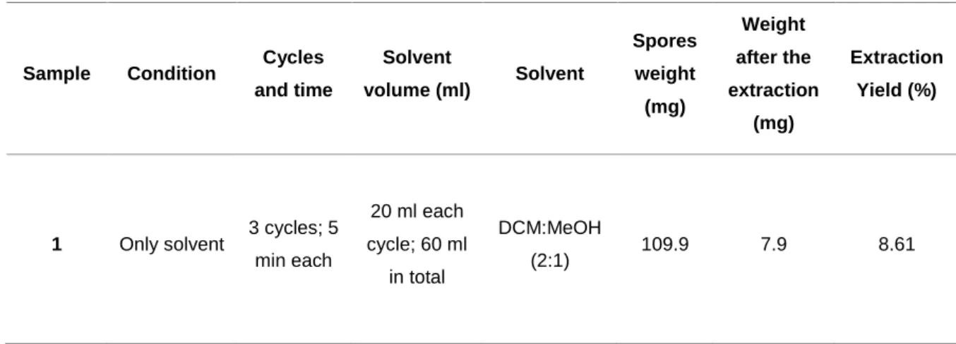
Improvement of the efficiency of the extraction was crucial to obtain sufficient mass to use a bioactivity assay guided isolation of compounds (using anticancer assays) and to perform the characterization of the same substances. The four-different conditions of extractions resulted in distinct extraction yields. Additionally, all the experimental conditions used the same volume of solvent mixture, submitted to the same time cycles, (with some slight differences in the weight of spore’s due to the scale sensitivity). Giving this, the standard extraction (sample 3) previously used by Alves et al., 2015 had resulted in 7.50 % of efficiency, being the lowest yield among the four, contrasting with the highest obtained with 40 glass beads plus sonication (17.71%). The use of only solvent to perform the extraction had given a yield of 8.61% and the use of the 40 glass beads resulted in an yield of 16.5%. Ideally, sample 4 would show the highest efficiency extraction and logical the first choice to be use in the final extraction, however the method used with sample 2 was chosen to perform the large-scale extraction.
Table 5. The four-different conditions of experimental extractions and respective recovered mass and efficency yield
Sample Condition Cycles and time Solvent volume (ml) Solvent Spores weight (mg) Weight after the extraction (mg) Extraction Yield (%)
1 Only solvent 3 cycles; 5
min each 20 ml each cycle; 60 ml in total DCM:MeOH (2:1) 109.9 7.9 8.61
23 2 40 glass beads 3 cycles; 5 min each with agitation by vortex 20 ml each cycle; 60 ml in total DCM:MeOH (2:1) 97.2 16.1 16.5 3 Sonication 3 cycles; 5 min submitted in sonication 20 ml each cycle; 60 ml in total DCM:MeOH (2:1) 96.2 7.8 7.50 4 40 glass beads; Sonication 3 cycles; 5 min with agitation by vortex; 2 min submitted to sonication 20 ml each cycle; 60 ml in total DCM:MeOH (2:1) 112 15.8 17.71
4.2. Fractionation of the E15094, PT01 and E16152 crude extract
Following the extraction method of Alves et al. (2015) it was possible to obtain two crude extracts, as cited above, with the codenames of E15094 and PT01, with yields of 1.9% and 2.2%, respectively. After a detail observation of the fractionation table data of E15094 (Table 6), it is possible to verify that 1.9% of the biomass was recovered with the standard protocol by Alves et al., 2015. Using this polarity gradient and looking to the results from Alves et al., 2015 this VLC was performed to obtain nine fractions with different weights between them. Fraction A and I represented more than half of the total weight, with 23.5% and 32.4%, respectively. Contrasting these results the fractions B (10.0 mg) and C (12.2 mg) denoted the lowest weight fractions.
24
Table 6. Data about E15094 fractionation through VLC and extraction procedure
Results from the extraction and respective fractionation of PT01 (Table 7) revealed a light increase of the yield by 0.3 % when compared with the E15094. This same extract was divided in nine fractions, following the same weight pattern, with the fraction B and I having most mass of the extract. Furthermore, the fractions D, E, F and G only totaling 13.35 mg, represent 1.3% of the total mass of the extract. The use of glass beads in the extraction process, almost duplicated the yield when compared with the previous extracts. With an efficiency of 4.0%, it was possible to recover 2388mg of extract, distributed in 9 fractions. After a more detailed observation of Tables 6, 7 and 8, it is possible to affirm, that the fractions A and I are the ones with more mass, comparing with F and G, which are the fraction that contain less mass. Nevertheless, these fractions have more mass when comparing with the ones from PT01.
Extract name Spores weight (g) Recovered mass (mg) and yield extraction (%)
Fractions Solvent Mixing ratio (%) Volume (mL) Fractions mass (mg) E15094 50.0 950; 1.9% A Hex- EtOAc 90-10 200 223.6 B Hex- EtOAc 80-20 100 10.0 C Hex- EtOAc 60-40 100 12.2 D Hex- EtOAc 50-50 100 34.3 E Hex- EtOAc 40-60 100 127.6 F Hex- EtOAc 20-80 100 104.5 G EtOAc 100 150 40.0 H EtOAc-MeOH 75-25 150 89..2 I MeOH 100 300 308.1
25
Table 7. Data about PT01 fractionation through VLC and extraction procedure
When compared with the glass beads extraction E16152 (2388mg) with a yield of 4.0%, the recovered weight of the E15094 and PT01 (950mg and 977mg) was considered very low, proving that this method improves the spore’s extractions in large-scale extractions.
Extract name Spores weight (g) Recovered mass (mg) and yield extraction (%)
Fractions Solvent Mixing ratio (%) Volume (mL) Fractions mass (mg) PT01 44.42 977mg 2.2% A Hex- EtOAc 90-10 400 139.93 B Hex- EtOAc 80-20 250 202.53 C Hex- EtOAc 60-40 250 12.46 D Hex- EtOAc 50-50 250 3.16 E Hex- EtOAc 40-60 250 4.95 F Hex- EtOAc 20-80 250 3.01 G EtOAc 100 250 2.23 H EtOAc-MeOH 75-25 250 47.81 I MeOH 100 500 597.11
26
Table 8. Data about E16152 fractionation through VLC procedure
Extract name Spores weight (g) Recovered mass (mg) and yield extraction (%)
Fractions Solvent Mixing ratio (%) Volume (mL) Fractions mass (mg) E16152 61.29 2388mg 4.0% A Hex- EtOAc 90-10 400 427.60 B Hex- EtOAc 80-20 300 159.94 C Hex- EtOAc 60-40 250 220.61 D Hex- EtOAc 50-50 250 129.9 E Hex- EtOAc 40-60 250 60.66 F Hex- EtOAc 20-80 250 25.47 G EtOAc 100 250 15.51 H EtOAc-MeOH 75-25 250 82.0 I MeOH 100 600 642.38
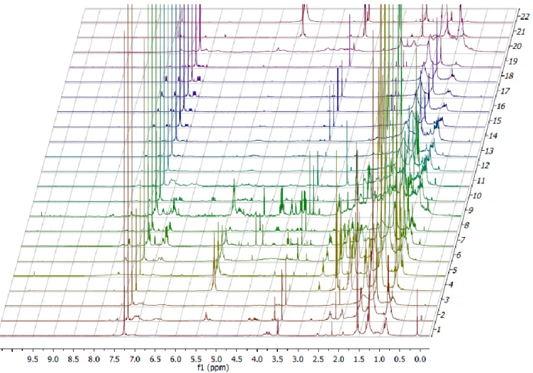
4.3. The NMR spectra from the PTAFCES.
The NMR analyses from PTAFCES fractions showed multiple peaks differing between the 22 fractions. Therefore, the analyses (Figure 4) revealed that the fractions still complex, with the presence of multiple compounds. However, some common peaks were found, generally between the fractions 12-19, with some variance in the strength of the signal. It
was possible to observe the presences of doublets at 7.90 δ, 7.59 δ and quartet 7.10 δ.
Additionally, in all PTAFCES NMR spectra it is possible to observe strong signals, with singlets at 7.26 δ and 3.49 δ
27
28
4.4. Results from the chromatograph scan
Figure 16 presents the chromatograph profile of the extraction collection PTAFCES, with the blue and the red line representing the absorbance at 280 nm and 254 nm, respectively. In the chromatograph is possible to verify the presence of numerous peaks in the two wavelengths, in specific between the 6:00min and 23:00min. For instance, the first detected peak had a one strong signal, noticed at 6:30 min, corresponding to the solvent injection peak.
280 nm (280.00 nm) 254 nm (254.00 nm) AU 0.0000 0.0002 0.0004 0.0006 0.0008 0.0010 0.0012 0.0014 0.0016 0.0018 0.0020 0.0022 0.0024 0.0026 0.0028 0.0030 0.0032 0.0034 0.0036 Minutes 2.00 4.00 6.00 8.00 10.00 12.00 14.00 16.00 18.00 20.00 22.00 24.00 26.00 28.00 30.00 32.00 34.00 36.00 38.00 40.00 42.00 44.00 46.00 48.00 50.00 52.00 54.00 56.00 58.00 60.00 62.00 64.00
Figure 16. Chromatographic profile of the PTAFCES 12-19 pool. The blue and the
29
Figure 5. Bioassay guided fractionation design used in this study
4.5. Cell viability results
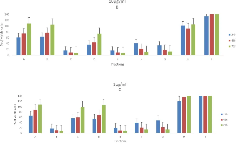
4.5.1. Cell viability results from E15094
The E15094 fractions were tested for cell viability at a concentration of 10µg/ml with time of exposure of 24h and 48h. Giving this, the figure 8 shows the effects of the fractions in the in the cancer and HCMEC/D3 cell viability. Regarding the T47D cell viability, the fraction A and I are the less toxic with 86% of the cells being viable after 48h. In other hand, only the 10% and 12% of the cells were viable when treated with the fraction F and G during 48h. The exposure of the RKO cell lines to the fraction A and I manifest less toxicity, with 73% and 74% of viability after 24h, with a slight increase 85% and 89% after
30 48h, respectively. Moreover, the fractions E and F demonstrate the strongest decrease of the cell viability (12% and 11% after 48h), with no relevant differences between the time exposure. Following the same pattern of the results obtained in HCMEC/D3. The data demonstrate that fractions A and I exhibit an increase of the number of the viable cells, superior to the control group in 120% after 24h of exposure, then dropping to 72% and 78% after an exposure of 48h, being the ones with less impact in the viability of the HCMEC/D3 cells. On contrary, the exposure of the fractions E and F result in only 38% and 37% of the cells were viable after 48h. In fact, by prolonging for 48h, the results are more acute with the viability being 13% and 14%, respectively.
31
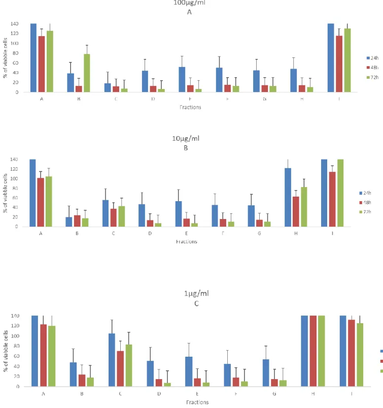
4.5.2. Cell viability results from PT01
The figures 7-10 illustrates the cell viability from exposure of nine fractions (in 100µg/ml, 10µg/ml and 1 µg/ml) from PT01 in four cell lines, during three-time periods. Concerning the effects of the fractions on viability of the T47D cells (Figure 7), it is clearly visible that the highest concentration provokes the lowest number of viable cells after 72h, more specifically the fractions C, D, E, F, and G. Yet, fractions E, F and G can still reduce drastically the viability the cell when exposed to the lowest concentration (1µg/ml) to 12% at 72h. It is also important to note, that in fraction I it was noticed an increase of the number of viable cells when compared with the control in all concentrations, more precisely 74% at 1µg/ml. It is also interesting results from fraction B, show to be more active in lowest concentration than other highest two.
Figure 6. Effects of fractions from E15094 in 10µg/ml in the viability of HDMC
(A), RKO (B) and T47D (C) Values are present as means ±SD of triplicate analyses when compared with control group (n = 3).
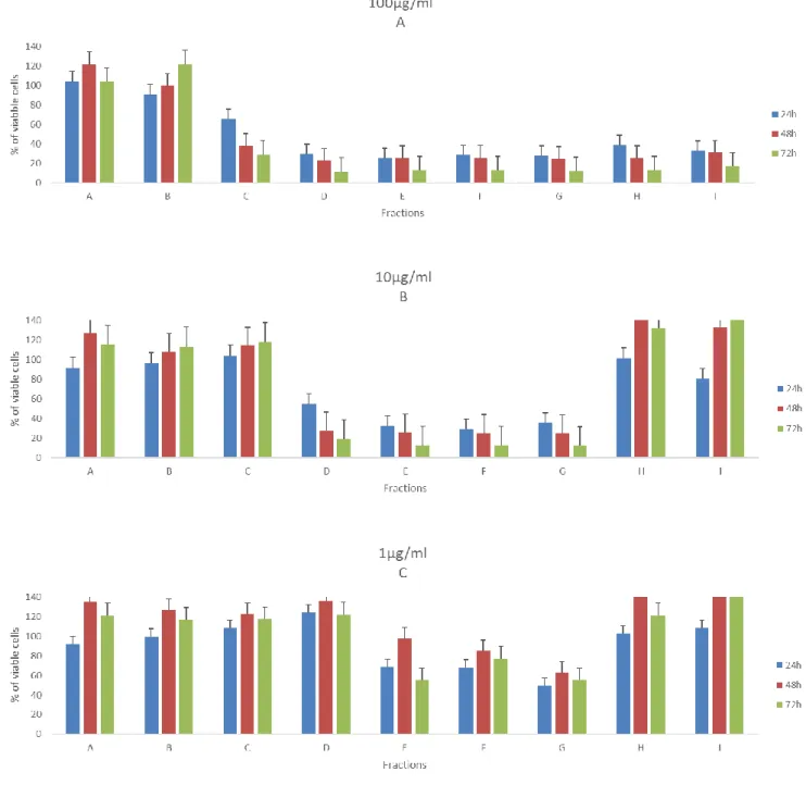
32 Regarding the RKO cell lines (figure 8), the most active fractions are the C, (the Fraction C in 1µg/ml showed low impact on the cell viability) D, E, F and G at 72h. On the contrary, it is possible to observe a cellular proliferation, when treated with the fractions A and I and compared with control, displaying no signs of being toxic at that concentration test.
Figure 7. Effects of fractions from PT01 in 100µg/ml (A), 10µg/ml (B) and 1µg/ml
(C) in the viability of T47D. Values are present as means ±SD of triplicate analyses when compared with control group (n = 3).