Antimicrobial activity of
Aspilia latissima
(Asteraceae)
Jeana M.E. Souza
1, Marilene R. Chang
2, Daniela Z. Brito
2, Katyuce S. Farias
1,
Geraldo A. Damasceno-Junior
3, Izabel C.C. Turatti
4, Norberto P. Lopes
4,
Edson A. Santos
5, Carlos A. Carollo
11Laboratório de Produtos Naturais e Espectrometria de Massas, Universidade Federal
de Mato Grosso do Sul, Campo Grande, MS, Brazil.
2Laboratório de Pesquisa em Microbiologia, Universidade Federal de Mato Grosso do Sul,
Campo Grande, MS, Brazil.
3Laboratório de Botânica, Universidade Federal de Mato Grosso do Sul, Campo Grande, MS, Brazil. 4Faculdade de Ciências Farmacêuticas de Ribeirão Preto, Universidade de São Paulo,
Ribeirão Preto, SP, Brazil.
5Coordenação do Curso de Licenciatura em Química, Universidade Tecnológica Federal do Paraná,
Apucarana, PR, Brazil.
Submitted: December 3, 2013; Approved: December 31, 2014.
Abstract
We evaluated the antimicrobial activity ofAspilia latissima- an abundant plant from the Brazilian
Pantanal region - againstCandida albicans, Candida parapsilosis, Candida krusei, Candida tro-picalis, Pseudomonas aeruginosa, Enterococcus faecalis, Escherichia coli and Staphylococcus aureus.The crude extracts and fractions showed activity in all tested microorganisms. The
chloro-form fraction of the leaves and roots showed the most antimicrobial activity againstS. aureus, with an
MIC of 500mg/mL. This fraction was submitted to bioautographic assays to characterize the activity of the compounds. Two bands from the leaves (L-A and L-B) and three bands from the roots (R-C, R-D and R-E) were bioactive. Within the root-derived bands, the terpene derivatives stigmasterol, kaurenoic acid and kaura-9(11), 16-dien-18-oic acid were identified. Antibiotic activity of A. latissimais reported for the first time.
Key words:Aspilia latissima,Staphylococcus aureus, bioautographic, MIC, terpenes.
Introduction
Drugs derived from natural products have made and continue to make huge contributions to human health. In-deed, they have been part of folk medicine for thousands of years (Kingston, 2011; Ortholand and Ganesan, 2004).
Natural products provide a diverse source of bio-active compounds, with an estimated 25% to 50% of mar-keted drugs having been obtained from natural sources (Newman and Cragg, 2012). This proportion increases to about two-thirds for antibacterial treatments, showing the importance of this class to infectious disease drug discov-ery (Roemeret al., 2011). According to Mishra and Tiwari
(2011), the treatment of infectious diseases with natural
products is efficient due to the ability of natural products to interact with specific targets within cells. Newman and Cragg (2012) showed that the pharmaceutical industry re-mains focused on treatments for infectious diseases, includ-ing microbial, parasitic and viral infections.
This interest in identifying new antimicrobial agents is due to the emergence of resistant microorganisms, which are a major problem in hospitals, particularly in intensive care units (ICUs). ICU patients are exposed to various antimicrobial agents, and this exposure provides a great op-portunity for the co-transmission of resistant bacteria from patient to patient (Rice, 2009).
There are approximately 270,000 species of plants all over the world, and Brazil is considered to have the greatest DOI: http://dx.doi.org/10.1590/S1517-838246420131281
Send correspondence to C.A. Carollo. Laboratório de Produtos Naturais e Espectrometria de Massa, Universidade Federal de Mato Grosso do Sul, Cam-po Grande, MS, Brazil. E-mail: [email protected].
Morin, 2003). Among the local flora, there are a large num-ber of species used in folk medicine as antimicrobial treat-ments, with some species belonging to the Asteraceae family. TheAspiliagenus (Asteraceae) exhibited such
bio-logical activities as antibacterial (Ambrosioet al., 2008)
and antifungal (Yongabiet al., 2009) effects, attributed to
the presence of kaurane-type diterpenoids and sesqui-terpene lactones (Bohlmannet al., 1981; Pageet al., 1992). Aspilia latissima, one of the species of theAspiliagenus, is
a semi-aquatic bush that grows in abundance on the banks of the Paraguay River in the Brazilian Pantanal region. The chemical or biological properties of this plant have not been studied yet. Given the emphasis on discovering new drugs, especially antibiotics, from plants, the present work fo-cused on evaluating the antimicrobial activity of A. latissimaand the chemical composition underlying those
effects.
Material and Methods
Plant material
Plant material (leaves and roots) was collected in March 2012 from adultA. latissimaplants on the banks of the Paraguay River, Corumbá, MS, Brazil (19°34’36”, S 57°1’11” O), with license from the Brazilian CGEN (n° 246/2009). The plant was identified by Prof. Dr. Geraldo Alves Damasceno Jr., and a voucher specimen was depos-ited at the GCMS Herbarium as number 5173.
Preparation of plant extracts
The dried leaves (430 g) were pulverized and perco-lated (ethanol:water 7:3) at 20 drops/min for 96 h. The per-colate was concentrated to obtain 190.06 g (44.2% yield) of the crude extract.
The dried roots (31 g) were extracted using a pressur-ized fluid extractor (ASE 150, Dionex) using ethanol:H2O (7:3) as the extraction solvent. The program consisted of 5 min of static time, a temperature of 100 °C, a washing vol-ume of 60% and a purging time of 100 s in two cycles on the 100 mL extraction cell. The extract was concentrated to ob-tain 7.21 g (yield 23.25%) of the crude extract.
The crude extracts from the leaves and roots were fractionated using the ASE extractor. The plant extract was adsorbed on 21 g of silica gel (70-230 mesh, Sigma, St. Louis, MO, USA), and a chromatographic column was pre-pared on the 100-mL extraction cell, with silica gel (22 g) in the bottom half and the adsorbed extract in the upper half. The solvent gradient used was hexane, chloroform, ethyl acetate, ethanol and ethanol:H2O (7:3). In the fractionation process, a 5-min static program was used, with a tempera-ture of 100 °C, a wash volume of 60% and a purge time of 100 s in two cycles. The fractions were concentrated and the yields calculated (Table 1).
Antimicrobial activity assay
The following strains from the American Type Cul-ture Collection (ATCC) were used for the antifungal and antibacterial evaluations: Candida albicans (ATCC 90028), Candida parapsilosis (ATCC 22019), Candida krusei (ATCC 6258), Candida tropicalis (ATCC 750),
Pseudomonas aeruginosa (ATCC 9027), Enterococcus faecalis(ATCC 29212), Escherichia coli (ATCC 25922) and Staphylococcus aureus (ATCC 25923), which were kindly provided by the Adolfo Lutz Institute-SP. The mi-croorganisms were maintained in BHI (brain heart infu-sion) broth with 15% glycerol to -20 °C. The tests were conducted from 24-h subcultures at 35 °C (± 2 °C) in Sabouraud Dextrose agar for fungi and Muller Hinton agar for bacteria.
Disc diffusion method
The evaluation of thein vitroantibacterial and
anti-fungal activity was performed using the disk diffusion method and following the guidelines of the Clinical and Laboratory Standards Institute in Doc. M44-A2 of the CLSI (2009) and M2-A8 of the NCCLS (2003). For the crude hydroethanolic extracts, two concentrations were used: 10 mg/mL and 40 mg/mL. For the fractions, only the 40 mg/mL concentration was used. The spherical discs were impregnated with 25mL, resulting in final concentra-tions of 250 mg/disc (10 mg/mL) and 1,000 mg/disc (40 mg/mL). As a negative growth control, discs impreg-nated with dimethylsulfoxide (DMSO) were used. As a ref-erence, discs containing antimycotic (fluconazole, 25mg) and antibacterial (ceftazidime, 30mg; oxacillin, 1mg; and vancomycin, 30mg) controls were used. The assays were performed in triplicate; the presence of a halo was consid-ered indicative of bioactivity. The diameter of inhibition zones were measured with a caliper.
Fraction Code Yield (%)
Leaves – hexane fraction L-HX 4.72
Leaves – chloroform fraction L-CHCl3 3.32 Leaves – ethyl acetate fraction L-EtAc 4.72 Leaves – ethanol fraction L-EtOH 61.2 Leaves – ethanol:water fraction L-EtOH:H2O 26.04
Roots – hexane fraction R-HX 0.22
Broth microdilution assay
The fractions with the highest activity in the disk dif-fusion were tested for their minimum inhibitory concentra-tion (MIC) using microbroth diluconcentra-tion, following the guidelines of the Clinical and Laboratory Standards Insti-tute in Doc. M27-A3 of the CLSI (2008) for fungi and M7-A6 of the NCCLS (2003) for bacteria.
The plant fractions were tested at final concentrations of 2,000, 1,000, 500, 250, 125, 62.5, 31.25, 15.125 and 7.8125 mg/mL. The interpretation of the tests was per-formed by visual inspection after 24 h at 35±2 °C for bac-teria and 48 h at 35±2 °C for fungi. To better visualize the growth of the micro-organisms, 30mL of resazurin solution were added to the cultures (100mg/mL) and incubated for 2 h at the same temperature.
Bioautography
Bioautography was performed using S. aureus for
thoseA. latissimaleaf and root fractions that showed low
MICs, using the methodology described by Valgaset al.
(2007) and Rahalison (1991). The thin layer chromatogra-phy (TLC) plates were eluted with CHCl3:EtAc (8:2) and dried completely before the experiment was conducted. The TLC plates were developed with a solution of p-iodo-nitrotetrazolium violet (INT) (2 mg/mL) and incubated for 2 h. The experiment was performed in duplicate. After in-cubation, inhibition zones were made visible (as clear ha-los).
Isolation and characterization of compounds
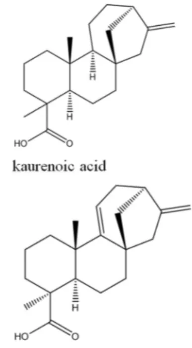
The fractions L-CHCl3and R-CHCl3were submitted to TLC on glass plates (20 cm x 20 cm) prepared with silica 60GF254, with 150 mg divided among three plates for each fraction were applied. The TLC plates were eluted using CHCl3:EtAc (8:2). The extraction was monitored by UV light at 257/365 nm. The bands corresponding to the com-pounds were removed from the plates and extracted with EtAc and MeOH. The preparative plates revealed five bands for L-CHCl3and six bands for R-CHCl3. Bioauto-graphy showed inhibition zones in two of the bands for L-CHCl3and three of the bands for R-CHCl3. The band R-C and R-E were identified as kaurenoic acid and acid kaura-9(11), 16-dien-18-oic (Figure 1).
Gas chromatography–mass spectrometry (GC-MS) Compounds from the active bands were subjected to analysis by GC-MS in a gas chromatograph (GC-MS-QP-2010, Shimadzu) with an AOC-20i autoinjector, a DB-5MS column (30 m x 0.25 mm x 0.25mm) using he-lium as carrier gas, a temperature injector at 250 °C and an injected sample volume of 1mL. We used the following temperature program: 200 °C for 10 min and 290 °C for 35 min. The mass spectra were obtained by electron ioniza-tion at 70 eV.
Nuclear magnetic resonance (NMR)
One-dimensional NMR1H and13C spectra were ob-tained using a Bruker DPX -300 (300/75 MHz) spectrome-ter and CDCl3as solvent.
Statistical analysis
±-Wallis/Dunn (p < 0.05) test was used as the data were not normally distributed (Gaussian). The GraphPad InStat computer program was used to perform the analyses.
Results
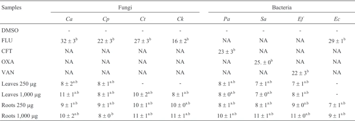
The anti-microbial activity of the crude extracts ofA. latissima was studied using two concentrations, 10 and
40 mg/mL. The results summarized in Table 2 show that the growth inhibition for both concentrations is similar when tested against C. albicans, C. parapsilosis, P. aeruginosa,S. aureusandE. faecalis.
The two test concentrations of the root extract inhib-ited the growth ofC. tropicalisandC. krusei. For those
mi-cro-organisms, the leaf extract showed activity only at the higher concentration, so it was considered dose-dependent. Only the root material showed activity againstE. coli.
We observed from the results shown in Table 3 that all fractions (with the exception of R-EtAc) showed activ-ity againstE. faecalis.We also highlight the R-EtOH:H2O as being the most effective againstC. parapsilosis,with its
halo of inhibition statistically similar to the positive con-trol.
We observed in Table 3 that the inhibition halos of the active fractions againstS. aureusshowed a broad spectrum
of inhibition. We noted that the activities of L-CHCl3and R-CHCl3, which caused halos of 15±0 mm and 20±1 mm, respectively, were closer to the activity of the positive con-trol (oxacillin), which caused a halo of 25 mm.
The fractions causing inhibition halos for all fungi (L-HX, L-CHCl3, L-EtAc and R-EtOH:H2O) and bacteria (L-HX, L-CHCl3, R- CHCl3and R-EtAc) were chosen for determining MICs. The only fractions that showed a
signif-icant MIC were L-CHCl3 and R-CHCl3, with an MIC of 500mg/mL when tested againstS. aureus.
Bioautography was used to characterize the com-pounds within L-CHCl3and R-CHCl3that contributed to the antimicrobial activity againstS. aureus. In Table 4 and
Figure 2, we summarize and show the antimicrobial activ-ity of the isolated bands.
Samples Fungi Bacteria
Ca Cp Ct Ck Pa Sa Ef Ec
DMSO - - -
-FLU 32±3b 22±3b 27±3b 16±2b NA NA NA 29±1b
CFT NA NA NA NA 23±3b NA NA NA
OXA NA NA NA NA NA 25.±0b NA NA
VAN NA NA NA NA NA NA 22±3b NA
Leaves 250mg 8±2a.b 8±1a.b - - 8±1a.b 7±1a.b 7±1a.b
-Leaves 1,000mg 11±1a.b 8±1a.b 10±2a.b 8±1a.b 8±0a.b 7±0a.b 8±1a.b -Roots 250mg 9±1a.b 9±1a.b 10±1a.b 10±0a.b 8±1a.b 8±1a.b 9±0a.b 7±1a.b Roots 1,000mg 10±2a.b 8±0.b 11±1a.b 11±1a.b 10±1a.b 11±1a.b 11±0a.b 9±1a.b
NA: Not applicable; Statistical Test: Kruskal-Wallis/Dunn (p < 0.05); Different letters indicate statistically significant differences, with comparisons made in a column-wise fashion. Ca:C. albicans; Cp:C. parapsilosis; Ct:C. tropicalis; Ck:C. krusei; Pa:P. aeruginosa; Ec:E. coli; Sa:S. aureus; Ef:E. faecalis.FLU: fluconazole (25mg), CFT: ceftazidime (30mg), OXA: oxacillin (1mg) and VAN: vancomycin (30mg).
Table 3- Antimicrobial activity ofA. latissimafractions (CHCl3, EtAc, EtOH and EtOH:H2O) obtained from crude extract of leaves and roots (1,000
mg/disc), evaluated by the disc diffusion assay. Inhibition Zone (mm).
Samples Fungi Bacteria
Ca Cp Ct Ck Pa Sa Ef Ec
DMSO - - -
-FLU 32±3b 25±0b 28±3b.c 16±2b NA NA NA NA
CFT NA NA NA NA 24±1b NA NA 29±1b
OXA NA NA NA NA NA 25±0b NA NA
VAN NA NA NA NA NA NA 21±1b NA
L-HX 9±1a.b 10±0a.b 13±1b 12±0a.b 10±0a.b 12±1a.b 11±1a.b 9±0a.b L-CHCl3 8±1a.b 10±0a.b 11±0a.b 11±2a.b 9±1a.b 15±0a.b 10±1a.b 9±1a.b
L-EtAc - - 10±0a.b 10±0a.b - - 10±1a.b
-L-EtOH 11±1.b 9±1a.b 10±1a.b 10±0a.b - - 11±0a.b 8±1a.b
L-EtOH:H2O 10±0a.b - 10±1a.b 8±0a.b - - 9±1a.b
-R-CHCl3 - - 8±1a.b 10±0a.b 10±1a.b 20±1a.b 10±1a.b 9±1a.b
R-EtAc 10±0a.b - 10±0a.b - 9±1a.b 15±1a.b 7±1a 10±1a.b
R-EtOH 9±1a.b 9±1a.b - 10±2a.b 10±0a.b - 11±1a.b
-R-EtOH:H2O 10±1a.b 11±1b 10±1a.b 10±2a.b 10±1a.b - 11±0a.b
Figure 2- Bioautography plates of L-CHCl3 and R-CHCl3 againstS. aureus, stained with INT (2 mg/mL). L-A=n-hexadecanoic acid, L-B= kaura-9(11), 16-dien-18-oic,R-C=mixture of kaurenoic acid and kaura-9(11), 16-dien-18-oic acid,R-D= stigmasterol,R-E= mixture of kaurenoic acid and kaura-9(11), 16-dien-18-oic acid.
Table 4- Secondary metabolites from L-CHCl3and R-CHCl3activity againstS. aureusobserved by bioautography test.
L-CHCl3 Activity Compounds R-CHCl3 Activity Compounds
L-A + n-Hexadecanoic acid R-A - NI
L-B + Stigmasterol kaura-9(11), 16-dien-18-oic kaurenoic acid R-B - NI
L-C - NI R-C + kaurenoic acid and acid
kaura-9(11), 16-dien-18-oic
L-D - NI R-D + Stigmasterol kaurenoic acid acid
kaura-9(11), 16-dien-18-oic
L-E - NI R-E + kaurenoic acid kaura-9(11), 16-dien-18-oic
acid
R-F - NI
GC-MS analysis. The compound L-A was characterized as
n-Hexadecanoic acid, with a molecular formula of
C16H32O2and anm/zof 256 [M.+].
Another bioactive band from the leaf extract was L-B, the major compound in which, representing 46% of the area on the chromatogram, was the steroid stigmasterol, having a molecular formula of C29H48O and anm/zof 412 [M.+]. Other compounds identified in L-B were the acid kaura-9(11), 16-dien-18-oic (C20H28O2) and kaurenoic acid (C20H30O2), with m/z ratios of 302 [M.+] and 300 [M.+], re-spectively.
In the root extracts, three bioactive bands were found. The band R-C and R-E were identified as kaurenoic acid and acid kaura-9(11), 16-dien-18-oic, each representing different areas of the chromatogram: kaurenoic acid from R-C accounted for 45.45% of the area, and kaurenoic acid from R-E accounted for 67.92% of the area; kaura-9(11), 16-dien-18-oic acid from R-C accounted for 43.78% of the area, and kaura-9(11),16-dien-18-oic acid from R-E ac-counted for 16.97% of area. The major compound within R-D, representing 60% of the area of the chromatogram, was stigmasterol, with an MSm/zratio of 412 [M.+].
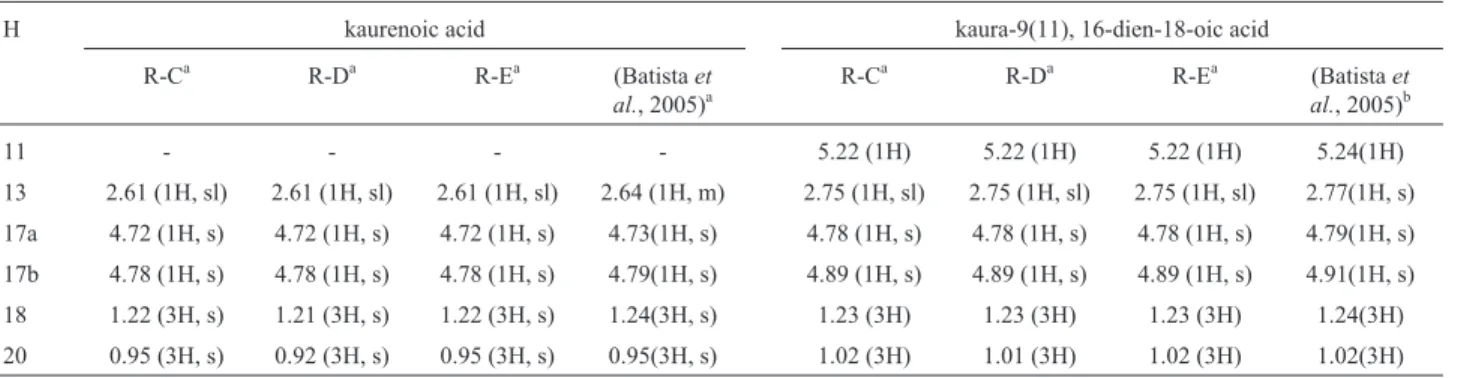
The kaurenoic acid and kaura-9(11), 16-dien-18-oic acid were characterized by1H-NMR and13C-NMR, and the results were compared with literature data (Batistaet al.,
2005). The results of that analysis are presented in Tables 5 and 6.
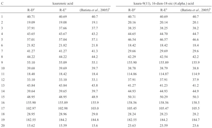
In the13C-NMR spectrum of R-E, we observed five signals of kaura-9(11), 16-dien-18-oic acid atd= 105.47, d = 114.87, d= 155.89,d= 158.56 andd= 184.20.
For stigmasterol from R-D, we also observed five sig-nals in the13C-NMR spectrum that were characteristic of its structure:d= 140.74,d= 138.3,d= 129.27,d= 121.71 and d= 71.82. In kaurenoic acid from R-D, we observed in the 13C-NMR spectrum three signals characteristic of its
struc-ture:d= 182.55,d= 155.9 andd= 102.97.
We observed in the disk diffusion screening test that the crude extracts were active against all tested micro-organisms and were considered contain potential anti-microbial compounds (Table 2). Those results suggested promising activity against the tested micro-organisms.
All of the fractions from the crude extract (except R-EtAc) showed activity againstC. krusei, which we con-sidered an important result because the intrinsic resistance of this fungus to fluconazole is a serious problem in the treatment of candidemias (Scorzoniet al., 2013). Another prominent result was that effectiveness of the R-EtOH:H2O againstC. parapsilosis, one of pathogens responsible for nosocomial infections, with its activity being statistically similar to that of the positive control.
ForE. faecalis, all tested fractions were active. This bacterium resides in the human intestine and is a principal cause of serious nosocomial infections, which are difficult to manage due to the intrinsic and acquired resistance to the main classes of antibiotics (Ranaet al., 2013)
Better results of the disk diffusion test were obtained for L-CHCl3and R-CHCl3againstS. aureus, with an inhi-bition similar to that caused by the positive control. This bacterium is considered an important food pathogen, and it is the primary bacterium in human infections, some of which can be fatal (Kelseyet al., 2006).
Confirming the results of the disk diffusion test, the MICs of L-CHCl3and R-CHCl3againstS. aureuswere the only ones with an interesting activity, given the article of Rios and Recio (2005) that suggests that extracts with MIC values above 1,000mg/mL should be discarded.
Bioautography was performed to identify antimicro-bial compounds and simplify the isolation process (Raha-lisonet al., 1991). The presence ofn-hexadecanoic acid in
L-A contributed to its antibacterial activity against S. aureusand, in previous work, plants containing this fatty
acid had exhibited strong activity against S. aureus and
other Gram-positive bacteria (Keawsa-ard et al., 2012;
Pasdaranet al., 2012; Yagiet al., 2012).
Table 5-1H-NMR (d) data for kaurenoic and kaura-9(11), 16-dien-18-oic, (4.alpha.) acids.
H kaurenoic acid kaura-9(11), 16-dien-18-oic acid
R-Ca R-Da R-Ea (Batistaet
al., 2005)a
R-Ca R-Da R-Ea (Batistaet
al., 2005)b
11 - - - - 5.22 (1H) 5.22 (1H) 5.22 (1H) 5.24(1H)
13 2.61 (1H, sl) 2.61 (1H, sl) 2.61 (1H, sl) 2.64 (1H, m) 2.75 (1H, sl) 2.75 (1H, sl) 2.75 (1H, sl) 2.77(1H, s) 17a 4.72 (1H, s) 4.72 (1H, s) 4.72 (1H, s) 4.73(1H, s) 4.78 (1H, s) 4.78 (1H, s) 4.78 (1H, s) 4.79(1H, s) 17b 4.78 (1H, s) 4.78 (1H, s) 4.78 (1H, s) 4.79(1H, s) 4.89 (1H, s) 4.89 (1H, s) 4.89 (1H, s) 4.91(1H, s) 18 1.22 (3H, s) 1.21 (3H, s) 1.22 (3H, s) 1.24(3H, s) 1.23 (3H) 1.23 (3H) 1.23 (3H) 1.24(3H) 20 0.95 (3H, s) 0.92 (3H, s) 0.95 (3H, s) 0.95(3H, s) 1.02 (3H) 1.01 (3H) 1.02 (3H) 1.02(3H)
a300 MHz, CDCl
In L-CHCl3, stigmasterol, acid kaura-9(11), 16-dien-18-oic, (4.alpha.) and kaurenoic acid (L-B) were identified. stigmasterol also contributed to antibacterial activity againstS. aureus(Guilhonet al., 2012; Yagiet al., 2012).
Diterpenes with a kaurene skeleton are known for their antimicrobial activity. In the study by Pereiraet al.
(2012), kaurenoic acid showed significant activity against
S. aureus. Indeed, Okoyeet al. (2012) demonstrated that
the antibacterial activity ofAnnona senegalensisis due to
kaurenoic acid.
The compounds identified in this work have other studies and demonstrate a wide variety of biological activi-ties, for example kaurenoic acid is found in several medici-nal plants such as Copaifera (Paiva et al., 2002). This
compound in addition to the antimicrobial activity pre-sented and discussed here has a great potential as a new drug. Among the studies, kaurenoic acid demonstrated strong antileishmanial activity (IC50values of 4.0mg/mL) (Santos et al., 2013) antimalarial activity (Batistaet al.,
2013) anti-inflammatory and antinociceptive effect (Mizo-kamiet al., 2012). The occurrence of active secondary
me-tabolites in this species, allows its application not only as a possible antimicrobial agent, but also with a new source of these compounds.
The present study demonstrated that crude extracts of the roots and leaves ofA. latissimashowed antimicrobial
activity against all tested microorganisms. Through the
identification of the active compounds by bioautography, we identified the same compounds in the leaves and roots, with the presence of stigmasterol in L-B and R-D; kau-renoic acid in L-B, R-C, R-D and R-E; and kaura-9(11), 16-dien-18-oic acid in R-C, R-D and R-E. These results show that these compounds accumulate in more than one part of the plant. In addition, we emphasize that bioauto-graphy is effective for isolating antimicrobial compounds, but the resolution is not high enough to achieve complete compound separation.
Finally, we conclude that the accumulation of the same compounds in the leaves and roots explains the simi-lar antibacterial activity of leaf and root extracts, and the presence of steroids and kaurene diterpenes are responsible for the bacteriostatic activity of the extracts and fractions. Moreover, this study contributed to the identification of some secondary metabolites in this plant species.
Acknowledgments
The authors are grateful to FUNDECT (Fundação de Apoio ao Desenvolvimento do Ensino, Ciência e Tecno-logia do Estado de Mato Grosso do Sul), CNPQ (Conselho Nacional de Desenvolvimento Científico e Tecnológico), UFMS (Universidade Federal de Mato Grosso do Sul) and INAU (Instituto Nacional de Áreas Úmidas) for financial support and to CAPES (Coordenação de Aperfeiçoamento Table 6-13C-NMR (d) data for kaurenoic and kaura-9(11), 16-dien-18-oic, (4.alpha.) acids.
C kaurenoic acid kaura-9(11), 16-dien-18-oic (4.alpha.) acid
R-Da R-Ea (Batistaet al., 2005)b R-Da R-Ea (Batistaet al., 2005)b
1 40.71 40.69 40.7 40.71 40.69 40.7
2 19.09 19.08 19.1 20.16 20.14 20.1
3 37.91 37.66 37.7 38.35 38.25 38.2
4 43.65 43.67 43.2 44.65 44.70 44.7
5 57.01 57.04 57.1 46.54 46.57 46.6
6 21.82 21.82 21.8 18.42 18.42 18.4
7 41.27 41.27 41.3 29.66 29.69 29.6
8 44.22 44.22 44.2 42.29 42.54 42.2
9 55.10 55.09 55.1 155.90 155.89 155.9
10 39.68 39.69 39.7 38.78 38.79 38.8
11 18.48 18.42 18.4 114.86 114.87 114.9
12 33.10 33.10 33.1 37.91 37.91 37.9
13 43.84 43.84 43.8 41.27 41.23 41.2
14 39.64 39.65 39.7 44.93 44.93 44.9
15 48.95 48.95 48.9 50.31 50.29 50.3
16 155.90 155.89 155.9 158.56 158.56 158.5
17 102.97 102.98 103.0 105.45 105.47 105.5
18 28.95 28.96 29.0 28.24 28.23 28.2
19 182.55 184.2 184.8 182.55 184.2 184.7
20 15.62 15.59 15.6 23.63 23.59 23.6
SOUZA, J. M. E.
References
Ambrosio SR, Furtado NAJC, Martins CHGet al.(2008) Anti-microbial activity of kaurane diterpenes against oral patho-gens. Z. Naturforsch. C: J Biosci 63:326-330.
Batista R, Braga FC, Oliveira AB (2005) Quantitative determina-tion by HPLC of ent-kaurenoic and grandiflorenic acids in aerial parts ofWedelia paludosaD.C. Rev Bras Farmacogn
15:119-125.
Batista R, Garcia PA, Castro MAet al.(2013) Synthesis,
cyto-toxicity and antiplasmodial activity of novel ent-kaurane de-rivatives. Eur J Med Chem 62:168-176.
Berdy J (2005) Bioactive microbial metabolites - A personal view. J Antibiot 58:1-26.
Bohlmann F, Ziesche J, King RMet al.(1981) Naturally-occur-ring terpene derivatives .300. eudesmanolides and diter-penes from wedelia-trilobata and an ent-kaurenic acid-deri-vative fromAspilia parvifolia. Phytochemistry 20:751-756.
CLSI (2008) M27-A3: Reference method for broth dilution anti-fungal susceptibily testing of yeasts. Approved Standard, third edition, 28:25.
CLSI (2009) Publication M44-A2: Method for antifungal disk dif-fusion susceptibility testing of yeasts. Approved guideline, 29:22.
Guilhon GMSP, da Silva ES, Santos LDet al.(2012) Volatile and non-volatile compounds and antimicrobial activity of
Mansoa difficilis (Cham.) Bureau & K. Schum.
(Bignoniaceae). Quim Nova 35:2249-2253.
Keawsa-ard S, Liawruangrath B, Liawruangrath Set al.(2012)
Chemical constituents and antioxidant and biological activi-ties of the essential oil from leaves ofSolanum spirale. Nat Prod Commun 7:955-958.
Kelsey JA, Bayles KW, Shafii Bet al.(2006) Fatty acids and monoacylglycerols inhibit growth of Staphylococcus aureus. Lipids 41:951-961.
Kingston DGI (2011) Modern natural products drug discovery and its relevance to biodiversity conservation. J Nat Prod 74:496-511.
Lewinsohn TM (2006) Avaliação do estado do conhecimento da biodiversidade brasileira. Ministerio do Meio Ambiente 2:145-192.
Mishra BB, Tiwari VK (2011) Natural products: An evolving role in future drug discovery. Eur J Med Chem 46:4769-4807. Mizokami SS, Arakawa NS, Ambrosio SRet al.(2012)
Kaur-enoic acid fromSphagneticola trilobataInhibits Inflamma-tory Pain: effect on cytokine production and activation of the NO-cyclic GMP-protein kinase G-ATP-sensitive potassium channel signaling pathway. J Nat Prod 75:896-904. NCCLS (2003) Methods for dilution antimicrobial susceptibility
tests for bacteria that grow aerobically. Approved Standard. Sixth Edition. NCCLS document M7-A6.
Newman DJ, Cragg GM (2012) Natural products as sources of new drugs over the 30 years from 1981 to 2010. J Nat Prod 75:311-335.
Okoye TC, Akah PA, Okoli COet al.(2012) Antimicrobial ef-fects of a lipophilic fraction and kaurenoic acid isolated from the root bark extracts ofAnnona senegalensis.
Evid-Based Compl Alt.
8:271-280.
Page JE, Balza F, Nishida Tet al. (1992) Biologically active
diterpenes fromAspilia mossambicensis, a chimpanzee
me-dicinal plant. Phytochemistry 31:3437-3439.
Paiva LA, Gurgel LA, Silva RMet al.(2002) Anti-inflammatory
effect of kaurenoic acid, a diterpene from Copaifera langsdorffi on acetic acid-induced colitis in rats. Vascul Pharmacol 39:303-307.
Pasdaran A, Delazar A, Nazemiyeh Het al. (2012) Chemical
composition, and antibacterial (against Staphylococcus aureus) and free-radical-scavenging activities of the essen-tial oil ofScrophularia amplexicaulisBenth. Rec Nat Prod 6:350-355.
Peixoto AL, Morin MP (2003) Coleções botânicas: documentação da biodiversidade brasileira. Ciência e Cultura 55:21-24. Pereira S, Taleb-Contini S, Coppede Jet al.(2012) An
ent-kau-rane-type diterpene inCroton antisyphiliticusMart.
Mole-cules 17:8851-8858.
Rahalison L, Hamburger M, Hostettmann K et al. (1991) A bioautographic agar overlay method for the detection of antifungal compounds from higher-plants. Phytochem Analysis 2:199-203.
Rana NF, Sauvageot N, Laplace JMet al.(2013) Redox balance via lactate dehydrogenase is important for multiple stress re-sistance and virulence in Enterococcus faecalis. Infect
Immun 81:2662-2668.
Rice LB (2009) The clinical consequences of antimicrobial resis-tance. Curr Opin Microbiol 12:476-481.
Rios JL, Recio MC (2005) Medicinal plants and antimicrobial ac-tivity. J Ethnopharmacol 100:80-84.
Roemer T, Xu DM, Singh SBet al.(2011) Confronting the
chal-lenges of natural product-based antifungal discovery. Chem Biol 18:148-164.
Ruiz LdS, Khouri S, Hahn RCet al.(2013) Candidemia by
spe-cies of theCandida parapsilosiscomplex in children’s
hos-pital: prevalence, biofilm production and antifungal suscep-tibility. Mycopathologia 175:231-239.
Santos AO, Izumi E, Ueda-Nakamura Tet al.(2013)
Antileish-manial activity of diterpene acids in copaiba oil. Memorias do Instituto Oswaldo Cruz 108:59-64.
Scorzoni L, Pilar dLM, Mesa-Arango ACet al.(2013) Antifungal
efficacy during Candida krusei infection in
non-conven-tional models correlates with the yeastin vitrosusceptibility profile. Plos One 8:e60047.
Valgas C, de Souza SM, Smania EFAet al.(2007) Screening
methods to determine antibacterial activity of natural prod-ucts. Braz J Microbiol 38:369-380.
Yagi S, Chretien F, Duval REet al.(2012) Antibacterial activity,
cytotoxicity and chemical constituents ofHydnora johannis
roots. South African Journal of Botany 78:228-234. Yongabi KA, Mbacham WF, Nubia KKet al.(2009) Yeast strains
isolated from HIV-seropositive patients in Cameroon and their sensitivity to extracts of eight medicinal plants. Afr J Microbiol Res 3:133-136.
Associate Editor: Ana Carolina Ramos Moreno