MINI REVIEW published: 18 February 2020 doi: 10.3389/fmats.2020.00024
Frontiers in Materials | www.frontiersin.org 1 February 2020 | Volume 7 | Article 24
Edited by: Antonella Motta, University of Trento, Italy Reviewed by: Vamsi Yadavalli, Virginia Commonwealth University, United States Biman B. Mandal, Indian Institute of Technology Guwahati, India Pornanong Aramwit, Chulalongkorn University, Thailand *Correspondence: Filipa Castro [email protected] Ana L. Oliveira [email protected] Specialty section: This article was submitted to Biomaterials, a section of the journal Frontiers in Materials Received: 19 September 2019 Accepted: 21 January 2020 Published: 18 February 2020 Citation: Veiga A, Castro F, Rocha F and Oliveira AL (2020) Recent Advances in Silk Sericin/Calcium Phosphate Biomaterials. Front. Mater. 7:24. doi: 10.3389/fmats.2020.00024
Recent Advances in Silk
Sericin/Calcium Phosphate
Biomaterials
Anabela Veiga1,2, Filipa Castro1*, Fernando Rocha1and Ana L. Oliveira2*
1LEPABE – Laboratory for Process Engineering, Environment, Biotechnology and Energy, Faculty of Engineering, University of Porto, Porto, Portugal,2Universidade Católica Portuguesa, CBQF - Centro de Biotecnologia e Química Fina - Laboratório Associado, Escola Superior de Biotecnologia, Porto, Portugal
Calcium phosphates (CaPs) have been widely used in the field of biomedical engineering as bone graft substitutes or as carriers for drug delivery applications. Recent developments have focused on combining CaPs with proteins to obtain functional biomaterials that accommodate a broader spectrum of functional requirements. Silk sericin was considered an unutilized protein by-product from the textile industry, generating tons of residues every year. However, much effort has been dedicated to its recovery after being associated with numerous biological properties such as antioxidant, antibacterial, anti-coagulation and regenerative activities. In the past years, sericin has also demonstrated to be suitable as a template for CaP mineralization. The present review focuses on the recent developments for the production of sericin/CaP composites, exploring their potential applications in bioengineering and opening new avenues in other research fields such as in the cosmetic, food and environmental sectors. In addition, this paper can also be useful as a guideline to design future research based on sericin/CaP biomaterials.
Keywords: biomaterials, bone tissue engineering (bone-TE), calcium phosphates (CaPs), drug delivery, hydroxyapatite (HAp), sericin
INTRODUCTION
Bone matrix consists of both a non-mineralized organic component (∼20% of the wet weight) and a mineralized inorganic component (∼65–70%), composed mainly by collagen and carbonated calcium phosphate (CaP) crystals, respectively. Crystallographic c-axis of these crystals are arranged parallelly to the longitudinal axis of the fibrous protein, forming a tough and flexible nanocomposite structure (Fuchs et al., 1997; Amini et al., 2012).
Accordingly, calcium phosphates (CaPs) from natural or synthetic nature, are widely used in bone-related applications due to their biocompatibility, low density, chemical stability and crystallinity (Vallet-Regí and González-Calbet, 2004). Among existing CaPs, hydroxyapatite (HAp) is the most studied biomaterial due to its close similarity to human hard tissue in terms of morphology and composition (Habraken et al., 2016).
In a quest to mimic bone tissue, several studies have focused on the synthesis of CaP composites using synthetic polymers [polyglycolic acid (PGA), poly L-lactic acid (PLLA), polylactic-coglycolic acid (PLGA), and polycaprolactone (PCL)] (Rezwan et al., 2006; Lee and Yuk, 2007; Chen
et al., 2014), natural polymers (alginate, chitosan, cellulose) (Pighinelli and Kucharska, 2013;
Cardoso et al., 2014; Salama, 2019), proteins (collagen, fibrin, keratin, and silk fibroin) (Dias
Veiga et al. Silk Sericin/Calcium Phosphate Biomaterials
or combinations of the foregoing (Chen et al., 2014). Synthetic polymers may induce local and systemic host reactions due to the release of chemicals and monomers from polymer degradation, while natural polymers may lead to products with different characteristics due to the differences in raw materials (Chen et al.,
2014).
Proteins have demonstrated to regulate the size, particle size distribution, morphology, and assembly method of nanostructured CaPs, being thus used to fabricate novel materials with biomimetic characteristics for hard tissue repair (Cai and Yao, 2010; Chen et al., 2014). Further, their natural origin confers unique biocompatibility, versatility, and biodegradability properties (Swetha et al., 2010). Collagen has been conventionally used in the synthesis of organic/CaP composites. Although collagen/CaP composites have a greater similarity to natural bone, the clinical application of collagen-type biomaterials is still limited due to its high cost, increased risk of cross-infection and fast degradability (Chen et al., 2014). Alternative protein systems can play a pivotal role during the mineralization of CaP, as a binding-matrix. Keratin/CaP and fibrin/CaP composites have biocompatibility and bioactivity properties suitable for bone tissue engineering. However, these biomaterials lack mechanical properties and processing properties, and have fast biodegrability (Gsib et al., 2017;
Shavandi et al., 2017).
In this context silk is a valuable alternative due to its excellent intrinsic properties such as non-toxicity, biodegradability, self-assembly, mechanical stability and controllable structure
(Altman et al., 2003; Ha et al., 2013). In nature, silk-like proteins
are produced by several organisms such as silkworms, spiders, mollusks, scorpions, bees, and ants (Holland et al., 2019). The use of silk from the domesticated silkworm Bombyx mori is well-established as suitable for biomedical applications due to its abundance, batch-to-batch stability, and clinical track record
(Ude et al., 2014). This type of silk comprises a fibrous
semi-crystalline silk core, silk fibroin, which is mainly responsible for the load-bearing capacity, and an outer layer of a globular protein, sericin, which serves as gluing agent and has a protective function. While fibroin is processed in large scale in the textile industry, sericin is a by-product generated during this process, in the so-called degumming procedure (Mondal et al., 2006).
Silk fibroin has been widely used and investigated for applications such as sutures (Saxena et al., 2014), artificial ligaments (Farè et al., 2013), tissue engineering constructs
(Kasoju and Bora, 2012), and substrates for cell culture (Liu
et al., 2012). Fibroin/CaP composites have been reported and are
considered suitable for load bearing applications (Yan et al., 2013;
Farokhi et al., 2018).
Silk sericin, on the other hand, was until recently considered unfit for biomedical use. Of 1 million tons of silkworm cocoons produced each year worldwide, ∼50,000 t of sericin are generated, leading to environmental and economic concerns
(Aramwit et al., 2012b). This protein, in its native state, was
found to elicit immune and allergic responses when present in virgin silk sutures (Soong and Kenyon, 1984). However, emerging evidence suggests that extracted sericin per se is biocompatible, similarly to silk fibroin or collagen (Chirila et al., 2013; Lamboni
et al., 2015). Therefore it has been gaining reputation in the
biomedical field with several works showing its potential, as revised elsewhere (Kunz et al., 2016; Ahsan et al., 2018). Thus, the use of sericin/CaP composites not only reduces the inherent environmental impact of sericin disposal but also allows the development of new functional biomaterials. This silk protein increases antioxidant, anti-tyrosinase and anti-inflammatory activity; stimulates collagen production, tumor inhibitory effects; induces the nucleation of bone-like CaPs; and promotes stability and prolonged release in drug delivery systems (Aramwit, 2014;
Lamboni et al., 2015).
The molecular weight distribution and even the amino acid composition of sericin were found to be dependent on the extraction method used (Yang et al., 2014c; Kunz et al., 2016). Sericin consists of 18 amino acids, among which serine, histidine, glutamic acid, aspartic acid, threonine, and tyrosine are normally present in higher percentages. Thus, this protein is highly hydrophilic and its molecular weight can range from 20 to 400 kDa (Kunz et al., 2016). Its polar side chains have several functional groups (carboxyl, amine, hydroxyl) responsible for moisturizing and oxidizing properties, allowing the interaction with other compounds through crosslinking, copolymerization or blending to form improved biomaterials (Ahsan et al., 2018). According to the literature, amino acids can highly influence the mineralization process of CaPs. Glutamic acid and aspartic acid play a critical role in controlling HAp nucleation and growth, by attracting calcium and phosphate ions and consequently increasing the local supersaturation, which results in the development of crystals. Histidine and other negatively charged amino acids are involved in HAp nucleation within extracellular matrix proteins (George and Veis, 2008; Tavafoghi
and Cerruti, 2016).
Sericin mainly adopts the form of an amorphous random spiral and may also present the form of a β-sheet organized
structure. The random spiral easily acquires β-sheet
conformation as a consequence of moisture absorption and mechanical elongation, forming a denser, organized, crystalline and less soluble structure. If the protein is dissolved in hot water and subsequently undergoes a temperature decrease, its random coil structure becomes β-sheet, acquiring a gelatinous form
(Chen et al., 2015). That is, with the change in temperature the
conformation of the protein changes. Upon heating the sericin dissolves in solution, upon cooling the sericin solution jellifies (Sol-Gel properties;Kunz et al., 2016).
SILK SERICIN/CALCIUM
PHOSPHATE-BASED MATERIALS
Recently, sericin has demonstrated to be suitable as a template for CaP mineralization. In the literature, sericin/CaP biomaterials are usually synthetized by wet mechanochemical processes, where mineralization occurs by spontaneous nucleation (Cai
et al., 2009, 2010) or using a surface as template (Takeuchi
et al., 2005b; Yang et al., 2014a). The resulting composites
generally present a poor crystallinity and the size of the particles generated can range from 20 to 500 nm in length to 3–80 nm
Veiga et al. Silk Sericin/Calcium Phosphate Biomaterials
in width (Cai et al., 2009, 2010; Veiga et al., 2019). The influence of several experimental conditions on the formation of these sericin/CaPs has been studied by several authors, namely different sericin extraction and storage procedures, mineralization time, sericin concentration, and temperature (Table 1). In the works ofTakeuchi et al. (2003a, 2005b), sericin was obtained by extraction in boiling water, using different temperatures and storage procedures. It was found that high molecular weight sericin and β-sheet structure induces apatite deposition. These conditions were achieved for higher extraction temperatures and sericin storage. It is well-known that the extraction method used influences the molecular weight and the amino acid concentration of sericin, which results in different physical and biological properties (Kunz et al., 2016). According
to Aramwit et al. (2010), extraction in boiling water is the
least toxic procedure to cells and activates the highest collagen production. Acid, alkaline, and urea extraction methods can also be used to obtain sericin that promotes the formation of bone organic matrix. However, these approaches can result in toxicity at higher sericin concentrations (Padamwar and Pawar, 2004). Moreover, acid and alkaline methods have a degrading effect on proteins and considerably increase its solubility. Although urea and enzymatic methods can be used to obtain unchanged sericin, the procedures are expensive and time consuming. On the other hand, hot-water extraction is a simple and environmentally friendly method that preserves the main characteristics of silk sericin (Gulrajani et al., 2000; Dash et al., 2008).
As for the influence of the mineralization time on apatite deposition, it was found that the optimum days for growth of nano HAp crystals with low crystallinity is 7 days. In contrast, after 30 days, the size of HAp crystals increases, and other CaP phases are formed (Sukjai et al., 2012). Further, it was also shown that nucleation of HAp mediated by sericin is a gradual process, since nanoneedle crystals with low crystallinity are only grown after at least 24 h of mineralization (Yang et al., 2014b). In the work ofZhang et al. (2014), the same conclusions were drawn for ethanol-treated sericin films immersed in SBF. A three-dimensional (3D) structure with globular carbonate apatite particles was observed after 7 days. Further, rigid and brittle properties of the film increased with mineralization time, since apatite particles were increasingly being deposited. While the elastic modulus increased significantly after 5 and 7 days (23.027 ±0.83 to 57.02 ± 1.04 MPa), the tensile strength (day 1: 1.58 ±0.53, day 7: 0.24 ± 0.18) and the elongation at break of the 3D film decreased (day 1: 112.83 ± 49.02%, day 7: 2.01 ± 1.81%). The increase of the mineralization time and sericin concentration was also reported to lead to an increase in the mean particle size of the particles synthetized (Cai et al., 2009). The recent developments in sericin/CaP composites and its potential for biomedical applications is presented in the next section.
RESEARCH APPLICATIONS
Biomineralization
Although proteins play a crucial role during mineral formation in biological systems, the biomineralization process is still far from being understood (Subburaman et al., 2006). Therefore,
most studies on sericin/CaP composites focus on contributing to the understanding of this phenomenon. Sericin/CaP nanoparticle systems obtained are generally nanorod-like crystals with poor crystallinity. Further, sericin promotes crystal growth of HAp along the c-axis, resembling natural biomineralization. Although in the work ofCai et al. (2010), HAp particles similar to mineral bone were achieved in the absence of sericin, this was only verified for specific temperature and pH conditions. Other works argue that the presence of sericin promotes a homogeneous assembly of HAp, under several experimental conditions (Cai
et al., 2009; Yang et al., 2014b). The proposed mechanisms
for the assembly of CaP crystals in the presence of sericin are usually based on changes in the protein conformation. When dissolved, sericin adopts an amorphous structure in which the hydrophilic sidechains are exposed. However, when submitted to different triggers (e.g., mechanical stretching properties, moisture absorption, temperature, dehydration, or using crosslinking agents), changes in random coil structure for β-sheet easily occur
(Teramoto and Miyazawa, 2003; Nayak et al., 2012; Kunz et al.,
2016).
According to Takeuchi, the induction sites for HAp nucleation in sericin films are governed by the arrangement of carboxyl groups on the protein (Takeuchi et al., 2005b). When a protein acquires a β-sheet conformation, the amino acid side chains alternate between the two faces of the sheet. Hydrophobic side-chains point in one direction and polar side-chains in the other, which can explain orientation of functional groups in β-sheet sericin (Boyle, 2018). For sericin/HAp particles, the presence of chelated calcium ions in the precipitation medium provides nucleation sites. The formed HAp crystals are rearranged along their c-axis by attaching to the sericin molecular chains (Cai et al., 2009).
The understanding of the biomineralization process can lead to numerous applications in biomedical engineering, namely for the development of advanced bone substitutes (Chen et al.,
2019; Veiga et al., 2019). Even though pure sericin has poor
mechanical properties, which hinders its utilization in bone tissue engineering (bone-TE), its biological properties can promote bone formation and induce the nucleation of bone-like HAp. In fact, sericin/CaP materials have shown the ability to promote cell differentiation and proliferation of bone marrow-derived mesenchymal stem cells (BMSCs) and of human osteosarcoma cells (MG-63; Table 1). Therefore, most of the developed works on sericin/CaP focus on demonstrating the potential of these composites in the field of bone-TE, as biomaterials for bone filling and repair or as drug delivery systems for tumor therapy, as illustrated in Figure 1.
Bone Tissue Engineering
Although some authors mention the potential of sericin/CaP as scaffolds for TE (Takeuchi et al., 2003b; Yang et al., 2014b), an ideal 3D scaffold should not only act as a template for tissue growth and has controlled resorbability, but should also elicit a regenerative effect and present adequate mechanical performance over time (Canillas et al., 2017). As previously mentioned, the last requirement is not fulfilled by using only sericin (Ahsan et al.,
2018).
V e ig a e t a l. S ilk S e ric in /C a lc iu m P h o sp h a te B io m a te ria ls
TABLE 1 | Articles on sericin/CaP biomaterials.
Objectives Methodology Experimental conditions
Studied variables Physicochemical properties Biological properties Applications References
Investigate the ability of natural silk and its related materials to facilitate apatite deposition under biomimetic conditions.
- Sericin extraction was performed in boiling water (105◦C, 1 h);
Soaking method: - Cloths made of raw silk,
normal silk fiber, and sericin films were soaked in 1.5 SBF.
pH = 7.25; T = 36.5◦C;
Mineralization time: 7 days.
– - The deposition of apatite microspheres was observed on both the surface of cloth made from raw silk fiber and in the sericin films; - CaP particles were not obtained on cloth
made from normal silk fibers;
- The apatite deposition on the raw silk fiber cloth was accelerated by presence of calcium ions (treatment with CaCl2).
– Present the
potential of sericin/HAp composites in the field of bone tissue engineering (as bone substitutes and scaffolds). Takeuchi et al., 2003b Study the structural effect of sericin on its apatite-forming ability using 1.5 SBF. Soaking method: - Sericin films were
immersed in 1.5 SBF.
Four Different types of sericin films were obtained: Extraction/Storage of sericin 105◦C 1 h/none: 105–0 d; 120◦C 1 h/none: 120–0 d; 105◦C 1 h/4◦C for 2 weeks: 105–2 w; 120◦C 1 h/4◦C for 2 weeks: 120–2 w.
- Molecular weight: 134, 106, 42, and 43 kDa for 105–0 d, 105–2 w, 120–0 d, and 120–2 w, respectively;
- After storage for 2 weeks at 4◦C, β-sheet
structure became dominant;
- Spherical HAp particles with low crystallinity were only observed on the surface of 105–2 w film after 7 days.
– Contributing for the understanding of biological mineralization. Takeuchi et al., 2003a, 2005b Investigate the optimum day for the growth of HAp in sericin-coated silk fibers using SBF.
Soaking method: - Sericin coated silk fibers
and non-sericin coated silk fibers were used were soaked in SBF of various concentrations. T = 37◦C. Mineralization time: 7 and 30 days. SBF concentration (1.5 and 1.0 SBF).
- In non-sericin coated silk fibers, flat shape CaP crystals were formed after 7 days. With the increase in SBF concentration, cauliflower shape HAp crystals were obtained; - In sericin coated silk fibers only cauliflower
shape HAp crystals were formed for 1.0 SBF concentrations (1–4 µm) and 1.5 SBF (0.1–0.2 µm). – Contributing for the understanding of biological mineralization. Sukjai et al., 2012 Investigate the mineralization of sericin by alternative soaking in calcium and phosphate. - Sericin (wt.%) = 8, extraction was performed in boiling water for 30 min; - The sericin film was
soaked in 90% (v/v) ethanol;
- Alternative soaking of the sericin film in calcium and phosphate solutions: 1◦) [CaCl 2] (100 mM); 2◦) [NaHCO 3] (60mM). pH = 8.7; T = 37◦C. Immersion cycles: 5, 10, 15 cycles.
- When the cycles of dip coating were set to be 5, 10, and 30, respectively, aggregation appeared on the coating and apatite spherical crystals were increased; - The coating thickness increased by
increasing cycles of dip coating.
- In vitro assays showed that the HAp/sericin composites obtained have higher cell adhesion and growth activity of MG-63 cells, when compared to HAp.
Present the potential of HAp/sericin composites in the field of bone tissue engineering (for bone regeneration). Yang et al., 2014a (Continued) F ro n tie rs in M a te ria ls |w w w .fr o n tie rs in .o rg 4 F e b ru a ry 2 0 2 0 | V o lu m e 7 | A rtic le 2 4
V e ig a e t a l. S ilk S e ric in /C a lc iu m P h o sp h a te B io m a te ria ls TABLE 1 | Continued
Objectives Methodology Experimental conditions
Studied variables Physicochemical properties Biological properties Applications References
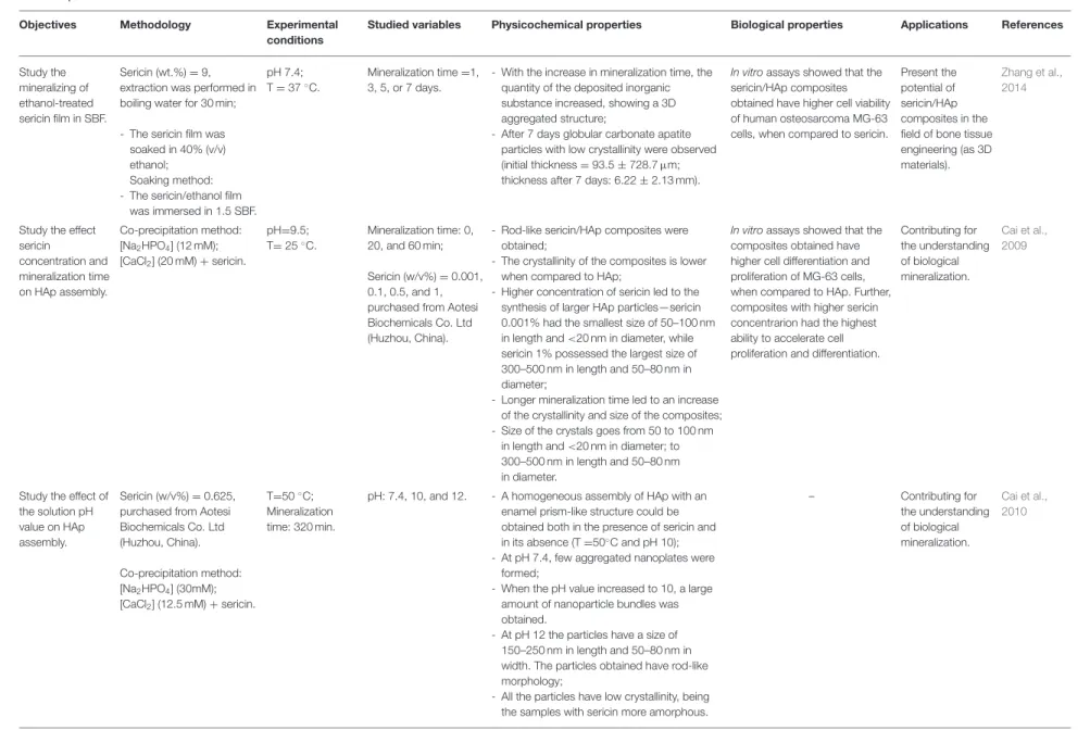
Study the mineralizing of ethanol-treated sericin film in SBF.
Sericin (wt.%) = 9, extraction was performed in boiling water for 30 min; - The sericin film was
soaked in 40% (v/v) ethanol;
Soaking method: - The sericin/ethanol film
was immersed in 1.5 SBF. pH 7.4; T = 37◦C.
Mineralization time =1, 3, 5, or 7 days.
- With the increase in mineralization time, the quantity of the deposited inorganic substance increased, showing a 3D aggregated structure;
- After 7 days globular carbonate apatite particles with low crystallinity were observed (initial thickness = 93.5 ± 728.7 µm; thickness after 7 days: 6.22 ± 2.13 mm).
In vitroassays showed that the sericin/HAp composites obtained have higher cell viability of human osteosarcoma MG-63 cells, when compared to sericin.
Present the potential of sericin/HAp composites in the field of bone tissue engineering (as 3D materials).
Zhang et al., 2014
Study the effect sericin concentration and mineralization time on HAp assembly. Co-precipitation method: [Na2HPO4] (12 mM); [CaCl2] (20 mM) + sericin. pH=9.5; T= 25◦C. Mineralization time: 0, 20, and 60 min; Sericin (w/v%) = 0.001, 0.1, 0.5, and 1, purchased from Aotesi Biochemicals Co. Ltd (Huzhou, China).
- Rod-like sericin/HAp composites were obtained;
- The crystallinity of the composites is lower when compared to HAp;
- Higher concentration of sericin led to the synthesis of larger HAp particles—sericin 0.001% had the smallest size of 50–100 nm in length and <20 nm in diameter, while sericin 1% possessed the largest size of 300–500 nm in length and 50–80 nm in diameter;
- Longer mineralization time led to an increase of the crystallinity and size of the composites; - Size of the crystals goes from 50 to 100 nm
in length and <20 nm in diameter; to 300–500 nm in length and 50–80 nm in diameter.
In vitroassays showed that the composites obtained have higher cell differentiation and proliferation of MG-63 cells, when compared to HAp. Further, composites with higher sericin concentrarion had the highest ability to accelerate cell proliferation and differentiation.
Contributing for the understanding of biological mineralization. Cai et al., 2009
Study the effect of the solution pH value on HAp assembly.
Sericin (w/v%) = 0.625, purchased from Aotesi Biochemicals Co. Ltd (Huzhou, China). Co-precipitation method: [Na2HPO4] (30mM); [CaCl2] (12.5 mM) + sericin. T=50◦C; Mineralization time: 320 min.
pH: 7.4, 10, and 12. - A homogeneous assembly of HAp with an enamel prism-like structure could be obtained both in the presence of sericin and in its absence (T =50◦C and pH 10);
- At pH 7.4, few aggregated nanoplates were formed;
- When the pH value increased to 10, a large amount of nanoparticle bundles was obtained.
- At pH 12 the particles have a size of 150–250 nm in length and 50–80 nm in width. The particles obtained have rod-like morphology;
- All the particles have low crystallinity, being the samples with sericin more amorphous.
– Contributing for the understanding of biological mineralization. Cai et al., 2010 (Continued) F ro n tie rs in M a te ria ls |w w w .fr o n tie rs in .o rg 5 F e b ru a ry 2 0 2 0 | V o lu m e 7 | A rtic le 2 4
V e ig a e t a l. S ilk S e ric in /C a lc iu m P h o sp h a te B io m a te ria ls TABLE 1 | Continued
Objectives Methodology Experimental conditions
Studied variables Physicochemical properties Biological properties Applications References
Study the effect of sericin
concentration and mineralization time on HAp assembly and its osteogenic potential.
Sericin (mg/mL) = 0.5, 2, and 8; extraction was performed in boiling water (120◦C for 30 min). Co-precipitation method: [Na2HPO4] (12 mM); CaCl2 (20 mM) + sericin. pH = 9.5; T = room temperature. Mineralization time: 2, 6, 12, and 24 h.
- A higher concentration of sericin promotes the formation of nanoneedle-like crystals; - Nucleation of HAp mediated by sericin is a
gradual process;
- After 2 h, a clump of starfish- like crystals was randomly scattered;
- After 6 h, spherical crystals were formed and aggregated;
- After 12 h, spindly crystals were heavily aggregated;
- After 24 h nanoneedle crystals with 20–40 nm in length and 3–5 nm in width were obtained.
In vitroassays showed that the mineralized sericin/HAp composites obtained have higher osteogenic differentiation of BMSCs, when compared to non-mineralized composites. Present the potential of sericin/HAp composites in the field of bone tissue engineering (as scaffolds). Yang et al., 2014b Present a new simple method to synthetize sericin/HAp composites and study the effect of SS concentration on HAp assembly. Co-precipitation method: [Na2HPO4] (0.012 M); [CaCl2.2H2O] (0.02 M) +sericin. T= 37◦C; pH ≈ 6; Mineralization time: ≈ 2 h 30 min. Sericin (g/L) = 0.1, 1, extraction was performed in boiling water (1 h, 150 rpm).
- The synthesized particles are single-phased nanometric HAp with low crystallinity; - The increase in sericin concentration is
associated with decreased crystallinity; - Sericin/HAp composites have a higher mean
particle size when compared to HAp. Further, the increase in sericin concentration leads to a size increase (Hap-−0.070 µm; sericin 0.1 g/L-−0.105 µm; sericin 1 g/L-−0.116 µm.); - The increase in sericin concentration also
leads to the formation of more particles with a plate-like structure.
In vitroassays showed that the sericin/HAp composites obtained do not elicit a cytotoxic response on human primary cells (HNDFs) and that at high concentrations (100 mg/mL) elicit a lower cytotoxic response than a commercially available HAp. Contributing for the understanding of biological mineralization. Veiga et al., 2019 Use sericin to regulate the mineralization of CaCO3. Sericin (g/L) = 2, purchased from Aotesi Biochemicals Co. Ltd (Huzhou, China). Co-precipitation method: [Na2CO3] (0.2 mol/L);
CaCl2 (0.2 mol/L) + sericin.
T = 20◦C. Mineralization time =
10, 20, 30, 40, and 50 min.
- With the increase in the mineralization time, the crystal phase of CaCO3transferred from
calcite dominated to vaterite dominated mixtures;
- The morphology of CaCO3changed from
disk-like calcite crystal to spherical vaterite crystal, the products with the reaction time of 10 min are mainly disk-like crystals and little spherical and cube-like crystals (5–15 µm). After 20 min small thorn spherical aggregates of rhombic crystal (with the diameter of 6–7 µm) and lamellar structured crystal (with the size of 3–4 µm) are formed. After 30 and 40 min flower-like crystals with the size of 4–5 µm and partly ellipsoidal crystals 5–6 µm in length and 2–3 µm in width and polyhedral structure of 3–4 µm are formed. After 50 min 4–5 µm sized spherical crystals assembled are obtained. – Contributing for the understanding of biological mineralization. Zhao et al., 2013 (Continued) F ro n tie rs in M a te ria ls |w w w .fr o n tie rs in .o rg 6 F e b ru a ry 2 0 2 0 | V o lu m e 7 | A rtic le 2 4
V e ig a e t a l. S ilk S e ric in /C a lc iu m P h o sp h a te B io m a te ria ls TABLE 1 | Continued
Objectives Methodology Experimental conditions
Studied variables Physicochemical properties Biological properties Applications References
Obtain HAp microspheres using CaCO3as
the template under the assistance of microwave irradiation.
Sericin = 2 g/L, purchased from Aotesi Biochemicals Co. Ltd (Huzhou, China). Precipitation method: 1◦) Synthesis of CaCO
3
particles through a co-precipitation method: - [CaCl2] (0.2 M) + sericin; 1◦) Synthesis of HAp particles: Addition of [Na2HPO4] (0.2 M). 1◦) Mixture was stirred for 50 min; T = 25◦C and pH =7; 2◦) Mixture was stirred for 180 min: T = 40◦C; pH =10. Ca and P ratios of 1:0.15, 1:0.3, 1:0.45, 1:2.5; Mineralization time = 1, 3, 5, 10, 15 min; Microwave powers = 160, 320, 480, 640, and 800 W.
- Weight percentages of HAp in the composites are 19, 25, 40, 45, and 56% corresponding to the microwave power values of 160, 320, 480, 640, and 800 W, respectively;
- With the increase of Ca/P ratio in the reaction solution from 1:0.15, 1:0.30, 1:0.45, 1:2.5 to 1:7, the content of HAp in the composite increases from 0.8, 1.4, 12.3, 50.0 to 99.0%; - The composite with higher HAp content
showed a slower degradation speed; - The average diameter of the spherical
particles is about 6.7 µm, which is close to the size of CaCO3template.
– Present the
potential of sericin/HAp composites in the field of bone tissue engineering (for bone repair and regeneration).
Yang et al., 2016
Obtain HAp using spherical CaCO3
fabricated in the presence of sericin.
Sericin (g/L) = 2, purchased from Aotesi Biochemicals Co. Ltd (Huzhou, China); 1◦) Synthesis of CaCO
3
particles through a co-precipitation method: - [CaCl2] (0.2 M) + sericin; 2◦) Addition of [Na 2HPO4] (0.2 M); 3◦) Synthesis of HAp particles using a hydrothermal method: • CaCO3powder was
added into [Na2HPO4] (0.1 M). 1◦) Mixture was stirred for 30 min at room temperature; 2◦) Mixture was stirred at 40◦C for 180 min and dried at 60◦C for 48 h. 2◦) Mixture was maintained in na autoclave at 140◦C for different times; Precipitate was dried at 60◦C for 48 h.
Time in the autoclave = 2, 4, 8, and 16 h.
- The sericin/CaCO3/HAp particles obtained
are homogeneous microspheres with a size distribution from 3 to 10 µm;
- Uniform porous HAp microspheres with high crystallinity were obtained:
- Increasing time in the autoclave has little impact on morphology of sample but results in more rough surfaces.
- In vitro assays showed that the sericin/CaCO3/HAp
composites obtained are not toxic to MG-63 cells and have similar proliferation rates. Further, with the increase in HAp content higher cell viability was observed.
- In vivo assays showed that sericin/CaCO3/HAp
composites are biodegradable and biocompatible when implanted into the groin subcutaneous tissue, using colloidal sodium alginate solution as a carrier. Additionaly, sericin/CaCO3
have a higher degradation rate.
Present the potential of sericin/CaCO3/HAp
composites in the field of bone tissue engineering (for bone repair). Zhong et al., 2016 Prepare sericin/monite composites using a cost-effective method.
- Sericin was obtained by extraction in boiling water. - Sericin was treated with
ethanol, subjected to membrane dialysis and lyophilized.
Co-precipitation method: [Na2HPO4]
(Sigma-Aldrich Chemicals Pvt Ltd; CAS Number 7558794) + sericin;
[CaCl2·2H2O] (1 M).
Misture was stirred for 3 h.
– - The particles obtained have a heterogeneous size distribution and different morphologies.
- In vitro assays showed that the sericin/monite composites do not elicit hemolysis of human blood. Further, the composites present cell viability and osteogenic properties and increase cell proliferation of MG-63 cells.
Present the potential of sericin/monite composites in the field of bone tissue engineering (as bone grafts).
Vedakumari et al., 2019 (Continued) F ro n tie rs in M a te ria ls |w w w .fr o n tie rs in .o rg 7 F e b ru a ry 2 0 2 0 | V o lu m e 7 | A rtic le 2 4
V e ig a e t a l. S ilk S e ric in /C a lc iu m P h o sp h a te B io m a te ria ls TABLE 1 | Continued
Objectives Methodology Experimental conditions
Studied variables Physicochemical properties Biological properties Applications References
Investigate the distribution and degradation of CaP/sericin nanoparticles in vivo. - Sericin (mg/mL) = 10, purchased from Aotesi Biochemicals Co. Ltd (Huzhou, China). Co-precipitation method: [Na2HPO4] (30 mM); [CaCl2] (50 mM) + sericin. pH = 9–10; T = 50◦C.
– - The sericin/CaP materials obtained adopt a spherical morphology with an average diameter of 80 nm;
- The particles have a smooth surface and a low degree of crystallinity.
In vivotests evaluated the distribution, degradability and tumor targeting of the sericin/CaP composites; - Distribution of sericin/CaP
nanoparticles in vivo were mostly verified in the liver of mice;
- The degradation time did not increase linearly with the amount of sericin/CaP nanoparticles injected in vivo; - Nanoparticles with an average
size of 80 nm can achieve tumor localization at 7 days after the intravenous injection.
Present the potential of sericin/CaP composites in the field of bone tissue engineering (for tumor therapy as a vehicle to deliver therapeutic medicine). Zhao et al., 2017
Study the stability of sericin/CaP microcapsules for stability release.
Sericin (w/v%) = 0.2, purchased from Aotesi Biochemicals Co. Ltd (Huzhou, China). - Sericin microcapsules
were obtained mixing the sericin solution with [CaCl2] (30 mM) and by the posterior addition of amylum (0.5%); Soaking method: - The sericin microcapsules
were added to a supersaturated calcium phosphate solution [Ammonium phosphate solution (5 mM); calcium nitrate solution (10 mM); citric acid (2.5 mM)]. – Mineralization time: 3, 5, 6.5, and 12 h; In vitroamylum release at different pH values: 3, 5, 7, and 9; T = 37◦C;
In vitroamylum release at different ionic strengths: Sodium chloride solutions with 0, 0.1, 0.4, and 0.7 M.
- An increase in the mineralization time led to an increase of the crystallinity and thickness of the microcapsules; Particles in the surface of the microparticles with a CaP salt-based composition and flake-like morphology after 12 h;
- When the pH value was 3 the amylum release rate was slow for the sericin microcapsules and the release amount increased gradually. After 100 h, the release rate decreased, and the release amount did not increase. pH 5 and 7 with similar behavior, characterized by a faster release rate. At pH 9, the composite became unstable;
- The release rate was very fast when the concentration of NaCl in the
controlled-releasing solution was 0.7 M at 37◦C, being slower for other concentrations.
Further, at lower pH values sericin/CaP microspheres release rate was rather slower when compared to sericin microspheres.
In vitroassays showed the composites promote the viability and proliferation of HapG-2 cells, when compared to sericin microcapsules.
Present the potential of sericin/HAp composites in the field of bone tissue engineering (for drug delivery and encapsulating bioactive molecule systems). Li et al., 2016 In vivo biodegradation evaluation of the α-TCP porous body coated with SS.
Sericin was extracted from raw silk fiber by an autoclave at 105◦C for
60 min and stored at 4◦C for
2 weeks. Soaking method: - A α-TCP block was
soaked into a sericin solution.
– – – In vivotests showed that after 4
weeks of implantation on bone defects made in rabbit tibiae and subcutaneous sites of rabbit backs, higher density of cortical bone was estimated for α-TCP coated with sericin than for mere α-TCP.
Present the potential of sericin/HAp composites in the field of bone tissue engineering (for bone repair as a biodegradable materials). Takeuchi et al., 2005a (Continued) F ro n tie rs in M a te ria ls |w w w .fr o n tie rs in .o rg 8 F e b ru a ry 2 0 2 0 | V o lu m e 7 | A rtic le 2 4
Veiga et al. Silk Sericin/Calcium Phosphate Biomaterials T A B L E 1 | C o n tin u e d O b je c ti v e s M e th o d o lo g y E x p e ri me n ta l c o n d it io n s S tu d ie d v a ri a b le s P h y s ic o c h e mi c a l p ro p e rt ie s B io lo g ic a l p ro p e rt ie s A p p li c a ti o n s R e fe re n c e s F a b ric a tio n o f h ie ra rc h ic a l b io -h yb rid a rc h ite c tu re s th ro u g h a g re e n a n d fa c ile m e th o d . S e ric in (m g /m L ) = 0 .1 , p u rc h a se d fr o m W a ko C h e m ic a ls , Ja p a n . C o -p re c ip ita tio n m e th o d : [C a C l2 ] (1 0 0 m M ) + se ric in P B S . T = 2 5 ◦C ; M in e ra liz a tio n tim e : 2 4 h . – -P o ro u s, u n ifo rm ly g ro w n flo w e r-lik e c o m p o si te s w e re o b ta in e d (a ve ra g e si ze 2 –5 µ m ); -T h e c o m p o si te s h a ve h ig h ly c ry st a lli n e c h a ra c te ris tic s w h e n c o m p a re d to p u re se ric in ; -T h e se c o m p o si te s h a ve a la rg e su rf a c e a re a a n d sh o w e xc e lle n t a d so rp tio n a c tiv ity fo r th e to xi c h e a vy m e ta li o n s o f L e a d (II) , c a d m iu m (II) , a n d m e rc u ry (II) fr o m w a st e w a te r. – P re se n t th e p o te n tia lo f se ric in /H A p c o m p o si te s fo r m u lti m o d a l p o ss ib le a p p lic a tio n s su c h a s a d so rp tio n o f to xi c h e a vy m e ta ls a n d h a za rd o u s d ye fr o m w a st e w a te r. K o le y e t a l., 2 0 1 6 T h e o bj e c ti ve s a n d s u g g e s te d a ppl ic a ti o n s o f th e re vi e w e d pa pe rs , a s w e ll a s th e m e th odol ogy , e xpe ri m e n ta lc on di ti on s , s tu di e d va ri a bl e s , a n d m a in re s u lts a re h igh ligh te d.
Therefore, sericin/CaP biomaterials can better perform as, for instances, a particulate system for bone filling rather than as a 3D regenerative material (Jones, 2013). Accordingly, studies focus on the synthesis of sericin/CaP nanoparticles
(Cai et al., 2009), and films (Jones, 2013; Zhang et al., 2014).
Sericin/CaP nanoparticles have been described as suitable for bone filling applications due to their biocompatibility, biodegradation, strong ability to promote cell differentiation and proliferation and possibility to deliver therapeutic medicine. Sericin/CaP composite films have also potential in bone-related applications, being biocompatible and promoting cell viability. Further, both particles and films can be used to for fundamental studies of cell–matrix interactions (Table 1).
HAp is a relatively inert material that is retained in vivo for prolonged periods of time due to its slow reabsorption rate (Campana et al., 2014). Thus, sericin/HAp composites have potential as coating in orthopedic or dental implants. This is in agreement with the studies ofKlein et al. (1991) andDhert
et al. (1993)where it was found that, when used as a coating
material, HAp exhibits excellent bone contact with implanted titanium prosthesis. By comparison, tricalcium phosphate (TCP) materials give rise to more bone remodeling lacunae along the implant surface (Klein et al., 1991). Additionally, it has also been reported that the use of sericin can improve the osseointegration and osteoconduction of orthopedic titanium implants (Nayak
et al., 2013), and that sericin/HAp composites have excellent
biocompatibility and cell viability (Yang et al., 2014a; Zhang et al.,
2014).
In contrast to HAp, TCP-based scaffolds are resorbable, making TCP composites specially suitable for the treatment
of small bone defects (Bohner, 2000). In the study of
Chasono et al., a TCP-based material led to complete new bone formation after 13–20 weeks of implantation
(Chazono et al., 2004). Also, TCP appears to have superior
osteoconductivity and bone remodeling, when compared to HAp (Ogose et al., 2005). The co-precipitation of sericin with TCP is demonstrated to decrease the degradation rate and improve the biological performance. According to the results of Takeuchi et al. (2005a), sericin/α-TCP materials show slower degradation in bone defect of rabbits, when compared to pure α-TCP. Other sericin/CaP composites, such as sericin/HAp/calcium carbonate (CaCO3), have also showed controllable degradability properties (Zhao et al.,
2013; Yang et al., 2016). In published works, the formation of
sericin/HAp composites, was achieved through the conversion of sericin/CaCO3 particles using the assistance of microwave irradiation (Yang et al., 2016) or via a hydrothermal method
(Zhong et al., 2016). A higher HAp content was also associated
with a slower degradation rate, being therefore appropriate for bone regeneration.
In vivo experiments showed that dicalcium phosphate anhydrous (DCPA), also known as monetite, can resorb faster than most CaP, supporting implant replacement by newly formed tissue (Tamimi et al., 2012). Thus, the sericin/monetite synthetized by Vedakumari et al. (2019) has potential as biodegradable materials.
Veiga et al. Silk Sericin/Calcium Phosphate Biomaterials
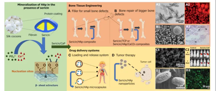
FIGURE 1 | Schematic representation of CaP mineralization process in the presence of sericin and research applications of sericin/CaP composites. Proposed biomineralization mechanism based on the works ofTakeuchi et al. (2005b),Cai et al. (2009), andVeiga et al. (2019). (A1) Sericin/HAp composite particles, (A2) cell morphology and differentiation of MG-63 cells after 5 days of contact with the composite reproduced fromCai et al. (2009)with permission from Royal Society of Chemistry; (B1) sericin/CaCO3composite particles, (B2) mice tissue after 12 weeks of composite implantation reproduced fromZhong et al. (2016)with permission from Elsevier Science & Technology Journals; (C1) sericin/CaP composite nanoparticles, (C2) distribution of the composite in vivo reproduced fromZhao et al. (2017)
with permission from John Wiley and Sons; (D1) sericin/HAp composite microcapsules, (D2) cell adhesion activity on HapG-2 cells after 7 days of contact with the composite reproduced fromLi et al. (2016)with permission from Royal Society of Chemistry.
Drug Delivery Systems
Sericin/CaPs materials have also been proposed as drug delivery systems. Due to their biodegradability and hydrophilicity sericin-based materials may be used as a vehicle to deliver therapeutic medicine (Wang et al., 2015). Ideally, these systems should have controlled degradation, enable easy binding of other molecules and have pH responsiveness (Kunz et al., 2016). Sericin/CaP composites can be designed as microsystems with a good loading and release capability for bioactive molecules to be applied in TE or as an anti-tumor therapeutic system (Table 1). The drug release potential of sericin/CaP microcapsules was studied in a paper by Li et al. (2016), by incubating sericin microcapsules with a supersaturated calcium phosphate solution containing citric acid. The CaP shell protected the inner sericin structure, and consequently the stored bioactive molecules until a pH change occurs. Not only did the capsules showed an improved stability and a longer storage time capability, but also increased cell viability.
In the work ofZhao et al. (2017), sericin/CaP nanoparticles were developed and injected into mice by intravenous injection, to allow particle distribution through the whole body. Analysis of the ex vivo organs showed that 80 nm spherical sericin/CaP nanoparticles are almost completely distributed in the liver cells of mice (≈90%) and can be biodegraded within 2 weeks. However, the degradation rate is not proportional to the amount of composites injected, as 100 and 200 µg of nanoparticles were degraded after 48 h and 13 days, respectively. The targeting percentage was 30% at 7 days post-injection. As no particles were found in other organs, it was concluded that these composites can target specific solid tumors in vivo.
Nowadays, nanoparticle pharmaceutical carriers are probably amongst the most studied sericin/CaP composites, making these very interesting composites in this field. The large surface area/volume ratio provides the capability to load large amounts of biomolecules and the ability to go across the cellular membrane
(Jia et al., 2013).
Other Applications
As the study of sericin/CaP composite materials is relatively recent, there are still several areas to be explored, taking advantage of the individual properties of sericin and CaP. For instance, calcium has an important role in maintaining homeostasis in mammalian skin, promoting wound healing
(Lansdown, 2002; Kawai et al., 2011). In particular calcium-based
nanoparticles have shown to have therapeutic benefits when used in cutaneous wound repair (Kawai et al., 2011). On the other hand, sericin has high antioxidant potential (Prasong, 2011;
Kumar and Mandal, 2017), antibacterial activity (Ramesan et al.,
2016), mitogenic effect on cells (Terada et al., 2005), potential to inhibit inflammation (Aramwit et al., 2012a), and ability to retain hydration (Padamwar et al., 2005). In the study of Aramwit et al., a sericin-based therapeutic cream formulation improved healing by promoting collagen production and rapid re-epithelialization in rat skin wounds (Aramwit et al., 2009, 2013). Both two-dimensional (films) and three-two-dimensional (scaffolds) sericin matrices, obtained using different blending methods, have been reported for skin tissue repair (Nayak et al., 2012; Lamboni
et al., 2015; Karahaliloglu et al., 2017). Therefore, a sericin/CaP
biomaterial could significantly accelerate and improves wound healing and soft tissue replacement. While sericin has been used
Veiga et al. Silk Sericin/Calcium Phosphate Biomaterials
in many cosmetic products over the years, CaP-based materials are only recently being explored in this area. CaP particles can be used as a bone filler in esthetical treatments for diminishing wrinkles by stimulating conjunctive tissue formation (Lopes
et al., 2009). Due to its affinity for biopolymers, crystallites of
HAp in the dermis can promote the self-assembly of collagen fibers, smoothing fine lines and even deeper wrinkles (Bell,
2012; Gupta et al., 2019). CaP-based sunscreens have also been
studied due to their biocompatible nature that does not generate allergies and other skin irritations, contrary to many organic sunscreens (de Araujo et al., 2010). These properties coupled with sericin strong suppressing activity against UVB-induced acute damage and tumor promotion (Zhaorigetu et al., 2003;
Kumar et al., 2018), anti-wrinkle effect and anti-tyrosinase
characteristics (Chlapanidas et al., 2013; Kumar and Mandal, 2019) justify the development of sericin/CaP biomaterials for the cosmetic industry.
Different CaP phases are used in food applications, as a supplement to increase calcium and phosphorus intake or as food additives to thicken and stabilize firm foods (Eliaz and
Metoki, 2017). Phosphate additives are widely used in processed
foods as preservatives, acidifying agents, acidity buffers, and emulsifying agents. Despite their applications, the use of non-naturally occurring phosphates in food can negatively affect the general health of the consumer. Synthetically produced phosphates are effectively absorbed in the gastrointestinal tract, whereas in “organic” phosphates, the absorption percentage ranges between 40 and 60%. This can lead to calcification in blood vessels and organs, being also potentially harmful for patients with chronic kidney disease (Sherman, 2007; Ritz
et al., 2012). Studies show that sericin can be used to prevent
functional gastrointestinal disorders, improving ion absorption and consequent bioavailability of these elements (zinc, iron, magnesium, and calcium ions) and improving constipation
(Sasaki et al., 2000a,b). These characteristics can inspire the
synthesis of sericin/CaP materials also for the food industry. Recent applications of sericin/CaP composites include the removal of micro-pollutants from wastewater. In the work of
Koley et al. (2016), sericin/HAp materials were used to purify
water, exhibiting excellent adsorption of toxic heavy metal ions. While HAp has stability, sericin is a water-soluble material, capable of forming hydrogen bonds and with strong metal binding properties (Khosa et al., 2014).
RECENT ADVANCES
Despite their properties of interest and numerous applications, sericin/CaP composites still have disadvantages to circumvent such as poor mechanical resistance under complex stress states. CaPs and pure sericin have a low mechanical profile when compared to bone tissue, which limits the application of sericin/CaP composites as 3D biomaterials for load-bearing applications (Kunz et al., 2016; Canillas et al., 2017). Of the reviewed papers, only Zhang et al. (2014) reported the mechanical properties of sericin/HAp composites, obtained by mineralization of a flexible ethanol-treated sericin film. Other
explored alternatives to enhance these characteristics include the integration of other components in the composite. Adding a structural organic component such as collagen (Griffanti et al.,
2019; Jang et al., 2019), chitosan (Chen et al., 2015), and alginate
(Zhang et al., 2015); or a synthetic polymer such as polymethyl
methacrylate (PMMA) (Tontowi et al., 2017) are examples of some of the most recent approaches. Structural integrity of the constructs can also be obtained by cross-linking the components with glutaraldehyde (GTA) (Bhowmick et al., 2016).
CONCLUSIONS
Up to recently, silk sericin was traditionally disposed during silk processing. Nowadays, there has been a growing interest in its recovery due to both its beneficial properties and environmental impact caused by its generation. Works on sericin/CaP composites focus mainly on understanding the biomineralization process, through the study of the effect of sericin in HAp nucleation. Other studies dedicate to the synthesis of these biomaterials for the substitution and regeneration of hard tissues and to the development of new drug delivery systems. Apart from the biomedical field, there are other areas where these composites have a lot of potential, such as in the cosmetic, food an environmental sector.
In the last decade, the number of published papers on sericin/CaP composites has increased substantially. Thus, in the near future, it is clear that the integration of silk sericin in CaP biomaterials will result in advanced composite systems with evident benefits on several research fields, particularly in biomedical and pharmaceutic-related applications.
AUTHOR CONTRIBUTIONS
All authors contributed to the analysis and writing of the manuscript, and approved it for publication.
FUNDING
This work was financially supported by: Base Funding – UIDB/00511/2020 of the Laboratory for Process Engineering, Environment, Biotechnology and Energy – LEPABE – funded by national funds through the FCT/MCTES (PIDDAC). The authors also acknowledge Portuguese National Funds from FCT—Fundação para a Ciência e a Tecnologia through project UID/Multi/04044/2019. This work was supported by project Biotherapies-Bioengineered Therapies for Infectious Diseases and Tissue Regeneration and Interreg V-A POCTEP Programme through FEDER funds from the European Union [0245_IBEROS_1_E] C. A.
ACKNOWLEDGMENTS
The authors also extend their deep appreciation to Department of Chemical Engineering at Faculty of Engineering of the University of Porto, and Centre for Biotechnology and Fine Chemistry at School of Biotechnology of the Portuguese Catholic University.
Veiga et al. Silk Sericin/Calcium Phosphate Biomaterials
REFERENCES
Ahsan, F., Ansari, T., Usmani, S., and Bagga, P. (2018). An insight on silk protein sericin: from processing to biomedical application. Drug Res. 68, 317–327. doi: 10.1055/s-0043-121464.
Altman, G., Diaz, F., Jakuba, C., Calabro, T., Horan, R. L., Chen, T., et al. (2003). Silk-based biomaterials. Biomaterials 24, 401–416. doi: 10.1016/S0142-9612(02)00353-8
Amini, A., Laurencin, C., and Nukavarapu, S. (2012). Bone tissue engineering: recent advances and challenges. Crit. Rev. Biomed. Eng. 40, 363–408. doi: 10.1615/CritRevBiomedEng.v40.i5.10.
Aramwit, P. (2014). “Bio-response to silk sericin,” in Silk Biomaterials for Tissue Engineering and Regenerative Medicine, ed S. C. Kundu (England, Woodhead Publishing; Elsevier), 299–329. doi: 10.1533/9780857097064.2.299
Aramwit, P., Kanokpanont, S., De-Eknamkul, W., and Srichana, T. (2009). Monitoring of inflammatory mediators induced by silk sericin. J. Biosci. Bioeng. 107, 556–561. doi: 10.1016/j.jbiosc.2008.12.012.
Aramwit, P., Kanokpanont, S., Nakpheng, T., and Srichana, T. (2010). The effect of sericin from various extraction methods on cell viability and collagen production. Int. J. Mol. Sci. 11, 2200–2211. doi: 10.3390/ijms11052200. Aramwit, P., Keongamaroon, O., Siritientong, T., Bang, N., and Supasyndh, O.
(2012a). Sericin cream reduces pruritus in hemodialysis patients: a randomized, double-blind, placebo-controlled experimental study. BMC Nephrol. 13:119. doi: 10.1186/1471-2369-13-119.
Aramwit, P., Palapinyo, S., Srichana, T., Chottanapund, S., and Muangman, P. (2013). Silk sericin ameliorates wound healing and its clinical efficacy in burn wounds. Arch. Dermatol. Res. 305, 585–594. doi: 10.1007/s00403-013-1371-4. Aramwit, P., Siritientong, T., and Srichana, T. (2012b). Potential applications of
silk sericin, a natural protein from textile industry by-products. Waste Manage. Res. 30, 217–224. doi: 10.1177/0734242X11404733.
Bell, S. (2012). Therapeutic Calcium Phosphate Particles in Use for Aesthetic of Cosmetic Medicine, and Methods of Manufacture and Use. U.S. Patent No 2012/0207803. Washington, DC: U.S. Patent and Trademark Office. Bhowmick, S., Scharnweber, D., and Koul, V. (2016). Co-cultivation
of keratinocyte-human mesenchymal stem cell (HMSC) on sericin
loaded electrospun nanofibrous composite scaffold (cationic
gelatin/hyaluronan/chondroitin sulfate) stimulates epithelial differentiation in HMSCs: in vitro study. Biomaterials 88, 83–96. doi: 10.1016/j.biomaterials.2016.02.034.
Bleek, K., and Taubert, A. (2013). New developments in polymer-controlled, bioinspired calcium phosphate mineralization from aqueous solution. Acta Biomater. 9, 6283–6321. doi: 10.1016/j.actbio.2012.12.027.
Bohner, M. (2000). Calcium orthophosphates in medicine: from ceramics to calcium phosphate cements. Int. J. Care Injured. 31: D37–D47. doi: 10.1016/S0020-1383(00)80022-4.
Boyle, L. (2018). “Applications of de novo designed peptides,” in Peptide Applications in Biomedicine, Biotechnology and Bioengineering, ed S. Koutsopoulos (England: Woodhead Publishing; Elsevier), 51–86. doi: 10.1016/B978-0-08-100736-5.00003-X
Brown, C., and Barker, T. (2014). Fibrin-based biomaterials: modulation of macroscopic properties through rational design at the molecular level. Acta Biomater. 10, 1502–1514. doi: 10.1016/j.actbio.2013.09.008.
Cai, Y., Jin, J., Mei, D., Xia, N., and Yao, J. (2009). Effect of silk sericin on assembly of hydroxyapatite nanocrystals into enamel prism-like structure. J. Mater. Chem. 19:5751. doi: 10.1039/b901620a.
Cai, Y., Mei, D., Jiang, T., and Yao, J. (2010). Synthesis of oriented hydroxyapatite crystals: effect of reaction conditions in the presence or absence of silk sericin. Mater. Lett. 64, 2676–2678. doi: 10.1016/j.matlet.2010.08.071
Cai, Y., and Yao, J. (2010). Effect of proteins on the synthesis and assembly of calcium phosphate nanomaterials. Nanoscale 2:1842. doi: 10.1039/c0nr00092b Campana, V., Milano, G., Pagano, E., Barba, M., Cicione, C., Salonna, G., et al.
(2014). Bone substitutes in orthopaedic surgery: from basic science to clinical practice. J. Mater. Sci. 25, 2445–2461. doi: 10.1007/s10856-014-5240-2 Canillas, M., Pena, P., de Aza, A. H., and Rodríguez, M. A. (2017). Calcium
phosphates for biomedical applications. Boletín de La Sociedad Española de Cerámica y Vidrio 56, 91–112. doi: 10.1016/j.bsecv.2017.05.001
Cardoso, D., Beucken, J., Both, L., Bender, J., Jansen, J., and Leeuwenburgh, S. C. (2014). Gelation and biocompatibility of injectable alginate-calcium phosphate
gels for bone regeneration. J. Biomed. Mater. Res. Part A 102, 808–817. doi: 10.1002/jbm.a.34754.
Chazono, M., Tanaka, T., Komaki, H., and Fujii, K. (2004). Bone formation and bioresorption after implantation of injectable β-tricalcium phosphate granules-hyaluronate complex in rabbit bone defects. J. Biomed. Mater. Res. 70A, 542–549. doi: 10.1002/jbm.a.30094
Chen, L., Hu, J., Ran, J., Shen, C., and Tong, H. (2015). A novel nanocomposite for bone tissue engineering based on chitosan-silk sericin/hydroxyapatite: biomimetic synthesis and its cytocompatibility. RSC Adv. 5, 56410–56422. doi: 10.1039/C5RA08216A
Chen, L., Hu, J., Ran, J., Shen, X., and Tong, H. (2014). Preparation and evaluation of collagen-silk fibroin/hydroxyapatite nanocomposites for bone tissue engineering. Int. J. Biol. Macromol. 65, 1–7. doi: 10.1016/j.ijbiomac.2014.01.003 Chen, Y., Feng, Y., Deveaux, J., Masoud, M., Chandra, F., Chen, H., et al. (2019). Biomineralization forming process and bio-inspired nanomaterials for biomedical application: a review. Minerals 9:68. doi: 10.3390/min9020068 Chirila, T., Suzuki, S., Bray, L., Barnett, N., and Harkin, D. (2013). Evaluation
of silk sericin as a biomaterial: in vitro growth of human corneal limbal epithelial cells on Bombyx mori sericin membranes. Progr. Biomater. 2:14. doi: 10.1186/2194-0517-2-14
Chlapanidas, C., Faragò, S., Lucconi, G., Perteghella, S., Galuzzi, M., Mantelli, M., et al. (2013). Sericins exhibit ROS-scavenging, anti-tyrosinase, anti-elastase, and in vitro immunomodulatory activities. Int. J. Biol. Macromol. 58, 47–56. doi: 10.1016/j.ijbiomac.2013.03.054
Dash, R., Mandal, M., Ghosh, S., and Kundu, S. C. (2008). Silk sericin protein of tropical tasar silkworm inhibits UVB-induced apoptosis in human skin keratinocytes. Mol. Cell. Biochem. 311, 111–119. doi: 10.1007/s11010-008-9702-z
de Araujo, T. S., Souza, S. O., and Sousa, E. M. B. (2010). Effect of Zn2+, Fe3+and Cr3+addition to hydroxyapatite for its application as an active constituent of sunscreens. J. Phys. 249:012012. doi: 10.1088/1742-6596/249/1/012012. Dhert, W. J. A., Klein, C. P. A. T., Jansen, J. A., Velde, E. A., Vriesde, R.
C., Rozing, P. M., et al. (1993). A histological and histomorphometrical investigation of fluorapatite, magnesiumwhitlockite, and hydroxylapatite plasma-sprayed coatings in goats. J. Biomed. Mater. Res. 27, 127–138. doi: 10.1002/jbm.820270116
Dias, G., Mahoney, P., Swain, M., Kelly, R., Smith, R., and Ali, M. A. (2010). Keratin-hydroxyapatite composites: biocompatibility, osseointegration, and physical properties in an ovine model. J. Biomed. Mater. Res. 95, 1084–1095. doi: 10.1002/jbm.a.32908.
Eliaz, N., and Metoki, N. (2017). Calcium phosphate bioceramics: a review of their history, structure, properties, coating technologies and biomedical applications. Materials 10:334. doi: 10.3390/ma10040334
Farè, S., Torricelli, P., Giavaresi, G., Bertoldi, S., Alessandrino, A., Villa, T., et al. (2013). In vitro study on silk fibroin textile structure for anterior cruciate ligament regeneration. Mater. Sci. Eng. C 33, 3601–3608. doi: 10.1016/j.msec.2013.04.027
Farokhi, M., Mottaghitalab, F., Samani, S., Shokrgozar, M., Kundu, S. C., Reis, R. L., et al. (2018). Silk fibroin/hydroxyapatite composites for bone tissue engineering. Biotechnol. Adv. 36, 68–91. doi: 10.1016/j.biotechadv.2017. 10.001
Fuchs, K., Thompson, W., and Warden, S. (1997). “Bone biology,” in Bone Repair Biomaterials, Vol. 11, eds K. M. Pawelec and J. A. Planell (England: Woodhead Publishing; Elsevier), 1–22. doi: 10.1016/S0950-351X(97)80473-9
George, A., and Veis, A. (2008). Phosphorylated proteins and control over apatite nucleation, crystal growth, and inhibition. Chem. Rev. 108, 4670–4693. doi: 10.1021/cr0782729
Griffanti, G., Jiang, W., and Nazhat, S. (2019). Bioinspired mineralization of a functionalized injectable dense collagen hydrogel through silk sericin incorporation. Biomater. Sci. 7, 1064–1077. doi: 10.1039/C8BM01060A Gsib, O., Egles, C., and Bencherif, S. (2017). Fibrin: an underrated biopolymer for
skin tissue engineering. J. Mol. Biol. Biotechnol. 2, 1–4.
Gulrajani, M. L., Agarwal, R., and Chand, S. (2000). Degumming of silk with a fungal protease. Indian J. Fibre Text. Res. 25, 138–142.
Gupta, N., Spiegel, O. L., and Spiegel, J. H. (2019). “RadiesseTM calcium hydroxylapatite injectable filler,” in Neurotoxins and Fillers in Facial Esthetic Surgery (Hoboken, NJ: John Wiley & Sons, Inc.), 71–74. doi: 10.1002/9781119294306.ch5
Veiga et al. Silk Sericin/Calcium Phosphate Biomaterials
Ha, T., Quan, T., Vu, D., and Si, D. (2013). “Naturally derived biomaterials: preparation and application,” in Regenerative Medicine and Tissue Engineering, ed J. Andrades (Rijeka: InTech), 247–269. doi: 10.5772/55668
Habraken, W., Habibovic, P., Epple, M., and Bohner, M. (2016). Calcium phosphates in biomedical applications: materials for the future? Mater. Today 19, 69–87. doi: 10.1016/j.mattod.2015.10.008
Holland, C., Numata, K., Rnjak-Kovacina, J., and Seib, F. (2019). The biomedical use of silk: past, present, future. Adv. Healthc. Mater. 8:1800465. doi: 10.1002/adhm.201800465
Jang, J., Lee, E., Kwon, G., and Seo, Y. (2019). The effect of coated nano-hydroxyapatite concentration on scaffolds for osteogenesis. J. Biomater. Appl. 34, 827–839. doi: 10.1177/0885328219875275
Jia, F., Liu, X., Li, L., Mallapragada, S., Narasimhan, B., and Wang, Q. (2013). Multifunctional nanoparticles for targeted delivery of immune activating and cancer therapeutic agents. J. Control. Rel. 172, 1020–1034. doi: 10.1016/j.jconrel.2013.10.012
Jones, J. (2013). “Scaffolds for Tissue Engineering,” in Biodegradable Polymer-Based Scaffolds for Bone Tissue Engineering, ed N. Sultana (Berlin; Heidelberg; Germany: Springer-Verlag; SpringerBriefs in Applied Sciences and Technology), 201–213. doi: 10.1007/978-3-642-34802-0_1
Karahaliloglu, Z., Kilicay, E., and Denkbas, E. (2017). Antibacterial chitosan/silk sericin 3D porous scaffolds as a wound dressing material. Artif. Cells Nanomed. Biotechnol. 45, 1172–1185. doi: 10.1080/21691401.2016.1203796
Kasoju, N., and Bora, U. (2012). Silk fibroin in tissue engineering. Adv. Healthc. Mater. 1, 393–412. doi: 10.1002/adhm.201200097
Kawai, K., Larson, B., Ishise, H., Carre, A., Nishimoto, S., Longaker, M., et al. (2011). Calcium-based nanoparticles accelerate skin wound healing. PLoS ONE 6:e27106. doi: 10.1371/journal.pone.0027106
Khosa, M., Shah, S., and Feng, X. (2014). Metal sericin complexation and ultrafiltration of heavy metals from aqueous solution. Chem. Eng. J. 244, 446–456. doi: 10.1016/j.cej.2014.01.091
Klein, C. P., Patka, P., van der Lubbe, H. B., Wolke, J. G., and de Groot, K. (1991). Plasma-sprayed coatings of tetracalciumphosphate, hydroxyl-apatite, and β-TCP on titanium alloy: an interface study. J. Biomed. Mater. Res. 25, 53–65. doi: 10.1002/jbm.820250105
Koley, P., Sakurai, M., Takei, T., and Aono, M. (2016). Facile fabrication of silk protein sericin-mediated hierarchical hydroxyapatite-based bio-hybrid architectures: excellent adsorption of toxic heavy metals and hazardous dye from wastewater. RSC Adv. 6, 86607–86616. doi: 10.1039/C6RA12818A Kumar, J., Alam, S., Jain, A., Ansari, K., and Mandal, B. (2018). Protective
activity of silk sericin against UV radiation-induced skin damage by downregulating oxidative stress. ACS Appl. Biomater. 1, 2120–2132. doi: 10.1021/acsabm.8b00558
Kumar, P., and Mandal, B. (2017). Antioxidant potential of mulberry and non-mulberry silk sericin and its implications in biomedicine. Free Radic. Biol. Med. 108, 803–818. doi: 10.1016/j.freeradbiomed.2017.05.002
Kumar, P., and Mandal, B. (2019). The inhibitory effect of silk sericin against ultraviolet-induced melanogenesis and its potential use in cosmeceutics as an anti-hyperpigmentation compound. Photochem. Photobiol. Sci. 18, 2497–2508. doi: 10.1039/C9PP00059C
Kunz, R., Brancalhão, R., Ribeiro, L., and Natali, M. (2016). Silkworm sericin: properties and biomedical applications. Biomed Res. Int. 2016:8175701. doi: 10.1155/2016/8175701
Lamboni, L., Gauthier, M., Yang, G., and Wang, Q. (2015). Silk sericin: a versatile material for tissue engineering and drug delivery. Biotechnol. Adv. 33, 1855–1867. doi: 10.1016/j.biotechadv.2015.10.014
Lansdown, B. G. (2002). Calcium: a potential central regulator in
wound healing in the skin. Wound Rep. Regener. 10, 271–285.
doi: 10.1046/j.1524-475X.2002.10502.x
Lee, Y., and Yuk, S. (2007). Polymeric protein delivery systems. Prog. Polym. Sci. 32, 669–697. doi: 10.1016/j.progpolymsci.2007.04.001
Li, W., Cai, Y., Zhong, Q., Yang, Y., Kundu, C., and Yao, J. (2016). Silk sericin microcapsules with hydroxyapatite shells: protection and modification of organic microcapsules by biomimetic mineralization. J. Mater. Chem. B 4, 340–347. doi: 10.1039/C5TB02328A
Liu, C. (2015). “Hydroxyapatite (HAp) for biomedical applications,” in Structure and Properties of Hydroxyapatite for Biomedical Applications,
ed M. Mucalo (England: Woodhead Publishing; Elsevier), 3–19. doi: 10.1016/B978-1-78242-033-0.00010-9
Liu, J., Lawrence, B., Liu, A., Schwab, I., Oliveira, L., and Rosenblatt, M. I. (2012). Silk fibroin as a biomaterial substrate for corneal epithelial cell sheet generation. Invest. Opthalmol. Vis. Sci. 53:4130. doi: 10.1167/iovs.12-9876
Lopes, B., Dias, M., Silva, V., Santos, P., Monteiro, F., and Gomes, P. (2009). Production Method for Calcium Phosphate Nano-Particles With High Purity and Their Use. U.S. Patent No 2009/0263497 A1. Washington, DC: U.S. Patent and Trademark Office.
Mondal, M., Trivedy, K., and Kumar, S. (2006). The silk proteins, sericin and fibroin in silkworm, bombyx mori linn., - a review. Caspian J. Environ. Sci. 25, 77–84.
Nayak, S., Dey, T., Naskar, D., and Kundu, S. (2013). The promotion of osseointegration of titanium surfaces by coating with silk protein sericin. Biomaterials 34, 2855–2864. doi: 10.1016/j.biomaterials.2013. 01.019
Nayak, S., Talukdar, S., and Kundu, S. (2012). Potential of 2D crosslinked sericin membranes with improved biostability for skin tissue engineering. Cell Tissue Res. 347, 783–794. doi: 10.1007/s00441-011-1269-4
Ogose, A., Hotta, T., Kawashima, H., Kondo, N., Gu, W., Kamura, T., et al. (2005). Comparison of hydroxyapatite and beta tricalcium phosphate as bone substitutes after excision of bone tumors. J. Biomed. Mater. Res. 72B, 94–101. doi: 10.1002/jbm.b.30136
Padamwar, M., Pawar, A., Daithankar, A., and Mahadik, K. R. (2005). Silk sericin as a moisturizer: an in vivo study. J. Cosmet. Dermatol. 4, 250–257. doi: 10.1111/j.1473-2165.2005.00200.x
Padamwar, M. N., and Pawar, A. P. (2004). Silk sericin and its application : a review. J. Sci. Ind. Res. 63, 323–329.
Pighinelli, L., and Kucharska, M. (2013). Chitosan-hydroxyapatite composites. Carbohydr. Polym. 93, 256–262. doi: 10.1016/j.carbpol.2012.06.004
Prasong, S. (2011). Screening of antioxidant activity of some samia ricini (eri) silks: comparison with Bombyx mori. J. Biol. Sci. 11, 336–339. doi: 10.3923/jbs.2011.336.339
Ramesan, M. T., Athira, V. K., Jayakrishnan, P., and Gopinathan, C. (2016). Preparation, characterization, electrical and antibacterial properties of sericin/poly(vinyl alcohol)/poly(vinyl pyrrolidone) composites. J. Appl. Polym. Sci. 43535. doi: 10.1002/app.43535
Rezwan, K., Chen, Q. Z., Blaker, J. J., and Boccaccini, A. (2006). Biodegradable and bioactive porous polymer/inorganic composite scaffolds for bone tissue engineering. Biomaterials 27, 3413–3431. doi: 10.1016/j.biomaterials.2006.01.039
Ritz, E., Hahn, K., Ketteler, M., Kuhlmann, M., and Mann, J. (2012). Phosphate additives in food. Deutsches Aerzteblatt Online 109, 49–55. doi: 10.3238/arztebl.2012.0049
Salama, A. (2019). Cellulose/Calcium phosphate hybrids: new materials for biomedical and environmental applications. Int. J. Biol. Macromol. 127, 606–617. doi: 10.1016/j.ijbiomac.2019.01.130.
Sasaki, M., Yamada, H., and Kato, N. (2000a). A resistant protein, sericin improves atropine-induced constipation in rats. Food Sci. Technol. Res. 6, 280–283. doi: 10.3136/fstr.6.280
Sasaki, M., Yamada, H., and Kato, N. (2000b). Consumption of silk protein, sericin elevates intestinal absorption of zinc, iron, magnesium and calcium in rats. Nutr. Res. 20, 1505–1511. doi: 10.1016/S0271-5317(00) 80031-7
Saxena, T., Karumbaiah, L., and Valmikinathan, C. (2014). “Proteins and poly(amino acids),” in Natural and Synthetic Biomedical Polymers, eds S. Kumbar and M. Deng (Netherlands: Elsevier Science; ScienceDirect), 43–65. doi: 10.1016/B978-0-12-396983-5.00003-X
Shavandi, A., Silva, T.i., Bekhit, A., and Bekhit, A. (2017). Keratin: dissolution, extraction and biomedical application. Biomater. Sci. 5, 1699–1735. doi: 10.1039/c7bm00411g
Sherman, R. (2007). Dietary phosphate restriction and protein intake in dialysis patients: a misdirected focus. Semin. Dial. 20, 16–18. doi: 10.1111/j.1525-139X.2007.00204.x
Soong, K., and Kenyon, K. (1984). Adverse reactions to virgin
silk sutures in cataract surgery. Ophthalmology 91, 479–483. doi: 10.1016/S0161-6420(84)34273-7