Case Report
Takotsubo Syndrome: A Pathway through the Pituitary Disease
Rui Plácido,
1Ana Filipa Martins,
2Susana Robalo Martins,
1Sónia do Vale,
2Ana G. Almeida,
1Fausto Pinto,
1and João Martin Martins
21Hospital Santa Maria, Cardiology Department, Lisbon Academic Medical Centre, CCUL, Lisbon, Portugal 2Hospital de Santa Maria, Avenida Professor Egas Moniz, 1649-035 Lisboa, Portugal
Correspondence should be addressed to Rui Pl´acido; [email protected] Received 24 November 2015; Accepted 2 February 2016
Academic Editor: Man-Hong Jim
Copyright © 2016 Rui Pl´acido et al. This is an open access article distributed under the Creative Commons Attribution License, which permits unrestricted use, distribution, and reproduction in any medium, provided the original work is properly cited. Takotsubo cardiomyopathy (TTC) is characterized by reversible left ventricular apical and/or midventricular hypokinesia with unknown etiology. The clinical presentation is similar to acute myocardial infarction in the absence of significant obstructive coronary artery disease. Various predisposing factors have been related to TTC, such as acute neurological illnesses, endocrine diseases, pain, and emotional stress. We present the first description of an association between TTC cardiomyopathy and panhypopituitarism. This case reinforces the connection between the hormonal and cardiovascular systems. Furthermore, it supports the importance of a comprehensive and integrated medical history in the approach of a patient with cardiac disease, towards clinical decision-making.
1. Introduction
Takotsubo cardiomyopathy (TTC) is a novel cardiomyopathy that has been firstly described by Sato et al. [1] as an important consideration in the differential diagnosis of acute coronary syndrome. The presenting features of TTC are similar to those of myocardial ischemia after acute plaque rupture, but the characteristic distinctions are regional wall motion abnormalities that extend beyond a single coronary vascular bed and the absence of epicardial coronary occlusion. A preceding emotional or physical stressor is common [2]. The presentation can include life-threatening symptoms and hemodynamic compromise. Recently a substantial rate of death and complications after the acute phase of TTC was shown, with a long-term follow-up revealing a significant rate of death from any cause (5.6% per patient-year) and a rate of major adverse cardiac and cerebrovascular events (9.9% per patient-year) [2].
Marked increased levels of catecholamines and meta-nephrines are found in the acute phase and they proba-bly account for a possible acute coronary spasm and/or focal myocardial dysfunction with contraction band necro-sis/myocytolysis underlying the clinical condition [3]. Since the prognosis is much better and a different pharmaco-logic approach is required, angiotensin-converting–enzyme
inhibitors or angiotensin receptor blockers are preferred [2], while inotropes should be avoided; the differential diagnosis is crucial [4]. Little is known about individual susceptibility to TTC. However increased sympathetic autonomic activity might be casually related. More rarely it may be related to a diseased hypothalamic-pituitary-adrenal axis (HPA), proba-bly requiring chronic increased compensatory hyperactivity of the sympathetic autonomic system [5]. We report a clinical case supporting that association.
2. Case Presentation
A 74-year-old man presented at the emergency department with chest pain, palpitations, and progressive dyspnea within the last 24 hours. He had a past history of high blood pressure and dyslipidemia and was being treated with lisino-pril/hydrochlorothiazide and simvastatin; an acquired atri-oventricular block was managed with DDDR pacing. In the last year, fatigue, asthenia, adynamia, cold intolerance, loss of muscular strength, anorexia, decreased libido, and loss of male hair pattern distribution were also reported, with blood pressure decreasing whereat lisinopril/hydrochlorothiazide was interrupted. On physical examination, blood pressure was 102/84 mmHg, heart rate 65 beats per minute, respiratory rate 28 breaths per minute, and normal oxygen saturation
Volume 2016, Article ID 9219018, 4 pages http://dx.doi.org/10.1155/2016/9219018
2 Case Reports in Cardiology
(a) (b)
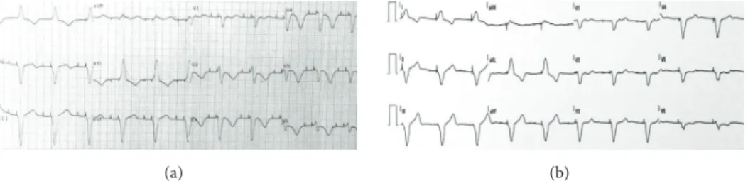
Figure 1: (a) Initial electrocardiogram showing an atrioventricular sequential paced rhythm with left bundle branch block morphology complexes, no ST-segment deviation, deeply inverted T waves on DI, aVL and precordial leads, and a prolonged QTc interval (560 ms). (b) Electrocardiogram 3 months after discharge.
(a) (b)
Figure 2: (a) Transthoracic echocardiography (apical four-chamber view) during the initial admission, demonstrating apical ballooning (white arrows). (b) Left ventriculography images in diastole and systole, showing typical type of takotsubo cardiomyopathy with apical ballooning, dyskinesia (white arrows), and left ventricle basal hypercontractility.
(ambient air) and temperature 36.6∘C. He had signs of heart failure (jugular venous distention, peripheral edema, and pulmonary congestion) and cardiac auscultation was remarkable for a protodiastolic gallop.
Electrocardiogram showed an atrioventricular sequential paced rhythm with left bundle branch block morphology complexes, no ST-segment deviation, deeply inverted T waves on DI, aVL and precordial leads, and a prolonged QTc interval (560 ms) (Figure 1(a)). Complete blood count, serum electrolytes, renal function parameters, and glycaemia were normal. Troponin I (<0.07 ng/mL) was mildly elevated (0.79 ng/mL). Echocardiography showed a nondilated left ventricle with apical dyskinesia and hyperdynamic basal contraction assuming a systolic ballooning pattern, and a moderately depressed (37%) ejection fraction (Figure 2(a); Video 1 in Supplementary Material available online at http://dx.doi.org/10.1155/2016/9219018).
Acute coronary syndrome and TTC were considered in the differential diagnosis. Coronarography showing no significant coronary artery disease and a left ventriculogram revealing the typical apical ballooning (Figure 2(b); Video 2) favor the former hypothesis. Cardiac magnetic resonance was not performed given the presence of a noncompatible pacemaker.
During hospitalization the patient was managed with diuretics, beta-blocker, and angiotensin-converting enzyme inhibitor. Laboratory data showed a peak troponin I of 1,38 ng/mL. Pheochromocytoma/paraganglioma was excluded by urinary meta- and normetanephrines measurement (High Performance Liquid Chromatography) and the patient was referred for endocrine consultation.
A formal pituitary dynamic test was performed after an overnight fast: growth hormone-releasing hormone
100𝜇g, corticotropin-releasing hormone 100 𝜇g, thyrotropin-releasing hormone 200𝜇g, and gonadotropin-releasing hor-mone 100𝜇g bolus (intravenous) at time 0; baseline/peak hormone levels were as follows: growth hormone (<2 ng/mL): 0.15/0.18; prolactin (2–18 ng/mL): 21/35; cortisol (4–23𝜇g/ dL): 1.5/4.1; follicle-stimulating hormone (1–18 U/L): 2.2/2.8; luteinizing hormone (3–35 U/L): 0.3/1.1; other baseline val-ues were insulin-like growth factor 1 (48–188 ng/mL): 33; adrenocorticotropin (0–46 pg/mL): 31; triiodothyronine (60– 181 ng/dL): 81; thyroxine (4.5–10.9𝜇g/dL): 2.2; free thyroxine (0.89–1.80 ng/dL): 0.42; total testosterone (188–772 ng/dL): <10. All measurements were obtained with standardized chemiluminescence immunoassays; age and gender specific reference values at 8.00–10.00 h am after an overnight fast are presented within square brackets. Thyroid autoantibodies were negative.
Brain magnetic resonance imaging revealed a normal sized and positioned pituitary gland with no morphological abnormalities and a centered pituitary stalk.
A diagnosis of idiopathic panhypopituitarism was estab-lished and the patient was prescribed hydrocortisone20 + 10 mg daily (oral), levothyroxine 100 𝜇g daily (oral), and testosterone enanthate 250 mg every fortnight (intramuscu-lar). The usual instructions were given orally and by writing regarding stressful conditions.
After 3 months of optimized heart failure treatment the patient showed clinical improvement, normalization of left ventricular function, and reversibility of T waves changes and QT prolongation (Figure 1(b)). Interestingly, in this case, the repolarization changes were clearly interpretable notwith-standing the presence of a right ventricular paced rhythm. In that time, lost weight was recovered and the patient regained normal physical vigour without other complaints.
3. Discussion
This case begins with an unusual presentation of TTC. There was no record for any particular acute emotional or physical stress; the patient is male while TTC is much more common in women and the presentation is that of an acute coronary syndrome with marked left ventricular dysfunction [3, 6].
TTC is an acute, reversible dysfunction of the left ventri-cle in the absence of significant obstructive coronary disease. Recently, Templin et al. [2] prospectively showed that the most common pattern was the apical type (81.7%) followed by the midventricular (14.6%), the basal (2.2%), and the focal (1.5%) types. The condition is especially prevalent in postmenopausal women but the true prevalence of TTC remains uncertain because of its underrecognition. The long-term prognosis is generally favorable. The pathophysiology of TTC is not established but is likely multifactorial, involving the vascular, endocrine, and central nervous systems. Several mechanisms have been postulated to link the sympathetic hyperactivity and myocardial dysfunction including diffuse coronary artery spasm, coronary microcirculation alter-ations, and direct catecholamine mediated myocyte injury [3, 7]. In 70% of the documented cases there is an apparent trigger leading to catecholamine-induced myocardial injury [6]. Further investigation is needed to define the connection between these triggers and their effect on the vulnerable myocardium.
In the acute phase, TTC is sometimes indistinguishable from acute myocardial infarction with respect to clinical symptoms, ECG changes, and cardiac biomarkers such as troponin and creatine kinase. Fast and accurate diagnosis on admission remains challenging, and exclusion of significant obstructive coronary artery disease is mandatory. Thus, there is a need for sensitive and specific biomarkers for the early diagnosis of TTC. MicroRNAs are a class of highly con-served and small noncoding posttranscriptional regulators of diverse cellular processes that have been recently considered as biomarkers in cardiovascular disease [8]. Jaguszewski et al. [9] recently described a signature of circulating microRNAs for sensitive and specific identification of TTC in the acute phase. The significant upregulation of stress- and depression-related microRNAs suggested a role of central and/or periph-eral nervous system in TTC.
Retrospectively the patient of our case presented com-plains suggesting pituitary failure—low blood pressure, cold intolerance, loss of physical vigour and motivation, decreased libido, scanty body hair—although this is a rare condition, and these symptoms are by themselves rather common in older men [7]. Endocrine evaluation unequivocally revealed complete anterior pituitary failure, but the reason is not apparent. Combined pituitary failure by itself is a very rare condition—estimated prevalence 46/100.000 and esti-mated annual incidence 4/100.000—and at least in adult/old patients most commonly depends on pituitary tumors [10, 11]. Other less common causes include head trauma [12], for which the patient had no recollection and besides pituitary stalk section generally occurs; hemochromatosis and other metabolic conditions, for which no evidence was found in this case (iron, transferrin, and ferritin levels were normal
and the pituitary was not enlarged); vascular, that would be likely in this age group, namely, subarachnoid, hemorrhage [13]. However, there was no evidence for pituitary apoplexy at the CT scan. In fact most other conditions leading to combined pituitary failure, namely, infections, inflammatory, granulomatous, immune, or neoplastic disorders, generally present with structural abnormalities in the pituitary gland that can be shown by MRI [7, 10, 11]. Of course a congenital or genetic and functional defect is mostly unlikely, given patient age and clear evidence for previous normal growth and development, including two fathered children. In short, unequivocal combined pituitary failure is present but the rea-son is unclear. This occurs in around 10–20% of the cases, and an etiology sometimes is apparent only many years later [14]. An acute and transient defect is also unlikely since the patient had previous evidence of pituitary failure and maintained that condition for as long as two years of follow-up requiring substitutive therapy. Also, the possibility of a neuroendocrine adaptation to an acute/chronic condition may be discarded. That adaptation, for which the nonthyroidal patient syn-drome is the best-known pattern, is associated with increased cortisol levels [15]. By the same token, a drug effect is unlikely.
Besides some perplexing features, a most interesting point can be made that progressive multiple pituitary failure may require an increased sustained sympathetic activity to cope with daily life physical and emotional challenges, since it is by itself certainly a stressful condition. Curiously enough, to our knowledge, other reports of TTC and endocrine disease always resolve around the theme of pheochromo-cytoma [16] or failure of the HPA axis [5, 17, 18]; to our knowledge, this is the first report of the association of TTC with adult onset multiple pituitary hormonal fail-ure. Chronic activation of the sympathetic nervous system may on the other hand favor the development of TTC, as has been suggested by some reports regarding behav-ioral medicine [3]. Regarding combined pituitary failure, increased sympathetic drive may be particularly deleterious, since androgens may downregulate the stress response and may present direct vasodilator effects on coronary arter-ies [19], and low testosterone predicts the development of atherosclerosis and cardiovascular events [20] while gluco-corticoids may play a supportive role on myocardial function [21] and hypothyroidism may impair coronary blood flow [22].
TTC represents an acute heart failure syndrome that is associated with a substantial risk for adverse events. The great variety of triggering factors in this disease needs to be illu-minated and new investigations are required. Furthermore, a heightened vigilance in the treatment of these patients is crucial to improve prognosis.
In a way, the gap is narrowing between “Voodoo death” [23] as described by Sir Cannon in the 40s and TTC reported in the 90s. It remains to be seen if this association of combined pituitary failure and TTC is also reported by other authors. TTC by itself was also a long present but unknown condition until Sato first formally described it.
4 Case Reports in Cardiology
Conflict of Interests
The authors declare that there is no conflict of interests regarding the publication of this paper.
References
[1] H. Sato, H. Tateishi, T. Uchida et al., “Takotsubo type car-diomyopathy due to multivessel spasm,” in Clinical Aspect of
Myocardial Injury: From Ischemia to Heart Failure, K. Kodama,
K. Haze, and M. Hon, Eds., pp. 56–64, Kagaku Hyoronsha, Tokyo, Japan, 1990.
[2] C. Templin, J. R. Ghadri, J. Diekmann et al., “Clinical features and outcomes of takotsubo (stress) cardiomyopathy,” The New
England Journal of Medicine, vol. 373, pp. 929–938, 2015.
[3] I. S. Wittstein, D. R. Thiemann, J. A. C. Lima et al., “Neurohu-moral features of myocardial stunning due to sudden emotional stress,” The New England Journal of Medicine, vol. 352, no. 6, pp. 539–548, 2005.
[4] J. R. Ghadri, F. Ruschitzka, T. F. L¨uscher, and C. Templin, “Takotsubo cardiomyopathy: still much more to learn,” Heart, vol. 100, no. 22, pp. 1804–1812, 2014.
[5] S. Sakihara, K. Kageyama, T. Nigawara, Y. Kidani, and T. Suda, “Ampulla (takotsubo) cardiomyopathy caused by secondary adrenal insufficiency in ACTH isolated deficiency,” Endocrine
Journal, vol. 54, no. 4, pp. 631–636, 2007.
[6] K. A. Bybee, T. Kara, A. Prasad et al., “Transient left ventricular apical ballooning: a syndrome that mimics ST-segment eleva-tion myocardial infarceleva-tion,” Annals of Internal Medicine, vol. 141, no. 11, pp. 858–865, 2004.
[7] M. G. Burt and K. K. Y. Ho, “Hypopituitarism and growth hormone deficiency,” in Endocrinology. Adult and Pediatric, J. L. Jameson, L. J. De Groot, D. M. Kretser et al., Eds., chapter 11, pp. 188–208, Elsevier Saunders, Philadelphia, Pa, USA, 7th edition, 2016.
[8] S. K. Gupta, C. Bang, and T. Thum, “Circulating MicroRNAs as biomarkers and potential paracrine mediators of cardiovascular disease,” Circulation: Cardiovascular Genetics, vol. 3, no. 5, pp. 484–488, 2010.
[9] M. Jaguszewski, J. Osipova, J.-R. Ghadri et al., “A signature of circulating microRNAs differentiates takotsubo cardiomyopa-thy from acute myocardial infarction,” European Heart Journal, vol. 35, no. 15, pp. 999–1006, 2014.
[10] M. Regal, C. P´aramo, J. M. Sierra, and R. V. Garci-Mayor, “Prevalence and incidence of hypopituitarism in an adult Cau-casian population in northwestern Spain,” Clinical
Endocrinol-ogy, vol. 55, no. 6, pp. 735–740, 2001.
[11] M. O. van Aken and S. W. J. Lamberts, “Diagnosis and treatment of hypopituitarism: an update,” Pituitary, vol. 8, no. 3-4, pp. 183– 191, 2005.
[12] E. Richmond and A. D. Rogol, “Traumatic brain injury: endo-crine consequences in children and adults,” Endoendo-crine, vol. 45, no. 1, pp. 3–8, 2014.
[13] I. Kreitschmann-Andermahr, “Subarachnoid hemorrhage as a cause of hypopituitarism,” Pituitary, vol. 8, no. 3-4, pp. 219–225, 2005.
[14] H. F. Nystr¨om, A. Saveanu, E. J. L. Barbosa et al., “Detection of genetic hypopituitarism in an adult population of idio-pathic pituitary insufficiency patients with growth hormone deficiency,” Pituitary, vol. 14, no. 3, pp. 208–216, 2011.
[15] G. Van den Berghe, “Endocrine aspects of critical care medicine,” in Endocrinology. Adult and Pediatric, J. L. Jameson, L. J. De Groot, D. M. Kretser et al., Eds., chapter 114, pp. 1987– 1999, Saunders Elsevier, Philadelphia, Pa, USA, 7th edition, 2016.
[16] E. Lassnig, T. Weber, J. Auer, R. N¨omeyer, and B. Eber, “Pheochromocytoma crisis presenting with shock and tako-tsubo-like cardiomyopathy,” International Journal of Cardiology, vol. 134, no. 3, pp. e138–e140, 2009.
[17] C. Ukita, H. Miyazaki, N. Toyoda, A. Kosaki, M. Nishikawa, and T. Iwasaka, “Takotsubo cardiomyopathy during acute adrenal crisis due to isolated adrenocorticotropin deficiency,” Internal
Medicine, vol. 48, no. 5, pp. 347–352, 2009.
[18] M. Murakami, N. Matsushita, R. Arai et al., “Isolated adrenocor-ticotropin deficiency associated with delirium and takotsubo cardiomyopathy,” Case Reports in Endocrinology, vol. 2012, Article ID 580481, 5 pages, 2012.
[19] R. D. Jones, K. M. English, T. Hugh Jones, and K. S. Channer, “Testosterone-induced coronary vasodilatation occurs via a non-genomic mechanism: evidence of a direct calcium antag-onism action,” Clinical Science, vol. 107, no. 2, pp. 149–158, 2004. [20] Z. Hyde, P. E. Norman, L. Flicker et al., “Low free testosterone predicts mortality from cardiovascular disease but not other causes: the Health in Men study,” Journal of Clinical
Endocrinol-ogy and Metabolism, vol. 97, no. 1, pp. 179–189, 2012.
[21] R. Ren, R. H. Oakley, D. Cruz-Topete, and J. A. Cidlowski, “Dual role for glucocorticoids in cardiomyocyte hypertrophy and apoptosis,” Endocrinology, vol. 153, no. 11, pp. 5346–5360, 2012.
[22] S. J. Lee, J. G. Kang, O. K. Ryu et al., “The relationship of thyroid hormone status with myocardial function in stress cardiomyopathy,” European Journal of Endocrinology, vol. 160, no. 5, pp. 799–806, 2009.
[23] W. B. Cannon, “‘Voodoo’ death,” American Anthropologist, vol. 44, pp. 169–181, 1942.
Submit your manuscripts at
http://www.hindawi.com
Stem Cells
International
Hindawi Publishing Corporationhttp://www.hindawi.com Volume 2014
Hindawi Publishing Corporation
http://www.hindawi.com Volume 2014
INFLAMMATION
Hindawi Publishing Corporation
http://www.hindawi.com Volume 2014
Behavioural
Neurology
Endocrinology
International Journal of Hindawi Publishing Corporationhttp://www.hindawi.com Volume 2014
Hindawi Publishing Corporation
http://www.hindawi.com Volume 2014
Disease Markers
Hindawi Publishing Corporation
http://www.hindawi.com Volume 2014
BioMed
Research International
Oncology
Journal ofHindawi Publishing Corporation
http://www.hindawi.com Volume 2014
Hindawi Publishing Corporation
http://www.hindawi.com Volume 2014 Oxidative Medicine and Cellular Longevity Hindawi Publishing Corporation
http://www.hindawi.com Volume 2014
PPAR Research
The Scientific
World Journal
Hindawi Publishing Corporation
http://www.hindawi.com Volume 2014
Immunology Research
Hindawi Publishing Corporation
http://www.hindawi.com Volume 2014
Journal of
Obesity
Journal ofHindawi Publishing Corporation
http://www.hindawi.com Volume 2014
Hindawi Publishing Corporation
http://www.hindawi.com Volume 2014 Computational and Mathematical Methods in Medicine
Ophthalmology
Journal ofHindawi Publishing Corporation
http://www.hindawi.com Volume 2014
Diabetes Research
Journal ofHindawi Publishing Corporation
http://www.hindawi.com Volume 2014
Hindawi Publishing Corporation
http://www.hindawi.com Volume 2014
Research and Treatment
AIDS
Hindawi Publishing Corporation
http://www.hindawi.com Volume 2014
Gastroenterology Research and Practice
Hindawi Publishing Corporation
http://www.hindawi.com Volume 2014