Universidade de Aveiro 2016
Departamento de Ciências Médicas
Inês de Albuquerque
Almeida Batista
Contribuição da metiltransferase SETD7 para a
proliferação e diferenciação celular
Contribution of SETD7 methyltransferase to cell
proliferation and differentiation
Universidade de Aveiro 2016
Departamento de Ciências Médicas
Inês de Albuquerque
Almeida Batista
Contribuição da metiltransferase SETD7 para a
proliferação e diferenciação celular
Contribution of SETD7 methyltransferase to cell
proliferation and differentiation
Dissertação apresentada à Universidade de Aveiro para cumprimento dos requisitos necessários à obtenção do grau de Mestre em Biomedicina Molecular, realizada sob a orientação científica da Doutora Luisa Alejandra Helguero, Professora Auxiliar Convidada do Departamento de Ciências Médicas da Universidade de Aveiro.
This work was supported by Federal funds through Programa Operacional Temático Factores de Competitividade (COMPETE) with co-participation from the European Community Fund (FEDER) and national funds through Fundação para a Ciência e Tecnología (FCT) under the project PTDC/SAU-ONC/118346/2010 and funding to iBiMED through UID/BIM/04501/2013.
O júri / The jury
Presidente / President Prof. Doutora Odete Abreu Beirão Da Cruz E Silva
Professora Auxiliar c/ Agregação da Universidade de Aveiro
Prof. Doutora Luisa Alejandra Helguero
Professora Auxiliar Convidada da Universidade de Aveiro (orientadora)
Prof. Doutora Rita Maria Pinho Ferreira
Agradecimentos /
Acknowledgements Em primeiro lugar gostaria de expressar a minha profunda gratidão à minha orientadora, Doutora Luisa Helguero, pela orientação científica e encorajamento dados ao longo deste projecto. Obrigada pela força e optimismo demonstrados, mesmo quando as experiencias não corriam tao bem. Agradeço sobretudo pela confiança depositada em mim, pelo conhecimento transmitido e pela disponibilidade para responder e discutir as minhas questões e por ter em conta as minhas ideias e opiniões ao longo do projecto.
Gostaria ainda de agradecer às minhas colegas de laboratório por todo o apoio prestado e companheirismo. Agradeço, em especial, à Carina Bernardo pela disponibilidade para responder a todas as minhas pequenas dúvidas, principalmente no início deste projecto, e pelas opiniões e criticas que me ajudaram a progredir neste projecto. À Beatriz Corte Real, obrigada pelo optimismo, pelos momentos de descompressão e pelas questões pertinentes que me foi colocando que só me ajudaram a aprofundar ainda mais o meu conhecimento sobre diversos temas. Obrigada ainda às minhas colegas de mestrado pela força e amizade. Agradeço sobretudo à Stephany Francisco pelas nossas conversas e pela paciência, compreensão e entreajuda.
Gostaria também de agradecer a todos os professores do Mestrado em Biomedicina Molecular pelo incentivo e entusiasmo e por todo o conhecimento transmitido.
Às minhas amigas, Filipa Almeida, Raquel Bártolo e Catarina Silva, pelas nossas conversas, por todos os conselhos e tranquilidade transmitida. Agradeço-vos muito pela amizade e apoio, e por estarem sempre lá nos bons e maus momentos.
Por fim, agradeço muito aos meus pais, irmão e avós pelo apoio, coragem e incentivo para seguir em frente e por não me deixarem desistir. Muito obrigado pelo amor, disponibilidade e confiança ao longo deste anos todos.
Palavras-chave SETD7; metilação; transcrição; proliferação; diferenciação.
Resumo A SETD7 foi originalmente identificada como uma metiltransferase da histona 3, catalisando a monometilação da sua lisina 4, o que, por sua vez, provoca a abertura da cromatina e activação da transcrição genética. Recentemente, tem sido demonstrado que várias outras proteínas, tais como ER, HIF-, STAT3 e DNMT, são alvo de metilação pela SETD7. Devido à grande variedade de funções desempenhadas pelos alvos moleculares da SETD7, esta enzima é um potencial regulador de vários processos vitais para as células, isto é, do ciclo celular, resposta a danos no ADN, diferenciação e proliferação. De facto, SETD7 já começou a ser o foco de muitos estudos. Ainda assim, pouco se sabe sobre como SETD7 influencia esses processos e sua contribuição para o desenvolvimento de cancro, facto que despertou a nossa curiosidade para o estudo do papel da SETD7 na proliferação e diferenciação celular. Aqui mostramos que SETD7 é regulada negativamente pelo EGF e pela consequente activação da via de MAPK em células epiteliais mamárias. Além disso, SETD7 também diminui o número de células epiteliais mamárias e a sua expressão parece ser induzida durante a diferenciação celular em resposta a hormonas lactogénicas. Adicionalmente, dois potenciais alvos de SETD7 (STAT3 e HOXB2) exibem padrões de expressão semelhantes a SETD7 em resposta ao EGF e hormonas lactogénicas, o que sugere que a SETD7 pode desempenhar um papel na sua regulação em células mamárias.
Keywords SETD7; methylation; gene transcription; proliferation; differentiation.
Abstract SETD7 was originally identified as a histone methyltransferase, catalyzing the monomethylation of histone 3 at its fourth lysine and, thereby, triggering the opening of chromatin and gene transcription activation. Recently, several other non-histone proteins, such as ER, HIF-, STAT3 and DNMT, have been also shown to be methylated by SETD7. Due to the great variety of functions played by SETD7 molecular targets, this enzyme has the potential to arise as an important regulator of vital cellular processes, namely cell cycle, DNA damage response, differentiation and proliferation. In fact, SETD7 has now begun to be the focus of many studies. Still, little is known about how SETD7 influences these processes and its contribution to cancer development, a fact which has ignited our curiosity to study SETD7 role in cell proliferation and differentiation. Here we show that SETD7 is negatively regulated in proliferative mammary epithelial cells by EGF and the activation of the MAPK pathway. Furthermore, SETD7 also decreases mammary epithelial cell number and its expression seems to be induced during differentiation in response to lactogenic hormones. Additionally, two potential targets of SETD7 (STAT3 and HOXB2) exhibit similar expression patterns to SETD7 in response to EGF and lactogenic hormones which suggests that SETD7 plays a role in their regulation in mammary cells.
i
Table of Contents
1.
State of the Art ... 1
1.1. Introduction ... 3
1.2. Mammary development... 3
1.2.1. Mammary development during Embryogenesis, Puberty and Pregnancy/Lactation ... 3
1.2.2. Mammary Stem Cells (MaSCs) and mammary epithelial hierarchy ... 5
1.2.3. Proliferation and differentiation of the mammary gland ... 6
1.2.3.1. Estrogen and EGFR signaling ... 7
1.2.3.2. Progesterone ... 8
1.2.3.3. Prolactin ... 8
1.2.3.4. Insulin ... 9
1.3. Tumorigenesis and cancer development ... 9
1.3.1. Cancer and Stem cells ... 9
1.4. Epigenetic modifications ... 10
1.5. Histone methyltransferases ... 10
1.5.1. Histone H3 methylation at K4 ... 11
1.5.1.1. SETD7 and its histone and non-histone substrates ... 13
1.5.1.1.1. SETD7 substrates and its potential association with cancer ... 19
1.5.1.1.2. SETD7 substrates ... 20
1.6. Future perspectives ... 21
2.
Aims of the Study ... 23
... 23
3.
Materials and Methods ... 27
3.1. Cell Culture ... 29
3.1.1. Cell lines ... 29
3.1.2. Cell culture ... 30
3.1.3. Treatments ... 30
3.1.3.1. Initial considerations ... 30
3.1.3.2. For Western Blot ... 31
3.1.3.3. For immunofluorescence ... 31
3.1.3.4. For cell counting ... 31
3.1.3.5. For BrdU assay ... 32
3.2. Western Blot analysis protocol ... 32
3.2.1. Preparation of cell lysates ... 32
3.2.2. Protein quantification ... 33
3.2.3. Preparation of samples for Western blot analysis ... 33
3.2.4. Protein separation by SDS-PAGE, blotting and detection ... 33
ii
3.4. Cell counting protocol ... 35
3.5. BrdU labelling and detection protocol ... 35
3.6. Statistical analysis ... 35
4.
Results... 37
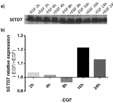
4.1. SETD7 protein levels increase in response to EGF deprivation ... 39
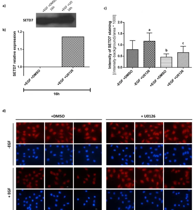
4.2. SETD7 protein levels increase upon MAPK inhibition ... 39
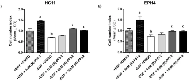
4.3. SETD7 activity inhibition by (R)-PFI-2 increases cell number ... 42
4.4. Cell proliferation is not significantly affected by (R)-PFI-2 ... 43
4.5. SETD7 levels increase upon inhibition of its methyltransferase activity by (R)-PFI-2 .. 44
4.6. ER, STAT3 and HOXB2 expression in response to EGF – correlation to SETD7 protein levels 45 4.7. SETD7 is regulated by lactogenic stimuli ... 47
4.8. (R)-PFI-2 is capable of reversing the negative effect of lactogenic hormones on cell number ... 48
4.9. STAT3 and HOXB2 regulation by lactogenic hormones ... 48
5.
Discussion ... 51
6.
Concluding Remarks ... 57
iii
List of Figures
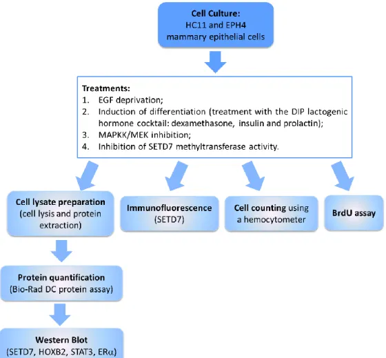
Figure 1 – Different stages of mammary development and TEBs structure. Adapted from (14).... 4 Figure 2 – Schematic representation of the human breast. Adapted from (15). ... 5 Figure 3 – Hypothetical model of the mammary epithelial hierarchy. The mammary cellular hierarchy is still poorly understood but it is hypothesized that there may exist a hierarchy of multiple stem and progenitor cells. MaSCs are multipotent, long-term, self-renewal adult stem cells that can differentiate into luminal and myoepithelial progenitor cells. Then, these progenitor cells commit to the luminal (ductal and alveolar) and myoepithelial lineages, respectively. ER, Estrogen receptor; LT-RC, Long-term repopulating stem cell; PR, Progesterone receptor; ST-RC, Short-term repopulating stem cell. Taken from (25). ... 6 Figure 4 – Main pathways activated downstream of EGFR. AKT, Protein kinase B; CCND1, gene encoding cyclin D1; CDKN1A, gene encoding p21; EGF, epidermal growth factor; EGFR, EGF receptor; ERK/MAPK, extracellular signal-regulated kinase; JAK, Janus kinase; MEK/MAPKK, ERK kinase; MKP1, MAPK phosphatase 1; PI3K, phosphoinositide 3 kinase; PKC, protein kinase C; PLC, phospholipase C; PTEN, phosphatase and tensin homologue; STAT, signal transducers and activators of transcription; TGFα, transforming growth factor-α; VEGF, vascular endothelial growth factor. Taken from (38). ... 8 Figure 5 – SETD7 substrates and gene expression. H3K4 methylation by SETD7 prevents chromatin condensation (i.e., chromatin adopts an “open” conformation – euchromatin) and enhances gene transcription. SETD7 also methylates non-histone proteins, including transcription factor. Methylation of ER, p53 and TAF10 results in their stabilization, thereby improving their activity as transcriptional activators. ER, Estrogen receptor ; SETD7, SET domain containing lysine methyltransferase 7; TAF10, TATA-box-binding protein associated factor 10. Taken from (93). ... 14 Figure 6 – STAT3 transcriptional targets and their effects during cancer development. Taken from (179). ... 21 Figure 7 – Schematic representation of the methodology used throughout this project. EGF, Epidermal growth factor; ER, Estrogen receptor ; HOXB2, Homeobox b2; MAPKK, Mitogen-activated protein kinase kinase; STAT3, Signal transducer and activator of transcription 3; SETD7, SET domain containing lysine methyltransferase 7. ... 29 Figure 8 – Effect of EGF on SETD7 expression. HC11 cells were depleted of EGF for the indicated time intervals. Afterwards, SETD7 protein levels were analyzed by Western Blot. (a) SETD7 expression throughout the different treatment periods with and without EGF. (b) Relative quantification of the intensity of the bands in the blot. For each treatment period, -EGF intensity values were related to those obtained for proliferating cells (+EGF controls), which were set to one. ... 39 Figure 9 – U0126 effect on SETD7 expression. (a) and (b) HC11 cells were co-treated with
EGF-free medium + 10 ng/mL EGF (+EGF) and 1 M U0126 (or DMSO – control) for 16h. Afterwards, SETD7 protein levels were analyzed by Western Blot. (a) SETD7 expression in undifferentiated cells treated with U0126. (b) Relative quantification of the intensity of the bands in the blot (the +DMSO/-U0126 control was set to one). For the immunofluorescence assay [(c) and (d)], HC11 cells were co-treated with EGF-free medium or +EGF medium and 1
iv
M of U0126 and incubated in this medium for 16h before fixation. DMSO was used as a vehicle control. SETD7 staining was then analyzed. (c) Fluorescence intensity of SETD7 staining. Mean SD of the values obtained is shown. a and b, p 0.0001 vs. -EGF +DMSO; c, p 0.05 vs. +EGF +DMSO. (d) Analysis of SETD7 expression in HC11 cells by immunofluorescence. In blue are the cell nuclei stained with DAPI and in red is SETD7 staining. Representative of one experiment. ... 41 Figure 10 – HC11 and EPH4 cell counting assay. Cells were treated with EGF-free medium, with or without 10 ng/mL EGF, and different concentrations of (R)-PFI-2 (1 nM, 8 nM and 10 nM). After 3 days, cells were counted in a Neubauer improved counting chamber and the relative cell number variation was calculated by setting untreated +EGF control values to 1. Mean SD from one experiment carried out in quadruplicates is shown for (a) HC11 (a and b, p 0.0001 vs. +EGF +DMSO; c, p 0.0001 vs. –EGF +DMSO and no significant differences vs. +EGF +DMSO) and (b) EPH4 (a, p 0.0001 vs. +EGF +DMSO; b, p 0.05 vs. +EGF +DMSO; c, p 0.05 vs. –EGF +DMSO and no significant differences vs. +EGF +DMSO) cells. Both graphs are representative of 2 independent experiments. ... 42 Figure 11 – BrdU labeling of HC11 and EPH4 cells. HC11 and EPH4 cells were cultured in EGF-free medium, with or without 10 ng/mL EGF, and 8 nM of (R)-PFI-2 or DMSO (vehicle control) for 2 days. Then, these experimental media were renewed and supplemented with 10 M BrdU. Cells were incubated overnight in these media, proceeding then with the Brdu assay. Absorbance was measured at 370 nm and 492 nm (reference wavelength). Mean SD from one experiment carried out in quadruplicates is shown for (a) HC11 and (b) EPH4 cells. No significant differences were found between treatments for either experiment. Both graphs are representative of 2 independent experiments. ... 43 Figure 12 – (R)-PFI-2 effect on SETD7 expression. (a) and (b) HC11 cells were co-treated with or without EGF and 8 nM (R)-PFI-2 (or DMSO – vehicle control) for 16h and 24h. Afterwards, SETD7 protein levels were analyzed by Western Blot. (a) SETD7 expression in proliferative (+EGF) and less-proliferative (-EGF) cells treated with (R)-PFI-2 for 16h and 24h. (b) Relative quantification of the intensity of the bands in the blot. Intensity values obtained for cells treated with (R)-PFI-2 were related to those obtained for untreated cells, which were set to one. Representative of 2 experiments. ... 44 Figure 13 – Effect of EGF on STAT3, ER and HOXB2 expression. HC11 cells were stimulated with or without EGF for the indicated time intervals. Afterwards, STAT3, ER and HOXB2 protein levels were analyzed by Western Blot. (a), (c) and (e) show STAT3, ER and HOXB2 expression (respectively) throughout the different treatment periods with EGF-free medium in comparison to +EGF controls. (b), (d) and (f) show the relative quantification of the intensity of the bands in the corresponding blot. For each treatment period, -EGF intensity values were related to those obtained for proliferating cells, which were set to one. ... 45 Figure 14 – (R)-PFI-2 effect on STAT3 expression. (a) and (b) HC11 cells were co-treated with or without EGF and 8 nM (R)-PFI-2 (or DMSO – vehicle control) for 16h and 24h. Afterwards, STAT3 protein levels were analyzed by Western Blot. (a) STAT3 expression in proliferative and competent (lower proliferation) cells treated with (R)-PFI-2 for 16h and 24h. (b) Relative quantification of the intensity of the bands in the blot. Intensity values obtained for cells
v
treated with (R)-PFI-2 were related to those obtained for untreated cells, which were set to one. ... 46 Figure 15 – Effect of lactogenic stimuli on SETD7 expression. HC11 cells were stimulated with DIP medium (+DIP) for the indicated time intervals. Controls were treated with EGF-free medium (-DIP). Afterwards, SETD7 protein levels were analyzed by Western Blot. (a) SETD7 expression throughout the different treatment periods with DIP medium in comparison to –DIP controls. (b) Relative quantification of the intensity of the bands in the blot. For each treatment period, +DIP intensity values were related to those obtained for cells treated with –DIP medium, which were set to one. ... 47 Figure 16 – HC11 cell counting assay. Cells were treated with EGF-free or DIP medium and different concentrations of (R)-PFI-2 (1 nM, 8 nM and 10 nM). After 3 days, cells were counted in a Neubauer improved counting chamber and the relative cell number variation was calculated by setting untreated -EGF control values to 1. Mean SD from one experiment carried out in quadruplicates is shown. a, p 0.05 vs. –EGF +DMSO; b, p 0.0001 vs. –EGF +DMSO; c, p 0.0001 vs. +DIP +DMSO and no significant differences vs. -EGF +DMSO. Representative of 2 experiments. ... 48 Figure 17 – Effect of lactogenic stimuli on STAT3 and HOXB2 protein levels. HC11 cells were stimulated with DIP medium (+DIP) for the indicated time intervals. Cells treated with EGF-free medium (-DIP) were used as controls. Afterwards, STAT3 and HOXB2 protein levels were analyzed by Western Blot. (a) and (c) show STAT3 and HOXB2 expression (respectively) throughout the different treatment periods with DIP medium in comparison to –DIP controls. (b) and (d) show the relative quantification of the intensity of the bands in the corresponding blot. For each treatment period, +DIP intensity values were related to those obtained for cells treated with –DIP medium, which were set to one. ... 49
vi
List of Tables
vii
List of Abbreviations
AKAP6 A kinase anchor protein 6
ASH1L Absent, Small or Homeotic 1-like protein AML Acute myeloid leukemia
ARTD1 ADP-ribosyltransferase diphtheriatoxin-like 1
AREG Amphiregulin
AR Androgen Receptor
BTC Betacellulin
CREB cAMP response element-binding protein CSC Cancer stem cells
CENPC1 Centromere protein C1
Co-REST Co-repressor of element-1 silencing transcription factor CBP CREB-binding protein
DEAF1 Deformed epidermal autoregulatory factor 1
DEX Dexamethasone
DMSO Dimethyl sulfoxide
dH2O Distilled water
DNMT1 DNA (cytosine-5)-methyltransferase 1 DNMT DNA methyltransferase
TTK Dual specificity protein kinase ELF5 E74-like factor 5
EGFR EGF receptor
ESC Embryonic stem cells
EZH2 Enhancer of zeste homolog 2 EGF Epidermal growth factor
EPR Epiregulin
MEK ERK kinase
E2 Estradiol
ER Estrogen receptor
ERK Extracellular signal-regulated kinase FXR Farnesoid X receptor
FoxO3 Forkhead Box O3
CCND1 gene encoding cyclin D1; CDKN1A gene encoding p21
HB-EGF Heparin-binding EGF-like growth factor HDM Histone demethylase
HMT Histone methyltransferase
HOX Homeobox
HIV-Tat Human immunodeficiency virus transactivator HIF-1 Hypoxia-inducible factor 1 alpha
IGF2 Insulin-like growth factor-2 IRF1 Interferon Regulatory Factor 1
viii
JAK Janus kinase
JARID1 Jumonji AT-rich interactive domain 1 LT-RC Long-term repopulating stem cell lum-SC Luminal unipotent stem cells KMT Lysine methyltransferases
LSD1 Lysine-specific histone demethylase 1
MaSC Mammary stem cell
MAPKK MAPK kinase
MKP1 MAPK phosphatase 1
MED1 Mediator Complex 1
MeCP2 Methyl-CpG Binding Protein 2 MAPK Mitogen-activated protein kinase MLL Mixed lineage leukemia protein MCP Monocyte chemoattractant protein MPS1 Monopolar spindle 1
MEF Mouse embryonic fibroblast
MMTV Mouse mammary tumor virus
MINT Msx2-interacting nuclear target protein
MYND Myeloid-Nervy-DEAF1
myo-SC Myoepithelial unipotent stem cells MyoD Myogenic differentiation protein NAD+ Nicotinamide adenine dinucleotide NF-B Nuclear factor-kappa B
PCAF p300/CBP-associated factor
PDX1 Pancreatic and duodenal homeobox 1 PRC2 PcG repressive complex 2
PPARBP Peroxisome proliferator-activated receptor binding protein PPAR- Peroxisome proliferator-activated receptor-gamma
PTEN Phosphatase and tensin homologue PI3K Phosphoinositide 3 kinase
PLC Phospholipase C gamma PARP1 Poly-ADP-ribose polymerase 1
PcG Polycomb group
PRDM9 Positive Regulatory-domain zinc finger protein 9 PGC-1 PPAR- co-activator alpha
PIC Pre-initiation complex formation PR Progesterone receptor
PRL Prolactin
PRLR Prolactin receptor AKT Protein kinase B PKC Protein kinase C
RANKL Receptor activator NF-B ligand
pRb Retinoblastoma tumor suppressor protein
ix SAM S-adenosylmethionine
SMYD SET and MYND domain-containing protein ST-RC Short-term repopulating stem cell
STAT Signal transducer and activator of transcription
SIRT1 Sirtuin 1
SDS Sodium dodecyl sulfate
SAGA Spt-Ada-Gcn5-acetyltransferase SET Su(var)3-9-enhancer of zeste-trithorax TAF TATA-box-binding protein associated factor TDLU Terminal ductal-lobular unit
TEB Terminal end bud
TFIID Transcription factor IID
TGF Transforming growth factor alpha TNF Tumor necrosis factor alpha VEGF Vascular endothelial growth factor
Wnt4 Wingless-type MMTV integration site family member 4 YAP Yes-associated protein
YY1 Yin yang 1
1. State of the Art
3
1.1.
Introduction
Cellular differentiation is an essential biological process by which cells become competent and commit to a specific cell lineage, being important during both embryonic development and adult life (1). It is also now known as an important feature to have in consideration in the clinical evaluation of tumor aggressiveness and potential response to treatment. Generally, a high degree of differentiation is associated with a better prognosis. In fact, differentiated cancer cells have a higher degree of similarity with each other and with their tissue of origin, are more cohesive and less invasive (2).
Increasing evidence suggests that cell differentiation is deeply regulated by chromatin remodeling and histone post-translational modifications (1). Specifically, methylation of histone H3 at its fourth lysine (K4) is now established as an active mark for gene transcription (functioning as a distinctive signal for the recruitment of specific transcription factors) and cell differentiation (3-7). SETD7 is a H3K4 methyltransferase (8, 9) also known to methylate several other histone and non-histone substrates involved in distinct cellular processes, including cell cycle regulation, DNA damage response, apoptosis, chromatin modulation, proliferation and differentiation (10-12). Thus, the potential of SETD7 as a regulator of these processes and, thereby, of cell’s homeostasis, is an interesting lead to follow. However, little is still known about SETD7 role in physiological and pathological processes. For this reason, we decided to study SETD7 expression in proliferation and differentiation of mammary epithelial cells. We believe this project to be of major importance as only by understanding how SETD7 influences these processes in normal cells, can one then establish its role in breast cancer development, progression and invasion.
In summary, this project main goal is to establish the correlation between SETD7 and cellular differentiation and proliferation, as well as its function and regulation, in mammary epithelial cells. In addition, the expression and potential role of some of SETD7 molecular targets during these processes will also be explored.
1.2.
Mammary development
1.2.1. Mammary
development
during
Embryogenesis,
Puberty
and
Pregnancy/Lactation
The mammary gland is a specialized epidermal appendage, whose main function is to produce and provide milk to the newborn. This not only assures the nutrition of the baby during its first months but also the provision of immune factors, which helps to protect the baby from infections. On the other hand, it is also important for the development of unique bonds between mother and infant during nursing (13).
This process of production and delivery of milk is called lactation, which is possible due to the existence of a complex network of branched ducts that constitute the mammary epithelium (14).
1. State of the Art
4
The ductal network is embedded within the mammary fat pad constituted by adipocytes. This stroma also comprises fibroblasts, immune cells and blood vessels (13).
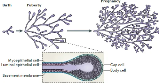
Mammary gland morphogenesis can be divided in five main stages: embryonic, pubertal, gestational, lactation and involution (or regression) (figure 1) (15). In the embryo, mammary development starts as a primary ectodermal thickening within the 4-6 months of gestation forming the mammary line, that then breaks up into individual placodes (a pair in humans) (16). At this stage, this primary bud contains two different cell populations: central and peripheral (or basal) (17). These placodes project into the underlying mesenchyme originating the epithelial buds (18). The epithelial buds then form branches and, at near term, the breast consists of a ductal tree of approximately five ducts embedded in the mammary fat pad and an external nipple that connects them to the body surface (13). Similarly to what occurs in the formation of other epithelial appendages, all of these changes rely on coordinated signaling between the epithelium and the underlying mesenchyme (19).
Figure 1 – Different stages of mammary development and TEBs structure. Adapted from (14).
After birth, the breasts involute and become quiescent until puberty (20), when, in females, the formation of secondary ductal branches takes place (17). In the pubertal stage, upon secretion of ovarian hormones like estrogen and progesterone, the mammary epithelium grows exponentially (21) and the immature ducts elongate, which leads to the formation of the terminal end buds (TEBs) (figure 1). Thereby, TEBs are structures that can be found at the end of the primary ducts and differ from them on three main features: they have an inner multilayer luminal epithelium (in contrast to the single layer present in the primary ducts) also delimited by an outer monolayer of myoepithelial cells, and own a large amount of mammary stem cells which, thus, allows them to have high levels of proliferation (22). It is important to notice that the primary TEBs, present in the beginning of the pubertal development, possess an outer layer of cap cells and an inner multilayer of body cells, which are highly proliferative and will give rise to the myoepithelium and luminal epithelium, respectively (23). Once deep in the fat pad, the TEBs bifurcate, forming new primary branches that then divide into secondary branches that will form a complex network of mammary branches through the entire fat pad. Simultaneously, stromal expansion can also be
1. State of the Art
5
observed in this phase (20). Moreover, lobular-alveolar structures, termed terminal ductal-lobular units (TDLUs) (figure 2), are formed at the end of these secondary ducts (19).
During pregnancy, an extensive and accelerated proliferation of the epithelium takes place, with further branching, lobuloalveolar development and terminal differentiation (24), which is triggered by progesterone and prolactin (23). These structures (alveoli) contain milk-producing alveolar cells that are responsible for the synthesis and delivery of milk during lactation (13). The myoepithelium that forms the outer layer of these structures is very important at this phase, consisting of specialized, contractile cells that permit the release of milk through the ducts (14). Upon lactation cessation, the mammary glands involute, i.e., they suffer a coordinated apoptosis and remodeling process, reversing the previous growth. The breast structure is now similar to the existing one before pregnancy (24).
Figure 2 – Schematic representation of the human breast. Adapted from (15).
1.2.2. Mammary Stem Cells (MaSCs) and mammary epithelial hierarchy
All of the changes described above, especially the ones that occur during pregnancy, and the fact that the human mammary gland maintains its regenerative capacity for decades, are thought to rely on the presence of mammary stem cells (MaSCs). As represented in figure 3, there is a hierarchy of stem and progenitor cells that ultimately give rise to the mature lobular and ductal structures (23).
There are different cell-surface markers that can be used to discriminate between the different mammary cell subpopulations, like CD24 (heat-stable antigen), CD29 (1-integrin) and CD49f ( 6-integrin) (25, 26). MaSCs have been shown to have higher levels of CD24, CD29 and CD49f and low levels of Sca-1, however, neither of these are exclusive of MaSCs which suggests that further research and discovery of specific markers is required (25). CD44 is a transmembrane receptor
1. State of the Art
6
that can serve as a stem cell marker as well, since it has been found to be highly present in MaSCs and continues to be expressed in myoepithelial cells, being involved in ductal outgrowth, cell-cell adhesion between luminal and myoepithelial cells and bilayer organization (27). It was also found that MaSCs don’t possess both estrogen (ER) and progesterone receptors (PR). However, as mentioned, it has been well established that these two steroid hormones are essential for mammary epithelial expansion and differentiation. Thus, these processes may be mediated by ER/PR positive (ER+/PR+) cells signaling to MaSCs via paracrine mechanisms. In other words, estrogen and progesterone stimulate ER-/PR-dependent gene transcription within the ER+/PR+ cells (that seem to be non-dividing cells from the luminal epithelium), promoting the release of certain factors. Then, these factors attach to specific receptors on the MaSCs surface and ultimately regulate MaSCs proliferation and differentiation (25).
Figure 3 – Hypothetical model of the mammary epithelial hierarchy. The mammary cellular hierarchy is
still poorly understood but it is hypothesized that there may exist a hierarchy of multiple stem and progenitor cells. MaSCs are multipotent, long-term, self-renewal adult stem cells that can differentiate into luminal and myoepithelial progenitor cells. Then, these progenitor cells commit to the luminal (ductal and alveolar) and myoepithelial lineages, respectively. ER, Estrogen receptor; LT-RC, Long-term repopulating stem cell; PR, Progesterone receptor; ST-RC, Short-term repopulating stem cell. Taken from (25).
1.2.3. Proliferation and differentiation of the mammary gland
All of the changes that occur during the different stages of mammary gland development are driven by different pathways, most of which are related to pituitary and ovarian hormones that
1. State of the Art
7
act in specific receptors located on the mammary gland. The following hormones are particularly relevant to guide and control the developmental changes occurring in puberty and adulthood (28).
1.2.3.1. Estrogen and EGFR signaling
Estrogen is a hormone released by the ovaries that has a major role in pubertal mammary growth during ductal morphogenesis through the activation of ER. However, ER is only expressed in a subpopulation of luminal cells (ER+ cells) and therefore it is thought that these cells drive proliferation of ER negative (ER-) cells through paracrine signals. In addition to its role in ductal morphogenesis, estrogen also promotes alveolar cells growth and maintenance during pregnancy (28).
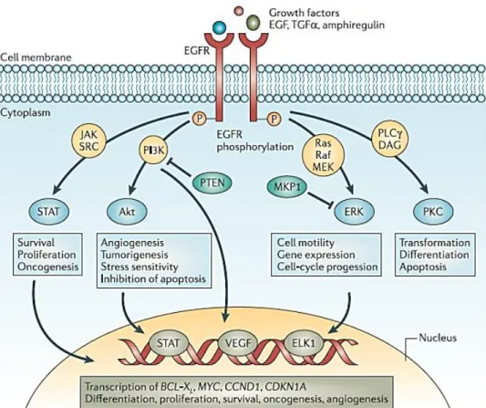
Epidermal growth factor receptor (EGFR), a transmembrane ERBB/HER receptor tyrosine kinase present in both epithelial and stromal cells (28), has long been shown to be implicated in mammary gland development and to mediate estrogen proliferative effects (29-31). In fact, one of EGFR ligands, Amphiregulin (AREG), is a strong candidate for the factor that is released by ER+ cells (28, 32). There is also evidence supporting a role for EGF as a mediator of estrogen paracrine signaling (30, 31, 33). In fact, EGF was shown to promote mammary epithelial cell proliferation in synergy with estrogen (33). Other members of the EGF-like family of growth factors include transforming growth factor- (TGF), heparin-binding EGF-like growth factor (HB-EGF), betacellulin (BTC), epiregulin (EPR), and epigen. All of these EGFR ligands are originally expressed as transmembrane precursors whose extracellular domain is proteolytically cleaved to release the mature growth factor that will activate EGFR (34). Activated EGFR can then activate multiple signaling pathways involved in cell proliferation and survival, such as the phosphoinositide 3 kinase/protein kinase B (PI3K/AKT), Janus kinase/signal transducers and activators of transcription (JAK/STAT) and mitogen-activated protein kinase (MAPK) pathways (figure 4) (35-37).
1. State of the Art
8
Figure 4 – Main pathways activated downstream of EGFR. AKT, Protein kinase B; CCND1, gene encoding
cyclin D1; CDKN1A, gene encoding p21; EGF, epidermal growth factor; EGFR, EGF receptor; ERK/MAPK, extracellular signal-regulated kinase; JAK, Janus kinase; MEK/MAPKK, ERK kinase; MKP1, MAPK phosphatase 1; PI3K, phosphoinositide 3 kinase; PKC, protein kinase C; PLC, phospholipase C; PTEN, phosphatase and tensin homologue; STAT, signal transducers and activators of transcription; TGFα, transforming growth factor-α; VEGF, vascular endothelial growth factor. Taken from (38).
1.2.3.2. Progesterone
Progesterone is an ovarian hormone that contributes to the development of side-branches and alveologenesis during pregnancy. For that reason it is crucial for the formation of mature structures capable of producing and secreting milk (that holds nutrients vital for the newborn). PR is only expressed in a subpopulation of luminal cells (about 30% of them) and, similarly to estrogen, this suggests that progesterone promotes cell proliferation through paracrine signals (28). Between the several possible paracrine mediators of the progesterone function, the receptor activator of nuclear factor kappa B1 (NF-B1) ligand (RANKL) and the Wingless-type mouse mammary tumor virus (MMTV) integration site family member 4 (Wnt4) seem to be the more promising.
1.2.3.3. Prolactin
Prolactin (PRL) is a hormone secreted by the pituitary gland that collaborates with progesterone in lobuloalveolar development, in order to generate a lactation-competent gland during pregnancy. It can also act indirectly by stimulating progesterone production in the ovaries. Prolactin binds to a member of the class I cytokine receptor superfamily, PRLR, which enhances
1. State of the Art
9
several signaling pathways such as JAK/STAT, MAPK and PI3K/AKT pathways. The interaction PRL-PRLR triggers JAK2/STAT5 signaling pathway activation (28), enhancing transcription via the transcription factor STAT5 phosphorylation (by JAK2) and its nuclear translocation. This cascade of events lead to induction of RANKL (in synergy with progesterone), which ultimately triggers mammary epithelial growth and alveologenesis. PRL also induces insulin-like growth factor 2 (IGF2), which accelerates alveolar growth (39). Prolactin, in association with insulin and cortisol, are also key factors in lactogenesis (that occurs from mild pregnancy to a few days before parturition), where the mammary epithelium differentiates and begins to produce all of the milk components. Further epithelial expansion and an increase in the expression of genes involved in milk secretion can occur after birth of the new infant as suckling begins (40-42). This process is also driven by STAT5 that appears to regulate the transcription and secretion of milk proteins (41).
1.2.3.4. Insulin
Insulin is essential for lactation since it enhances milk protein production in synergy with PRL. Recent data suggests that insulin increases the transcription of E74-like factor 5 (ELF5) and, thus, of STAT5 (since ELF5 is a co-regulator and amplifier of STAT5 activity), which furthers its phosphorylation in result to PRL signaling and leads to an increase in the transcription of milk protein genes such as -lactalbumin and -casein (40, 43).
1.3.
Tumorigenesis and cancer development
Tumorigenesis is a multistep process, which depends on accumulation of DNA mutations. However, only a small portion of these mutations will contribute for malignant transformation and, therefore, for cancer initiation and progression. Also, to drive this transformation, these mutations may intensify cells proliferation rate, make them resistant to apoptosis and activate specific pathways involved in cell growth and division, giving these cells a selective proliferative advantage (44). Moreover, nowadays it is acknowledged that cancer cells main features are: they are self-sufficient in growth signals, insensitive to anti-growth and pro-apoptotic signals, thereby having unlimited self-renewal potential. They are also capable of stimulating blood vessels growth (angiogenesis) and of invading other tissues and form metastases (45). Additionally, cancer development can be instigated not only by genetic alterations but also by dysregulation of the epigenetic machinery. Accordingly, epigenetic and genetic alteration can cause aberrant gene expression and genomic instability and, possibly, predispose to cancer (46).
1.3.1. Cancer and Stem cells
Considering the current knowledge about cancer pathology, it is thought that cancer originates from stem cells or cells that have acquired stem cell properties. One of stem cells’ key characteristics is the fact that they have a long lifespan, which makes them major candidates for the accumulation of mutations and subsequent malignant transformation. On the other hand,
1. State of the Art
10
cancer stem cells are non-differentiated cells that can give rise to specific cell lineages, thus perpetuating mutations through uncontrolled mitotic divisions. The cancer stem cells capacity to differentiate is also a probable cause for cancer heterogeneity and therapeutic resistance (44). Currently it is not established if cancer stem cells originate from normal adult stem cells or result from de-differentiation of cancer cells (47-49).
1.4.
Epigenetic modifications
Epigenetic modifications are hereditable changes in the genetic expression patterns mediated by epigenetic regulatory mechanisms, without a change in DNA sequence. These mechanisms comprise DNA methylation, histone modifications and RNA silencing by noncoding RNAs (50). Most of these changes are generated during cellular differentiation and can be passed through to descendant cells, being maintained even after multiple cycles of cell division (51).
DNA methylation, catalyzed by DNA methyltransferases (DNMTs), occurs on cytosine residues predominantly in the sequence CpG (52). This causes the silencing of the genetic material (genes and non-coding regions), which is particularly important during embryogenesis (50). On the other hand, histone modifications consist of enzyme-dependent histone residues posttranslational changes, including acetylation, phosphorylation, methylation, ubiquitylation and sumoylation (50, 53). This kind of epigenetic modification will be further explained ahead. Lastly, RNA silencing consists of posttranscriptional mRNA silencing by small non-coding RNAs, commonly called microRNAs (50). MicroRNAs are partially or completely complementary to their target mRNA, which allows them to recognize and bond with specific mRNAs. Then, this may cause mRNA degradation or inhibition, preventing mRNA translation into proteins. This is important for the maintenance and control of the global gene expression patterns (51).
Epigenetic changes play an important role in the regulation of gene expression and chromosomal stability. In other words, they ensure that the correct genes are transcribed, at the right time and amount within each specific tissue. For these reasons, it is plausible to assume that any perturbation of this process can lead to the dysregulation of gene transcription and of normal cellular processes and indirectly contribute to cancer initiation and progression. Moreover, alteration of the normal methylation patterns due to changes in the enzymatic activities of the histone methyltransferases (HMTs) and histone demethylases (HDMs) has been associated with breast, prostate, lung and brain cancers as well as leukemia (52).
1.5.
Histone methyltransferases
In eukaryotic cells the genetic material is enclosed in the nucleus and packaged into chromatin, a complex structure composed of DNA and proteins. Moreover, in mammalian cells, chromatin is arranged into compacted and highly organized structures, the chromosomes (54). Chromatin’s fundamental unit is the nucleosome formed by 145-147 base pairs of DNA, wrapped around an
1. State of the Art
11
octamer of four core histones: H2A, H2B, H3 and H4. Each nucleosome core is then stabilized by a linker histone (e.g., H1), that in cooperation with other non-histone proteins further direct nucleosome packaging and organization into chromosomes (55, 56).
Histones can suffer posttranslational modifications, including acetylation, phosphorylation and methylation. These modifications (that mostly occur on the N-terminal tail domains) are catalyzed by specific histone modifying enzymes and can alter the interactions between nucleosomes, DNA-nucleosomes and DNA-nucleosomes/DNA-regulatory factors, leading to the regulation of chromatin organization, mitosis and DNA transcription, replication, recombination and repair (53, 55, 57). Histone methylation is catalyzed by HMTs that transfer a methyl group to a lysine or to an arginine. All the lysine methyltransferases (KMTs), except Dot1 that methylates H3 at K79, contain a Su(var)3-9-Enhancer of Zeste-Trithorax (SET) domain that is responsible for catalysis of cofactor S-adenosylmethionine (SAM), with subsequent transference of a methyl group to a lysine. Similarly, arginine methyltransferases (PRMTs) also catalyze the transference of a methyl group from SAM to an arginine residue within its substrates. Contrarily to other histone post-translational modifications, methylation does not alter histone charge, having little to no effect on DNA-histone interactions. Methylation can rather serve as a distinctive signal for the recruitment of methylation “reader” proteins that can trigger transcriptional activation or suppression depending on the modified histone and residue position in the histone primary structure. Lysines and arginines can be either mono- or di-methylated. Additionally, lysines can also be tri-methylated (6, 53, 58).
Due to its involvement on gene expression and chromatin architecture regulation, epigenetic modifications can be implicated on cell malignant transformation and subsequent aberrant proliferation. Thus, the dysregulation of histone modifying enzymes’ activity may lead to aberrant histone modification and give rise to cancer through inhibition of tumor suppressors and/or oncogenes activation (51, 53). In fact, because of HTMs ability to control gene transcription, the correct regulation of HTMs expression and activity is crucial for the normal function and fate determination of cells. Therefore, dysregulation of HTMs is nowadays increasingly acknowledged as a hallmark for cancer development and progression, and therefore several HTMs are currently in the spotlight of cancer research because of their potential oncogenic or tumor suppressor roles (52, 59, 60). One example is SETD7 which has been found to be associated with but have divergent functions in various cancers (61-65).
1.5.1. Histone H3 methylation at K4
Epigenetic modifications, such as histone modifications, are responsible for the control of the genetic material expression or silencing, being crucial during the different development stages, apoptosis and aging. H3 methylation at K4 mark is enriched at actively transcribed regions, and has been shown to be important for an efficient control of normal development, nucleosomal function and homeostasis. Thereby, dysregulation of this modification can lead to impairment of normal cellular regulatory mechanisms and is related to cancer development. Furthermore,
1. State of the Art
12
H3K4me1/me2/me3 dysregulation may mediate anti-apoptotic, proliferative, tumor-induced angiogenesis and inflammation pathways activation (66). Currently, H3K4 methylation modifiers (methyltransferases and demethylases) are known to have a role in embryonic development and regulation of stem cells’ fates, being required for stem cells self-renewal or differentiation and important for future cancer stem cells studies (6, 7, 67).
H3 methylation in its fourth lysine is often found near the promoters of actively transcribed genes (68). This is emphasized by the fact that H3K4me2 are often found in coding regions and about 75% of all human promoters are marked by H3K4me3 (6, 69). H3K4 methylation is present in the intergenic regions within Homeobox (HOX) gene clusters, which are some of the most important genes regulated by H3K4 methylation (68). HOX genes encode a family of transcriptional regulators that are involved in embryonic development. During the initial stages of development HOX genes are downregulated, whereas it can become increasingly activated during embryogenic development, coordinating tissue-specific cell proliferation and differentiation (with HOX loss-of-function resulting in embryonic lethality) (70-72). Similarly to embryogenesis, HOX expression during adulthood is tissue-dependent. In fact, the different HOX genes exhibit a tissue-specific expression pattern, regulating cell renewal (73), hematopoiesis (74), cell fate and differentiation and tissue homeostasis (75-77). Nonetheless, HOX genes dysregulation has been associated with cancer. Interestingly, HOX genes can either be upregulated or downregulated in cancer and either have an oncogenic or a tumor suppressor effect, depending on the tissue context and cancer type. For example, HOXB13 was found to be upregulated in ER+ breast cancers from patients that had been treated with tamoxifen, being responsible for tamoxifen resistance and an increase in cancer cells proliferation, migration and invasion (77-79). On the other hand, HOXB13 is downregulated in colorectal and prostate cancers, functioning as a tumor suppressor (78). HOXB2 was also shown to function as a tumor suppressor (inhibiting tumor growth and promoting apoptosis) in breast cancer (80) and acute myeloid leukemia (AML) (81). In fact, HOXB2 is correlated with better a prognosis in breast cancer patients (80). Conversely, HOXB2 overexpression has been associated with cervical (82, 83), lung (84), and pancreatic (85) cancers progression, invasiveness and recurrence. On the other hand, H3K4 methylation antagonizes Enhancer of zeste homolog 2 (EZH2) repression of Polycomb group (PcG)-regulated genes. EZH2 is the catalytic subunit of PcG repressive complex 2 (PRC2) (initially described as a repressor of the HOX genes by catalyzing H3K27 trimethylation) (86-88). PRC2 is essential for the regulation of transcriptional silencing during embryonic development, especially in the early stages, mediating lineage decisions (89, 90).
The forth lysine of H3 histones can be methylated by several SET-domain containing enzymes such as SET1A, SET1B, mixed lineage leukemia proteins 1 to 5 (MLL1 to MLL5), SET7/9, SET and Myeloid-Nervy-DEAF1 (MYND) domain-containing protein 1 to 3 (SMYD1 to SMYD3), Absent, Small or Homeotic 1-like protein (ASH1L), SET domain and Mariner transposase fusion protein (SETMAR) and Positive Regulatory-domain zinc finger protein 9 (PRDM9) (6, 91). These can methylate H3 at K4 into all three states, being involved in transcriptional activation (6, 69).
1. State of the Art
13
H3K4 demethylation is controlled by lysine demethylase 5/Jumonji adenine-thymine (AT)-rich interactive domain 1 (KDM5/JARID1) and lysine-specific histone demethylase 1 (LSD1), that act as transcriptional repressors (67).
1.5.1.1. SETD7 and its histone and non-histone substrates
SETD7 (also known as SET7 or SET9) is a lysine methyltransferase that comprises a SET domain and it catalyzes the transference of a methyl group to a lysine residue of various histones and non-histone subtracts. These potential SETD7 substrates are involved in distinct cellular processes, for instance, in cell cycle regulation, DNA damage response, RNA polymerase II dependent gene transcription, chromatin modulation and differentiation. Hence, SETD7 can play a critical role in several physiological and pathological processes (10-12). Moreover, SETD7 knockdown has been shown to cause embryonic lethality, which indicated that it plays an important role in development. On the other hand, SETD7 was also shown to be important for -cells’ function and skeletal muscle differentiation, as described below (92).
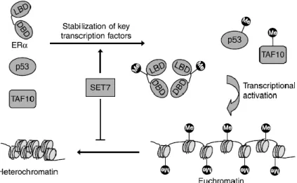
Histone H3 can be monomethylated by SETD7 at its lysine 4, which can lead to transcriptional activation by preventing chromatin condensation and by facilitating transcriptional factors binding to the promoter regions of specific genes (figure 5) (93). For example, SETD7-driven methylation of H3 at K4 takes part in the expression of the tumor necrosis factor alpha (TNF-) gene by facilitating NFB-p65 subunit binding to gene promoters of NFB-dependent inflammatory genes, such as TNF, monocyte chemoattractant protein-1 (MCP-1) and interleukin 8 (IL-8) (whose dysregulation can give rise to several inflammatory diseases, including atherosclerosis, insulin resistance, diabetes and metabolic syndrome, as well as cancer). H3K4 methylation is also associated with RELA expression, which encodes for NFB-p65. Additionally, SETD7 also directly methylates p65 at K37, restricting p65 to the nucleus and promoting its association with NFκB-dependent promoters, or at K315 and K316, resulting in negative regulation of p65 translocation to the nucleus (92, 94).
A recent study suggests that SETD7 is required for skeletal muscle gene expression and myogenic differentiation by directly interacting with Myogenic differentiation (MyoD) protein, a transcriptional factor that binds to the regulatory region of muscle differentiation genes to activate gene expression. The interaction between MyoD and SETD7 facilitates SETD7 access to the silencing nucleosome and monomethylation of H3K4, leading to increased affinity of MyoD to the myogenic regulatory regions. SETD7 also prevents Suv39h1 (a methyltransferase that transfers three methyl group to H3K9) association with MyoD (by competing with Suv39h1 for MyoD association), preventing its inhibitory effect over differentiation genes through H3K9 methylation (12).
1. State of the Art
14
Figure 5 – SETD7 substrates and gene expression. H3K4 methylation by SETD7 prevents chromatin
condensation (i.e., chromatin adopts an “open” conformation – euchromatin) and enhances gene transcription. SETD7 also methylates non-histone proteins, including transcription factor. Methylation of ER, p53 and TAF10 results in their stabilization, thereby improving their activity as transcriptional activators. ER, Estrogen receptor ; SETD7, SET domain containing lysine methyltransferase 7; TAF10, TATA-box-binding protein associated factor 10. Taken from (93).
SETD7 is also required for normal pancreatic and duodenal homeobox 1 (Pdx1)-mediated insulin synthesis by pancreatic -cells, since the interaction between SETD7 and this transcriptional factor, Pdx1, leads to the recruitment of SETD7 and consequent H3K4 methylation in the promoter region of the Ins1/2 gene (with its subsequent activation and preproinsulin expression) (92, 95-97). Having this in mind, SETD7 may play an important role in development through cellular differentiation and tissue-specific gene activation (92).
As described in Table 1, methylation of specific lysine residues on non-histone proteins (such as ER, p53 and TAF10) by SETD7 has also been described, improving their ability to serve as transcriptional activators (figure 5) (93). Recent studies have demonstrated that SETD7 also methylates several lysines of H2A and H1.4, as well as, the lysine K15 of H2B (92).
These findings highlight the importance of SETD7 and H3K4 methylation in several physiological processes and the basis of its mechanism of action. Furthermore, one can question if this enzyme is also linked to pathological processes, like cancer, diabetes or inflammatory diseases. Therefore, it would be interesting to conduct additional research to further explore SETD7 importance and expression patterns in these diseases.
Table 1 - SETD7 targets and respective effects upon SETD7-mediated methylation.
SETD7 targets Effects Ref.s
Histones H3 H3 methylation at K4, enhances transcriptional
activation by preventing chromatin condensation.
(93)
H2A H2B
Methylates histones H2A and H2B when in a free state. Little is known about the functional effect of this modification.
1. State of the Art
15
H1.4 H1.4 is methylated by SETD7 in six different lysine residues (K121, K129, K159, K171, K177 and K192) which can influence H1 binding to DNA and its function in chromatin compaction.
(92, 99) Chromatin associated MeCP2 (Methyl-CpG Binding Protein 2)
MeCP2 is a nuclear protein that recognizes and binds methylated DNA, functioning as a promoter repressor. SETD7 was reported to methylate MeCP2 at K347, although the effects of this methylation are still a mystery. (92, 100) PPARBP (Peroxisome Proliferator-Activated Receptor Binding Protein) or MED1 (Mediator Complex 1)
PPARBP has been demonstrated to be methylation by SETD7 at K1006, however, the functional purpose of this modification is still to be defined. PPARBP is a coactivator complex of the transcription machinery that is recruited in order to enhance the expression of RNA polymerase II transcribed genes, regulating the cell cycle, DNA repair, peroxisome proliferation, differentiation, proliferation and apoptosis. Med1 gene was shown to be overexpressed in breast, prostate and hepatic cancer.
(98, 101, 102)
CENPC1
(Centromere protein C1)
CENPC1-K414 monomethylation is catalyzed by SETD7. Little is known about the biologic effects of this modification, with the need to further study how SETD7 can influence CENPC1 function. CENPC1 associate with centromeric DNA and assists the assembly of the kinetochore to the centromere.
(92, 98, 103)
Transcriptional factors and
Co-activators /
repressors
p53 SETD7 methylation at K372 results in the stabilization of p53 and inhibition of p53 nuclear export, which leads to transcriptional activation of the p53 target genes. (92, 104, 105) TAF10 (TATA-box-binding protein associated factor 10)
TAF10 is methylated at K189, which increases TAF10-RNA polymerase II affinity and, as a result, stimulates TAF10 mediated transcription. TAFs are transcription factors, part of the Transcription Factor IID (TFIID) complex, that bind gene promoters and trigger pre-initiation complex formation (PIC), which regulates gene transcription initiation by RNA polymerase II. TAFs enhance transcription by interacting with transcriptional activators and as readers of epigenetic marks.
(92, 93, 106-108)
TAF7 Similarly to TAF10, TAF7 is also monomethylated by SETD7 at K5.
(92, 105, 109)
ER Enhances ER-driven transcription by altering ER recruitment and binding to target gene regulatory sequences through K302 methylation.
(92, 93, 110)
E2F1 E2F1 is destabilized by methylation at K185 by SETD7, which prevents E2F1 accumulation during DNA damage and the activation of its pro-apoptotic target gene p73. This highlights SETD7 potential role in cell cycle regulation (and, by association, in cancer proliferation). However, other reports suggest that E2F1 methylation at K185 by SETD7 also enhances E2F1-dependent transcriptional activation (although it enhances E2F1 proteosomal degradation).
(92, 111-113)
1. State of the Art
16 MINT
(Msx2-interacting nuclear target protein)
SETD7 methylates MINT at K2076. MINT functions as transcription repressor involved in the regulation of several processes such as cell cycle, craniofacial development, neural cell fate and apoptosis. Yet, MINT methylation function is still unknown.
(98, 114)
IRF1 (Interferon Regulatory Factor 1)
Lysine K126 of IRF1 seems to be methylated by SETD7. IRF1 is a transcription regulator of immune responses and hematopoietic development. Further studies should focus on the discovery of SETD7 modification effects over IRF1 function.
(98, 115)
HIV-Tat (Human immunodeficiency virus
transactivator)
By methylating Tat at K51, SETD7 enhances Tat-dependent transactivation of several viral and cellular genes, contributing to viral replication and HIV-1 pathogenesis.
(92, 116, 117)
FoxO3 (Forkhead
Box O3)
FoxO3 is an activator of genes involved in several cellular regulatory pathways, such as stress resistance, cell cycle arrest, differentiation, apoptosis and metabolism. It also is described in the literature as a tumor suppressor. FoxO3 is methylated at K271, which leads to an increase in its transcriptional activity.
(92, 118)
NFB-p65 SETD7-driven methylation of H3 at K4 or of p65 at K37 facilitates NFB-p65 subunit binding to gene promoters of NFB-dependent inflammatory genes, such as TNF-, MCP-1 and IL-8.
(92, 94, 119)
AR (Androgen Receptor)
SETD7 methylates AR (nuclear and cytoplasmic) at K630, which leads to enhanced transcriptional activity of AR and subsequent expression of PSA and NKX3-1.
(92, 120)
FXR (Farnesoid X
receptor)
FXR is a nuclear receptor that regulates bile acid homeostasis (by inhibiting its synthesis) and lipid, cholesterol and glucose metabolism in the liver and intestines. FXR also promotes liver regeneration and inhibits transcription of pro-inflammatory genes which may explain why FXR-knockout in mice significantly increases liver tumor incidence. SETD7-dependent methylation of FXR at K206 enhances transcription of two FXR target genes, SHP and BSEP. FXR lysines K210 and K460 are also putatively methylated by SETD7, although this still needs to be proven.
(121-128) pRb (Retinoblastoma tumor suppressor protein)
pRb is monomethylated at K873 by SETD7, which is necessary for Rb-dependent cell cycle arrest, transcriptional repression, and Rb-dependent differentiation.
(92, 105, 129)
STAT3 STAT3, a transcription factor normally activated in response to several cytokines and growth factors involved in cell growth and survival, is dimethylated at K140 by SETD7 in response to IL-6 signaling, which inhibit STAT3 binding to DNA promoters.
(92, 105, 130)
HIF-1
(Hypoxia-inducible factor 1)
Under hypoxic conditions HIF is activated, inducing the transcription of genes involved in cell’s adaptation to cellular hypoxia. More specifically, it can alter cellular energy metabolism and promote angiogenesis, maintaining tissue integrity and homeostasis. Recent studies (65, 131) show that SETD7 stabilizes HIF-1 and induces HIF-1-dependent gene transcription by
(131-133)
1. State of the Art
17
methylating HIF-1 at K32 and inhibiting its ubiquitination and degradation. SETD7 also methylates H3K4 at the promoters of HIF-1-activated genes. This may suggest that SETD7 is involved in metabolic adaptation in hypoxic cancer cells. By contrast, a different study (132) states that SETD7 may negatively regulate HIF-1 transcriptional activity. Therefore, additional research must be conducted in other to clarify SETD7 effects over HIF-1 function.
PGC-1 (Peroxisome proliferator-activated receptor-gamma (PPAR-) co-activator )
PGC-1 is a transcription co-activator that interacts with nuclear receptors, including PPAR-, and transcription factors to facilitate gene transcription. It is required for the regulation of mitochondrial biogenesis, energy metabolism and adaptive thermogenesis. PGC-1 has also an anti-inflammatory role in muscle tissue.
PGC-1 is methylated at K779, which is essential for the recruitment of Mediator and Spt-Ada-Gcn5-acetyltransferase (SAGA) complexes and, thus, for the transcription of PGC-1 target genes. In fact, SETD7 knockdown decreased PGC-1 capacity to bind MED1, MED17 and the SAGA complex component CCDC101/SGF29, which consequently impaired PGC-1 capacity to stimulate transcription.
(134-137)
YAP
(Yes-associated protein)
YAP is a transcriptional coactivator of proliferation and anti-apoptotic genes, whose activity is inhibited upon activation of the Hippo signaling pathway by cell-cell contact. YAP is methylated by SETD7 at K494, which prevents YAP translocation to the nucleus, decreasing YAP target genes (Ctgf and Cdc20) expression. Moreover, SETD7+/+ mouse embryonic fibroblasts (MEFs) were more sensitive to contact inhibition of proliferation than SETD7-/- MEFs, which may be related to YAP methylation and consequent cytoplasmic sequestration. (138, 139) PDX1 (Pancreatic And Duodenal Homeobox protein 1)
PDX1 is a transcription factor essential for pancreatic development, promoting cells differentiation. PDX1 also promotes pancreatic regeneration and regulates mature -cells function, proliferation and survival as well as -cell-related genes transcription (such as insulin). PDX1 has been associated with diabetes and cancer development, being overexpressed in several cancers, such as pancreatic and gastric cancer.
Maganti et al. (2015) found that PDX1 is methylated at K123 and K131, with K131 been necessary for PDX1 transcriptional activity. They then proposed that the methylation of these two lysines may be catalyzed by SETD7 as PDX1 transcriptional activity is significantly increased (by about 40%) by SETD7. However, there is not enough evidence to prove this hypothesis.
(140-147)
YY1 (Yin Yang 1) YY1 is a transcription factor that is methylated by SETD7 at two lysine residues, K173 and K411. YY1 can either activate or repress gene expression, regulating
(148-150)
1. State of the Art
18
cell proliferation and differentiation, DNA repair and apoptosis. Evidence suggests that YY1 methylation by SETD7 promotes YY1-dependent transactivation of specific genes involved in cell cycle regulation and cell proliferation.
Moreover, alike other SETD7 substrates YY1 plays a role in cancer development and progression. Interestingly, YY1 can function either as an oncogene (promoting cancer proliferation) or a tumor-suppressor, depending on the type of cancer. Therefore, SETD7 is a putative regulator of YY1 oncogenic or tumor-suppressor functions.
Enzymes ZDHC8 (Zinc finger
DHHC (Asp-His-His-Cys) domain-containing protein 8)
ZDHC8 is an S-acyltransferase that mediates the attachment of fatty acids on to cysteine residues (S-acylation), which regulates their solubility, attachment to membranes, distribution over specific organelles, folding and stability. ZDHC8 methylation by SETD7 occurs at K300. Up to now, no functional effect has been described. (98, 151, 152) TTK (Dual specificity protein kinase) or MPS1 (Monopolar spindle 1)
It was verified that TTK is methylated by SETD7 at K708, however, the functional effects of this modification still need to be cleared. TTK is a dual-specificity protein kinase that phosphorylates several proteins involved in DNA damage-induced cell cycle arrest, spindle pole duplication and chromosome alignment and segregation during mitosis. Dysregulation of this mitotic kinase may be involved in cancer development since it would result in uncontrolled proliferation. (98, 153, 154) DNMT1 (DNA cytosine-5-methyltransferase 1)
Methylation of DNMT1 (a maintenance DNA methyltransferase that specifically methylates DNA at CpG residues, normally involved in transcriptional repression) at K142 by SETD7 results in a decrease in DNMT1 by facilitating its polyubiquitination and subsequent proteasome-mediated degradation.
(92)
SIRT1 (Sirtuin 1) In response to DNA damage, SIRT1 methylation at K233, K235, K236 and K238 is catalyzed by SETD7, which inhibits SIRT1-p53 association and, consequently, enhances p53 acetylation and transactivation. This ultimately leads to apoptotic cell death, which indicates that SIRT1 is an oncogenic protein and in the absence of SETD7 protects cells from apoptosis.
(92, 155)
PCAF
(p300/CBP-associated factor)
STED7 methylates six different lysine residues of PCAF (K78, K89, K638, K671, K672 and K692) – an acetyltransferase implicated in several cellular processes –, regulating PCAF localization (methylated PCAF was found to localize to the nucleus) and possibly its function.
(92, 156) PARP1 (Poly-ADP-ribose polymerase 1) or ARTD1 (ADP-ribosyltransferase diphtheria
PARP1is a nuclear enzyme that transfers ADP-ribose from nicotinamide adenine dinucleotide (NAD+) to nuclear proteins (e.g., histones, transcription factors or PARP1 itself – auto-inhibition). PARP1 is best known as a DNA damage sensor, promoting DNA repair and
(157-163)