Relevant coexpression of
STMN1
,
MELK
and
FOXM1
in
glioblastoma and review of the impact of
STMN1
in
cancer biology
Fernanda de Oliveira Serachi
I, Suely Kazue Nagahashi Marie
I,II, Sueli Mieko Oba-Shinjo
II Universidade de Sao Paulo, Faculdade de Medicina (FMUSP), Department of Neurology, Laboratory of Molecular and Cellular Biology (LIM 15), Sao Paulo, SP, Brazil II Universidade de Sao Paulo, Center for Studies of Cellular and Molecular Therapy (NAP-NETCEM-NUCEL), Sao Paulo, Brazil
OBJECTIVE:
To analyze the associated expression of
STMN1
,
MELK
and
FOXM1
in search of alternative drugable
target in glioblastoma (GBM) and to review relevant functional roles of
STMN1
in cancer biology.
METHOD:
STMN1
,
MELK
and
FOXM1
expressions were studied by quantitative PCR and their coexpressions were
analyzed in two independent glioblastoma cohorts. A review of articles in indexed journals that addressed the
multiple functional aspects of
STMN1
was conducted, focusing on the most recent reports discussing its role in
cancer, in chemoresistance and in upstream pathways involving
MELK
and
FOXM1
.
RESULTS:
Signiicant associated expressions of
MELK
and
FOXM1
were observed with
STMN1
in GBM. Additionally,
the literature review highlighted the relevance of
STMN1
in cancer progression.
CONCLUSION:
STMN1
is very important to induce events in cancer development and progression, as cellular
proliferation, migration, and drug resistance. Therefore,
STMN1
can be an important therapeutic target for a large
number of human cancers. In glioblastoma, the most aggressive brain tumor, the
MELK
/
FOXM1
/
STMN1
presented
signiicant associated expressions, thus pointing
MELK
and
FOXM1
as alternative targets for therapy instead of
STMN1
, which is highly expressed in normal brain tissue. Continuous functional research to understand the
STMN1
signaling pathway is worthwhile to improve the therapeutic approaches in cancer.
KEYWORDS:
Stathmin, cytoskeleton, microtubules, glioblastoma.
Serachi FO, MarieI SKN, Oba-Shinjo SM. Relevant coexpression of STMN1, MELK and FOXM1 in glioblastoma and review of the impact of STMN1
in cancer biology. MedicalExpress (São Paulo, online). 2017 Oct; 4(5):M170506
Received for Publication on July 4, 2017; First review on August 4, 2017; Accepted for publication on October 18, 2017; Online on October 30, 2017
E-mail: [email protected]
■
INTRODUCTION
Cell proliferation and migration are two relevant
features in cancer biology determining tumor growth
and invasion/metastasis. The subcellular cytoskeleton
is essential to control these processes.
1This includes the
microtubule dynamic behavior involving rapid switches
between periods of polymerization (growth) and
de-polymerization (shrinkage) at the microtubule extremity
(named dynamic instability).
Currently, several proteins are known to be related
to microtubule interaction with tubulin, and participate
in microtubule dynamics. The stathmin (STMN) family
members are among those proteins that inhibit
micro-tubule polymerization. Four members of evolutionarily
conserved cytosolic proteins compose this family, namely
STMN1 to 4. STMN1 and STMN3 are ubiquitously
expres-sed in different cells, while STMN2 and STMN4 are more
restricted to the nervous system.
2These proteins share
up to 70% of sequence homology in a highly conserved
C-terminus within the tubulin-binding stathmin-like
domain, and in the N-terminus region containing the
phosphorylation sites, which also dictates their cellular
localization.
3,4■
MATERIALS AND METHOS
Analysis of Cases and Gene Expression
Eighty-seven astrocytomas grade IV or GBM and 22
non-neoplastic (NN) brain anonymized tissues from epilepsy
patients subjected to temporal lobectomy were obtained
during therapeutic surgery from patients treated by the
Neu-rosurgery Group of the Department of Neurology at Hospital
das Clínicas of the Faculdade de Medicina da Universidade
de São Paulo. Written informed consents were obtained
from all patients in accordance with ethical guidelines. This
project was approved by the Ethic Committee of Faculdade
de Medicina da Universidade de Sao Paulo (case # 0263/07).
Samples were immediately snap-frozen in liquid
nitrogen and necrotic and non-neoplastic areas were
re-moved by microdissection from the tumoral blocks prior to
RNA extraction. Total RNA extraction, reverse transcription
and qRT-PCR (Sybr Green approach) were performed as
previously described.
17Quantitative data were normalized
using the geometric mean of three reference genes suitable
for the analysis: hypoxanthine phosphoribosyltransferase
(HPRT), glucuronidase beta (GUSB) and TATA box-binding
protein (TBP), as previously demonstrated by our group.
20Primers of housekeeping genes, STMN1 and MELK are
described in our previous report.
17Primers for FOXM1
were synthesized by (Integrated DNA Technologies, IDT,
Coralville, IA) as follows (5′ to 3′): FOXM1 F: GAAGAACTC
-CATCCGCCACA, FOXM1 R: TCAAGTAGCGGTTGGCACTG. All
reactions were performed in duplicates and and the 2
−ΔCtmethod was applied to calculate gene expression levels,
where ΔCt = [Ct target gene] - [geometric mean Ct of re
-ference genes] and Ct is the cycle threshold. The median
values of gene expression were used to divide samples with
high and low expression.
Analysis of The Cancer Genome Atlas (TCGA) GBM
gene expression database
STMN1
,
MELK
and
FOXM1
gene expression levels
were analyzed in an independent cohort at the cBio Portal
for Cancer Genomics database (http://www.cbioportal.
org).
21RNAseq data set of 154 cases of GBM
22was used to
assess coexpression analysis of mRNA levels (z-score, RNA
Seq V2 RSEM).
Statistical analyses
Mann Whitney tests were performed to compare
STMN1
,
MELK
and
FOXM1
expression levels between GBM
and NN samples. Correlations between gene expression
values in different groups of tumors were assessed using
the Spearman-rho correlation tests (non-parametric test).
and migration. STMN1 has been described as associated to
a wide range of malignancies and is a target for alternative
therapy in cancer treatment.
5STMN2, also known as the superior cervical
gan-glion-10 protein (SCG10), has been reported as a
neuron--specific growth-associated phosphoprotein, abundant
in the growth cone of neurons.
In particular, STMN2 is
described as a neuronal marker at an early stage of neural
development, playing a regulatory role in the control of
neu-ronal differentiation.
6Previous studies have also described
its role in osteogenesis.
7In liver tumorigenesis, it has been
described as a target of β-catenin/TCF-mediated trans
-cription in the Wnt dependent regulation of microtubule
dynamics in hepatoma cells.
8Moreover, STMN2 plays a role
in promoting the invasive potential of gastric cancer cells.
9STMN3, also known as SCLIP, is involved in the
de-velopment of the central nervous system, including axonal
branching and dendritic differentiation of Purkinje cells.
10,11STMN3 is highly expressed in glioma samples, and has been
associated to migration and invasion of glioma cells.
12Addi-tionally, STMN3 has been described as a modulator of the
sensitivity of ovarian cancer cells to microtubule-targeting
drugs by preventing the formation of the spindle and
con-sequently promoting mitosis arrest.
13STMN4, also known as RB3, presents two splice
variants, RB3’ and RB3’’.
14STMN4 has a putative role in
neuronal morphogenesis and plasticity.
15,16Among the members of the stathmin family, STMN1
is the most studied member, and cumulative evidence
singles out STMN1 as a candidate target for cancer
therapy. However, STMN1 can hardly become an eligible
drug for brain tumors, as its expression is high in brain
tissue,
17and consequently undesirable side effects would
be expected. Therefore, the search for more suitable up or
downstream targets in the STMN1 signaling pathway is an
alternative strategy. On this rationale, we previously linked
MELK upstream to STMN1 in glioma cells,
17and have also
demonstrated the importance of MELK in astrocytoma
progression, mainly in GBM.
18More recently, AKT/FOXM1/
STMN1 pathway has been reported to confer multidrug
resistance phenotype in non-small cell lung cancer.
19FOXM1
is a transcription factor of the forkhead family that plays
critical roles in cell cycle progression and cell fate decision.
In the present work, we analyzed the
STMN1
,
MELK
and
FOXM1
associated expressions in our GBM cohort, and
validated our results in an expanded independent public
cohort
in silico
. To highlight the relevance of this pathway in
cancer biology, we also present a
review
focused on the role
Literature review focused in STMN1
A literature search was conducted in the PubMed
database using the following terms: “stathmin”, “cancer”.
Only articles in English were selected, with a search ending
in September 2017. We selected reviews and articles that
described STMN1 and cancer. We focused specially in the
most recent data of STMN1 role in cancer treatment and
chemoresistance.
■
RESULTS
We aimed to analyze the association of
STMN1
,
MELK
and
FOXM1
in two independent cohorts of GBM. Initially,
we analyzed
STMN1
,
MELK
and
FOXM1
expression levels
in 87 GBM samples compared to 22 non-neoplastic (NN)
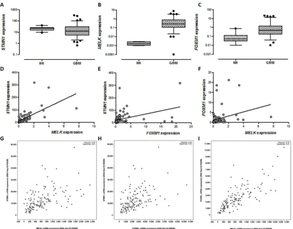
brain samples in our case database (Figure 1A, B and C).
Coexpression analyses were significantly positive
for
STMN1
and
MELK
,
STMN1
and
FOXM1
and
MELK
and
FOXM1
(Figure 1D, E and F). Our results were validated in
a larger independent public database of TCGA
corrobora-ting the tight association among
MELK
,
FOXM1
and
STMN1
(Figure 1G, H and I).
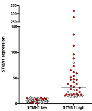
Additionally, we divided the GBM cases of our cohort
in low (44 cases) and high (43 cases)
STMN1
expression. In
the first group, there were 11 cases (25%) that presented
high
FOXM1
expression, while in the second group there
were 32 cases (73%) with high
FOXM1
expression (Figure
2). Our data indicate that overexpression of
STMN1
corre-lates to an overexpression of
FOXM1
.
■
DISCUSSION
Altogether, our data demonstrate that
MELK
and
FOXM1
expressions are significantly associated to
STMN1
expression, pointing them as promising alternative targets
in the STMN1 signaling pathway for glioma therapy.
A schematic illustration of the signaling pathway
involving STMN1, MELK and FOXM1 is proposed in Figure
3, implementing the interaction network recently
publi-shed.
19STMN1 plays a central role in the regulation of cell
cycle, proliferation, epithelial mesenchymal transition and
chemoresistance, crucial processes in cancer progression.
The details of these STMN1 roles are reviewed and
pre-sented below.
Figure 2. STMN1 and FOXM1 expression analysis of our GBM cohort. We divided the
GBM samples according to STMN1 expression (low and high, based on median gene
expression values). The horizontal bars represent the median expression of each group. Red dots represent cases with high FOXM1 expression levels (cases above median of
FOXM1 expression level).
Figure 3. STMN1, MELK and FOXM1 signaling. Activation of a tyrosine kinase receptor (TKR) activates PI3K/AKT and RAS/MAPK signaling pathways. Both pathways are activated through formation of the complex GRB/SOS (growth factor receptor-bound protein 2/ Son of Sevenless homologs), which binds to phosphorylated TKR. Both PI3K and RAS/MAPK pathways result in STMN1 phosphorylation, through MELK and FOXM1.
Phosphorylation inactivates STMN1 and allows the association of tubulin dimers and
polymerization of microtubules. STMN1 dephosphorilation activates the protein,
cau-sing in the sequestration of tubulin. This dynamics of STMN1 activation/inactivation results in depolymerization/polymerization of microtubules and consequently cell cycle progression/proliferation, epithelial-mesenchymal transition and chemoresistance. (Figure adapted from Marie et al., 2016)
STMN1 and microtubule dynamics
Tight regulation of cytoskeletal microtubule
dyna-mics in living cells is essential for many cellular functions.
Microtubules are a network of filaments comprising hetero
-dimer α/β-tubulin subunits that play a key role during cell
events such as proliferation, migration and differentiation.
The dynamic reorganization of microtubules in cells is
regu-lated by proteins that promote their assembly (stabilizers)
or disassembly (destabilizers). Microtubules (dis)assembly
is partially determined by the concentration of free tubulin
heterodimers in the cytoplasm, where it determines the
growth rate of microtubule by incorporation of tubulin at
its ends. STMN1 is one of the most prominent and rapid
microtubule regulators in response to cell needs. STMN1
downregulation increases the concentration of microtubule
polymers and decreases the concentration of free tubulin
heterodimers.
23STMN1 (de)phosphorylation and cell cycle
The dynamic regulation of the tubulin assembly
by STMN1 is performed by its four extremely conserved
phosphorylation sites within the N-terminal domain: Ser16,
Ser25, Ser38 and Ser63.
24,25The dephosphorylated (active)
STMN1 promotes the depolymerization of microtubules by
sequestering tubulin heterodimers into a ternary complex
T2S where one STMN1 molecule interacts with two
mole-cules of α,β-tubulin through the stathmin-like domain.
26On the other hand, the phosphorylated (inactive) STMN1
impacts negatively on STMN-tubulin association, and
the-refore promotes microtubule stabilization and formation of
mitotic spindle.
27This post-translational phosphorylation
of STMN1 by multiple kinases is largely dependent on
specific stimulus especially during cell cycle progression,
and migration.
28In mitotic cells, sequential phosphorylation of the
four residues of STMN1 blocks tubulin binding to T2S and
terminates depolymerization activity, consequently
allo-wing the spindle formation.
29Initially, a moderate STMN1
inactivation is achieved by Ser25 and Ser38
phosphoryla-tion by MAPK and p34
cdk2during G1/S phase. Next, for the
metaphase initiation, these two residues are
phosphoryla-ted by CDK1, a master regulator of M phase progression,
and also by CDK2 and CDK5.
30,31protein kinase families, particularly Ca
+2/Calmodulin-de-pendent protein kinase type Gr which is mainly expressed at
high levels in neural cells and CD4-positive T lymphocytes.
And, as consequence STMN1 is phosphorylated.
37,38Therefore, STMN1 is able to integrate multiple
ex-tracellular inputs through hormone peptides, ion channels,
and growth factor receptors to intracellular molecular
ne-tworks, regulating multiple cellular activities and signaling
pathways.
STMN1 and cell migration
Cell migration is a complex cellular behavior that
results from the coordinated changes in the regulation of
microtubule dynamics.
52Therefore, STMN1, a master
mi-crotubule regulator, is also involved in cell migration, with
crucial role in cytoskeletal rearrangements for formation
and dispersal of adhesion sites between cells and
extracellu-lar matrix. Intrinsically, STMN1 is involved in extension and
retraction of leading edges which depend on polymerization
of actin microfilaments, and microtubule assembly (stabi
-lity) and disassembly (instabi-lity).
53,54Microtubules may participate in cell migration in
a Rac1- and p21-activated kinase-dependent manner. In
the advancing cell edge of the migrating cells, there is a
Rac1 mediated microtubule net growth dependent on Pak
kinase activity. Pak1 can directly phosphorylate STMN1 at
S16 residue, in EGF-stimulated cells. This leads to
downre-gulation of the STMN1 inhibitory activity on bulk tubulin
polymerization with consequent microtubule growth.
55Moreover, STMN1 may interact with p27 and Cdk2/
Cdk5, leading to enhanced protein phosphorylation and
consequent tubulin stabilization and inhibition of cell
migration.
56Another recent study has demonstrated that STMN1
phosphorylation at Ser25 and Ser38 is required to maintain
the migration properties of breast cancer cells through
in-teraction with glucose-regulated protein of molecular mass
78 (GRP78). Furthermore, this interaction is regulated by
MEK kinase-dependent phosphorylation of STMN1, which
has an important role in cell proliferation, differentiation,
migration and invasion of breast cancer cells with impact
on tumor recurrence and metastasis.
57Similarly, STMN1
was described as playing a fundamental role in
neuroblas-toma cells by regulating the invasion and transendothelial
migration by RhoA/ROCK signaling, in a
microtubule--independent manner.
58Association of STMN1 expression
with metastasis has also been reported in other types of
tumor, indicating STMN1 as a molecular biomarker for the
risk of metastasis.
5STMN1 and cancer
STMN1 modifications have been frequently reported
activity of STMN1 is restored by dephosphorilation to
disas-semble the mitotic spindle. At this point, different serine/
threonine protein phosphatases, as protein phosphatase 1
(PP1), protein phosphatase 2A (PP2A) and protein
phos-phatase 2B (PP2B), dephosphorylate STMN1.
41,42Additionally, STMN1 phosphorylation at Ser28 and
Ser38 residues is also mediated by c-Jun N-terminal kinase
(JNK).
43,44The JNKs are stress-activated serine/threonine
kinases that regulate both cell death and cell proliferation,
and they are also regulators of critical processes such as
inflammation and metabolism. c-JUN overexpression also
stimulates STMN1 transcription via direct activation of its
promoter by the activating transcription factor (ATF)-like
or by indirect activation of the E2F activity.
45Extracellular
signal-regulated kinases (ERK), also known as
mitogen--activated kinase (MAPK) act as an integration point for
multiple biochemical signals, and also phosphorylate
STMN1of unknown function, that is frequently up-regulated
in transformed cells. Stimulation of various cell-surface
receptors results in extensive phosphorylation of Op18 and
this protein has, therefore, previously been implicated in
intracellular signaling. In the present study, by expression of
specific Op18 cDNA mutant constructs and phosphopeptide
mapping, we have identified in vivo phosphorylation sites.
In conjunction with in vitro phosphorylation experiments,
using purified wild-type and mutant Op18 proteins in
combination with a series of kinases, these results have
identified two distinct proline-directed kinase families
that phosphorylate Op18 with overlapping but distinct
site preference. These two kinase families, mitogen
acti-vated protein (MAP.
4Also in the MAPK cascade, apoptosis
signal-regulating kinase (ASK1) activates the JNK and p38
MAPK cascades through STNM1 phosphorylation and ASK1
is involved in a broad range of activities including cell
di-fferentiation and stress-induced apoptosis.
45-48Moreover,
ribosomal protein S6 kinase A3 (RPS6KA3, RSK2) can
reduce microtubule depolymerization by phosphorylation
of STMN1 specifically at Ser16 residue.
49Additionally, peptide hormones as
gonadotropin--releasing hormone (LHRH) secreted from hypothalamic
neurons, regulators of LH and FSH synthesis and release,
have also been described to induce STMN1 phosphorylation
in a PKC-dependent pathway.
50More recently, thyroid
hor-mone receptor (THR) has been reported as a transcription
regulator of STMN1 in hepatocellular carcinoma. Thyroxine
(T3) binds to nuclear TRHs to exert numerous
physiologi-cal processes, including ontogenesis, cell growth, cellular
differentiation and metabolism. Clinical and experimental
observations suggest that T3 might regulate microtubule
network assembly through repression of STMN1
expres-sion.
51migration foci and vascular invasion, with negative impact
in recurrence free survival of diffuse type of gastric
carci-noma. The same group demonstrated the oncogenic role
of STMN1 by the decrease of proliferation rate, migration
and invasion of gastric cancer cells
in vitro
through STMN1
inhibition.
59Henceforth, STMN1 has been considered as a
mitotic regulator oncoprotein that modulates microtubule
stability.
60STMN1 expression is also upregulated in various
hu-man malignancies, including colorectal,
61ovarian,
62,63hepa-tocellular,
64,65gastric,
66,67cutaneous,
68prostate,
69breast,
70,71cervical,
72lung,
73-75bladder,
76colorectal,
61,77pancreas,
78,79nasopharyngeal,
80esophageal,
81,82oral squamous cell,
83gallbladder,
84endometrial cancer,
85,86choleoangiocarcino-ma,
87GBM,
17medulloblastoma,
88,89meningeomas
90,91and
acute leukemia.
92Upregulated STMN1 expression and/
or activity (phosphorylation status) have been correlated
with tumor grade, tumor progression, invasion/ metastasis,
poor survival and drug resistance in several types of
malig-nanciesfirstly identified as the downstream target of many
signal transduction pathways. Several studies then
indica-ted that stathmin is overexpressed in many types of human
malignancies, thus deserving the name of Oncoprotein 18
(Op18,
5,28highlighting the central role of STMN1 in tumor
onset and progression. Accordingly, cumulative evidences
have demonstrated reduction of important features of
tu-mor, such as cell proliferation, motility, and metastasis by
STMN1 downregulation.
In cancer, the most common and studied mechanism
of STMN1 activation is mediated by phosphorylation by
several intracellular signaling kinases, as mentioned above,
but it can be also mediated by protein sequestration. The
p27
Kip1, a cyclin-dependent kinase (CDK) inhibitor,
55,93,94and
STAT3, a transcription factor signal transducer and activator
transcription 3,
95,96are both able to bind to STNM1, and
consequently preventing its ability to sequester free tubulin
heterodimers. FOXM1 is another transcription factor able
to activate STMN1, as demonstrated recently in non-small
cell lung cancer.
19Recently, we identified STMN1 as one of
the proteins downstream the maternal embryonic leucine
zipper kinase (MELK) pathway in GBM cell lines.
17Similar
to STMN1, MELK is involved in tumor cell cycle,
prolifera-tion and differentiaprolifera-tion in several human cancers.
97MELK
silencing has led to the decrease of
STMN1
expression.
17And, MELK directly binds to FOXM1 and regulates its
phosphorylation,
98and consequently FOXM1 modulates
STMN1 expression.
17On the other hand, STMN1 downregulation can the
modulated by
TP53
, a transcription factor that represses
STMN1
transcription
and regulates cell cycle arrest at the
G2/M and G1/S checkpoints.
99,100There are cumulative
evidences that STMN1 is the key downstream target of
p53, mainly in cells harboring mutant p53 protein, as in
hepatocellular carcinoma patients. And such condition is
associated to a poorer prognosis.
101Corroborating these
observations,
STMN1
inhibition in cancer cells harboring
TP53 mutation has decreased cell proliferation and
viabi-lity, increased apoptosis and suppressed tumorigenicity,
suggesting that STMN1 is required for the survival of
p53--mutant cells.
102-105Recently, it was suggested that
overex-pressed STMN1 interacts with p53 and contributes to the
gain-of-function of p53.
105Altogether, these data suggest
that targeting STMN1 may be an interesting approach to
treat different types of cancers with aberrant p53 function.
Additionally, small non-coding RNAs, micro-RNAs
(miRNAs) also modulate STMN1 expression. STMN1 is
negatively regulated by the oncogene miR-221 during
epithelial-mesenchymal transition (EMT) in bladder cancer
cells
106and by miR-34a in osteosarcoma.
107Downregula-tion of miR-223 has been described to increase STMN1
expression in liver cancer, stimulating tumor cell growth
and mobility.
108These cumulative evidences corroborating the
onco-protein properties of STMN1 turn it a potential therapeutic
target.
STMN1 as potential therapeutic target
STMN1 expression may be modulated interfering
in the several mechanisms enumerated above, and also
through Nf-κB in pancreatic cancer, where
STMN1
silencing
reduced cell viability and promoted cell cycle arrest at G2/M
phase.
78Or, by inhibiting of
HIF-1α
and
VEGF
mRNA levels
through the decrease of AKT phosphorylation in the PI3K/
AKT/mTOR signaling pathway, as in ovarian cancer.
109Of note, STMN1 modulation may be of interest to
approach multidrug resistance. Chemoresistance of several
solid cancers, including non-small cell lung (NSCLC),
eso-phageal, breast, gastric, endometrial, bladder,
retinoblasto-ma, glioretinoblasto-ma, osteosarcoma and colorectal cancers has been
related to overexpression of
STMN1
. This association was
described especially for microtubule-destabilizing drugs,
as taxol, paclitaxel and docetaxel, but also for platinum,
temozolamide, doxorubicin, arsenic acid, gefitinib and zo
-ledronic acid.
110-112More recently, upregulation of
STMN1
expression by FOXM1 has been described in NSCLC. STMN1
overexpression was related to EMT and conferred
multi-drug tyrosine kinase inhibitors (TKIs) resistance on these
cells. Mechanistically TKIs, the first group of target-based
compounds used as therapy for large numbers of cancer,
activate AKT/FOXM1/STMN1 pathway that has conferred
multidrug resistance phenotype.
19■
SUMMARY
can be an important candidate target for a large number of
human cancers. In GBM, the most aggressive brain tumor,
the MELK/FOXM1/STMN1 presented significant associated
expressions, thus pointing MELK and FOXM1 as alternative
targets for therapy instead of STMN1, which is highly
ex-pressed in normal brain tissue. In conclusion, continuous
research to better elucidate the interacting mechanism
with STMN1 looking for new therapeutic strategies is
worthwhile.
■
ACKNOWLEDGEMENTS
We thank the Sao Paulo Research Foundation
(FA-PESP), grants 2013/02162-8 and 2015/03614-5, Conselho
Nacional de Pesquisa (CNPq), Fundação Faculdade de
Me-dicina (FFM) and Faculdade de MeMe-dicina da USP (FMUSP)
for financial support.
■
AUTHOR CONTRIBUTION
F.O.S, S.K.N.M. and S.M.O.S. conceived and wrote the
manuscript.
■
CONFLICT OF INTEREST
All the authors declare that have no conflicts of in
-terest with respect to this manuscript.
COEXPRESSÃO RELEVANTE DE
STMN1
,
MELK
E
FOXM1
EM GLIOBLASTOMA E REVISÃO DO
IM-PACTO DE STMN1 NA BIOLOGIA DO CÂNCER
OBJETIVO
: Analisar as expressões associadas de
STMN1
,
MELK
e
FOXM1
na procura de alvos alternativos
de tratamento em glioblastoma (GBM) e revisar os papeis
funcionais relevantes de STMN1 na biologia do câncer.
MÉTODO
: As expressões de
STMN1
,
MELK
e
FOXM1
foram estudadas
por PCR quantitativo e suas coexpressões
foram analisadas em dois coortes independentes de GBM.
A revisão dos artigos publicados em revistas indexadas na
procura dos aspectos funcionais múltiplos de STMN1 foi
conduzida focando-se nos estudos mais recentes discutindo
o seu papel em câncer, quimiorresistência e vias de
sinali-zação envolvendo MELK e FOXM1.
RESULTADOS
: Observou-se expressões associadas
significantes de
MELK
e
FOXM1
com
STMN1
.
Adicionalmen-te, a revisão da literatura salientou a relevância do STMN1
na progressão do câncer.
CONCLUSÃO
: STMN1 é muito importante nos
eventos relacionados ao desenvolvimento e progressão do
Em GBM, o tumor cerebral mais agressivo, MELK/FOXM1/
STMN1 apresentaram significativa associação em suas ex
-pressões gênicas, indicando, portanto, MELK e FOXM1 como
alvos alternativos para terapia em substituição ao STMN1,
que apresenta alta expressão no tecido cerebral normal.
Perseverar nos estudos funcionais para o entendimento da
via de sinalização do STMN1 é relevante para melhorar os
esquemas terapêuticos para câncer.
PALAVRAS-CHAVE
: Stathmin, citoesqueleto,
micro-túbulos, glioblastoma
■
REFERENCES
1. Gardner MK, Zanic M, Howard J. Microtubule catastrophe and rescue. Curr Opin Cell Biol. 2013;25(1):1-9. DOI:10.1016/j.ceb.2012.09.006 2. Bièche I, Maucuer A, Laurendeau I, Lachkar S, Spano AJ, Frankfurter
A, et al. Expression of stathmin family genes in human tissues: Non--neural-restricted expression for SCLIP. Genomics. 2003;81(4):400-10. DOI:10.1016/S0888-7543(03)00031-4
3. Cassimeris L. The oncoprotein 18/stathmin family of microtubule destabilizers. Curr. Opin. Cell Biol. 2002;14(1):18-24. DOI:10.1016/ S0955-0674(01)00289-7
4. Lin X, Liao Y, Chen X, Long D, Yu T, Shen F. Regulation of Oncoprotein 18/Stathmin Signaling by ERK Concerns the Resistance to Taxol in Nonsmall Cell Lung Cancer Cells. Cancer Biother Radiopharm. 2016;31(2):37-43. DOI:10.1089/cbr.2015.1921
5. Biaoxue R, Hua L, Wenlong G, Shuanying Y. Overexpression of stathmin promotes metastasis and growth of malignant solid tumors: a syste-mic review and meta-analysis. Oncotarget. 2016;7(48):78994-9007. DOI:10.18632/oncotarget.12982
6. Grenningloh G, Soehrman S, Bondallaz P, Ruchti E, Cadas H. Role of the microtubule destabilizing proteins SCG10 and stathmin in neuronal growth. J Neurobiol. 2004;58(1):60-9. DOI:10.1002/neu.10279 7. Chiellini C, Grenningloh G, Cochet O, Scheideler M, Trajanoski Z, Ailhaud
G, et al. Stathmin-like 2, a developmentally-associated neuronal marker, is expressed and modulated during osteogenesis of human mesen-chymal stem cells. Biochem Biophys Res Commun. 2008;374(1):64-8. DOI:10.1016/j.bbrc.2008.06.121
8. Lee H-S, Lee DC, Park M-H, Yang S-J, Lee JJ, Kim DM, et al. STMN2 is a novel target of beta-catenin/TCF-mediated transcription in human hepatoma cells. Biochem Biophys Res Commun. 2006;345(3):1059-67. DOI:10.1016/j.bbrc.2006.05.017
9. Guo Q, Su N, Zhang J, Li X, Miao Z, Wang G, et al. PAK4 kinase-mediated SCG10 phosphorylation involved in gastric cancer metastasis. Onco-gene. 2014;33(25):3277-87. DOI:10.1038/onc.2013.296
10. Poulain FE, Sobel A. The “SCG10-LIke Protein” SCLIP is a novel regu-lator of axonal branching in hippocampal neurons, unlike SCG10. Mol Cell Neurosci. 2007;34(2):137–46. DOI:10.1016/j.mcn.2006.10.012 11. Poulain FE, Chauvin S, Wehrlé R, Desclaux M, Mallet J, Vodjdani G, et
al. SCLIP is crucial for the formation and development of the Purkinje cell dendritic arbor. J Neurosci. 2008;28(29):7387-98. DOI:10.1523/ JNEUROSCI.1942-08.2008
12. Zhang Y, Ni S, Huang B, Wang L, Zhang X, Li X, et al. Overexpression of SCLIP promotes growth and motility in glioblastoma cells. Cancer Biol Ther. 2015;16(1):97-105. DOI:10.4161/15384047.2014.987037 13. Xie X, Bartholomeusz C, Ahmed AA, Kazansky A, Diao L, Baggerly KA, et al. Bisphosphorylated PEA-15 sensitizes ovarian cancer cells to paclitaxel by impairing the microtubule-destabilizing effect of SCLIP. Mol Cancer Ther. 2013;12(6):1099–111. DOI:10.1158/1535-7163. MCT-12-0737
15. Beilharz EJ, Zhukovsky E, Lanahan AA, Worley PF, Nikolich K, Good-man LJ. Neuronal activity induction of the stathmin-like gene RB3 in the rat hippocampus: possible role in neuronal plasticity. J Neurosci. 1998;18(23):9780-9.
16. Nakao C, Itoh TJ, Hotani H, Mori N. Modulation of the
stathmin--like microtubule destabilizing activity of RB3, a neuron-specific
member of the SCG10 family, by its N-terminal domain. J Biol Chem. 2004;279(22):23014-21. DOI:10.1074/jbc.M313693200
17. Marie SK, Oba-Shinjo SM, da Silva R, Gimenez M, Nunes Reis G, Tassan JP et al. Stathmin involvement in the maternal embryonic leucine zipper kinase pathway in glioblastoma. Proteome Sci. 2016;14:6. DOI:10.1186/s12953-016-0094-9.
18. Marie SK, Okamoto OK, Uno M, Hasegawa AP, Oba-Shinjo SM, Cohen T et al. Maternal embryonic leucine zipper kinase transcript abundance correlates with malignancy grade in human astrocytomas. Int J Cancer. 2008;122(4):807-15. DOI:10.1002/ijc.23189
19. Li M, Yang J, Zhou W, Ren Y, Wang X, Chen H et al. Activation of an AKT/ FOXM1/STMN1 pathway drives resistance to tyrosine kinase inhibi-tors in lung cancer. Br J Cancer. 2017;117(7):974-83. DOI:10.1038/ bjc.2017.292.
20. Valente V, Teixeira SA, Neder L, Okamoto OK, Oba-Shinjo SM, Marie SK et al. Selection of suitable housekeeping genes for expression analysis in glioblastoma using quantitative RT-PCR. BMC Mol Biol. 2009;10:17. DOI:10.1186/1471-2199-10-17
21. Gao J, Aksoy BA, Dogrusoz U, Dresdner G, Gross B, Sumer SO, et al.
Integrative analysis of complex cancer genomics and clinical profi
-les using the cBioPortal. Sci Signal. 2013;6(269):pl1. DOI:10.1126/ scisignal.2004088.
22. Brennan CW, Verhaak RG, McKenna A, Campos B, Noushmehr H, Salama SR, et al. TCGA Research Network. The somatic genomic landscape of glioblastoma. Cell. 2013;155: 462-77. DOI:10.1016/j.cell.2013.09.034. 23. Holmfeldt P, Brännström K, Stenmark S, Gullberg M. Aneugenic activity of Op18/stathmin is potentiated by the somatic Q18-->e mutation in leukemic cells. Mol Biol Cell. 2006;17(7):2921-30. DOI:10.1091/ mbc.E06-02-0165
24. Berettas L, Dobransky T. Multiple phosphorylation of stathmin.
Identification of four sites phosphorylated in intact cells and in vitro
by cyclic AMP-dependent protein kinase and p34cdc2. J Biol Chem. 1993;268(27):20076–84.
25. Curmi PA, Gavet O, Charbaut E, Ozon S, Lachkar-Colmerauer S, Manceau V, et al. Stathmin and its phosphoprotein family: general properties, biochemical and functional interaction with tubulin. Cell Struct Funct. 1999;24(5):345-57.
26. Belmont LD, Mitchison TJ. Identification of a protein that interacts with
tubulin dimers and increases the catastrophe rate of microtubules. Cell. 1996;23;84(4):623-31. DOI:10.1016/S0092-8674(00)81037-5 27. Honnappa S, Jahnke W, Seelig J, Steinmetz MO. Control of intrinsically
disordered stathmin by multisite phosphorylation. J Biol Chem. 2006;281(23):16078-83. DOI:10.1074/jbc.M513524200
28. Belletti B, Baldassarre G. Stathmin: a protein with many tasks. New biomarker and potential target in cancer. Expert Opin Ther Targets. 2011;15(11):1249-66. DOI:10.1517/14728222.2011.620951 29. Strahler JR, Lamb BJ, Ungar DR, Fox DA, Hanash SM. Cell cycle
progres-sion is associated with distinct patterns of phosphorylation of Op18. Biochem Biophys Res Commun. 1992;185(1):197-203. DOI:10.1016/ S0006-291X(05)80975-1
30. Brattsand G, Marklund U, Nylander K, Roos G, Gullberg M. Cell-cycle-re-gulated phosphorylation of oncoprotein 18 on Ser16, Ser25 and Ser38. Eur J Biochem. 1994;220(2):359-68. DOI:10.1111/j.1432-1033.1994. tb18632.x
31. Hayashi K, Pan Y, Shu H, Ohshima T, Kansy JW, White CL, et al. Phos-phorylation of the tubulin-binding protein, stathmin, by Cdk5 and MAP kinases in the brain. J Neurochem. 2006;99(1):237-50. DOI:10.1111/ j.1471-4159.2006.04113.x
32. Larsson N, Marklund U, Gradin HM, Brattsand G, Gullberg M. Control of microtubule dynamics by oncoprotein 18: dissection of the regu-latory role of multisite phosphorylation during mitosis. Mol Cell Biol. 1997;17(9):5530-9. DOI:10.1128/MCB.17.9.5530
33. Andersen SS, Ashford AJ, Tournebize R, Gavet O, Sobel A, Hyman AA, et al. Mitotic chromatin regulates phosphorylation of Stathmin/Op18. Nature. 1997;389(6651):640-3. DOI:10.1038/39382
34. Schubart UK, Alago W, Danoff A. Properties of p19, a novel
cAMP--dependent protein kinase substrate protein purified from bovine
brain. J Biol Chem. 1987;262(24):11871-7.
35. Gadea BB, Ruderman J V. Aurora B is required for mitotic chromatin--induced phosphorylation of Op18/Stathmin. Proc Natl Acad Sci U S A. 2006;103(12):4493–8. DOI:10.1073/pnas.0600702103
36. Daub H, Gevaert K, Vandekerckhove J, Sobel A, Hall A. Rac/Cdc42 and p65PAK regulate the microtubule-destabilizing protein stathmin through phosphorylation at serine 16. J Biol Chem. 2001;276(3):1677– 80. DOI:10.1074/jbc.C000635200
37. Marklund U, Larsson N, Brattsand G, Osterman O, Chatila TA, Gullberg M. Serine 16 of oncoprotein 18 is a major cytosolic target for the Ca2+/ calmodulin-dependent kinase-Gr. Eur J Biochem. 1994;225(1):53-60. DOI:10.1111/j.1432-1033.1994.00053.x
38. le Gouvello S, Manceau V, Sobel A. Serine 16 of stathmin as a cytosolic target for Ca2+/calmodulin-dependent kinase II after CD2 triggering of human T lymphocytes. J Immunol. 1998;161(3):1113-22. 39. Holmfeldt P, Larsson N, Segerman B, Howell B, Morabito J, Cassimeris
L, et al. The catastrophe-promoting activity of ectopic Op18/stathmin is required for disruption of mitotic spindles but not interphase micro-tubules. Mol Biol Cell. 2001;12(1):73–83. DOI:10.1091/mbc.12.1.73 40. Iancu C, Mistry SJ, Arkin S, Wallenstein S, Atweh GF. Effects of stathmin
inhibition on the mitotic spindle. J Cell Sci. 2001;114(Pt 5):909-16. 41. Tournebize R, Andersen SS, Verde F, Dorée M, Karsenti E, Hyman AA.
Distinct roles of PP1 and PP2A-like phosphatases in control of mi-crotubule dynamics during mitosis. EMBO J. 1997;16(18):5537-49. DOI:10.1093/emboj/16.18.5537
42. Mistry SJ, Li HC, Atweh GF. Role for protein phosphatases in the cell--cycle-regulated phosphorylation of stathmin. Biochem J. 1998;334(Pt 1):23-9.
43. Yeap YYC, Ng IH, Badrian B, Nguyen T-V, Yip YY, Dhillon AS, et al. c-Jun
N-terminal kinase/c-Jun inhibits fibroblast proliferation by nega
-tively regulating the levels of stathmin/oncoprotein 18. Biochem J. 2010;430(2):345–54. DOI:10.1042/BJ20100425
44. Yip YY, Yeap YYC, Bogoyevitch MA, Ng DC. Differences in c-Jun N-terminal kinase recognition and phosphorylation of closely re-lated stathmin-family members. Biochem Biophys Res Commun. 2014;446(1):248-54. DOI:10.1016/j.bbrc.2014.02.101
45. Kinoshita I, Leaner V, Katabami M, Manzano RG, Dent P, Sabichi A
et al. Identification of cJun-responsive genes in Rat-1a cells using
multiple techniques: increased expression of stathmin is necessa-ry for cJun-mediated anchorage-independent growth. Oncogene. 2003;22(18):2710-22. DOI:10.1038/sj.onc.1206371
46. Parker CG, Hunt J, Diener K, McGinley M, Soriano B, Keesler GA,
et al. Identification of stathmin as a novel substrate for p38 delta.
Biochem Biophys Res Commun. 1998;249(3):791-6. DOI:10.1006/ bbrc.1998.9250
47. Mizumura K, Takeda K, Hashimoto S, Horie T, Ichijo H. Identification of
Op18/stathmin as a potential target of ASK1-p38 MAP kinase cascade. J Cell Physiol. 2006;206(2):363-70. DOI:10.1002/jcp.20465 48. Hu JY, Chu ZG, Han J, Dang YM, Yan H, Zhang Q, et al. The p38/MAPK
pa-thway regulates microtubule polymerization through phosphorylation of MAP4 and Op18 in hypoxic cells. Cell Mol Life Sci. 2010;67(2):321-33. DOI:10.1007/s00018-009-0187-z
49. Alesi GN, Jin L, Li D, Magliocca KR, Kang Y, Chen ZG, et al. RSK2 signals through stathmin to promote microtubule dynamics and tumor me-tastasis. Oncogene. 2016;35(41):5412-21. DOI:10.1038/onc.2016.79 50. Drouva SV, Poulin B, Manceau V, Sobel A. Luteinizing hormone-releasing hormone-signal transduction and stathmin phosphorylation in the gonadotrope alphaT3-1 cell line. Endocrinology. 1998;139(5):2235-9. DOI:10.1210/endo.139.5.5995
52. Hunter AW, Wordeman L. How motor proteins influence microtubule
polymerization dynamics. J Cell Sci. 2000;113 Pt 24:4379-89. 53. Rakic P, Knyihar-Csillik E, Csillik B. Polarity of microtubule
as-semblies during neuronal cell migration. Proc Natl Acad Sci U S A. 1996;93(17):9218-22. DOI:10.1073/pnas.93.17.9218
54. Schimmack S, Taylor A, Lawrence B, Schmitz-Winnenthal H, Fischer L, Büchler MW, et al. Stathmin in pancreatic neuroendocrine neo-plasms: a marker of proliferation and PI3K signaling. Tumor Biol. 2014;36(1):399-408. DOI:10.1007/s13277-014-2629-y
55. Baldassarre G, Belletti B, Nicoloso MS, Schiappacassi M, Vecchione
A, Spessotto P et al . p27(Kip1)-stathmin interaction influences
sarcoma cell migration and invasion. Cancer Cell. 2005;7(1):51-63. DOI:10.1016/j.ccr.2004.11.025
56. Nadeem L, Brkic J, Chen YF, Bui T, Munir S, Peng C. Cytoplasmic mis-localization of p27 and CDK2 mediates the migratory and anti--proliferative effects of Nodal in human trophoblast cells. J Cell Sci. 2013;126(Pt 2):445-53. DOI:10.1242/jcs.110197
57. Kuang XY, Jiang HS, Li K, Zheng YZ, Liu YR, Qiao F et al. The
phosphoryla-tion-specific association of STMN1 with GRP78 promotes breast
cancer metastasis. Cancer Lett. 2016;377(1):87-96. DOI:10.1016/j. canlet.2016.04.035
58. Fife CM, Sagnella SM, Teo WS, Po’uha ST, Byrne FL, Yeap YY, et al. Sta-thmin mediates neuroblastoma metastasis in a tubulin-independent manner via RhoA/ROCK signaling and enhanced transendothelial migration. Oncogene. 2017;36(4):501-11. DOI:10.1038/onc.2016.220 59. Jeon T-Y, Han M-E, Lee Y-W, Lee Y-S, Kim G-H, Song G-A, et al. Overex-pression of stathmin1 in the diffuse type of gastric cancer and its roles in proliferation and migration of gastric cancer cells. Br J Cancer. 2010;102(4):710-8. DOI:10.1038/sj.bjc.6605537
60. Rubin CI, Atweh GF. The role of stathmin in the regulation of the cell cycle. J Cell Biochem. 2004;93(2):242-50. DOI:10.1002/jcb.20187 61. Tan HT, Wu W, Ng YZ, Zhang X, Yan B, Ong CW, et al. Proteomic
analy-sis of colorectal cancer metastaanaly-sis: stathmin-1 revealed as a player in cancer cell migration and prognostic marker. J Proteome Res. 2012;11(2):1433-45. DOI:10.1021/pr2010956
62. Wei SH, Lin F, Wang X, Gao P, Zhang HZ. Prognostic significance of
stathmin expression in correlation with metastasis and clinicopatho-logical characteristics in human ovarian carcinoma. Acta Histochem. 2008;110(1):59-65. DOI:10.1016/j.acthis.2007.06.002
63. Reyes HD, Miecznikowski J, Gonzalez-Bosquet J, Devor EJ, Zhang Y, Thiel KW, et al. High stathmin expression is a marker for poor clinical outcome in endometrial cancer: An NRG oncology group/gyneco-logic oncology group study. Gynecol Oncol. 2017;146(2):247-53. DOI:10.1016/j.ygyno.2017.05.017
64. Hsieh SY, Huang SF, Yu MC, Yeh T Sen, Chen TC, Lin YJ, et al. Stathmin1 overexpression associated with polyploidy, tumor-cell invasion, early recurrence, and poor prognosis in human hepatoma. Mol Carcinog. 2010;49(5):476-87. DOI:10.1002/mc.20627
65. Chen YL, Uen YH, Li CF, Horng KC, Chen LR, Wu WR, et al. The E2F transcription factor 1 transactives stathmin 1 in hepatocellular carcinoma. Ann Surg Oncol 2013;20(12):4041-54. DOI:10.1245/ s10434-012-2519-8
66. Jeon T-Y, Han M-E, Lee Y-W, Lee Y-S, Kim G-H, Song G-A, et al. Overex-pression of stathmin1 in the diffuse type of gastric cancer and its roles in proliferation and migration of gastric cancer cells. Br J Cancer. 2010;102(4):710-8. DOI:10.1038/sj.bjc.6605537
67. Liu X, Liu H, Liang J, Yin B, Xiao J, Li J, et al. Stathmin is a potential molecular marker and target for the treatment of gastric cancer. Int J Clin Exp Med. 2015;8(4):6502-9.
68. Li X, Wang L, Li T, You B, Shan Y, Shi S, et al. STMN1 overexpression correlates with biological behavior in human cutaneous squamous cell carcinoma. Pathol Res Pract. 2015;211(11):816-23. DOI:10.1016/j. prp.2015.07.009
69. Sabherwal Y, Mahajan N, Helseth DL, Gassmann M, Shi H, Zhang M.
70. Baquero MT, Hanna JA, Neumeister V, Cheng H, Molinaro AM, Harris LN, et al. Stathmin expression and its relationship to microtubule--associated protein tau and outcome in breast cancer. Cancer. 2012;118(19):4660-9. DOI:10.1002/cncr.27453
71. Procházková I, Lenčo J, Fučíková A, Dresler J, Čápková L, Hrstka R, Ne
-nutil R, Bouchal P. Targeted proteomics driven verification of biomarker
candidates associated with breast cancer aggressiveness. Biochim Bio-phys Acta. 2017;1865(5):488-98. DOI:10.1016/j.bbapap.2017.02.012 72. Xi W, Rui W, Fang L, Ke D, Ping G, Hui-Zhong Z. Expression of
stath-min/op18 as a significant prognostic factor for cervical carcinoma
patients. J Cancer Res Clin Oncol. 2009;135(6):837-46. DOI:10.1007/ s00432-008-0520-1
73. Nie W, Xu M-D, Gan L, Huang H, Xiu Q, Li B. Overexpression of stathmin 1 is a poor prognostic biomarker in non-small cell lung cancer. Lab Investig. 2014;95(1):56-64. DOI:10.1038/labinvest.2014.124 74. Sun R, Liu Z, Wang L, Lv W, Liu J, Ding C, et al. Overexpression of
sta-thmin is resistant to paclitaxel treatment in patients with non-small cell lung cancer. Tumour Biol. 2015;36(9):7195-204. DOI:10.1007/ s13277-015-3361-y
75. Yurong L, Biaoxue R, Wei L, Zongjuan M, Hongyang S, Ping F et al. Stathmin overexpression is associated with growth, invasion and metastasis of lung adenocarcinoma. Oncotarget. 2017;8(16):26000-12. DOI:10.18632/oncotarget
76. Hemdan T, Lindén M, Lind SB, Namuduri A V, Sjöstedt E, de Stahl TD, et al. The prognostic value and therapeutic target role of stath-min-1 in urinary bladder cancer. Br J Cancer. 2014;111(6):1180-7. DOI:10.1038/bjc.2014.427
77. Zhang HQ, Guo X, Guo SQ, Wang Q, Chen XQ, Li XN, et al. STMN1 in colon cancer: expression and prognosis in Chinese patients. Eur Rev Med Pharmacol Sci. 2016;20(10):2038-44.
78. Lu Y, Liu C, Cheng H, Xu Y, Jiang J, Xu J et al. Stathmin, interacting with
Nf-κB, promotes tumor growth and predicts poor prognosis of pan
-creatic cancer. Curr Mol Med. 2014;14(3):328-39.
79. Watanabe A, Araki K, Yokobori T, Altan B, Ishii N, Tsukagoshi M et al. Stathmin 1 promotes the proliferation and malignant transformation of pancreatic intraductal papillary mucinous neoplasms. Oncol Lett. 2017;13(3):1783-88. DOI:10.3892/ol.2017.5603
80. Hsu HP, Li CF, Lee SW, Wu WR, Chen TJ, Chang KY et al. Overexpression of stathmin 1 confers an independent prognostic indicator in nasopha-ryngeal carcinoma. Tumour Biol. 2014;35(3):2619-29. DOI:10.1007/ s13277-013-1345-3
81. Wang F, Xuan XY, Yang X, Cao L, Pang LN, Zhou R, et al. Stathmin is a marker of progression and poor prognosis in esophageal carci-noma. Asian Pac J Cancer Prev. 2014;15(8):3613-8. DOI:10.7314/ APJCP.2014.15.8.3613
82. Suzuki S, Yokobori T, Altan B, Hara K, Ozawa D, Tanaka N, et al. High stathmin1 expression is associated with poor prognosis and chemora-diation resistance in esophageal squamous cell carcinoma. Int J Oncol.
2017. DOI:10.3892/ijo.2017.3899. [Epub ahead of print]
83. Ma HL, Jin SF, Tao WJ, Zhang ML, Zhang ZY. Overexpression of stath-min/oncoprotein 18 correlates with poorer prognosis and interacts with p53 in oral squamous cell carcinoma. J. Cranio-Maxillofacial Surg.2016;44(10):1725–32. DOI:10.1016/j.jcms.2016.07.033 84. Wang J, Yao Y, Ming Y, Shen S, Wu N, Liu J, et al. Downregulation of
stathmin 1 in human gallbladder carcinoma inhibits tumor growth in vitro and in vivo. Sci Rep. 2016;28;6:28833. DOI:10.1038/srep28833 85. Trovik J, Wik E, Stefansson IM, Marcickiewicz J, Tingulstad S, Staff
AC, et al. Stathmin overexpression identifies high-risk patients and
lymph node metastasis in endometrial cancer. Clin Cancer Res. 2011;17(10):3368-77. DOI:10.1158/1078-0432.CCR-10-2412 86. He X, Liao Y, Lu W, Xu G, Tong H, Ke J, et al. Elevated STMN1 promotes
tumor growth and invasion in endometrial carcinoma. Tumour Biol. 2016;37(7):9951-8. DOI:10.1007/s13277-016-4869-5
88. Neben K, Korshunov A, Benner A, Wrobel G, Hahn M, Kokocinski F, et al. Microarray-based screening for molecular markers in medulloblas-toma revealed STK15 as independent predictor for survival. Cancer Res. 2004;64(9):3103-11. DOI:10.1158/0008-5472.CAN-03-3968 89. Zanini C, Mandili G, Bertin D, Cerutti F, Baci D, Leone M, et al. Analysis
of different medulloblastoma histotypes by two-dimensional gel and MALDI-TOF. Child’s Nerv Syst 2011;27(12):2077-85. DOI:10.1007/ s00381-011-1515-9
90. Liu H, Li Y, Li Y, Zhou L, Bie L. STMN1 as a candidate gene associated aty-pical meningioma progression. Clin Neurol Neurosurg. 2017;159:107-10. DOI:2017;159:107-10.1016/j.clineuro.2017.06.003
91. Wang H, Li W, Wang G, Zhang S, Bie L. Overexpression of STMN1 is associated with the prognosis of meningioma patients. Neurosci Lett. 2017;654:1-5. DOI:10.1016/j.neulet.2017.06.020
92. Hanash SM, Strahler JR, Kuick R, Chu EHY, Nichols D. Identification
of a polypeptide associated with the malignant phenotype in acute leukemia. J Biol Chem. 1988;263(26):12813-5.
93. Schiappacassi M, Lovat F, Canzonieri V, Belletti B, Berton S, Di Stefano D et al. p27Kip1 expression inhibits glioblastoma growth, invasion, and tumor-induced neoangiogenesis. Mol Cancer Ther. 2008;7(5):1164-75. DOI:10.1158/1535-7163.MCT-07-2154
94. Schiappacassi M, Lovisa S, Lovat F, Fabris L, Colombatti A, Belletti B et
al. Role of T198 modification in the regulation of p27(Kip1) protein
stability and function. PLoS One. 2011;6(3):e17673. DOI:10.1371/ journal.pone.0017673
95. Ng DCH, Bao HL, Cheh PL, Huang G, Zhang T, Poli V, et al. Stat3 regu-lates microtubules by antagonizing the depolymerization activity of stathmin. J Cell Biol 2006;172(2):245-57. DOI:10.1083/jcb.200503021 96. Verma NK, Dourlat J, Davies AM, Long A, Liu WQ, Garbay C, et al.
STAT3-stathmin interactions control microtubule dynamics in mi-grating T-cells. J Biol Chem 2009;284(18):12349-62. DOI:10.1074/ jbc.M807761200
97. Ganguly R, Hong CS, Smith LG, Kornblum HI, Nakano I. Maternal embryonic leucine zipper kinase: key kinase for stem cell phenotype in glioma and other cancers. Mol Cancer Ther. 2014;13(6):1393-8. DOI:10.1158/1535-7163.MCT-13-0764
98. Joshi K, Banasavadi-Siddegowda Y, Mo X, Kim SH, Mao P, Kig C et al. MELK-dependent FOXM1 phosphorylation is essential for proliferation of glioma stem cells. Stem Cells. 2013;31(6):1051-63. DOI:10.1002/ stem.1358
99. Johnsen JI, Aurelio ON, Kwaja Z, Jörgensen GE, Pellegata NS, Plattner R, et al. p53-mediated negative regulation of stathmin/Op18 expression is associated with G2/M cell-cycle arrest. Int J Cancer. 2000;88(5):685-91. DOI:10.1002/1097-0215(20001201)88:5<685::AID-IJC1>3.0.CO;2-Z 100. Fang L, Min L, Lin Y, Ping G, Rui W, Ying Z, et al. Downregulation of
stathmin expression is mediated directly by Egr1 and associated with p53 activity in lung cancer cell line A549. Cell Signal. 2010;22(1):166-73. DOI:10.1016/j.cellsig.2009.09.030
101. Yuan RH, Jeng YM, Chen HL, Lai PL, Pan HW, Hsieh FJ, et al. Stath-min overexpression cooperates with p53 mutation and osteopontin overexpression, and is associated with tumour progression, early recurrence, and poor prognosis in hepatocellular carcinoma. J Pathol. 2006;209(4):549–58. DOI:10.1002/path.2011
102. Alli E, Yang J-M, Hait WN. Silencing of stathmin induces tumor--suppressor function in breast cancer cell lines harboring mutant p53. Oncogene. 2007;26(7):1003–12. DOI:10.1038/sj.onc.1209864 103. Sonego M, Schiappacassi M, Lovisa S, Dall’Acqua A, Bagnoli M, Lovat F,
et al. Stathmin regulates mutant p53 stability and transcriptional activi-ty in ovarian cancer. EMBO Mol Med. 2013;5(5):707–22. DOI:10.1002/ emmm.201201504
104. Silva VC, Plooster M, Leung JC, Cassimeris L. A delay prior to mitotic
entry triggers caspase 8-dependent cell death in p53-deficient hela
and hct-116 cells. Cell Cycle. 2015;14(7):1070-81. DOI:10.1080/15 384101.2015.1007781
105. Ma HL, Jin SF, Ju WT, Fu Y, Tu YY, Wang LZ et al. Stathmin is overexpres-sed and regulated by mutant p53 in oral squamous cell carcinoma. J Exp Clin Cancer Res. 2017;36(1):109. DOI:10.1186/s13046-017-0575-4 106. Liu J, Cao J, Zhao X. miR-221 facilitates the TGFbeta1-induced
epithe-lial-mesenchymal transition in human bladder cancer cells by targeting STMN1. BMC Urol. 2015;15:36. DOI:10.1186/s12894-015-0028-3 107. Vetter NS, Kolb EA, Mills CC, Sampson VB. The Microtubule Network
and Cell Death Are Regulated by an miR-34a/Stathmin 1/βIII-Tubulin
Axis. Mol Cancer Res. 2017;15(7):953-64. DOI:10.1158/1541-7786. MCR-16-0372
108. Wong QW, Lung RW, Law PT, Lai PB, Chan KY, To KF, et al. MicroR-NA-223 is commonly repressed in hepatocellular carcinoma and poten-tiates expression of Stathmin1. Gastroenterology. 2008;135(1):257-69. DOI:10.1053/j.gastro.2008.04.003
109. Tamura K, Yoshie M, Miyajima E, Kano M, Tachikawa E. Stathmin
Regu-lates Hypoxia-Inducible Factor-1α Expression through the Mammalian
Target of Rapamycin Pathway in Ovarian Clear Cell Adenocarcinoma. ISRN Pharmacol. 2013;2013:279593. DOI:10.1155/2013/279593 110. McGrogan BT, Gilmartin B, Carney DN, McCann A. Taxanes,
microtu-bules and chemoresistant breast cancer. Biochim Biophys Acta - Rev Cancer 2008;1785(2):96-132. DOI:10.1016/j.bbcan.2007.10.004 111. Biaoxue R, Xiguang C, Hua L, Shuanying Y. Stathmin-dependent
mole-cular targeting therapy for malignant tumor: the latest 5 years’ disco-veries and developments. J Transl Med. 2016;14(1):279. DOI:10.1186/ s12967-016-1000-z