Alkali-free bioactive glasses for bone regeneration Page i
Universidade de Aveiro
2014
Departamento de Engenharia de Materiais e Cerâmica
Saurabh Kapoor
Vidros bioactivos isentos de alcalinos para
regeneração óssea
Alkali-Free Bioactive Glasses For Bone
Regeneration
Alkali-free bioactive glasses for bone regeneration Page iii
Universidade de Aveiro 2014
Departamento de Engenharia de Materiais e Cerâmica
Saurabh Kapoor
Alkali-Free Bioactive Glasses For Bone Regeneration
Tese apresentada à Universidade de Aveiro para cumprimento dos requisitos necessários à obtenção do grau de Doutor em Ciência e Engenharia de Materiais, realizada sob a orientação científica do José Maria da Fonte Ferreira, Professor Associado com Agregação do Departamento de Engenharia Cerâmica e do Vidro da Universidade de Aveiro, e do Doutor Ashutosh Goel, Professor adjunto da Universidade Rutgers New Jersey, USA.
Alkali-free bioactive glasses for bone regeneration Page iv
Apoio financeiro do POCTI no âmbito do III Quadro Comunitário de Apoio, através dos projectos:
PTDC/CTM-CER/114209/2009-FCOMP-01-0124
PTDC/CTM/099489-FCOMP-01-0124-FEDER-099371
Alkali-free bioactive glasses for bone regeneration Page v
Alkali-free bioactive glasses for bone regeneration Page vi
o júri
presidente Prof. Doutor Luís Filipe Pinheiro de Castro
Professor Catedrático da Universidadede Aveiro
Prof. Doutor João Carlos Matias Celestino Gomes da Rocha
Professor Catedrático da Universidade de Aveiro
Prof. Doutora Maria Filomena Rabaça Roque Botelho
Professora Catedrática da Faculdade de Medicina da universidade de Coimbra
Prof. Doutor José Maria da Fonte Ferreira
Professor Associado com Agregação da Universidade de Aveiro (Orientador)
Doutor Pedro Lopes Granja
Investigador Principal do Instituto de Engenharia Biomédica da Universidade do Porto
Prof. Doutora Maria Clara Henriques Batista Gonçalves
Alkali-free bioactive glasses for bone regeneration Page vii
I would like to express my heartiest appreciation and thanks to my supervisor Professor J.M.F Ferreira, for supporting me during these past 3 years, you have been a tremendous mentor for me. He has been actively interested in my work and has always been available to advise me. I am very grateful for his patience, motivation, enthusiasm, and immense knowledge in bioactive glasses that, taken together, make him a great mentor. I would like to thank you for encouraging my research and for providing all possible facilities and funding for me to complete my PhD. He is an excellent example a person with so much humility and trying to help in whatever possible way he can.
I would like to express my immense gratitude to Dr. Ashutosh Goel for believing in me since i met you in 2010 Amritsar, for being a brother to me and guiding me in every possible way during my PhD, always trying make things work for me. His patience, flexibility, genuine caring and concern, and faith in me during the dissertation process enabled me to enjoy my Ph.D. He’s been motivating, encouraging, has never judged nor pushed when he knew I needed to juggle priorities.
I would like to thank Dr. Rajan Saini for being a friend, mentor during my undergraduate and post graduate days and for encouraging me to pursue research during my masters.
My heartiest thanks to Dr. Rohit Chopra and Dr. Jaskirat Singh for being there and helping and supporting me throughout my academic years
My gratitude is also extended to Dr. Susana Olhero for her friendship and support. Without her I would not have been able to complete my work.
I would like to express my gratitude to Dr. Ajay Kaushal (also laboratory Colleague) for his friendship since the first day of my arrival in Aveiro and for teaching me important aspects of research and life.
Appreciation also goes to my laboratory colleagues and friends, whose expertise and knowledge added considerably to my experience. Their friendship was a great support in the most difficult times and doubts: Anuraag Gaddam, Allu Amarnath Reddy, Rupam Sen, Budhendra K. Singh, Paula Torres, Catarina Marques, Hugo Fernandes and B C Jamaliah.
My sincere thanks to Prof. Hae-Won Kim, Prof. Vikram Dhuna and Prof. Pedro L Granja for performing cell-culture studies the glasses samples which were helpful in showing the efficiency of our glasses.
My immense gratitude to Professor Maria J. Pascual for supporting by performing HSM measurements on my samples which were very helpful in understanding the sintering and crystallization behaviour of studied glasses in this dissertation.
To all colleagues and technicians of DEMaC and CICECO for the support to this work.
Alkali-free bioactive glasses for bone regeneration Page viii palavras-chave Vidros, vitrocerâmicos, estrutura, bioactividade, sinterização.
Alkali-free bioactive glasses for bone regeneration Page ix
resumo Os vidros e vitrocerâmicos bioactivos fazem parte da chamada terceira geração de biomateriais, i.e., materiais que estimulam uma resposta especial quando em contacto com fluidos biológicos, capaz de conduzir ao estabelecimento de ligações fortes entre a sua superfície e os tecidos vivos. O presente estudo visou o estudo e desenvolvimento de vidros bioactivos à base de diópsido e isentos de metais alcalinos que apresentem um bom comportamento na sinterização, elevados índices de bioactividade, e taxas de dissolução / degradação compatíveis com as almejadas aplicações em regeneração óssea e em engenharia de tecidos. Procurou-se ainda entender as relações entre a estrutura e as propriedades dos vidros bioactivos estudados. De acordo com esta perspectiva, estudaram-se várias composições de vidros bioactivos pertencentes ao sistema Diópsido (CaMgSi2O6) – Fluorapatite (Ca5(PO4)3F) – Fosfato de tricálcico (3CaO•P2O5).
Todas as composições vítreas foram preparados por fusão, seguida de fritagem em água fria, e caracterizados através de um conjunto de técnicas complementares de caracterização. Os vitrocerâmicos foram obtidos por sinterização das fritas de vidro moídas e compactadas, seguida de tratamento térmico adequado para promover os fenómenos de nucleação e cristalização. Além disso, algumas composições vítreas seleccionadas foram dopadas com vários iões funcionais e os seus efeitos na estrutura vítrea, na sua propensão para a sinterização, e nos comportamentos in vitro em termos de bio-degradação e bio-mineralização foram avaliados. Os efeitos das mesmas variáveis no processo de devitrificação (nucleação e cristalização) dos vidros e formação de materiais vitrocerâmicos foram também investigados. Algumas composições de vítreas apresentaram taxas de bio-mineralização elevadas, expressas através da formação de camadas superficiais de hidroxiapatite após 1-12 h de imersão num fluido fisiológico simulado (SBF).
Todas as composições vítreas apresentaram taxas de degradação mais baixas quando comparadas com a do 45S5 Bioglass®. Alguns vidros bioactivos revelaram comportamentos in vitro excelentes, sendo a taxa de bio-mineralização dos co-dopados com zinco e estrôncio dependente da dose incorporada de dopantes. Os materiais estudados demostraram boa aptidão para aplicações em regeneração óssea e para o fabrico de estruturas de suporte em engenharia de tecidos.
Alkali-free bioactive glasses for bone regeneration Page x keywords Glasses, glass-ceramics, structure, bioactivity, sintering.
Alkali-free bioactive glasses for bone regeneration Page xi
abstract Bioactive glasses and glass-ceramics are a class of third generation biomaterials which elicit a special response on their surface when in contact with biological fluids, leading to strong bonding to living tissues. The purpose of the present study was to develop diopside based alkali-free bioactive glasses in order to achieve good sintering behaviour, high bioactivity, and a dissolution/ degradation rates compatible with the target applications in bone regeneration and tissue engineering. Another aim was to understand the structure-property relationships in the investigated bioactive glasses. In this quest, various glass compositions within the Diopside (CaMgSi2O6) – Fluorapatite (Ca5(PO4)3F) –
Tricalcium phosphate (3CaO•P2O5) system have been investigated. All the
glasses were prepared by melt-quenching technique and characterized by a wide array of complementary characterization techniques. The glass-ceramics were produced by sintering of glass powders compacts followed by a suitable heat treatment to promote the nucleation and crystallization phenomena. Furthermore, selected parent glass compositions were doped with several functional ions and an attempt to understand their effects on the glass structure, sintering ability and on the in vitro degradation and bio-mineralization behaviours of the glasses was made. The effects of the same variables on the devitrification (nucleation and crystallization) behaviour of glasses to form bioactive glass-ceramics were also investigated. Some of the glasses exhibited high bio-mineralization rates, expressed by the formation of a surface hydroxyapatite layer within 1–12 h of immersion in a simulated body fluid (SBF) solution. All the glasses showed relatively lower degradation rates in comparison to that of 45S5 Bioglass®. Some of the glasses showed very good in vitro behaviour and the glasses co-doped with zinc and strontium showed an in vitro dose dependent behaviour. The as-designed bioactive glasses and glass–ceramic materials are excellent candidates for applications in bone regeneration and for the fabrication of scaffolds for tissue engineering.
Alkali-free bioactive glasses for bone regeneration Page xiii
Contents
List of figures ... xix
List of tables ... xxv
List of abbreviations ... xxvii
List of publications ... xxix
1. Introduction ... 3
2. State of the art ... 7
2.1 Need for Biomaterials for bone regeneration ... 7
2.2 Biomaterials for bone repair ... 8
2.3 Bioactive glasses ... 10
2.4 Glass structure and bioactivity ... 13
2.5 Mechanism of HCA layer deposition ... 15
2.6 Role of various functional ions ... 16
2.6.1 Silicon ... 16 2.6.2 Strontium ... 17 2.6.3 Zinc ... 17 2.6.4 Magnesium ... 18 2.6.5 Calcium ... 18 2.6.6 Phosphorus ... 19
2.7 Introduction to the present work ... 20
2.7.1 Aim of the thesis ... 23
3. Experimental ... 27
3.1 Glass synthesis ... 27
3.2 Density and Molar volume ... 27
3.3 Structural characterization of glasses ... 27
3.3.1 Magic angle spinning (MAS) – Nuclear magnetic resonance (NMR) spectroscopy . 27 3.4 Biodegradation of glasses ... 28
Alkali-free bioactive glasses for bone regeneration Page xiv
3.5 Thermal analysis ... 29
3.6 Preparation of glass-ceramics ... 30
3.7 Characterization of glass-ceramics ... 30
3.7.1 Mechanical Properties of sintered glass-ceramics ... 30
3.7.2 Microstructural characterization- SEM and EDS ... 30
3.8 In vitro studies ... 31
3.8.1 Alkali-free glasses for bone tissue engineering: A preliminary investigation ... 31
3.8.1.1 Mesenchymal stem cell proliferation ... 31
3.8.2 Influence of ZnO/MgO substitution on sintering, crystallization, and bio-activity of alkali-free glass-ceramics ... 32
3.8.3 Role of glass structure in defining the chemical dissolution behaviour, bioactivity and antioxidant properties of zinc- and strontium- co-doped alkali-free phosphosilicate glasses ... 32
3.8.3.1 In vitro cellular tests ... 32
3.8.3.1 Cell Culture studies ... 32
3.8.4 An experimental approach towards understanding the composition-structure-bioactivity relationships in silicate glasses ... 34
3.8.4.1 Cell Culture Studies ... 34
3.8.4.2 Cell metabolic activity ... 34
4 Results and Discussion ... 37
4.1 Alkali-free glasses for bone tissue engineering: A preliminary investigation ... 37
4.1.1 Introduction ... 37
4.1.2 Results and discussions ... 38
4.1.2.1 Glass forming ability ... 38
4.1.2.2 Structure of glasses (MAS – NMR spectroscopy) ... 39
4.1.2.3 Apatite formation and chemical degradation of glasses ... 40
4.1.2.4 Differential thermal analysis ... 45
Alkali-free bioactive glasses for bone regeneration Page xv
4.1.2.6 Cellular response to sintered glass powder compacts ... 51
4.2.2 Conclusions ... 55
4.2 Structural role of zinc in biodegradation of alkali-free bioactive glasses ... 57
4.2.1. Introduction ... 57
4.2.2 Results and discussion ... 58
4.2.2.1 Glass forming ability ... 58
4.2.2.2 Glass structure ... 59
4.2.2.3 Apatite forming ability of glasses ... 60
4.2.2.4 Chemical degradation of glasses in Tris-HCl ... 63
4.2.2.5 Discussion ... 65
4.2.3 Conclusions ... 67
4.3 Influence of ZnO/MgO substitution on sintering, crystallization and bio-activity of alkali-free glass-ceramics ... 69
4.3.1 Introduction ... 69
4.3.2 Results and discussion ... 69
4.3.2.1 Glass-forming ability ... 69
4.3.2.2 Thermal analysis ... 69
4.3.2.3 Sintering and Crystallization behaviour ... 70
4.3.2.4 Effect of heat treatment of the structure ... 73
4.3.2.4.1 Crystalline phase evolution by XRD ... 73
4.3.2.4.2 MAS-NMR of glass-ceramics ... 74
4.3.2.5 Density and molar volume ... 75
4.3.2.6 Mechanical Behaviour ... 76
4.3.2.7 Apatite forming ability of glass-ceramics ... 77
4.3.2.8 Mesenchymal cell activity and alkaline phosphatase activity ... 79
Alkali-free bioactive glasses for bone regeneration Page xvi 4.4 Role of glass structure in defining the chemical dissolution behaviour, bioactivity and antioxidant properties of zinc- and strontium- co-doped alkali-free phosphosilicate glasses
... 85
4.4.1 Introduction ... 85
4.4.2 Results ... 86
4.4.2.1 Glass forming ability ... 86
4.4.2.2 Structure of glasses ... 87
4.4.2.3 SBF immersion studies ... 90
4.4.2.4 Chemical degradation of glasses in Tris-HCl ... 95
4.4.2.5 Cellular response on bioactive glasses ... 97
4.4.2.5.1 In vitro proliferation of MG63 cells ... 97
4.4.3 Discussion ... 98
4.4.4 Conclusions ... 101
4.5 Thermo-mechanical behaviour of alkali-free bioactive glass-ceramics co-doped with strontium and zinc ... 103
4.5.1 Introduction ... 103
4.5.2. Results and Discussion ... 103
4.5.2.1 Glass-forming ability ... 103
4.5.2.2 Thermal analysis ... 104
4.5.2.3 Sintering and Crystallization behaviour ... 105
4.5.2.4 Structural transformation in glasses during heat treatment: ... 108
4.5.2.4.1 XRD ... 108
4.5.2.5 Mechanical Behaviour: ... 110
4.6 Understanding the influence of composition on structure bioactive and thermal behaviour of Diopside – Tricalcium phosphate based glasses ... 113
4.6.1 Introduction ... 113
4.6.2 Results ... 113
Alkali-free bioactive glasses for bone regeneration Page xvii
4.6.2.2 Structure of glasses by MAS-NMR ... 114
4.6.2.3 SBF immersion studies ... 115
4.6.2.4 Chemical degradation of glasses in Tris-HCl ... 119
4.6.2.5 Thermal analysis ... 120
4.6.2.6 Sintering and crystallization behaviours ... 121
4.6.2.7 Structural transformations during heat treatment ... 125
4.6.2.8 Density ... 125
4.6.2.9 Mechanical properties ... 126
4.6.2.10 Cell culture studies ... 126
4.6.3 Discussions ... 127
4.6.4 Conclusions ... 131
5. General conclusions ... 135
6. Future works ... 139
Alkali-free bioactive glasses for bone regeneration Page xix
List of figures
Figure 2.1 Compositional dependence of bioactivity in Na2O-CaO-SiO2-P2O5 glass system
[2]. ... 11
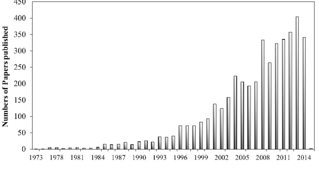
Figure 2.2 2-dimensional representation of structure of silica glass [1]. ... 13 Figure 2.3 Number of papers published per year in the field of ‘‘bioactive glass’’ (compiled
from a literature search SCOPUS carried out in November 2014). ... 20
Figure 4.1.1 X-ray diffractograms of as-quenched glass frits... 39 Figure 4.1.2 MAS NMR spectra of investigated glasses showing the peak positions of (a)
29
Si and (b) 31P. ... 40
Figure 4.1.3 X-ray diffractograms of glass powders after immersion in SBF solution for (a) 1
h; (c) 3 h; (c) 12 h, (d) 3 days and (e) 7 days. C refers to calcite while HA refers to hydroxyapatite... 41
Figure 4.1.4 FTIR spectra of glass powder (TCP-20) before and after immersion in SBF
solution for time durations varying between 1 h – 12 h. ... 43
Figure 4.1.5 Graph depicting changes in pH of solution and weight loss of glass powders
after immersion in Tris-HCl for 120 h. ... 45
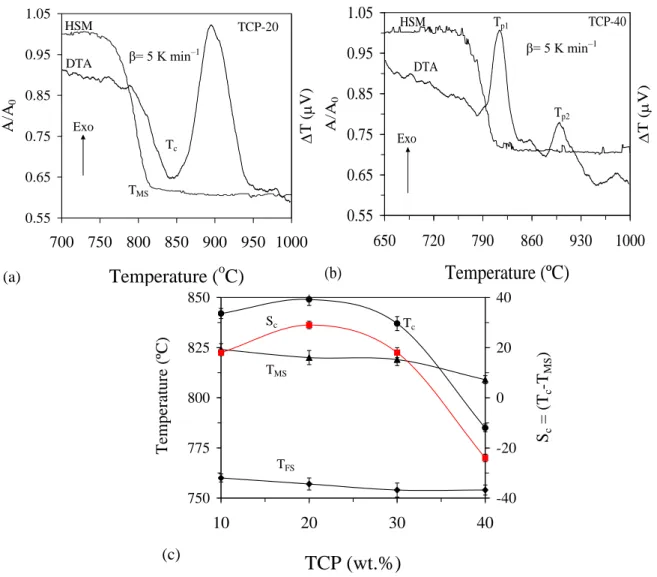
Figure 4.1.6 ... 46 Figure 4.1.7 Comparison of DTA and HSM curves under the same heating rate (5 K min1) for compositions (a) TCP-20 and (b) TCP-40 at the heating rate of 5 K min1; (c) influence of TCP content on different thermal parameters of glasses obtained from DTA and HSM, respectively ... 48
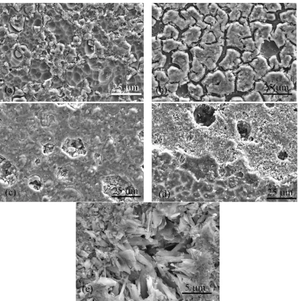
Figure 4.1.8. SEM images of glass powder compacts (a) TCP-10, (b) TCP-20, (c) TCP-30,
after sintering at 800 ºC for 1 h, respectively while (d) and (e) represent glass composition TCP-10 after sintering at 900 ºC for 1 h. ... 49
Figure 4.1.9 X-ray diffractograms of glass compositions after sintering at (a) 800 ºC; (b) 900
ºC and 1000 ºC, respectively. ... 50
Figure 4.1.10 Proliferation behaviour of the MSCs cultured on the glass compacts (TCP-10
and TCP-20) and the tissue culture plastic used as a control during the periods for 3, 7 and 14 days. MSCs derived from rat bone marrow were used for the assay. Statistically significance difference was noticed between the groups; control vs. TCP-10 at day 3; control vs. TCP-10 or TCP-20 at day 7 (p < 0.05, ANOVA, n = 3) ... 52
Figure 4.1.11 SEM images of the MSCs grown on the sintered glass powder compacts
Alkali-free bioactive glasses for bone regeneration Page xx contacts with the underlying substrates and have active cytoskeleton processes with highly elongated filopodia. Some surface cracks associated with the sample treatment for SEM are observed. ... 53
Figure 4.1.12 Alkaline phosphatase activity of the MSCs during culture for 7 and 14 days on
the glass compacts (TCP-10 and TCP-20) and tissue culture plastic control. Statistically significant higher level was noticed on the glass samples with respect to control at both periods; *p < 0.05, ANOVA, n = 3. ... 54
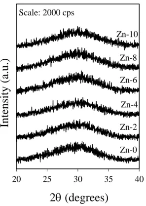
Figure 4.2.1 X-ray diffractograms of as-quenched glasses. ... 58 Figure 4.2.2 MAS NMR spectra of investigated glasses showing the peak positions of (a)
29
Si and (b) 31P. ... 59
Figure 4.2.3 X-ray diffractograms of glass powders before and after immersion in SBF
solution for (a) as quenched glasses (b) 1 h (c) 6 h (d) 3 days and (e) 7 days. HA refers to hydroxyapatite... 61
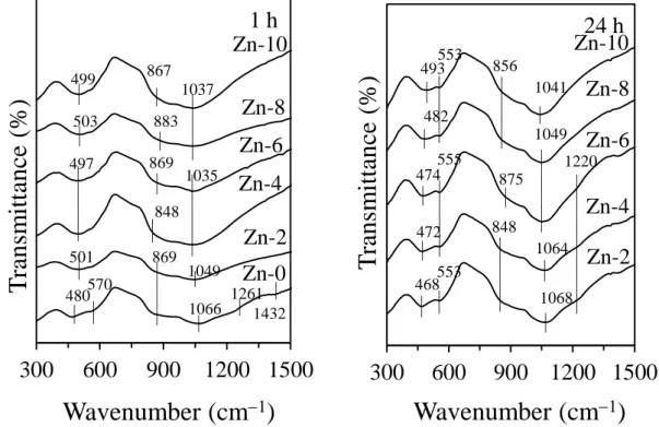
Figure 4.2.4 FTIR spectra of glass powders after immersion in SBF solution for (a) 1 h and
(b) 24 h. ... 62
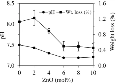
Figure 4.2.5 Graphs depicting the change in solution pH and weight loss of glass powders
after immersion in Tris–HCl. ... 63
Figure 4.2.6 X-ray diffractograms of glass powder after 120 days of immersion in
TRIS-HCL... 64
Figure 4.2.7 ICP-AES plots of elemental concentrations of Ca, Mg, P, Si and Zn in Tris–HCl
after 120 h of immersion of glass powder. It should be noted that Si refers to SiO44 and P
refers to PO43 species... 65
Figure 4.3.1 DTA thermographs of glasses at a heating rate of 20 K min−1. ... 70
Figure 4.3.2 Comparison of DTA and HSM curves under the same heating rate (5 K min1) for compositions: (a) Zn2, (b) Zn4, (c) Zn6, (d) Zn8 and (e) Zn10. ... 72
Figure 4.3.3 X-ray diffractograms of glass-ceramics heat treated at 850 ºC for 1 h. ... 74 Figure 4.3.4 (a) 29Si MAS-NMR and (b) 31P MAS-NMR spectra of glass-ceramics heat treated at 850 ºC for 1 h. ... 75
Figure 4.3.5 Flexural strength of the glass-ceramics heat treated at 850 ºC for 1 h ... 77 Figure 4.3.6a SEM/EDS micrographs images of unpolished and non-etched glass-ceramics
samples before soaking in SBF: Zn-0, Zn-4,and Zn-8. ... 78
Figure 4.3.6b SEM/EDS micrographs images of unpolished and non-etched glass-ceramics
Alkali-free bioactive glasses for bone regeneration Page xxi
Figure 4.3.7 Influence of ZnO content in glass-ceramics on cell viability during culture for
up to 7 days, as assessed by CCK method. Glass-ceramics with 4 mol% ZnO showed highest growth with respect to tissue culture plastic control as well as to that of other investigated glass-ceramics. ... 80
Figure 4.3.8 SEM images of the MSCs grown on the sintered glass powder compacts (Zn0,
Zn4 and Zn8) during culture for 3 and 7 days. After 7 days, cell proliferation on glass-ceramics was better in comparison to that observed after 3 days. Glass-ceramic Zn4 exhibited the highest rate of cell proliferation. ... 81
Figure 4.3.9 Alkaline phosphatase activity of the MSCs during culture for 7 and 14 days on
the glass compacts (Zn0 to Zn8) and on tissue culture plastic control. For all the investigated glass-ceramics, irrespective of their ZnO content, ALP levels were always higher in comparison to that of control (p < 0.05, ANOVA, n=3) ... 82
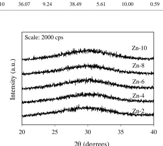
Figure 4.4.1 X-ray diffractograms of glass powders ... 87 Figure 4.4.2 MAS NMR spectra of investigated glasses showing the peak positions of (a)
29
Si and (b) 31P. ... 88 Thus NMR results shows that Zn2+/Mg2+ and Ca2+/Sr2+ substitutions do not affect the silicate network connectivity of these glasses. ... 88
Figure 4.4.3 (a) 29Si MAS NMR spectra and (b) 31P MAS NMR spectra after deconvolution of investigated glasses... 89
Figure 4.4.4 FTIR spectra of glass powders (a) before, after (b) 1h, (c) 24h, and (d)14 days
immersion in SBF. ... 91
Figure 4.4.5 X-ray diffractograms of glass powders after immersing the glass powders in
SBF solution for (a) 1 h (b) 3 h (c) 6 h (d) 24 h (e) 3 days and (e) 14 days. ... 92
Figure 4.4.6 ICP plots of elemental concentration of (a) Ca, (b) P, (c) Mg, (d) Sr, (e) Si and
(f) Zn, in SBF solution versus immersion time for the investigated glass powders. ... 94
Figure 4.4.7 X-ray diffractograms of glass powder after 120 days of immersion in
TRIS-HCL... 95
Figure 4.4.8 (a) Graphs depicting the change in solution pH and weight loss of glass powder
samples (with respect to variation in ZnO and SrO content in glasses) after immersion in Tris–HCl; (b) ICPAES plots of elemental concentrations of Ca, Mg, P, Si, Sr and Zn in Tris–HCl after 120 h of immersion of glass powder. It should be noted that Si refers to SiO44
Alkali-free bioactive glasses for bone regeneration Page xxii
Figure 4.4.9 Plots showing the cell growth kinetics of the glasses (ZS–2, ZS–4, ZS–6, and
ZS–8) (a) under normal condition during culture after 4 days under normal conditions (b) under H2O2 (250 μM) induced oxidative stress in MG63 cell after 3 days. *(C for control) .. 98
Figure 4.5.1 DTA thermographs of glasses at a heating rate of 20 K min−1. ... 104
Figure 4.5.2 Oxygen density plotted against percentage zinc and strontium substitution. ... 105 Figure 4.5.3 Comparison of DTA and HSM curves under the same heating rate (5 K min1) for compositions: (a) ZS-2, (b) ZS-4, (c) ZS-6, (d) ZS-8 and (e) ZS-10. ... 106
Figure 4.5.4 X-ray diffractograms of glass-ceramics heat treated for 1 h at: (a) 800 ºC, (b)
850 ºC and (c) 900 ºC. ... 108
Figure 4.5.5 Intensity ratio of the main XRD intensity peaks of Sr-diopside to Sr-fluorapatite
plotted against percentage of zinc and strontium substitution for glass-ceramics heat treated for 1 h at 850 ºC and 900 ºC. ... 109
Figure 4.5.6 SEM images and EDS results of glass-ceramic ZS–8 sintered for 1 h at: (a) 800
ºC, (b) 850 ºC, (c) 900 ºC, after a heating rate ramp of 5 K min−1. ... 110
Figure 4.5.7 Flexural strength of glass-ceramics heat treated for 1 h at: (a) 800 ºC, (b) 850 ºC
and (c) 900 ºC, using a heating rate of 5 K min−1. ... 111
Figure 4.6.1 MAS NMR spectra of investigated glasses showing the peak positions of (a)
29
Si and (b) 31P. ... 114
Figure 4.6.2 FTIR spectra of the investigated glasses ... 115 Figure 4.6.3 FTIR spectra of glasses powders after immersion in SBF solution for (a) 1 h, (b)
24 h, (c) 3 days, and (d) 14 h. ... 117
Figure 4.6.4 X-ray diffractograms of glass powders before and after immersion in SBF
solution for (a) 1 h (b) 24 h (c) 3 days and (d) 14 days. HA refers to hydroxyapatite ... 118
Figure 4.6.5 Variations in pH and weight loss of glass powders upon immersion in Tris-HCl
solution for 7 days ... 119
Figure 4.6.6 X-ray diffractograms of glass powder after 120 days of immersion in
TRIS-HCL... 120
Figure 4.6.7 DTA thermographs of glasses at a heating rate of 20 K min−1 ... 121
Figure 4.6.8 Comparison of DTA and HSM curves under the same heating rate (5 K min1) for compositions: (a) Di50, (b) Di60 (c) Di70, (d) Di80 (e) Di90 and (f) Di100 ... 123
Figure 4.6.10 Flexural strength of glass-ceramics heat treated for 1 h at (a) 900 ºC (b) 1000
Alkali-free bioactive glasses for bone regeneration Page xxiii
Figure 4.6.11 Metabolic activities of hMSCs on Di–60, Di–70 and Di–90 and TCPS after 1,
Alkali-free bioactive glasses for bone regeneration Page xxv
List of tables
Table 2.1 Selected bioactive glass compositions (mol%). ... 21 Table 3.1 Infrared bands of functional groups of bioactive glasses. ... 28 Table 4.1.1 Nominal composition of the as-designed glasses (mol%). ... 37 Table 4.2.1 Nominal composition of the as-designed glasses (mol%). ... 58 Table 4.3.1 Thermal parameters measured from DTA and HSM at 5 K min1 *β = 20 K min–
1
. ... 73
Table 4.4.1 Nominal composition of the as-designed glasses (mol%). ... 86 Table 4.4.2 Qn (Si) distribution for glasses ZS-4 and ZS-10 obtained by NMR de-convolution. ... 88
Table 4.4.3 Qn (P) distribution for glasses ZS-4 and ZS-10 obtained by NMR deconvolution. ... 89
Table 4.5.1 Thermal parameters measured from DTA at 20 K min1... 105
Table 4.6.1 Nominal composition of the as-designed glasses (mol%). ... 113 Table 4.6.2 Thermal parameters measured from DTA at 20 K min1... 120
Table 4.6.3 Thermal parameters (ºC) and A/Ao ratio derived from DTA and HSM at 5 K
min1. ... 122
Alkali-free bioactive glasses for bone regeneration Page xxvii
List of abbreviations
A-W Apatite-Wollastonite
ALP Alkaline Phosphatase Activity
BO Bridging Oxygen
BMD Bone Mineral Density CaSR calcium-sensing receptor CCK Cell counting kit
Di Diopside
DMEM Dulbecco’s Modified Eagle Medium DMSO Dimethyl Sulfoxide
DNA Deoxyribonucleic acid
DTA Differential Thermal Analysis EthD-1 Ethidium Homodimer-1
EDTA Ethylene diamine tetra acetic acid EDS Energy-Dispersive X-Ray Spectroscopy ELISA Enzyme-linked immunosorbent assay FA Fluorapatite
FBS Foetal bovine serum
FTIR Fourier Transform Infrared Spectroscopy GPCR G-protein coupled receptor
HA Hydroxyapatite
hMSCs Human Mesenchymal Stem Cells HCA Hydroxy Carbonate Apatite ICP Inductively Coupled Plasma
MAS-NMR Magic Angle Spinning– Nuclear Magnetic Resonance MSCs Mesenchymal Stem Cells
NBO Non-Bridging Oxygen NC Network connectivity PBS Phosphate Buffered Saline PGA Polyglycolic acid
PLA Polylactic acid
SBF Simulated Body Fluid
Alkali-free bioactive glasses for bone regeneration Page xxviii Tris Tris (Hydroxymethyl) Aminomethane
TE Tissue Engineering
TCPS Tissue Culture Polysterene TCP Tricalcium Phosphate XRD X-Ray Diffraction
Alkali-free bioactive glasses for bone regeneration Page xxix
List of publications
1. Saurabh Kapoor, Ashutosh Goel, Antonio Tilocca Raghu, Vikram Dhuna, Gaurav
Bhatia, Kshitija Dhuna, Jose M. F. Ferreira, Role of glass structure in defining the chemical dissolution behaviour, bioactivity and antioxidant properties of zinc- and strontium- co-doped alkali-free phosphosilicate glasses. Acta Biomaterialia, 2014; 10: 3264–78.
2. Saurabh Kapoor, Ashutosh Goel, Ana Filipa Correia, Maria J. Pascual, Hye-Young Lee,
Hae-Won Kim, José M.F. Ferreira. Synthesis and Characterization of Bioactive Glass-Ceramics Material Science and engineering C (accepted).
3. Saurabh Kapoor, Angela Semitela, Ashutosh Goel, YeXiang, Jincheng Du, Ana H
Lourenço, Daniela M Sousa, Pedro L Ganja, José M. F. Ferreira. Understanding the composition-structure-bioactivity relationships in diopside (CaO*MgO*2SiO2) –
tricalcium phosphate (3CaO*P2O5) glass system, Acta Bioamaterlia (accepted
4. Saurabh Kapoor, Ashutosh Goel, Maria J. Pascual, José M. F. Ferreira,
Thermo-mechanical behaviour of alkali free bioactive glass-ceramics co-doped with strontium and zinc, Journal of Non-crystalline Solids, 2013; 375:74-82.
5. Saurabh Kapoor, Ashutosh Goel, Maria J. Pascual and José M. F. Ferreira, Alkali-free
bioactive Diopside – Tricalcium phosphate glass ceramics for scaffolds fabrication: sintering and crystallization behaviour. Journal of Non-crystalline Solids (Invited paper for special issue on health care).
6. Ashutosh Goel, Saurabh Kapoor, Antonio Tilocca Raghu, Raman Rajagopal, Jose M. F. Ferreira, Structural role of zinc in biodegradation of alkali-free bioactive glasses,
Journal of Material Chemistry B, 2013;1:3073-82.
7. Ashutosh Goel, Saurabh Kapoor, Raghu Raman Rajagopal, Maria J. Pascual, Hae-Won Kim, Jose M. F. Ferreira, Alkali-Free bioactive glasses for bone tissue engineering: A preliminary investigation, Acta Biomaterialia, 2012; 8: 361-72.
Other publications
1. Allu Amarnath Reddy, Dilshat U. Tulyaganov, Ashutosh Goel, Saurabh Kapoor, Maria J. Pascual, José M.F. Ferreira, Sintering and devitrification of glass-powder compacts in the akermanite-gehlenite system, Journal of Materials Science,2013;48:4128–36. 2. Allu Amarnath Reddy, Ashutosh Goel, Dilshat U. Tulyaganov, Saurabh Kapoor, K
calcium-magnesium-aluminum-Alkali-free bioactive glasses for bone regeneration Page xxx silicate (CMAS) glass and glass-ceramic sealant for solid oxide fuel cell, Journal of
Power Sources,2013;231:203–12.
3. Allu Amarnath Reddy, Dilshat U. Tulyaganov, Saurabh Kapoor , Ashutosh Goel , Maria J. Pascual Vladislav V. Kharton and José M. F. Ferreira, Study of melilite based glasses and glass–ceramics nucleated by Bi2O3 for functional application, RSC
Alkali-free bioactive glasses for bone regeneration Page 1
Chapter 1
Alkali-free bioactive glasses for bone regeneration Page 3
1. Introduction
A glass can be defined as an amorphous solid completely lacking in long range, periodic atomic structure, and exhibiting a region of glass transformation behaviour. Any material, inorganic, organic, or metallic formed by any technique, which exhibits glass transformation behaviour is considered to be a glass [1].
Humans have been using glass for thousands of years, from the first uses of natural glass (e.g. obsidian, a volcanic glass) for tools and arrow heads, to the early man-made glass beads and drinking vessels of the Egyptians. Since then, glass has fascinated and attracted much interest both scientifically and technologically. The multiple forms and uses of glasses are becoming increasingly important in science, industry and in general daily life. During the last century, glasses and glass-ceramics became widely used for decorative articles, optics, architectural purposes (from windows to whole glass facades), and glassware for chemical reactions or fibres for telecommunication applications. The major advantage of glasses and glass-ceramics among its ceramic counterparts is their ability to accommodate various functional ions in their amorphous or crystalline phase, thus providing the flexibility to tailor and optimize their compositions with respect to different technological applications.
In particular bioactive glasses have been a relatively young group of materials discovered in 1970´s by Larry Hench. Bioactive glasses and glass-ceramics constitute a class of bioactive materials, i.e. “Materials which elicit a special response on their surface when in contact with biological fluids, leading to strong bonding to living tissue”. Since its discovery, 45S5 Bioglass® has been used in >650,000 human patients and is being marketed for various dental and orthopaedic applications under different commercial brand names [2-4]. Due to number of attractive properties for bone regeneration and tissue engineering (TE), increasing efforts have been made towards expanding the range of potential applications and understanding the fundamental science governing the physical, chemical and thermal properties of bioactive glasses in order to develop novel compositions with superior set of properties for biomedical applications [5-10]. Irrespective of this huge success, high alkali-containing glasses face some drawbacks which do restrict their application in some of the most advanced areas of human biomedicine. High alkali-content in glasses increase their crystallization tendency upon sintering, hindering the densification process and limiting the mechanical strength of sintered materials. Further, the relatively high chemical dissolution rates of such glasses in aqueous media decrease their in vitro and in vivo efficacy [11]. All these features constitute serious drawbacks in terms of fabrication of porous scaffolds or
Alkali-free bioactive glasses for bone regeneration Page 4 porous coatings, and mitigate the potential benefits that could be drawn from their usage as biomedical devices for bone regeneration and TE.
Despite many comprehensive studies leading to the development of bioactive glasses from different systems, the majority of these studies were based on 45S5 Bioglass® or compositions inspired by it. Therefore, this work aims at designing new alkali-free bioactive glasses in the Diopside (Hereafter referred as Di) – Tricalcium phosphate (Hereafter referred as TCP) – Fluorapatite (Hereafter referred as FA) system having good bioactive properties and to understand the structure–property relationships in the as designed glasses.
In the light of the above mentioned perspective, this dissertation comprises of five chapters. The first chapter, i.e., the Introduction, provides an outlook of the content of each chapter; the second chapter will focus on the literature survey in the field of bioactive glasses for biomedical applications, with special emphasis on alkali-free silicate systems. The second chapter has been divided into different subsections; the first section aims at providing a historical background on the development and evolution of bioactive glasses in general, while the second section outlines the motivations and purpose behind the present work. The third chapter deals with the description of the general experimental procedures and methodologies used along the thesis. It provides details about all the experimental techniques and procedures employed in order to synthesise, characterize and test our samples. Chapter four is the most important part of this work as it presents all the experimental results obtained on alkali-free glasses and glass-ceramics during past 3 years along with the pertaining discussions. In chapter five we have tried to conclude all the results and achievements obtained during this work and chapter six provides future directions in this field of research.
Several experimental techniques were used throughout this investigation aiming at a better understanding of the glass structure, biodegradation behaviour and the sintering and crystallization behaviour of the corresponding glass powder compacts. Some properties of glasses and glass-ceramics such as density, mechanical strength, were also evaluated to achieve a better understanding concerning the structure-properties relations.
Alkali-free bioactive glasses for bone regeneration Page 5
Chapter 2
Alkali-free bioactive glasses for bone regeneration Page 7
2. State of the art
2.1 Need for Biomaterials for bone regeneration
Human bone is a dynamic, highly vascularised connective tissue which provides structural support and act as a protective casing for the delicate internal organs of the body [12]. It is a tissue that is constantly remodelling to adapt to mechanical stresses imposed and repair minor injuries occurring during the lifetime. However, if the injuries suffered are extensive leading to very large bone defects, the use of bone grafts is required in order to repair the damaged part.
Worldwide, millions of people are affected by degenerative and inflammatory problems related to bone and joint. In fact, they account for half of all chronic diseases in people over 50 years of age in developed countries. Further, every year there are roughly 8.9 million cases of fractures, which occur due to osteoporosis only [13]. Also, approximately 1.6 million hip fractures occur worldwide each year, by 2050 this number could reach between 4.5 million [14] and 6.3 million [15]. Current treatments for filling bone defects and subsequent repair are based on autograft and allografts. Autologous grafts are the ones which contain essential elements for bone regeneration (osteogenic cells, osteoinductive growth factors and a matrix that supports adhesion and bone growth), are collected from the individual, minimizing the risk of rejection. Although autograft present relatively good percentages of success, however the range of applications is restricted, mainly due to the limited availability of living tissues and due to donor site morbidity [16]. Further, allograft bone introduces the possibilities of immune rejection and of pathogen transmission from donor to host. Although infrequent, infections could occur in the recipient’s body after the transplantation [16]. Therefore there is a severe need to search for new bone regeneration strategies in order to meet the increasing medical and socioeconomic challenge of our aging population. Hence, materials that enhance bone regeneration have a wealth of potential clinical applications from the treatment of non-union fractures to spinal fusion. Synthetic biomaterials, developed in an effort to overcome the inherent limitations of autograft and allograft, represents an alternative strategy.
Alkali-free bioactive glasses for bone regeneration Page 8
2.2 Biomaterials for bone repair
A biomaterial according to the American National Institute of Health is “Any substance (other than a drug) or combination of substances, synthetic or natural in origin, which can be used for any period of time, as a whole or as a part of a system which treats, augments, or replaces any tissue, organ, or function of the body”. The development of novel biomaterials is an iterative process that involves the creation of increasingly safer, more reliable, less expensive and more physiologically appropriate replacements for damaged or diseased human tissues. In the last 60 years, biomaterials for orthopaedic applications have evolved from materials available from different industrial applications into the materials with inherent capabilities to interact with the biological environment and to elicit specific biological responses. Most of the problems that orthopaedic surgery has to face have not basically changed, and are practically the same that orthopaedics had to face 50 years ago; however, the choice of possible solutions has been greatly expanded because new materials have allowed the design of innovative devices. Based on the evolution of biomaterials, these can be roughly divided into three broad categories depending upon the interaction mechanisms of the materials; namely inert materials (first generation biomaterials). Bioactive and biodegradable materials (second generation), and materials designed to stimulate specific cellular responses at the molecular level (third generation) [17]. This division represent the development in the field of biomaterials based on the requirements and properties of the materials involved. The present research is still devoted to the first or the second generation depending upon their properties and application. This means that the materials that each new generation brings in do not necessarily outweigh the existing materials.
First generation biomaterials: Developed in the early 1950s [18], the principle underlying the development of first generation biomaterials was that they should be as chemically inert as possible in order to minimize the immune response to the foreign body [17]. Initially, the first generation of biomaterials consisted of already functional materials which were being applied in industrial applications (for e.g. aerospace) where mechanical properties were the main benchmark for the selection of candidate materials for implant manufacture. These biomaterials are mainly metallic or alloys (Co–Cr–Mo alloys, Ti6Al4V, etc.), ceramics (alumina, zirconia, etc.) and polymers (silicone rubber, acrylic resins) [19]. Though bio-inert in nature, these tend to form a thin non-adherent fibrous capsule over their surface with the passage of time after implantation which inhibits further interaction with tissue [20]. The thickness of the layer developed is a function of the level of reactivity of the
Alkali-free bioactive glasses for bone regeneration Page 9 implant. Formation of this thin layer is attributed to the absorption of unspecific proteins that results in unspecific signalling to the cellular environment. This thin layer leads to loosening and deterioration of the mechanical fit. Eventually, surgical removal of the device is required. Therefore the development of bioactive interfaces eliciting a specific biological response and avoiding any fibrous layer was one of the main driving forces behind the development of second generation biomaterials.
Second generation of biomaterials: Developed between the years 1970-1980. These biomaterials mainly include ceramics (bioactive glasses, glass-ceramics and calcium phosphates), biodegradable polymers (polylactic acid (PLA) and polyglycolic acid (PGA) etc.) [20]. Bioactive glasses discovered by Larry Hench in 1969-1971 [21] provided an alternative to the bio-inert materials used in orthopaedic applications. These second generation biomaterials were defined by their ability to interact with the biological environment in a controlled manner to enhance the biological response and the tissue/surface bonding. Further progress of the second generation biomaterials was the development of bioresorbable or bio-absorbable materials with the ability to undergo a progressive degradation while new tissue regenerates and heals [20, 22]. Bioactivity is defined as the ability of the material to bond to bone tissue via the formation of a bone-like hydroxyapatite (HA) layer on its surface [23]. The HA phase that forms on bioactive implants is equivalent chemically and structurally to the mineral phase in the bone, and is responsible for strong interfacial bonding, allowing for bone regeneration rather than replacement.
Second generation biomaterials have been in clinical use since 1980 in many orthopaedics and dental applications, including various compositions of bioactive glasses, ceramics, glass-ceramics, and composites. Synthetic calcium phosphate ceramics have been routinely used as bone cements and coatings over metallic prosthesis to facilitate bioactive fixation [20]. Different phases of calcium phosphate ceramics are used depending upon whether a bioresorbable or bioactive material is desired. Bioactive glasses and glass-ceramics are used as middle-ear prostheses to restore the ossicular chain and treat conductive hearing loss and as oral implants to preserve the alveolar ridge from the bone resorption that follows tooth extraction [20, 24, 25]. In comparison to the bioactive glasses and glass-ceramics, calcium phosphate based cements have been used in many orthopaedic applications such as bone substitution, repair of bone fractures (including ligament fixation), cartilage, meniscus and intervertebral disc.
Alkali-free bioactive glasses for bone regeneration Page 10 Third generation of biomaterials: Developed in the 21st century, third generation of biomaterials brought together separate concepts of bioresorbable and bioactive materials of second generation. The motive behind the design and development of these materials was to enhance the self-healing capabilities of the body, by the systematic stimulation of specific cellular responses at the molecular level [20]. Primarily, third generation biomaterials were developed by interchanging and intermixing of the properties of bioresorbable and bioactive materials. Consequently, these tailored biomaterials with new levels of bio-functionality have led to new approaches for the treatment of bone defects. Two main strategies have been followed during the last two decades with respect to tissue regeneration are TE and in situ tissue regeneration.
TE is one of the approaches in which cells planted in the scaffolds are made to proliferate and differentiate in vitro. These tissue-engineered constructs are then implanted into the patients to replace diseased or damaged tissues. Clinical applications include repair of articular cartilage, skin, and the vascular system, although stability of the repaired tissues needs improvement. Another method was in situ tissue regeneration which involved the use of biomaterials directly implanted into the body in the form of powders, solutions, or doped micro-particles to stimulate local tissue repair. These materials release controlled amount of ionic dissolution products or growth factors such as bone morphogenic protein (BMP) by diffusion or network breakdown.
2.3 Bioactive glasses
Bioactive glasses are amorphous materials, compatible with the human body; bond to bone and can stimulate new bone growth while dissolving over time. They therefore have the potential to restore diseased or damaged bone to its original state and function (bone regeneration). Bioactive glasses belong to class A of bioactive materials which are osteogenic and osteoconductive materials. The first bioactive glass (silicate based) was discovered by Larry Hench and colleagues in 1969 using melt-quench method, at the University of Florida [26]. The glass composition of the first bioactive glass was 46.1% SiO2, 24.4% NaO, 26.9%
CaO and 2.6% P2O5 (mol%), termed 45S5 Bioglass®, which is now a trademarked name
Alkali-free bioactive glasses for bone regeneration Page 11
Figure 2.1 Compositional dependence of bioactivity in Na2O-CaO-SiO2-P2O5 glass system
[2].
When in contact with a physiological fluid, bioactive glasses undergo a specific set of reactions, leading to the formation of an amorphous calcium phosphate (ACP) or crystalline HA phase on the surface of the glass, which is responsible for their strong bonding with the surrounding tissue [20]. Further, ionic dissolution products released from bioactive glasses (e.g. Si, Ca, P) are also reported to activate osteogenic gene expression [27, 28] and to stimulate angiogenesis [29, 30]. In addition to it, bioactive glasses offer remarkable advantages such as the ease of controlling chemical composition and, thus, the rate of degradation which make them attractive materials for orthopaedic applications. The structure and chemistry of glasses can be tailored over a wide range by changing either composition, or thermal or environmental processing history. Therefore, it is possible to design bioactive glasses with variable degradation rates to match that of bone ingrowth and remodelling.
Based on the type of glass former, there are mainly three types of bioactive glasses: the conventional silicate-based glasses; phosphate-based glasses; and borate-based glasses. Recently, there has been an increase in interest in borate based bioactive glasses [31, 32], largely due its potential ability to heal chronic wounds that would not heal under conventional treatment [33]. The soft tissue response may be due to their lower chemical durability (faster dissolution) in comparison to the silica-based counterparts [31]. Also phosphate-based glasses have shown great potential in regenerative medicine [31], as the
Alkali-free bioactive glasses for bone regeneration Page 12 solubility of these glasses can be controlled by modifying their composition; therefore these glasses show additional clinical potential as resorbable materials. Thus, benefits of phosphate glasses are probably related to their very rapid solubility which is strongly composition dependent rather than bioactivity [34].
Oxide glasses are conventionally produced by melting the precursors (inorganic oxides, carbonates, fluorides and others) in precious metal or ceramic crucibles at high temperatures (for bioactive glasses typically above 1000 ºC, depending on the composition). If the cooling is rapid enough (with the necessary cooling rates depending, again, on the composition) crystallization will be inhibited and an amorphous material, a glass, will result. Another common way of preparing bioactive glasses is by a polycondensation reaction from organic precursors, alkoxides such as tetraethyl orthosilicate [35]. Synthesized in the early 1990s [35], this new class of bioactive glasses displays higher compositional range of bioactivity: bioactive sol-gel glass compositions have been synthesized within the ternary SiO2:CaO:P2O5 or even within the binary SiO2:CaO systems, and might contain between 60
and 90 mol% of silica [36, 37]. As the synthesis of glasses is highly dependent on composition and external conditions, sol-gel method gives better control over the surface and structural properties (such as surface area and porosity) of the resultant glass in comparison to melt-quenching method. The introduction of sol-gel synthesis technique has opened the research to new types of biomaterials. Many possible dopants can be introduced in a material synthesized via sol-gel. For example, it has been shown that sol-gel derived glasses containing 5 mol% ZnO resulted in increased alkaline phosphatase activity (ALP) activity and osteoblast proliferation [38].
However, in the frame of the present thesis, melt-quenching was used to prepare glasses and sol-gel derived glasses are beyond the scope of this PhD work program. Hence, the subsequent discussion will solely be based on the silicate based glasses prepared by melt-quenching.
Alkali-free bioactive glasses for bone regeneration Page 13
2.4 Glass structure and bioactivity
Within the glass structure, three different components are usually described. Network formers are able to form amorphous materials without the need for additional components. The basic building unit of silicate glasses is the SiO4 tetrahedron, which can be connected to
neighbouring SiO4 tetrahedra via Si—O—Si bonds, known as bridging oxygens (BO).
Silicon possess a charge of 4+ thus in the glasses when SiO4 tetrahedral forms a network these
building units can share all their four oxygen atoms to give the stoichiometry (SiO4/2 or SiO2
which is charge-balanced (assuming a charge of 2– on the oxygen).
Figure 2.2 2-dimensional representation of structure of silica glass [1].
Each oxygen atom is shared between two silicon atoms, which occupy the centres of linked tetrahedra. Randomness in the structure is facilitated by the variability in the Si–O–Si angle connecting adjacent tetrahedra. Further rotation of adjacent tetrahedra around the point occupied by the oxygen atom linking the tetrahedra, and by rotation of the tetrahedra around the line connecting the linking oxygen with one of the silicon atoms results in augmentation in disorder in the structure. These tetrahedra are commonly referred to as Qn (n = 1, 2, 3, 4) units, where n describes the number of BO connected to the tetrahedron. Silica glass therefore consists of Q4 units only. A 2-dimensional representation of such a structure is shown in Figure 2.2, where the fourth oxygen, which would sit directly above the small silicon ion, is not shown.
Network modifiers, on the other hand, alter the glass structure by turning BO’s (predominantly covalent in character) into non-bridging oxygens (NBO’s) (Si—O- M+
Alkali-free bioactive glasses for bone regeneration Page 14 linkages, predominantly ionic in character, where M+ is a modifier cation). Typical modifiers include the oxides of alkali or alkaline-earth metals having co-ordination number 6 or more. The more modifiers we have in the glass composition, lower is the average number of SiO4
tetrahedra each tetrahedron is linked to. At the same time, the number of NBO’s in the glass structure increases. In conventional silicate glasses, we find large concentrations of Q4 and Q3 units, i.e. SiO4 tetrahedra connected to neighbouring ones in four or three directions, forming
a silicate network. The relative concentrations of BO’s and NBO’s have an important influence on the structure and properties of the glasses. The rigidity of the glass network decreases gradually by replacing bridging atoms by non-bridging atoms.
Intermediates on the other hand can either act like typical network modifiers or enter the backbone of the glass structure, acting more like a network former. These have a co-ordination ranging from 4 to 6.
Bioactive glasses, by contrast, contain larger concentrations of network modifiers and smaller amounts of silica than conventional soda-lime silicate glasses, and they therefore consist of a more disrupted silicate network of mostly Q2 units. According to Tilocca [5] maximum bioactivity for bioactive glasses is achieved if the structure is dominated by chains of Q2 metasilicates, which are occasionally cross-linked through Q3 units, whereas the Q1 species terminate the chain. Further, the structure of glass can also be estimated based on the glass composition. Different parameters have been suggested for describing the glass structure in the literature [39-41]. Strnad [40] introduced the concept of network connectivity (NC) model to describe the average number of bridging oxygens per network forming element and to correlate the molecular structure of silicate glasses with their bioactivity (apatite forming ability). In a pure silica glass (containing no network modifiers) we have only Q4 units, thus has a NC = 4. With the introduction of the network modifiers the NC decreases as the BO’s are converted into NBO’s. Bioactive glasses with NC value in the range of 2 and 3 are believed to show good bioactive properties (45S5 Bioglass® has a NC of 1.90 [39], corresponding to a structure consisting of silicate).
However, while NC may be a useful as a qualitative and a preliminary tool to predict the bioactivity of glasses, its predictive power rapidly decreases when: (i) a wider range of glass compositions are considered; (ii) cations of similar charge are partially substituted in the glass compositions. It has been shown that there is no straightforward correlation between the NC and the degradation behaviour bioactive glasses. For example, glass compositions with NC similar to that of 45S5 Bioglass® (NC = ~1.95) have been shown to exhibit slower chemical dissolution behaviour in aqueous solutions than the latter while partial substitution
Alkali-free bioactive glasses for bone regeneration Page 15 of Zn2+ in bioactive glasses at the expense of Mg2+ retards the apatite forming ability of bioactive glasses despite their NC being unchanged [42].
2.5 Mechanism of HCA layer deposition
Even though all the composition of silicate based bioactive glasses synthesized till date are quite different, the mechanism of HA or HCA (hydroxycarbonate apatite) formation are analogous for all of them. The apatite layer forms following solution-mediated dissolution of the glass with a mechanism very similar to conventional glass corrosion [43]. Accumulation of dissolution products causes both the chemical composition and the pH of the solution to change, providing surface sites and a pH conducive to apatite nucleation. There are five proposed stages for apatite formation in body fluid in vivo or in SBF in vitro [20].
STEP 1 – Reactions for the rapid exchange of ions between the modifiers in the glass network (Na+ and Ca2+) with H+ ions or H3O+ in solution , leading to hydrolysis of the groups
of silica and creating silanol groups (Si–OH ) on the glass surface. This process results in a net increase of pH of the solution due to the increasing OH ions.
Si − O − Na++ H+ → Si − OH++ Na+ (1)
STEP 2 – The increase of pH leads to attack on the glass network, breaking Si–O–Si bonds. Soluble silica is lost in the form of Si–OH to the solution, leaving more Si–OH (silanols) at the glass–solution interface. These silanol groups play a vital role as the nucleation centres of the apatite formation.
Si − O − Si + H2O → Si − OH + OH − Si (2)
STEP 3 – Condensation and repolymerization of SiO2-rich layer on the glass surface depleted
in alkali and alkaline earth cations.
STEP 4 – Migration of Ca2+ and PO43– groups to the surface through the silica-rich layer,
forming a film rich in amorphous CaO–P2O5 on the silica-rich layer.
Si O O O H O Si OH O O O + Si + H 2 O O O O Si O O O O (3)
Alkali-free bioactive glasses for bone regeneration Page 16 STEP 5 – Incorporation of hydroxyls and carbonate from solution and crystallization of the CaO–P2O5 film to form apatite. After the apatite layer is formed the adsorption of growth
factors, attachment, proliferation and differentiation of osteoprogenitor cells are the biological mechanisms of bonding to bone. In addition to this, adsorption of adhesion proteins (e.g., fibronectin, vitronectin, etc.) is necessary condition for cellular attachment.
2.6 Role of various functional ions
The processes of bone formation and bone resorption (bone remodelling) are regulated by variety of systematic and local regulated agents including growth factors and hormones. Single inorganic ions are known to be involved in the bone formation and bone resorption (bone remodelling) and play a physiological role in angiogenesis, growth and mineralization of bone tissues. In particular, some metal ions act as enzyme co-factors and therefore influence signalling pathways and stimulate metabolic effects during tissue formation [44]. These properties make metal ions attractive for use as therapeutic agents in the fields of hard and soft TE.
Further, bioactive glasses have the ability to exhibit surface reactivity when in contact with body fluids leading to the release of ionic dissolution products (example: Si, Ca, P, Mg, F) which further stimulate the various vital mechanisms in human body like gene expression, osteoblast proliferation, angiogenesis and also provide bacterial as well as anti-inflammatory effects [28, 30, 45]. In fact, it has been well established that ionic dissolution products are key to understand the behaviour of parent inorganic materials in vitro and in vivo, especially in context of TE applications [46].
2.6.1 Silicon
Silicon (Si) is a non-metallic element with an atomic weight of 28. It is the second most abundant element in the Earth's crust at 28 wt%. Si is known to be an essential element for metabolic processes related with the formation and calcification of bone tissue [44]. Silicon is present in all body tissues, but the tissues with the highest concentrations of silicon are bone and other connective tissue including skin, hair, arteries, and nails [47]. Silicon plays an important role in the formation of cross-links between collagen and proteoglycans [48-50]. In vitro studies have demonstrated that silicon stimulates type I collagen synthesis and osteoblast differentiation [51]. Studies in rats have demonstrated that silicon at physiological levels improves calcium incorporation in bone when compared to rats that are
Alkali-free bioactive glasses for bone regeneration Page 17 deficient in silicon [52-54]. Additionally, dietary Si intake was shown to increase the bone mineral density (BMD) in men and premenopausal women. Further Si intake has shown to improve BMD by bone resorption in case of calcium deficiency in rats [55]. Moreover, it has been reported that Si has a biochemical function in bone growth processes affecting bone collagen turn over and sialic acid-containing ECM proteins like osteopontin. Moreover, orthosilicate acid (Si(OH)4) at physiological concentration of 10 mmol has been shown to
stimulate collagen I formation in human osteoblast cells (HOC) and to stimulate osteoblastic differentiation [44].
2.6.2 Strontium
Strontium (Sr) is an abundant and widely distributed element in the geosphere, natural water and human tissues. The amount of Sr in the skeleton is only 0.335% of its Ca content [56]. The biological properties of strontium are related to its chemical similarity to calcium. Sr can accumulate in bone by exchanging with Ca in the HA crystal lattice Thus strontium accumulates to a high degree in bone, can displace calcium in hard tissue metabolic processes and at high concentrations interferes with normal bone development. Low levels of Sr have been known to be associated with low energy fracture sites. Presence of strontium lactate enhances Ca deposition in the bone and reduces bone pain in osteoporosis patients. Sr ions have been shown to stimulate osteoblastic bone formation and to inhibit osteoclastic bone resorption both in vitro and in vivo [56, 57]. Sr drew attention as a drug for the management of osteoporosis in the 1950s. Indeed, strontium ranelate (Protelos) is a drug approved for treatment and prevention of osteoporosis. Owing to the above-mentioned beneficial aspects of Sr in bone regeneration and considering the ion releasing ability of glasses in aqueous medium, bioactive glasses incorporated with Sr have gained considerable attention in the recent past for various orthopaedic applications. Sr-containing bioactive glasses were shown to combine the known bone regenerative properties of bioactive glasses with the anabolic and anti-catabolic effects of Sr cations in vitro [58].
2.6.3 Zinc
Zinc (Zn) plays a vital role in bone formation, resorption and TE as it directly activates aminoacyl–tRNAsynthetase (a rate-limiting enzyme at translational process of protein synthesis) in osteoblastic cells and stimulates cellular protein synthesis [42]. Zn has also been shown to stimulate gene expression of the transcription factors: runt-related transcription factor 2 (Runx2) that is related to differentiation into osteoblastic cells.
Alkali-free bioactive glasses for bone regeneration Page 18 Moreover, Zn inhibits osteoclastic bone resorption by inhibiting osteoclast–like cell formation from bone marrow cells and stimulating apoptotic cell death of mature osteoclasts [42].
In case of soft tissue regeneration, Zn has been proved to be an essential element for wound healing [30, 44, 45]. It is an essential trace mineral for DNA synthesis, cell division and protein synthesis during the proliferative phase of wound healing [42]. Zn deficiency has been associated with poor wound healing and decreased breaking strength of animal wounds [44] which can result from decreased protein and collagen synthesis during healing found in Zn deficient animals. Agren et al [59] demonstrated the beneficial aspect of topically applied Zn on leg ulcer healing and wound healing in animal models. It was shown that topical zinc oxide promotes cleansing, re–epithelialisation and inhibits bacterial growth.
2.6.4 Magnesium
Magnesium (Mg) is the fourth most abundant cation in human body with approximately half of the total physiological Mg stored in bone tissue [60]. It is essential to bone metabolism and has been shown to have stimulating effects on new bone formation [61]. Mg is a co-factor for many enzymes, and stabilizes the structures of DNA and RNA. Depelition of Mg in body results in impaired bone growth, increased bone resorption and loss in trabecular bone [62, 63]. The level of Mg in the extracellular fluid ranges between 17 ppm and 25.5 ppm, where homeostasis is maintained by the kidneys and intestine [60, 64]. Although Mg levels exceeding 25.5 ppm can lead to muscular paralysis, hypotension and respiratory distress and cardiac arrest occurs for severely high serum levels of 145–170 ppm, the incidence of hyper- Mg is rare due to the efficient excretion of the element in the urine [60, 64, 65].
2.6.5 Calcium
Calcium (Ca) accounts for 1-2 % of the adult human body weight [66], and a major component of mineralized tissues, approximately 99% of body Ca is found in bone, where it serves a key role as a component of HA. Ca plays an important role in osteoblast proliferation and bone remodelling by directly activating intracellular mechanisms by affecting Ca-sensing receptors in osteoblastic cells. The influence of Ca in the body is highly concentration dependent. Maeno et al. [67] found that Ca concentrations in blood varying within the range of 48 mmol are suitable for osteoblast proliferation, differentiation and extracellular matrix
![Figure 2.2 2-dimensional representation of structure of silica glass [1].](https://thumb-eu.123doks.com/thumbv2/123dok_br/15746841.1073215/43.892.265.636.392.691/figure-dimensional-representation-structure-silica-glass.webp)