© 2008 The Authors Doi: 10.1111/j.1742-7843.2008.00301.x Journal compilation © 2008 Nordic Pharmacological Society. Basic & Clinical Pharmacology & Toxicology, 103, 461– 467
Blackwell Publishing Ltd
In vivo
Skin Irritation Potential of a
Castanea sativa
(Chestnut) Leaf
Extract, a Putative Natural Antioxidant for Topical Application
Isabel F. Almeida1, Patrícia Valentão2, Paula B. Andrade2, Rosa M. Seabra2, Teresa M. Pereira3, M. Helena Amaral1,Paulo C. Costa1 and M. Fernanda Bahia1
1Department of Pharmaceutical Technology, and 2REQUIMTE, Department of Pharmacognosy,
Faculty of Pharmacy, University of Porto, Porto, Portugal, and 3Department of
Dermatology and Venereology, São Marcos Hospital, Braga, Portugal. (Received March 14, 2008; Accepted May 13, 2008)
Abstract: Topical application of natural antioxidants has proven to be effective in protecting the skin against ultraviolet-mediated oxidative damage and provides a straightforward way to strengthen the endogenous protection system. However, natural products can provoke skin adverse effects, such as allergic and irritant contact dermatitis. Skin irritation potential of Castanea sativa leaf ethanol:water (7:3) extract was investigated by performing an in vivo patch test in 20 volunteers. Before performing the irritation test, the selection of the solvent and extraction method was guided by the 1,1-diphenyl-2-picryl hydrazyl (DPPH) free radical scavenging test and polyphenols extraction (measured by the Folin Ciocalteu assay). Iron-chelating activity and the phenolic composition (high performance liquid chromatography/diode array detection) were evaluated for the extract obtained under optimized conditions. The extraction method adopted consisted in 5 short extractions
(10 min.) with ethanol:water (7:3), performed at 40°. The IC50 found for the iron chelation and DPPH scavenging assays
were 132.94 ± 9.72 and 12.58 ± 0.54 μg/ml (mean ± S.E.M.), respectively. The total phenolic content was found to be
283.8 ± 8.74 mg GAE/g extract (mean ± S.E.M.). Five phenolic compounds were identified in the extract, namely, chlorogenic acid, ellagic acid, rutin, isoquercitrin and hyperoside. The patch test carried out showed that, with respect to irritant effects, this extract can be regarded as safe for topical application.
Many plant extracts are used in cosmetic formulations as additives (fragrances, colorants, etc.) or are used due to their functional properties (emollient, anti-irritant, moisturizing, slimming, anti-ageing, etc.). Botanical products are also used in topical pharmaceutical preparations in a wide variety of skin disorders. However, allergic and irritant contact dermatitis and phytophotodermatitis are included among the topical adverse effects of natural products [1]. Members of the Ranunculaceae, Euphorbiaceae and Asteraceae (Compositae) plant families are specially involved in irritant contact dermatitis. Phytophotodermatitis is caused by plants containing furocoumarins, which notably include the members of the Umbelifereae, Rutaceae and Moraceae plant families. Allergic contact dermatitis is induced primarily by plants from the Asteraceae family, which contain ses-quiterpene lactones, but other families containing this class of compounds, specially the Lauraceae, Magnoliaceae and Jubulaceae families, can also produce allergic reactions. Besides studying the activity of plant extracts, suitable tolerance studies are therefore required before any possible practical application.
The skin is exposed to a broad variety of biological, chemical and physical attacks. Among them is solar ultraviolet
(UV) radiation that is responsible for skin damage, which includes erythema, photoageing and cancer. Following UV exposure, reactive oxygen species (ROS) are produced and are believed to be largely responsible for some of the mentioned deleterious effects through interaction with proteins, lipids and DNA [2,3]. A number of skin diseases are believed to be associated with oxidative stress, including psoriasis, acne and cutaneous vasculitis [4]. There is also evidence that ROS are involved in allergic and irritant contact dermatitis [5–7]. In this regard, antioxidants from natural products present novel possibilities for the treatment and prevention of oxidative stress-mediated skin diseases. Castanea sativa (Fagaceae) is a species of chestnut native to Southeastern Europe and Asia Minor. Chestnut leaves are used in traditional medicine in the treatment of several diseases, such as bronchitis and cough [8]. Radical scavenging activity and scavenging activity against superoxide anion and hydroxyl radical were previously reported for C. sativa leaves [9]. No reports of metal-chelating effects were found in the literature. The reported antioxidant activity was suggested to be related with the phenolic composition [9]. Rutin, hesperidin, quercetrin, apigenin, morin, galangin, kaempferol [10] and isoquercitrin [9] have been identified in C. sativa leaves.
The aim of the present study was to evaluate the skin irritation potential of an extract of C. sativa leaves, a putative topical antioxidant. Therefore, only skin-compatible solvents (water, ethanol and hydroalcoholic mixtures) were selected for the preparation of the extract. The optimization of the
Author for correspondence: M. Fernanda Bahia, Department of Pharmaceutical Technology, Faculty of Pharmacy, University of Porto, Rua Aníbal Cunha 164, 4099-030 Porto, Portugal (fax +35 1222003977, e-mail [email protected]).
462 ISABEL F. ALMEIDA ET AL.
extraction method was performed in order to obtain the extract with the highest antioxidant activity. Free radical scavenging activity and ability to chelate iron ions were evaluated for the extract obtained under optimized condi-tions. In vivo skin irritation potential observed after single application under occlusion was assessed along with sodium lauryl sulfate (SLS) solution (2%) as irritant model.
Materials and Methods
Chemicals. The standards, Folin Ciocalteu reagent and 1,1-diphenyl-2-picryl hydrazyl (DPPH) were purchased from Sigma (St. Louis, MO, USA) and Extrasynthése (Genay, France). Methanol, ascorbic acid, sodium carbonate, formic acid and disodium EDTA were obtained from Merck (Darmstadt, Germany). Absolute ethanol and ethanol 96% were purchased from AGA (Sacavém, Portugal). Ferrozine and ferrous chloride were purchased from Aldrich (St. Louis, MO, USA). SLS (purity > 99%) was purchased from Fluka Chemie (Steinheim, Germany). The water was purified with ion-exchange resins.
Plant material. Castanea sativa leaves were collected during July 2003 in Mirandela, northern Portugal, and dried at room temperature for 3 weeks. A voucher specimen is preserved in our laboratory for further reference.
Preparation of the extracts.The dried leaves (2 g) were grounded
(500 μm) and extracted three times (20 min., 500 r.p.m.) with
each of the following solvents (50 ml) water, ethanol:water (4:6), ethanol:water (7:3) and absolute ethanol. The extracts were
gathered, ethanol was evaporated at 40° under vacuum and then the
resulting aqueous mixtures were lyophilized (Labconco, FreeZone 4.5, Kansas City, MO, USA).
Optimisation of the extraction procedure. The powdered leaves (4 g) were extracted 12 times (10 min., 500 r.p.m.) with 100 ml of the chosen solvent. Extract absorbance (280 and 350 nm) was measured after each extraction step. The cumulative absorbance was considered as 100% of extraction.
For studying the effect of time, one extraction (500 r.p.m.) was carried out, with samples being withdrawn at 5, 10 and 15 min., followed by two more extractions with samples collected at 5, 10, 15, 30, 45, 60 and 90 min. Absorbance (280 and 350 nm) was measured at each time interval.
After selecting the number and duration of the extractions, the influence of temperature was assessed by performing the extractions at 40°.
Determination of total phenolics.The amount of total phenolics in the extract was determined using the Folin Ciocalteu colorimetric method, according to a described procedure [11]. Briefly, 1 ml of
Folin Ciocalteu reagent was added to 300 μl of extract dissolved in
ethanol:water (7:3) (500 μg/ml), followed by the addition of 5 ml of
20% sodium carbonate solution. The mixture was made up to 10 ml with water and thorough shaking, and the absorbance was read after 20 min., at 735 nm (V 530, UV-VIS Spectrophotometer; Jasco Corporation, Tokyo, Japan). The contents were expressed as milligrams of gallic acid equivalents (GAE) per gram of dry extract. The measurements were performed in triplicate.
Qualitative analysis. The lyophilized extract was dissolved in
ethanol:water (7:3) and filtered (0.22 μm) before being analysed.
Identification of phenolic compounds was achieved with an analytical high performance liquid chromatography (HPLC) unit (Gilson Medical Electronics, Villiers le Bel, France) using a reversed-phase
Spherisorb ODS-2 column (250 × 4.6 mm, 5 μm particle size; Merck)
with a C18 guard column. The solvent system was a gradient of water/formic acid (19:1) (A) and methanol (B), starting with 5% methanol and installing a gradient to obtain 15% B at 3 min., 25% B at 13 min., 30% B at 25 min., 45% B at 39 min., 45% B at 42 min., 50% B at 44 min., 55% B at 47 min., 70% B at 50 min., 75% B at 56 min. and 80% B at 60 min., at a solvent flow rate of 0.9 ml/min. [12]. Detection was achieved with a Gilson diode array detector. Spectral data from all peaks were accumulated in the 200– 400 nm range and chromatograms were recorded at 280, 320 and 350 nm. The phenolic compounds were identified by their UV spectra and their HPLC retention times. For identity confirmation, co-injection with the standards was carried out. Data were processed on Unipoint® system software (Gilson Medical Electronics, Villiers le Bel, France).
Quantitative analysis. The extract (20 μl) was analysed on an analytical HPLC unit (Varian Inc., Palo Alto, CA, USA) coupled to a UV detector (Varian Inc.) using the column and solvent system described above. The extract was analysed in triplicate. Phenolic compounds quantification was carried out using linear calibration graphs obtained from the correspondent standard solutions. The quantification was conducted at 320 nm for chlorogenic acid and at 350 nm for the other polyphenols. Data were processed on the Star Chromatography workstation software, version 6.3 (Varian Inc.).
DPPH scavenging assay. The scavenging of the stable DPPH free radical was measured by monitoring its reduction, reflected in the absorbance decrease at 515 nm, according to a described procedure [13], with modifications. Reaction mixtures contained DPPH
(190 μM) and the extract at different concentrations, dissolved in
ethanol:water (7:3), in a final volume of 200 μl. After 20 min., the
absorbance was measured at 515 nm in a microplate reader (ELX 808 IU, Bio-Tek Instruments, Burlington, VT, USA). The effects were expressed as the percentual inhibition of the DPPH reduction. Ascorbic acid and rutin were used as positive controls. Three experiments were performed in duplicate.
Iron chelating assay. The chelation of ferrous ions was estimated according to a described procedure [14] with modifications.
Reaction mixtures contained FeCl2 (0.05 mM), ferrozine (0.1 mM),
methanol (60%) and the extract at different concentrations dissolved in ethanol:water (7:3) solution. After 10 min., the absorbance was measured at 562 nm (V 530, UV-VIS Spectrophotometer; Jasco Corporation). The effects are expressed as the percentual chelating
effect of Fe2+. Disodium EDTA was used as positive control. Four
experiments were performed in duplicate.
Skin irritation. Twenty healthy individuals (13 women and 7 men) with a mean age of 31 ± 9 years, with no known dermatological diseases or allergy to substances in topical products, participated in a blind study, in accordance with Declaration of Helsinki. Informed consent was obtained from all volunteers. The volunteers were asked not to apply any topical products in the forearms 24 hr before the beginning and throughout the test period. Additionally, solar exposure and use of occlusive clothes on the test area were forbidden. Along with the extract (5% w/v water dispersion), SLS (2% w/v aqueous solution) and purified water were also assayed. Three sites were marked in the inner forearm. Fifty microlitres of the test solution were applied on a filter paper disc and occlusion was achieved with aluminium chambers (12 mm, Finn Chambers, Epitest Ltd., Tuusula, Finland) fixed with adhesive tape (Scanpore,
Norgeplaster, Oslo, Norway). After 24 hr, the patches were
removed, and the skin was gently cleansed with water. The assay was performed in accordance to published guidelines [15].
Visual and instrumental assessments. The visual assessment of the degree of irritation was made 2 and 24 hr after patch removal and graded by a experienced dermatologist according to the following scale, similar to that used by Jibry and Murdan [16]; 0, no reaction;
IN VIVO SKIN IRRITATION POTENTIAL OF A CHESTNUT LEAF EXTRACT 463
1, weak, spotty erythema; 2, weak but well perceptible erytema covering the total exposure area; 3, moderate erythema; 4, severe erythema with oedema; and 5, very severe erythema with epidermal defects (vesicles, erosions, etc.).
Non-invasive biophysical measurements were also performed. All measurements were made in a draught-free room, with controlled
temperature (18.0–20.6°) and relative humidity (55–67%). The
erythema index was determined with a Derma Spectrometer (Cortex Technologies, Hadsund, Denmark) by performing triplicate measurements. Transepidermal water loss (TEWL) measurements were performed with a Tewameter (Courage & Khazaka, Cologne, Germany). After 2 min., the mean value for the last 20 sec. was recorded and used for further calculations. Measurements of TEWL and erythema were performed on each site before application of the patches and 2 and 24 hr after patch removal.
Statistics. Statistical evaluation of erythema and TEWL data (variation from basal values) was performed using the Friedman
test (α = 0.05). Paired comparisons with purified water (negative
control) were assessed with Wilcoxon test with Bonferroni correction (α = 0.025).
Results and Discussion
The DPPH assay, originally developed by Blois [17], is widely used for the measurement of free radical scavenging capacity. DPPH is a free radical that easily accepts an electron or hydrogen radical to become a stable diamagnetic molecule.
The IC50s for DPPH scavenging activity and results of the
Folin Ciocalteu assay found for the extracts prepared with the different solvents are presented in table 1. The extractive solvent which resulted in the highest activity was ethanol:water (7:3), and thus it was selected for further preparation of
C. sativa extract. Even though the antioxidant activity of
polyphenols is widely reported [18,19] and the extract with high activity presented the highest phenolic content and the one with the lowest phenolic content was the least active (table 1), a linear correlation between DPPH scavenging activity and polyphenolic content was not found. One explanation could be the presence of other reducing com-pounds such as sugars, aminoacids and specially ascorbic acid that are known to interfere with the Folin Ciocalteu
assay [20]. Additionally, contribution of non-phenolic compounds to antioxidant effects should also be taken into consideration.
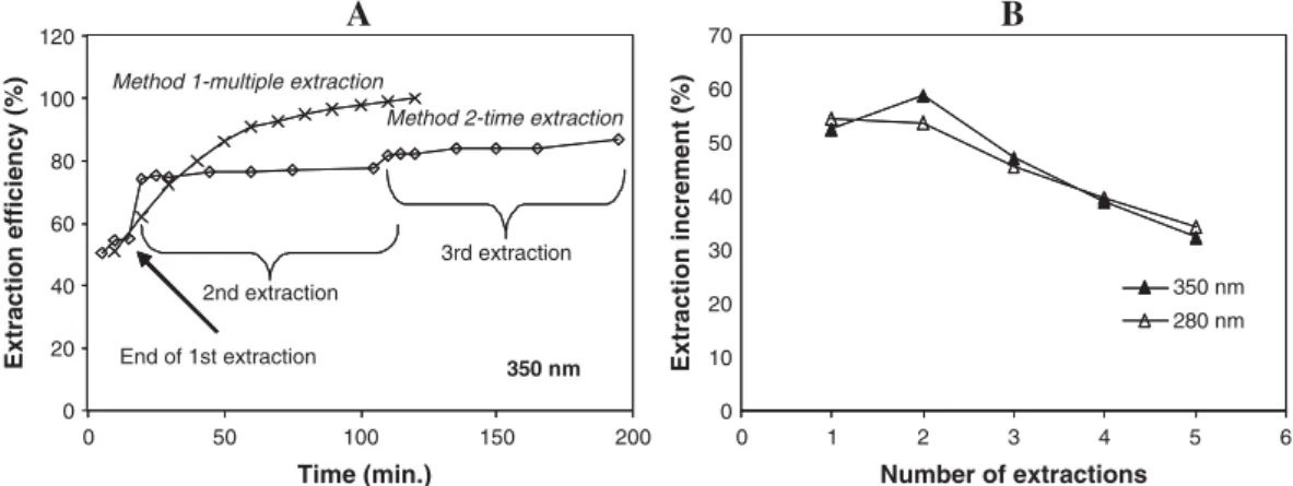
The UV spectra of most flavonoids exhibit two major absorption peaks. Band I usually occurs in the range 300– 350 nm and Band II in the range 240–285 nm [21]. Benzoic acids present absorption maxima at around 280 nm [22]. The extraction method was selected based on the absorbance at the wavelengths 280 and 350 nm, which was taken as an indicator of polyphenol extraction. The influence of the number of extraction steps and extraction time on the extraction efficiency is shown in fig. 1A. The effect of the time on the extraction efficiency was less important than the number of extractions. After performing 12 short extractions (120 min.), higher extraction efficiency was obtained than after three longer extractions (195 min., around 85% of the previous). Results obtained at 280 nm were similar to those at 350 nm (data not shown). After five extractions, a result of around 86% extraction efficiency was obtained. Further extraction steps resulted in minor increments. Temperature was shown to influence the extraction process as higher extraction efficiency was observed at 40° than at room temperature, being this effect more relevant on the first three extractions (fig. 1B). Similar relative increases were found at
Table 1.
DPPH scavenging activity (IC50) and total phenols of the tested
extracts.1
Castanea sativa leaf extract
DPPH IC50
(μg/ml)
Total phenolic content (mg GAE/g extract) Water extract 17.7 ± 0.2 269.2 ± 4.9 Ethanol:water (4:6) extract 14.6 ± 0.8 273.9 ± 4.0 Ethanol:water (7:3) extract 13.9 ± 0.8 245.3 ± 2.7 Ethanolic extract 23.0 ± 0.7 195.9 ± 0.9 Ethanol:water (7:3) optimized extract 12.6 ± 0.5 283.8 ± 8.7 Rutin 9.9 ± 0.5 − Ascorbic acid 4.9 ± 0.4 −
1Results are expressed as mean ± S.E.M. of three determinations.
Fig. 1. Influence of time and number of extraction steps (A) and temperature (B) on the extraction efficiency. The cumulative absorbance
464 ISABEL F. ALMEIDA ET AL.
280 and 350 nm. From these results, the extraction method adopted consisted in five short extractions, of 10 min. each, performed at 40°. When preparing the extract following this optimized method, higher DPPH scavenging activity and higher total phenolic content were obtained (table 1), thus confirming the validity of the undertaken optimization approach.
The C. sativa leaf ethanol:water (7:3) extract presented an
IC50 for the scavenging of DPPH of 12.58 ± 0.54 μg/ml
(mean ± S.E.M.), while rutin and ascorbic acid presented IC50s
of 9.92 ± 0.53 and 4.93 ± 0.36 μg/ml, respectively. Ferrozine quantitatively forms complexes with Fe2+. In the presence of
chelating agents, the complex formation is disrupted result-ing in a decrease of the complex red colour [23]. The IC50 found
for the iron chelation assay was 132.9 ± 9.72 μg/ml (mean ± S.E.M.) and EDTA presented an IC50 of 12.6 ± 0.29 μg/ml.
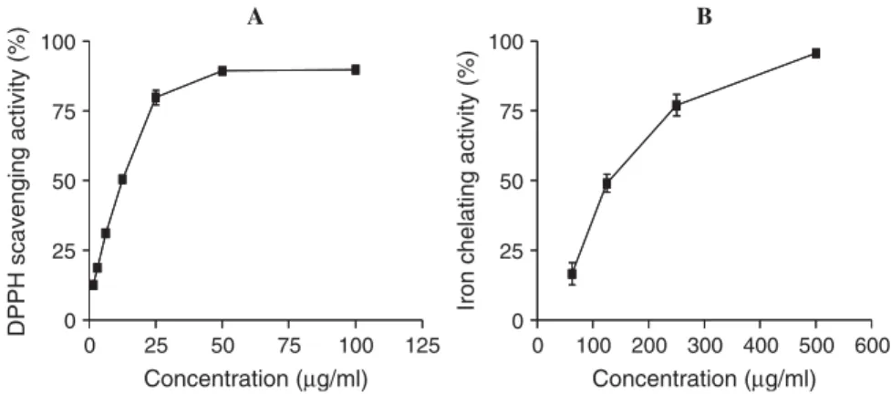
The optimized C. sativa leaf ethanol:water (7:3) extract prevented DPPH discoloration (fig. 2A) and chelated iron ions (fig. 2B) in a concentration-dependent manner.
It is interesting to compare data obtained on C. sativa
with other plant species obtained using the same assaying systems. This study has revealed that the DPPH antioxidant activity of C. sativa leaf ethanol:water (7:3) extract is relatively high compared to results in similar studies performed with Lamiaceae species (IC50s range from 300 to 600 μg/ml) [24],
with Brazilian plants (IC50s range from 11.6 to 158.9 μg/ml)
[25] and with Bolivian plants (IC50s range from 15.0 to
124.0 μg/ml) [26]. The DPPH scavenging activity of the extract studied in this work is comparable to green tea (IC50 = 11.8 μg/ml). The observed metal-chelating activity is
also relatively potent when compared to results obtained with similar assay performed with Cinnamomum verum leaf extract (IC50 in the range 400–600 μg/ml) [14] and with
Lamiaceae species (IC50s range from 0.8 to 1.40 mg/ml) [24].
The extract also presents strong absorption at 280 nm (UVB). This evidence suggests the possible effectiveness of chestnut leaf extract topical administration to prevent UV radiation-induced skin damage with a multiple mechanism.
The rational basis for the application of antioxidants and metal chelators in dermatology is well documented [6,23,27]. The inflammatory response following acute UV irradiation and the degenerative processes related to chronic UV radiation skin exposure are largely mediated by the overproduction of ROS, such as superoxide anion and hydrogen peroxide, and by impairment of the antioxidant endogenous system. ROS are, therefore, considered to play a major role in photoageing [2]. Irritants have been shown to induce the release of ROS event at non-cytotoxic concentrations [6]. It has also been mentioned that generation of ROS may be relevant in case allergens bind protein through a radical-mediated pathway like using limonene hydroperoxide [27]. Contact dermatitis also causes a potent increase of iron level in acute and chronic lesional area, as well in healthy skin [28]. ROS can release iron from intracellular iron storage protein, ferritin and trigger a rapid reduction of ferritin synthesis. On the other side, iron promotes the formation of ROS, contributing to lipid peroxidation [28]. The contact hypersensitivity response to metal in nickel-, cobalt- and copper-sensitive patients was shown to be reduced after topical application of chelating agents [29].
Botanical antioxidants have been used successfully in the treatment and prevention of photoageing, as reviewed by Afaq and Mukhtar [30], as well as of contact dermatitis. Phenolics and terpenoids are considered to be the most effective inhibitors against contact dermatitis [1]. As an example, the flavonoid wogonin was found to inhibit the skin inflammation of this disease [31]. These compounds may act by means of a non-specific mechanism (e.g. antioxidant), but may also act via specific mechanisms, such as the inhibition of the mediators implicated in the immune response [1]. Paradoxically, botanicals can be used to treat contact dermatitis, but may also be responsible for such inflammatory response. Many reports concerning irritant or allergic effects of plant extracts on the skin are available in the literature [32–34]. Together with sesquiterpene lactones, other compounds, such as alkaloids, flavonoids and terpenoids, can cause allergic reactions [1].
Fig. 2. DPPH scavenging (A) and iron chelating (B) activities. Each point represents the values obtained from four experiments, performed in duplicate (mean ± S.E.M.).
IN VIVO SKIN IRRITATION POTENTIAL OF A CHESTNUT LEAF EXTRACT 465
Several phenolic compounds were identified in C. sativa
leaf extract obtained under optimized conditions, namely, phenolic acids (chlorogenic acid and ellagic acid) and flavonoids (rutin, isoquercitrin and hyperoside) (fig. 3). Their amounts are expressed in table 2. The major component was found to be rutin. DPPH scavenging activity has been found for these phenolic compounds [13,35,36], and thus their putative contribution to the free radical scavenging activity of the whole extract might be inferred. No skin adverse effects were previously reported for these phenolic com-pounds. In fact, a preventive effect against photo-oxidative stress induced by UVA radiation has been described for rutin, the major phenolic compound found in this extract [37]. Cytotoxicity studies performed with McCoy mouse fibroblast cells also showed that at a concentration of 500 μg/ml, rutin failed to produce any overt signs of toxicity [38]. In accordance, a comparative study between the cyto-tocixity of nine flavonoids towards cultured human normal cells showed that rutin was among the least toxic compounds [39]. Despite the absence of reports of adverse effects of
C. sativa leaves or of the polyphenolic compounds found in
its composition, safety can not be assumed, and suitable tolerance tests should be carried out. Patch testing after a single application is a widely used procedure to evaluate acute
irritant reactions. Initially, evaluation of irritancy testing was based on visual scoring only. This type of evaluation, although subjective, can be a sensitive, reliable and repro-ducible method [40]. Several bioengineering techniques have been developed to provide objective and quantitative data. Although they present significant advantages over visual assessment, non-invasive evaluation methods of skin irrita-tion measure only one of its particular aspects. TEWL is considered to be the first choice to evaluate slight skin reactions, as it detects disrupted epidermal barrier that results in a higher loss of transepidermal water [41]. However, water barrier function and water evaporation from the skin is neither felt nor seen and, therefore, findings based on TEWL measurements are indirect and not automatically clinically relevant [15]. Erythema, sometimes associated with infiltration and sometimes with superficial erosion of the epithelium, is the main feature of acute inflammatory reaction. The evaluation of irritant reactions was carried out with a combined approach in order to cover the different aspects of skin irritation.
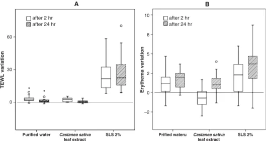
No significant differences were observed for TEWL measurements, in comparison with purified water 2 hr after patch removal (P = 0.4326), which is an indicator of an absence of barrier disruption. Interestingly, 24 hr after patch removal, the TEWL variation from basal values was lower than the variation observed for purified water (fig. 4A), the results being statistically significant (P = 0.0206). A positive influence in barrier integrity might be speculated. As the test was performed in healthy skin, healing effects can not be considered. Regarding erythema, differences were observed but the results for the extract were lower than those of puri-fied water (fig. 4B), which could be explained by a slight modification of the skin colour that interfered in the ery-thema measurements. This colour modification tended to dissipate, as it was only statistically significant 2 hr after patch removal (P = 0.001). Clinical scores were zero at both times for all volunteers, with the exception of one individual
Fig. 3. High performance liquid chromatography phenolic profile of Castanea sativa leaf ethanol:water (4:6) optimized extract. Detection at 350 nm. (1) Chlorogenic acid; (2) hyperoside; (3) rutin; (4) isoquercitrin; and (5) ellagic acid.
Table 2.
Phenolic composition of Castanea sativa leaf ethanol:water (7:3)
extract (mg/g lyophilized extract).1
Phenolic compound Optimized ethanol:water (7:3) extract
Chlorogenic acid 2.8 ± 0.1
Hyperoside 3.5 ± 1.3
Isoquercitrin2 3.0 ± 1.0
Rutin 5.9 ± 0.1
Ellagic acid 4.2 ± 0.1
1Results are expressed as mean ± S.E. of three determinations.
466 ISABEL F. ALMEIDA ET AL.
that experienced a very slight reaction 24 hr after the removal of the patch (fig. 5). None of the individuals reported subjective irritation symptoms, like stinging or itching. For the positive control (SLS 2%), significant differences were observed in TEWL (P < 0.001) and the irritation scores were different from zero for all volunteers (fig. 5). The results for erythema were not significantly different from purified water 2 hr after patch removal (P = 0.11623), although a clear increase of the median can be observed (fig. 4B). This is probably a result of the measuring method limitations, especially in case of spotty erythema. The patch test carried out, revealed that, with respect to irritant effects, the extract can be regarded as safe for application on the skin.
In conclusion, the optimized C. sativa leaf ethanol:water (7:3) extract presents interesting features that could be relevant
for topical application in the prevention and treatment of oxidative stress-mediated diseases and photoageing. Furthermore, the good skin tolerance found after a single application under occlusion reinforces its putative interest as topical antioxidant, after incorporation in suitable and safe topical bases.
Acknowledgement
The authors greatly acknowledge José Madureira for the collection of C. sativa leaves.
References
1 Rios JL, Bas E, Recio MC. Effects of natural products on contact dermatitis. Curr Med Chem Anti-inflammatory & Anti-allergy
Agents 2005;4:65– 80.
Fig. 4. Transepidermal water loss (TEWL) (A) and erythema index (B) variation from basal values. Outliers are presented as circles or asterisks.
IN VIVO SKIN IRRITATION POTENTIAL OF A CHESTNUT LEAF EXTRACT 467
2 Nishigori C, Hattori Y, Ariam Y, Miyavho Y. Photoaging and
oxidative stress. Exp Dermatol 2003;12:18 –21.
3 Matsumura Y, Ananthaswamy HN. Toxic effects of ultraviolet
radiation on the skin. Toxicol Appl Pharmacol 2004;195:298 –
308.
4 Myachi Y. Skin diseases associated with oxidative injury. In: Fuchs J, Parcker L (eds). Oxidative Stress in Dermatology. Marcel Dekker, New York, 1993;323 –31.
5 Willis CM, Reiche L, Wilkinson JD. Immunocytochemical demonstration of reduced Cu,Zn-superoxide dismutase levels following topical application of dithranol and sodium lauryl sulphate; an indication of the role of oxidative stress in acute
irritant contact dermatitis. Eur J Dermatol 1998;8:8 –12.
6 Mehrota T, Mishra KP, Raman G, Banerjee G. Differential regulation of free radicals (reactive oxygen and nitrogen species) by contact allergens and irritants in human keratinocyte cell
line. Toxicol Mech Methods 2005;15:343 –50.
7 Fuchs J, Mibradt R, Shurer N. Environmental stress and reactive oxidants in skin toxicology. In: Fuchs J, Parcker L (eds). Oxidative Stress in Dermatology. Marcel Dekker, New York, 1993;413–55.
8 Fleming T. Physicians Desk Reference (PDR) for Herbal Medi-cines, 1st edn. Medical Economics Company, Montvale, NJ, 1998;1078 –9.
9 Calliste CA, Trouillas P, Allais DP, Duroux JL. Castanea sativa
Mill. leaves as new sources of natural antioxidant; an electronic
spin resonance study. J Agric Food Chem 2005;53:282– 8.
10 Basile A, Sorbo S, Giordano S, Ricciardi L, Ferrara S, Montesano D et al. Antibacterial and allelopathic activity of extract from
Castanea sativa leaves. Fitoterapia 2000;71:S110– 6.
11 Wang C-K, Lee W-H, Peng C-H. Contents of phenolics and alkaloids in Areca Catechu Linn. during maturation. J Agric Food Chem 1997;45:1185 – 8.
12 Silva BM, Andrade PB, Ferreres F, Domingues A, Seabra RM, Ferreira MA. Phenolic profile of quince fruit (Cydonia oblonga Miller) (pulp and peel). J Agric Food Chem 2002;50:4615 – 8. 13 Fukomoto LR, Mazza G. Assessing antioxidant and prooxidant
activities of phenolic compounds. J Agric Food Chem 2000;48:3597– 604.
14 Mathew S, Abraham TE. In vitro antioxidant activity and scavenging effects of Cinnamomum verum leaf extract assayed by different methodologies. Food Chem Toxicol 2006;44:198–206. 15 Tupker RA, Willis C, Berardesca E, Lee CH, Fartasch M,
Agner T et al. Guidelines on sodium lauryl sulfate (SLS) exposure tests. A report from the Standardization Group of the European Society of Contact Dermatitis. Contact Derm 1997;37:53 – 69. 16 Jibry N, Murdan S. In vivo investigation, in mice and man, into
the irritation potential of novel amphiphilogels being studied as transdermal drug carriers. Eur J Pharm Biopharm 2004;58:107– 19.
17 Blois MS. Antioxidant determination by the use of a stable free radical. Nature 1958;181:1199–200.
18 Neergheen VS, Soobrattee MA, Bahorun T, Aruoma OI. Char-acterization of the phenolic constituents in Mauritian endemic plants as determinants of their antioxidant activities in vitro. J Plant Physiol 2006;163:787–99.
19 Kim D-O, Lee CY. Comprehensive study on vitamin C equivalent antioxidant capacity (VCEAC) of various polyphenolics in scavenging a free radical and its structural relationship. Crit Rev Food Sci Nutr 2004;44:253–73.
20 Georgé S, Brat P, Alter P, Amiot, MJ. Rapid determination of polyphenols and vitamin C in plant-derived products. J Agric Food Chem 2005;53:1370–3.
21 Harborne JB, Mabry TJ, Mabry H. The Flavonoids. Chapman and Hall, London, 1975.
22 Harborne JB. General procedures and measurement of total phenols. In: Harborne JB, Dey PM (eds). Methods in Plant
Biochemistry, vol. 1, Plant Phenolics. Academic Press, London, 1989;87–112.
23 Gulcin I, Buyukokuroglu ME, Kufrevioglu OI. Metal chelating and hydrogen peroxide scavenging effects of melatonin. J Pineal Res 2003;34:278–81.
24 Dorman HJ, Bachmayer O, Kosar M, Hiltunen R. Antioxidant properties of aqueous extracts from selected lamiaceae species grown in Turkey. J Agric Food Chem 2004;52:762–70. 25 Mensor LL, Menezes FS, Leitao GG, Reis AS, dos Santos TC,
Coube CS et al. Screening of Brazilian plant extracts for antioxidant activity by the use of DPPH free radical method. Phytother Res 2001;15:127–30.
26 Parejo I, Viladomat F, Bastida J, Rosas-Romero A, Saavedra G, Murcia MA et al. Investigation of Bolivian plant extracts for their radical scavenging activity and antioxidant activity. Life Sci 2003;73:1667–81.
27 Gafvert E, Nilsson JL, Hagelthorn G, Karlberg AT. Free radicals in antigen formation; reduction of contact allergic response to hydroperoxides by epidermal treatment with anti-oxidants. Br J Dermatol 2002;146:649–56.
28 Kaur S, Zilmer M, Eisen M, Kullisaar T, Rehema A, Vihalemm T. Patients with allergic and irritant contact dermatitis are char-acterized by striking change of iron and oxidized glutathione status in nonlesional area of the skin. J Invest Dermatol 2001;116: 886–90.
29 Wohrl S, Kriechbaumer N, Hemmer W, Focke M, Brannath W, Gotz M et al. A cream containing the chelator DTPA (diethylenetriaminepenta-acetic acid) can prevent contact allergic reactions to metals. Contact Derm 2001;44:224 –8.
30 Afaq F, Mukhtar H. Botanical antioxidants in the prevention of photocarcinogenesis and photoaging. Exp Dermatol 2006;15:678– 84.
31 Lim H, Park H, Kim HP. Inhibition of contact dermatitis in animal models and suppression of proinflammatory gene expression by topically applied flavonoid, wogonin. Arch Pharm Res 2004;27:442–8.
32 Thomson KF, Wilkinson SM. Allergic contact dermatitis to plant extracts in patients with cosmetic dermatitis. Br J Derm 2000;142:84 –8.
33 Ortiz KJ, Yaniin JA. Contact dermatitis to cosmetics, fragrances and botanicals. Dermatol Ther 2004;17:264–71.
34 Paulsen E. Contact sensitization from Compositae-containing herbal remedies and cosmetics. Contact Derm 2002;47:189– 98.
35 Zou Y, Lu Y, Wei D. Antioxidant activity of a flavonoid-rich extract of Hypericum perforatum L. in vitro. J Agric Food Chem 2004; 52:5032–9.
36 Lou X-D, Basile MJ, Kennely EJ. Polyphenolic antioxidants from the fruits of Chrysophyllum cainito L. (Star apple). J Agric Food Chem 2002;50:1379–82.
37 Filipe P, Silva JN, Haigle J, Freitas JP, Fernandes A, Santus R
et al. Contrasting action of flavonoids on phototoxic effects
induced in human skin fibroblasts by UVA alone or UVA plus cyamemazine, a phototoxic neuroleptic. Photochem Photobiol Sci 2005;4:420 –8.
38 Soares VC, Varanda EA, Raddi MS. In vitro basal and metabolism-mediated cytotoxicity of flavonoids. Food Chem Toxicol 2006;44:835 – 8.
39 Matsuo M, Sasaki N, Saga K, Kaneko T. Cytotoxicity of flavonoids toward cultured normal human cells. Biol Pharm Bull 2005;28:253 –9.
40 Basketter D, Reynolds F, Rowson M, Talbot C, Whittle E. Visual assessment of human skin irritation; a sensitive and reproducible tool. Contact Derm 1997;37:218–220.
41 Aramaki J, Effendy I, Happle R, Kawana S, Loffler C, Loffler H. Which bioengineering assay is appropriate for irritant patch testing with sodium lauryl sulfate? Contact Derm 2001;45:286–90.