Development of yeast-based assays to study p53 family proteins: identification of new small molecule modulators
166
0
0
Texto
(2)
(3) Mariana Valência Castanheira Ferreira Leão. DEVELOPMENT OF YEAST-BASED ASSAYS TO STUDY P53 FAMILY PROTEINS: IDENTIFICATION OF NEW SMALL MOLECULE MODULATORS. Tese do 3º ciclo de estudos conducente ao grau de doutoramento em Ciências Farmacêuticas, ramo Microbiologia. Orientadora. Professora Doutora Lucília Helena Ataíde Saraiva. Co-Orientadora. Doutora Clara Isabel Ferreira Pereira. October 2014.
(4) Thesis cover figures: 1- Structure of p53 family proteins [Adapted from (Wei et al., 2012)]; 2- Yeast p53 family assay; 3- MDM2 and MDMX: schematic representation of major domains [Adapted from (Lenos and Jochemsen, 2011)]; 4- Yeast p53 family-MDMs assay; 5- Pyranoxanthone 1 in the binding site of MDM2.. DE ACORDO COM A LEGISLAÇÃO EM VIGOR, NÃO É PERMITIDA A REPRODUÇÃO DE QUALQUER PARTE DESTA TESE.. ii.
(5) Acknowledgements “Science knows no country, because knowledge belongs to humanity, and is the torch which illuminates the world. Science is the highest personification of the nation because that nation will remain the first which carries the furthest the works of thought and intelligence”. Louis Pasteur First of all I want to thank to my PhD supervisor Prof Doutora Lucília Saraiva for giving me the opportunity to perform my PhD thesis in her research group. Thank you for all your support and for all the things that I have learned with you. Thanks for sharing your scientific knowledge and expertise in yeast field. Thanks also for the time invested in me. Thank you for your friendship and also thanks for helping me grow both as a scientist and as a person. I also thanks to my PhD co-supervisor Doutora Clara Pereira for all the support provided during my PhD. Thank you for all the technical help and for all the things that I have learned with you. Thanks for sharing your scientific knowledge and expertise in yeast field. Thank you for your friendship during these four years. I am deeply grateful to Prof Doutor Alberto Inga who gave me the possibility to work in his group in Trento. Thanks for all the things that I have learned with you and thanks for sharing your scientific knowledge and expertise in yeast and human cell lines fields. His contributions of time, ideas and support made my stage in Trento highly productive. I also thank to his collaborators Yari Ciribilli, Alessandra Bisio e Mattia Lion for sharing their scientific knowledge in different laboratorial techniques and in human cell lines. I would like to express my gratitude to the CEQUIMED group from the Faculty of Pharmacy of Porto. To Prof Doutora Madalena Pinto, Prof Doutora Emília Sousa and Ana Mafalda Paiva, I thank for all the support in the organic chemistry field. I also acknowledge to the Cancer Drug Resistance group from IPATIMUP, particularly to Prof Doutora Helena Vasconcelos who gave me the possibility to work in her research group. Thanks also to Doutora Raquel Lima and Diana Sousa for all the support and knowledge in human cell lines field. Many thanks to my dear lab colleagues Joana Soares, Cláudia Bessa, Sara Gomes and Cláudia Maciel for all the ideas, support, scientific discussions and friendship, during my PhD. To Sara, I would like to express my heartfelt gratitude for all the support in the two last years of my PhD. Thanks with all my heart. Thanks also to Doutora Isabel iii.
(6) Coutinho who gave me the knowledge in different laboratorial techniques in the beginning of my PhD and also thanks to Diana Costa for her support in a part of my PhD program. Many thanks to the Department of Pharmacology, particularly to Prof Doutora Glória Queiroz for all the support in the human cell lines field. Thanks also to the lab technicians Céu and Mónica. I would also like to acknowledge to the lab technicians, Nuno and Cristina, from the Department of Microbiology for all the support along the years. Special thanks to my parents, Miguel and Gabriela, and to my grandmother Emília, for all the support along my PhD years. Thank you for encourage me to do my PhD and also for all inspiration and enthusiasm. At last, a special thanks to my husband Francisco who gave me all the support along these four years. Thank you for all your patience, motivation and enthusiasm. Thanks for encourage me to do my PhD and for making me believe more in myself. I also thank to FCT (Fundação para a Ciência e Tecnologia) for the financial support of my doctoral fellowship (SFRH/BD/64184/2009) and to the Faculty of Farmacy for provided the logistical support. This work was supported by FCT through REQUIMTE (PEst-C/EQB/LA0006/2011), FEDER funds through the COMPETE program under the project FCOMP-01-0124-FEDER-015752 (PTDC/SAU-FAR/110848/2009) and by the Italian Association for Cancer Research, AIRC (IG#12869 to AI).. iv.
(7) Abstract The p53 family consists of three members, p53, p63 and p73 with a central role in the control of cell differentiation, proliferation and death, among others. This family of proteins is therefore considered a key therapeutic target in cancer. Although the intense research around these proteins, the biological and pharmacological profile of several p53 family members remains far from being completely understood, particularly due to the high complexity of the p53 pathway in mammalian cells. The function of these proteins is negatively regulated by interactors, such as murine double minute (MDM)2 and MDMX. However, contrary to p53 and p73, the regulation of p63 by MDM2/MDMX is still controversial. In spite of this, the inhibition of these interactions with MDM2/MDMX represents a promising therapeutic strategy to reactivate p53 family proteins in tumours. Despite the huge number of inhibitors of the p53-MDM2 interaction discovered in the last few years, only few inhibitors of the p53-MDMX interaction and dual inhibitors of the p53MDM2/MDMX interaction have been reported. Similarly, inhibitors of the p63 and 73 interaction with MDM2/MDMX remain mostly unknown. In the present thesis, the yeast Saccharomyces cerevisiae was used as a cell model for an independent analysis of human p53, p63 (full length and truncated forms) and p73 (full length) in a simplified eukaryotic cell system. Using this approach, several aspects of the biology of p53 family proteins were studied, particularly their regulation by MDM2/MDMX. Several similarities among the activities of the different p53 family proteins expressed in yeast were observed. In fact, all of them induced growth inhibition associated with S-phase cell cycle arrest and an autophagic cell death, reactive oxygen species generation, increase of actin depolarization and protein expression levels. Despite this, different sensitivities to the autophagosome pathway, to the inhibitory effect of MDM2 and MDMX, and to small molecule modulators of their interactions with MDM2/MDMX clearly emerged. The applicability of the yeast cell models developed for p53 family proteins, alone and combined with MDM2/MDMX, as a drug screening approach was also validated in this work with the discovery of promising modulators of the activity of these proteins. Particularly, with the developed yeast p53-MDM2 screening assay, the pyranoxanthone 1 (LEM1) was identified as the first small molecule inhibitor of the p53-MDM2 interaction with a xanthone scaffold. In fact, with the validation of the molecular mechanism of action of this compound in human tumour cells, a proof of concept was therefore provided for the effectiveness of this cell system for the screening of inhibitors of the p53 interaction with its negative regulators. Thereafter, using the same yeast assay, α-mangostin and gambogic acid, were identified as potential inhibitors of the p53-MDM2 interaction. This v.
(8) study contributed to the elucidation of the molecular mechanism of action of these two xanthones well-known by their potent antitumor properties. Moreover, with the developed yeast p73-MDM2 interaction assay, the xanthone LEM2 was identified as an activator of the p73 pathway through potential MDM2 inhibition. An additional outcome of this thesis was the identification of ACT1 as a putative p53 family proteins target gene in yeast. This study may lead to the development of a simplified yeast p53 family proteins transactivation assay for the analysis of the p53 family proteins transcriptional activity in alternative to the more complex artificial yeast transactivation reporter assays tradicionally used. In conclusion, the data emerged from this thesis confirm the enormous potential of the yeast cell model to the study of human disease-associated proteins. Several relevant insights about the biology and potential druggability of these human proteins are revealed for the first time in yeast. Additionally, new yeast target-directed approaches are provided for future studies of the p53 family network, particularly for the identification of new regulators of the activity of p53 family proteins. These assays have proved to be highly effective as a first-line approach, before studies in more complex cell systems. It is therefore anticipated that, for the near future, the use of these cell systems holds great promise in the discovery of novel anticancer agents with therapeutic applicability.. Keywords: Cancer; MDM2/MDMX; p53 family proteins; Target-directed screening assays; Yeast. vi.
(9) Resumo A família da p53 consiste em três membros, p53, p63 e p73, os quais desempenham um papel central no controlo da diferenciação, proliferação e morte celular, entre outros processos. Neste sentido, esta família de proteínas é considerada um alvo terapêutico chave no cancro. Apesar da investigação intensiva de que têm sido alvo, o perfil biológico e farmacológico dos diversos membros da família da p53 está longe. de. ser. completamente. compreendido,. principalmente. devido. à. elevada. complexidade das suas vias em células de mamífero. A função destas proteínas é modulada negativamente por reguladores endógenos, como as proteínas murine double minute (MDM)2 e MDMX. No entanto, contrariamente ao observado com p53 e p73, a regulação da p63 pelas proteínas MDM2/MDMX é ainda controversa. Apesar disto, a inibição das suas interações com MDM2/MDMX representa uma estratégia terapêutica promissora na reativação das proteínas da família da p53 em tumores. Apesar do número elevado de inibidores da interação p53-MDM2 descobertos nos últimos anos, poucos foram os inibidores da interação p53-MDMX e os inibidores duais das interações p53MDM2/MDMX descritos até à presente data. De forma semelhante, inibidores da interação da p63 ou p73 com as proteínas MDM2/MDMX permanecem quase desconhecidos. Na presente tese, a levedura Saccharomyces cerevisiae foi utilizada como modelo celular para uma análise das proteínas p53, p63 (forma completa e formas truncadas) e p73 (forma completa) expressas individualmente num sistema celular eucariótico simplificado. Usando esta abordagem, diversos aspetos da biologia das proteínas da família da p53 foram estudados, particularmente a sua regulação pelas proteínas MDM2/MDMX. Diversas similaridades entre as atividades das diferentes proteínas da família da p53 expressas em levedura foram observadas. De facto, todas elas induziram uma inibição do crescimento associada a uma retenção do ciclo celular na fase S e morte celular autofágica, geração de espécies reativas de oxigénio, aumento da despolarização e dos níveis de expressão da actina. Apesar disto, diferentes sensibilidades para a via do autofagossoma, para o efeito inibitório das proteínas MDM2 e MDMX e para os compostos moduladores das suas interações com as proteínas MDM2 e MDMX, foram observados no âmbito da presente tese. A aplicabilidade dos modelos de levedura desenvolvidos para as proteínas da família da p53, expressas isoladamente ou combinadas com MDM2/MDMX, na pesquisa de novos fármacos, foi também validada neste trabalho com a descoberta de moduladores promissores da actividade destas proteínas. Em particular, com o ensaio de pesquisa de levedura desenvolvido para p53-MDM2, a piranoxantona 1 (LEM1) foi vii.
(10) identificada como o primeiro inibidor da interação p53-MDM2 com um núcleo xantónico. De facto, com a validação do mecanismo molecular de ação deste composto em células tumorais humanas, uma prova de conceito foi portanto obtida para a eficácia deste sistema celular na pesquisa de inibidores da interação da p53 com os seus reguladores negativos. Num trabalho subsequente, usando o mesmo ensaio de levedura, a αmangostina e o ácido gambógico foram identificados como potenciais inibidores da interação p53-MDM2. Este estudo contribuiu para a elucidação do mecanismo molecular de ação destas duas xantonas conhecidas pelas suas potentes propriedades antitumorais. Adicionalmente, com o ensaio de levedura desenvolvido para a interação p73MDM2, a xantona LEM2 foi identificada como um ativador da via da p73 através de uma potencial inibição da proteína MDM2. Com esta tese foi ainda possível identificar o gene ACT1 como um potencial alvo das proteínas da família da p53 na levedura. Este estudo abriu caminho a um novo ensaio simplificado de transactivação das proteínas da família da p53 em levedura para a análise das suas atividades transcricionais. Este ensaio poderá ser utilizado em alternativa aos ensaios de transactivação tradicionais que têm por base um sistema de levedura artificial que utiliza um gene repórter. Em conclusão, os dados obtidos nesta tese confirmam o enorme potencial do modelo de levedura no estudo de proteínas associadas a doenças humanas. Informações relevantes. acerca. da. biologia. destas. proteínas,. assim. como. do. potencial. desenvolvimento de seus moduladores farmacológicos, são reveladas pela primeira vez em levedura. Adicionalmente, novos ensaios de levedura direcionados para um determinado alvo são implementados, os quais poderão ser utlizados em estudos futuros das proteínas da família da p53, particularmente na identificação de novos reguladores das suas atividades. Estes ensaios provaram ser altamente eficientes quando utilizados numa fase inicial da pesquisa, antes dos tradicionais ensaios em sistemas celulares mais complexos. O uso destes sistemas celulares revela-se, desta forma, muito promissor na descoberta de novos agentes anticancerígenos com aplicabilidade terapêutica num futuro próximo.. Palavras-chave: Cancro; Ensaios de pesquisa direcionados para um alvo; Levedura; MDM2/MDMX; Proteínas da família da p53. viii.
(11) Index. Acknowledgements .......................................................................................................... iii Abstract .............................................................................................................................. v Resumo ............................................................................................................................. vii Index................................................................................................................................... ix Index of figures ............................................................................................................... xiii Table Index ...................................................................................................................... xvi Abbreviations ................................................................................................................. xvii CHAPTER 1 - INTRODUCTION…………………………………………….………………… ..1 1.1. The p53 family proteins ................................................................................................ 3 1.1.1. Structural organization ............................................................................... 4 1.1.2. Biological functions .................................................................................... 5 1.1.2.1. Cell cycle………………………………………………………...7 1.1.2.2. Apoptosis………………………………………………………..9 1.1.2.3. Autophagy……...................................................................12 1.1.3. Therapeutic aplications ............................................................................ 14 1.1.3.1. MDM2 and MDMX: Endogenous negative regulators…….15 1.1.3.2. Regulation of p53 activity by MDM2/MDMX……………….19 1.1.3.3. Regulation of p63 and p73 activity by MDM2/MDMX……..26 1.2. Yeast as a cell model .................................................................................................. 27 1.2.1. Yeast as a cell model to study human proteins ....................................... 27 1.2.2. Yeast as a valuable screening tool in anticancer drug research ............. 28 1.2.3. Yeast as a cell model to study p53 family proteins .................................. 30 1.3. Scope of this thesis ..................................................................................................... 34 CHAPTER 2 - MATERIAL AND METHODS……...…………………………………………. 53 2.1. Compounds................................................................................................................. 38 2.2. Yeast Cells .................................................................................................................. 38 2.2.1. Plasmids .................................................................................................. 38 2.2.2. Yeast strains ............................................................................................ 38 2.2.3. Yeast transformation ............................................................................... 40 ix.
(12) 2.2.4. Growth conditions of transformed yeast cells .......................................... 40 2.2.5. Effect of compounds on yeast cell growth ............................................... 40 2.2.6. Yeast cell cycle analysis .......................................................................... 41 2.2.7. Yeast cell death assays ........................................................................... 41 2.2.8. Assessment of reactive oxigen species (ROS) production ...................... 42 2.2.9. Immunofluorescence assays ................................................................... 42 2.2.10. Analysis of actin depolarization ............................................................. 42 2.2.11. Dual-luciferase yeast p53 transactivation assay ................................... 43 2.2.12. Quantification of mRNA levels ............................................................... 43 2.2.13. Fluorescence microscopy ...................................................................... 44 2.3. Human tumour cell lines ............................................................................................. 45 2.3.1. Human tumour cell lines and growth conditions ...................................... 45 2.3.2. Effect of compounds on in vitro cell growth ............................................. 45 2.3.3. Dual-luciferase reporter assay ................................................................. 45 2.3.4. Analysis of cell cycle ................................................................................ 46 2.4. Yeast and human tumour cells ................................................................................... 46 2.4.1. Western blot analysis .............................................................................. 46 2.4.2. Analysis of phosphatidylserine exposure ................................................ 47 2.4.3. Flow cytometric data acquisition and analysis ......................................... 47 2.4.4. Statistical analysis ................................................................................... 47 2.5. Computational chemistry: docking of a library of xanthone derivatives in MDM2 ....... 48 CHAPTER 3 - RESULTS .................................................................................................. 51 3.1. Discovery of a new small molecule inhibitor of the p53-MDM2 interaction using a yeast-based approach ....................................................................................................... 53 3.1.1. Development of a yeast phenotypic assay for the screening of inhibitors of the p53-MDM2 interaction ............................................................................. 54 3.1.2. Virtual screening of a library of xanthone derivatives to search for potential MDM2 ligands ..................................................................................... 56 3.1.3. Identification of pyranoxanthone 1 as a promising inhibitor of the p53MDM2 interaction using the yeast approach ..................................................... 59 3.1.4. Pyranoxanthone 1 reactivates the p53 activity and downstream cell signalling in human tumour cells ....................................................................... 61 3.1.5. Analysis of the predicted binding model of pyranoxanthone 1 to MDM2 supports that pyranoxanthone 1 binds to MDM2 ............................................... 64 3.1.6. Discussion ............................................................................................... 65 x.
(13) 3.2. α‑Mangostin and gambogic acid as potential inhibitors of the p53−MDM2 interaction revealed by a yeast approach…………………………………………………………………..67 3.2.1. Identification of α-mangostin and gambogic acid as potential inhibitors of the p53-MDM2 interaction using the yeast approach ........................................ 68 3.2.2. α-Mangostin and gambogic acid reactivate the p53 activity in human tumour cells ....................................................................................................... 70 3.2.3. α-Mangostin and gambogic acid do not interfere with mutant p53 activity in yeast .............................................................................................................. 71 3.2.4. Analysis of the predicted binding model of α-mangostin and gambogic acid to MDM2 supports that α-mangostin and gambogic acid binds to MDM2 . 72 3.2.5. Discussion ............................................................................................... 73 3.3. Novel simplified yeast-based assays of regulators of the p53-MDMX interaction and p53 transcriptional activity.................................................................................................. 75 3.3.1. Development of a yeast-based assay to search for inhibitors of the p53MDMX interaction .............................................................................................. 76 3.3.2. Expression of p53 in yeast increases actin protein and mRNA levels and induces actin depolarization .............................................................................. 78 3.3.3. Discussion ............................................................................................... 83 3.4. Studying p53 family proteins in yeast: induction of autophagic cell death and modulation by interactors and small molecules ................................................................. 85 3.4.1. TAp63, ΔNp63 and TAp73 induce yeast growth inhibition and cell cycle arrest associated with an increase of actin expression levels and depolarization ........................................................................................................................... 86 3.4.2. Induction of an autophagic cell death by p53 family proteins in yeast ..... 89 3.4.3. MDM2 and MDMX inhibit the activity of TAp63, ΔNp63 and TAp73 in yeast .................................................................................................................. 92 3.4.4. Nutlin-3a and SJ-172550 inhibit the negative effect of MDM2 and MDMX, respectively, on TAp73 ...................................................................................... 94 3.4.5. Discussion ............................................................................................... 96 3.5. LEM2: a small molecule activator of the p73 pathway by potential MDM2 inhibition ......................................................................................................................................... 101 3.5.1. Identification of LEM2 as potential inhibitor of the p53-MDM2 interaction using the yeast approach ................................................................................ 102 3.5.2. LEM2 acts through a p53-independent mechanism in human tumour cell lines ................................................................................................................. 103 3.5.3. Identification of LEM2 as potential inhibitor of the p73-MDM2 interaction using the yeast approach ................................................................................ 104 3.5.4. LEM2 activates the p73 pathway in human tumour cells by potential MDM2 inhibition ............................................................................................... 105 xi.
(14) 3.5.5. Discussion ............................................................................................. 106 CHAPTER 4 - DISCUSSION ........................................................................................... 109 4.1. Development of yeast assays for biological and pharmacological studies of p53 family proteins ............................................................................................................................ 110 4.2. Identification of new small molecule modulators of p53 family proteins using the developed yeast target-directed screening assays .......................................................... 111 4.3. Final conclusions and future perspectives ................................................................ 113 CHAPTER 5 - REFERENCES ......................................................................................... 115 CHAPTER 6 - APPENDIX ............................................................................................... 139 . xii.
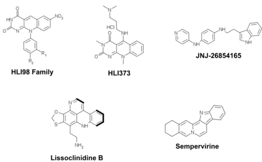
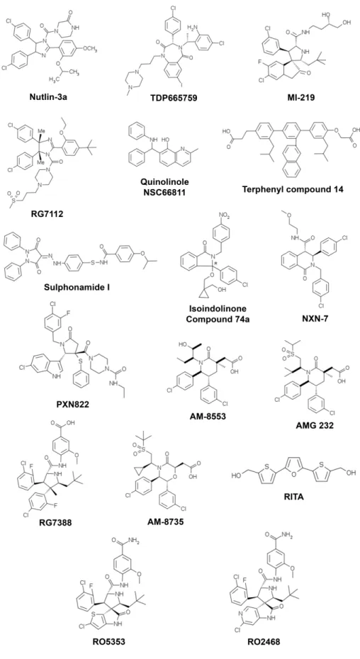
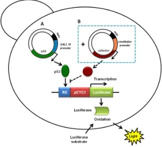
(15) Index of figures Figure 1. Structural organization of p53 family proteins ...................................................... 4 Figure 2. Regulation of the cell cycle by p53 family proteins .............................................. 8 Figure 3. Regulation of intrinsic and extrinsic apoptotic pathways by p53 ........................ 11 Figure 4. p53 distinctly regulates autophagy depending on its subcellular localization .... 14 Figure 5. Schematic representation of the major domains found in MDM2 and MDMX proteins .............................................................................................................................. 15 Figure 6. Regulation of p53 activity and stability by MDM2 and MDMX. .......................... 17 Figure 7. Chemical structures of modulators of MDMX expression .................................. 20 Figure 8. Chemical structures of inhibitors of E3 MDM2 ubiquitin ligase activity .............. 21 Figure 9. Chemical structures of inhibitors of the p53-MDM2 interaction. ........................ 23 Figure 10. Chemical structures of the inhibitors of the p53-MDMX interaction ................. 25 Figure 11. Chemical structures of the dual inhibitors of the p53-MDM2/MDMX interaction ........................................................................................................................................... 26 Figure 12. Yeast-based p53 dual-luciferase transactivation assay using cells ................. 31 Figure 13. Chemical structure of 3,4-dihydro-12-hydroxy-2,2-dimethyl-2H,6H-pyrano[3,2b]xanthen-6-one (pyranoxanthone 1) ................................................................................ 54 Figure 14. Nutlin-3a reduces the negative effect of MDM2 on p53 activity in yeast ......... 55 Figure 15. Pyranoxanthone 1 reduces the negative effect of MDM2 on p53 activity in yeast .................................................................................................................................. 60 Figure 16. Pyranoxanthone 1 increases the p53-dependent transcriptional activity in human tumour cells............................................................................................................ 62 Figure 17. Pyranoxanthone 1 leads to p53 stabilization, increases p21 and Bax levels, and enhances procaspase-7 cleavage to active caspase-7 in HCT116 p53+/+ cells.. ....... 63 Figure 18. Predicted binding model using computational docking for the hit pyranoxanthone 1 .............................................................................................................. 65 Figure 19. Chemical structure of α-mangostin (15) and gambogic acid (16) .................... 68 Figure 20. α-Mangostin (15) and gambogic acid (16) revert the inhibitory effect of MDM2 on p53-induced growth inhibition and S-phase cell cycle arrest, as well as on p53dependent transcriptional activity in yeast. ........................................................................ 69 xiii.
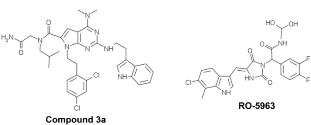
(16) Figure 21. α-Mangostin (15) and gambogic acid (16) increase the p53-dependent transcriptional activity in MCF-7 tumour cell line. .............................................................. 71 Figure 22. α-Mangostin (15) and gambogic acid (16) do not interfere with the activity of mutant p53 ......................................................................................................................... 72 Figure 23. Predicted binding model using computational docking for α-mangostin (15) and gambogic acid (16) ............................................................................................................ 73 Figure 24. MDMX inhibits the impact of p53 on the growth of yeast, an effect abolished by SJ-172550 ......................................................................................................................... 77 Figure 25. Contrary to R273H, wt p53 and V122A increase the actin protein levels ........ 78 Figure 26. Reduction of p53-induced actin protein levels by chemical (PFT-α) and natural (MDM2 and MDMX) inhibitors of p53 activity .................................................................... 80 Figure 27. Expression of p53 in yeast increases actin mRNA levels in total RNA and polysome-associated RNAs, stimulate transcription in ACT1-promoter reporter assays, and induces actin depolarization ....................................................................................... 82 Figure 28. Nuclear localization of p53 family members expressed in yeast ..................... 86 Figure 29. Expression of p53 family members in yeast induces growth inhibition associated with cell cycle arrest, an increase of actin expression levels and depolarization ........................................................................................................................................... 88 Figure 30. p53 family members induce ROS production and an autophagic cell death in yeast .................................................................................................................................. 91 Figure 31. MDM2 and MDMX reduce the TAp63-, ΔNp63- and TAp73-induced growth inhibition, cell cycle arrest, and increase of actin expression in yeast ............................... 93 Figure 32. Nutlin-3a and SJ-172550 revert the inhibitory effect of MDM2 and MDMX, respectively, on TAp73, but not on TAp63 and ΔNp63 ...................................................... 95 Figure 33. Chemical structure of 1-carbaldehyde-3,4-dimethoxy-9H-xanthen-9-one (LEM2) ............................................................................................................................. 102 Figure 34. LEM2 reverts the inhibitory effect of MDM2 on p53-induced growth inhibition ......................................................................................................................................... 102 Figure 35. LEM2 induces cell cycle arrest and apoptosis in HCT116 p53-/- cells ........... 104 Figure 36. LEM2 reduces the negative effect of MDM2 on p73 activity in yeast. ........... 105 Figure 37. LEM2 increased the expression levels of p73 and of p73 target genes (MDM2, p21 and Bax) in HCT116 p53-/- tumour cells .................................................................... 106 xiv.
(17) Figure 38. Novel yeast target-directed screening assays developed to search for modulators of p53 family proteins: Yeast growth-inhibitory (analysis of cell growth) and transactivation (analysis of actin protein expression levels) assays. ............................... 111 Figure 39. New small molecule modulators of the activity of p53 family proteins emerged from the developed yeast target-directed screening assays ........................................... 113 Figure A1. Grid dimensions and position in the MDM2 in PyRxAutodockVina...............140. xv.
(18) Table Index Table 1. Advantages and drawbacks of yeast as a screening tool over cell-free and human cell-based assays. ................................................................................................. 28 Table 2. Yeast expression vectors used in the yeast assays. ........................................... 39 Table 3. S. cerevisiae strains used in yeast assays. ......................................................... 39 Table 4. Antibodies used in Western blot. ......................................................................... 48 Table 5. Effects of compounds on the inhibitory activity of MDM2 on p53-induced yeast growth inhibition. ................................................................................................................ 58 Table 6. GI50 values obtained for LEM2 in human colon adenocarcinoma HCT116 tumour cell lines. .......................................................................................................................... 103 Table A1. Results of docking simulations for MDM2 (with grid box) perfomed in PyRx/AutoDockVina ........................................................................................................141 Table A2. Results of docking simulations for MDM2 (total protein) done in PyRx/AutoDockVina ........................................................................................................142 Table A3. Results of docking stimulations for MDM2 (PDB code: 1YCR) performed with PyRx/AutoDockVina ........................................................................................................144. xvi.
(19) Abbreviations ABP140. Actin binding protein 140. AIF. Apoptosis inducing factor. AMPK. Adenosine monophosphate activated protein kinase. Apaf-1. Apoptosis protease-activating factor 1. Bax. Bcl-2 associated X protein. Bak. Bcl-2-antagonist/killer. Bid. BH3 interacting-domain death agonist. CAK. CDK-activating kinase. CBP. CREB-binding protein. CDK. Cyclin-dependent kinase. CFU. Colony-forming unit. Cyt c. Cytochrome c. DAPK-1. Death associated protein kinase 1. DBD. DNA-binding domain. DIABLO. Direct IAP-binding protein with low pI. DISC. Deat-inducing signalling complex. DMSO. Dimethyl sulfoxide. EndoG. Endonuclease G. FADD. Fas-associated protein with death domain. FASAY. Functional analysis of separated alleles. GADD45. Growth arrest and DNA-damage inducible protein. GFP. Green fluorescent protein. HTS. High-throughput screening. H 2O 2. Hydrogen peroxide. IAP. Inhibitor of apoptosis. MDM2. Murine double minute 2. MDMX/4. Murine double minute X/4. MOMP. Mitochondrial outer membrane permeabilization. mtCLIC. Mitochondrial chloride intracellular channel. mTOR. Molecular target of rapamycin. mt. Mutant. OD. Oligomerization domain. OD600. Optical density at 600 nm. p53AIP1. p53-regulated apoptosis inducing protein 1. p53RE. p53 responsive element xvii.
(20) PBS. Phosphate-buffered saline. PCR. Polymerase chain reaction. PFT-α/µ. Pifthrin-α/µ. Pgk1p. Phosphoglycerate kinase. PI. Propidium iodide. pI. Isoelectric point. PUMA. p53 upregulated modulator of apoptosis. Q-PCR. Quantitative PCR. Rb. Retinoblastoma protein. RE. Response element. RLU. Relative light units. ROS. Reactive oxygen species. SAM. Sterile-alpha motif. SESN2. Sestrin 2. SCF. Skp, Cullin, F-box containing complex. SMAC. Second mitochondria-derived activator of caspases. SRB. Sulforhodamine B. TAD. Transactivation domain. TID. Transactivational inhibitory domain. TNF. Tumour necrosis factor. TRAIL. Tumour necrosis factor-related apoptosis-inducing ligand. TSC2. Tuberous sclerosis protein 2. TUNEL. Terminal deoxynucleotidyl transferase dUTP nick end labeling. wt. Wild-type. Y2H. Yeas two-hybrid. xviii.
(21) Chapter 1. Introduction. Leão M, Pereira C, Soares J, Bessa C, Saraiva L. Curr Pharm Des 2012 Sep; 18 (27): 4223-35.. Leão M, Soares J, Bessa C, Gomes S, Maciel C, Raimundo L, Harilal N, Saraiva L. Portuguese Society for Microbiology Magazine 2013. Number 3..
(22) Cancer is among the leading causes of death in economically developed countries. Despite the improvement in the relative survival rates for many types of cancer, the cancer incidence is increasing as a result of population aging and cancer-associated lifestyle choices. According to the World Health Organization, in 2012, there were 14.1 million new cases of cancer, 8.2 million cancer deaths and 32.6 million people living with cancer (within 5 years of diagnosis). In the same year, in Europe, 3.7 million new cases of cancer were diagnosed and 1.9 million people died with the disease (Bray et al., 2012; Ferley et al., 2012). These statistics data may justify why cancer has been the focus of an intense worldwide scientific research. In fact, during the last years, an enormous effort has been dedicated to the improvement of diagnostic techniques, accuracy of prognosis and cancer treatment [reviewed in (Hanahan and Weinberg, 2011)]. Carcinogenesis is a multistep process that involves dynamic changes in the genome. Usually, this process is associated with an abnormal cell growth and a defective cell death due to the oncogene activation and/or tumour suppressor gene inactivation. These genetic alterations drive the progressive transformation of normal cells into highly malignant derivatives [reviewed in (Hanahan and Weinberg, 2000)]. Durign this transformation process, normal cells acquire several capabilities, called hallmarks of cancer. In 2000, Hanahan and Weinberg proposed six hallmarks of cancer: selfsufficiency in growth signals, insensitivity to anti-growth signals, evasion of apoptosis, limitless replicative potential, sustained angiogenesis, tissue invasion and metastasis [reviewed in (Hanahan and Weinberg, 2000)] . Recently, two emerging hallmarks were added to this list: the capability to reprogram the energy metabolism, in order to control the neoplasic transformation, and the capability of cancer cells to evade immunological destruction [reviewed in (Hanahan and Weinberg, 2011)]. Due to its central role in cell differentiation, proliferation and death, the p53 tumour suppressor protein has been a major therapeutic target in cancer [reviewed in (Lane et al., 2010)]. In fact, during the last years, a p53-targeted approach has yielded very promising therapeutic tools, particularly small molecules that target the p53-MDM2 interaction (e.g., Nutlin-3a) [reviewed in (Wade et al., 2013)]. However, the complexity of the p53 pathway has grown significantly, and we should no longer think of p53 as a ‘lone warrior’ in tumour suppression, mostly because p63 and p73 proteins are also implicated in cancer progression and metastasis [reviewed in (Candi et al., 2014)]. For these reasons, the development of innovative therapeutic approaches targeting not only p53, but also other p53 family members reveals to be a promising strategy to minimize the emergence of resistance and to achieve maximal therapeutic responses in cancer therapy.. 2.
(23) 1.1. THE P53 FAMILY PROTEINS The p53 protein was discovered in 1979 as a protein that binds to the SV40 tumour-virus oncoprotein, the large T-antigen (Linzer and Levine, 1979; Lane and Crawford, 1979). At this time, p53 was considered an oncogene. However, this idea changed because the p53 initially discovered was in fact a mutant form of p53. Additionally, subsequent studies demonstrated that different types of tumours highly express a wild-type (wt) p53 form which, contrary to mutant p53, acts as a tumour suppressor protein [reviewed in (Lane and Levine, 2010)]. It was also found that p53 is a sequence-specific DNA-binding transcription factor, which controls the transcription of an assortment of genes involved in different cellular processes in order to prevent tumour formation [reviewed in (Lane and Levine, 2010)]. Due to its relevance as a tumour suppressor protein, p53 is also called “The Guardian of the Genome” (Lane, 1992). Latter, two TP53-related genes were identified: TP73 that encondes p73 (Kaghad et al., 1997) and TP63 that encodes p63 (Yang et al., 1998). In spite of their structural homologies, the overlap in cellular functions between p53, p63 and p73 is limited. In fact, studies in knockout mice revealed that each protein of the p53 family has its unique functions [reviewed in (Murray-Zmijewski et al., 2006)]. p53-null mice are viable but die at an early age due to spontaneous cancers (Donehower et al., 1992). p73-null mice display neurological, pheromonal and inflammatory defects resulting in death within two months (Yang et al., 2000), while p63-null mice die at birth and exhibit growth abnormalities (Mills et al., 1999). The p53 family members also appear to have different functions in human biology. p53 is a well-established tumour suppressor and is one of the most frequently mutated proteins in sporadic cancers. TP53 germline mutations are associated with the development of the cancer-prone Li-Fraumeni and Li-Fraumeni-like syndromes [reviewed in (Malkin, 2011)]. Conversely, p63 is critical for the correct development of ectodermalderived tissues, and p63 germline mutations are associated with a subset of ectodermal dysplasia syndromes [reviewed in (Rinne et al., 2007)]. p73 contributes to neural and immune systems functions [reviewed in (Bourdon, 2007)] and, to date, no syndromes associated with p73 germline mutations have been identified. Contrary to TP53, mutations in TP63 and TP73 genes are rare. The role of these proteins in tumourigenesis is higly complex mainly due to the existence of several isoforms in a same cellular context. However, evidence emerged that both p63 and p73 exert a tumour suppression function in many human tumours [reviewed in (Candi et al., 2014)].. 3.
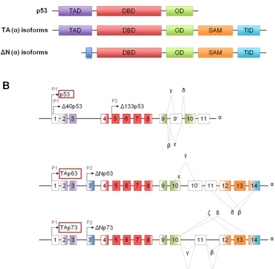
(24) 1.1.1. Structural organization The TP53, TP63 and TP73 genes are located in different chromosomes, 17p13.1, 3q27-29 and 1p36.2-3, respectively. Each gene encodes proteins with a high degree of structural homology, sharing three domains that are essential for their functions: a transactivation domain (TAD), a DNA binding domain (DBD) and an oligomerization domain (OD). The highest homology is found in the DBD (Fig. 1) [reviewed in (Wei et al., 2012)].. Figure 1. Structural organization of p53 family proteins. (A) Structure of p53 family proteins. p53, p63 and p73 consist of an amino-terminal transactivation domain (TAD), a central DNA binding domain (DBD) and a carboxy-terminal oligomerization domain (OD). α- Isoforms of p63 and p73 encode an additional Sterile Alpha Motif (SAM) domain followed by a transactivational inhibitory domain (TID). (B) Architecture of human TP53, TP63 and TP73 genes. Transcription of p53 family genes is controlled by two promoters, P1 and P2. TP53 presents and additional promoter, P1’. N- or C- terminal alternative mRNA splicing leads to the generation of different. 4.
(25) isoforms of each protein. Numbered boxes indicate exons. C-terminal splicing events for all p53 family members are indicated by dotted lines and Greek letter designation [Adapted from (Wei et al., 2012)].. Using different promoters (P1 and P2) and alternative mRNA splicing, TP53, TP63 and TP73 genes give rise to multiple protein isoforms with different functions (Fig. 1). In fact, while proteins resulting from the P1 promoter contain a N-terminal acidic TAD and are called TA proteins (TAp63 and TAp73), proteins resulting from the P2 promoter lack the entire N-terminal TAD and are called ΔN proteins (ΔNp63 and ΔNp73). Concerning the TP53 gene, it includes three promoters: the P1’ promoter that generates proteins containing the full TAD, the P1 promoter that gives rise to proteins lacking the first 40 amino acide residues (Δ40p53; containing a N-terminal acidic TAD), and the P2 promoter that produces proteins lacking the first 133 amino acids (Δ133p53; lacking all or part of the TAD) (Fig. 1) [reviewed in (Wei et al., 2012)]. Additional complexity is caused by alternative splicing at the C-terminal of p63 and p73 transcripts, generating a variety of TA and ΔN isoforms: eight splice variants for p53 (α, β, γ, δ, ε, ζ, Δp53 and ΔE6), five for p63 (α, β, γ, δ and ε) and nine for p73 (α, β, γ, δ, ε, θ, ζ, η and η1) (Fig. 1). Among the different generated isoforms, α, β and γ have been considered the most relevant [reviewed in (Wei et al., 2012)]. Moreover, contrarey to p53, α isoforms of p63 and p73 have an additional Sterile Alpha Motif (SAM) domain in the C-terminal that is responsible for protein-protein interactions and transcriptional repression (Fig. 1) [reviewed in (Wei et al., 2012; Candi et al., 2014)]. Additionally, p63α and p73α have an additional domain designated as transactivational inhibitory domain (TID) (Fig. 1). The presence of this domain in p63α and p73α proteins reduces their transactivation activitythrough intra- or intermolecular associations with the TAD [reviewed in (Candi et al., 2014)]. Besides, the transactivation potential of TAp63 and TAp73 depend on the isoform. In fact, TAp63γ and TAp73β have been shown to be as potent as p53 regarding the induction of gene transcription and apoptosis [reviewed in (Wei et al., 2012)].. 1.1.2. Biological functions The p53 protein is a potent transcriptional regulator that binds to specific DNA sequences, the p53-responsive elements (p53REs), regulating an assortment of genes involved in major cellular pathways [reviewed in Riley et al., 2008)]. In unstressed cells, 5.
(26) p53 is maintained at low levels due to the presence of its main negative regulator murine double minute 2 (MDM2) that target it for proteassomal degradation. In response to different stress signals such as, DNA damage, hyperproliferative signals, hypoxia, oxidative stress, ribonucleotide depletion and nutrient starvation, p53 is displaced from MDM2 being stabilized and activated (Aylon and Oren, 2011; Bieging et al., 2014). Once activated, p53 accumulates in the nucleus and induces cell cycle arrest, DNA repair, senescence, apoptosis, and autophagy, among others [reviewed in (Ozaki and Nakagawara, 2011; Sui et al., 2011; Stegh 2012)]. Additionally, it is well established that p53 can exert its tumour suppressive function by transcription-independent mechanisms [reviewed in (Stegh 2012)]. Several functions initially attributed to p53 only are now shared by p63 and p73. Due to their structural homology with p53, particularly in the DBD, TAp63 and TAp73 are able to transactivate p53REs. Indeed, similarly to p53, in response to DNA damage, p63 and p73 induce cell cycle arrest, senescence and apoptosis [reviewed in (Khoury and Bourdon, 2010; Dötsch et al., 2010)]. Despite the important role of TAp63 and TAp73 in tumourigenesis, in unstressed cells, these proteins have unique biological functions, such as the regulation of differentiation and development [reviewed in (Dötsch et al., 2010)]. In contrast, it is known that the roles of ΔNp63 and ΔNp73 isoforms are multifaceted. In fact, ΔN isoforms can function as dominant-negative inhibitors of p53, TAp63 and TAp73 counterparts through promoter competition or hetero-complex formation. In promoter competition, ΔN isoforms compete with TA isoforms for their target gene promoters, preventing their transactivation function. In the hetero-complex formation mechanism, ΔN isoforms can inhibit TA isoforms by forming transcriptionally inactive hetero-oligomers with TA variants [reviewed in (Vilgelm et al., 2008; Wei et al., 2012)]. Additionally, a negative feedback loop is established between TA and ΔN isoforms. For instance, TA isoforms induce the transcription of ΔN isoforms by direct activation of the P2 promoter. In turn, ΔN isoforms inhibit TA isoforms [reviewed in (Dötsch et al., 2010; Wei et al., 2012)]. In fact, it was demonstrated that the expression levels of ΔNp73 are regulated by both p53 and p73 by direct activation of the P2 promoter of p73, creating an auto-regulatory feedback loop (Grob et al., 2001; Kartasheva et al., 2002; Nakagawa et al., 2002). In addition, it was also reported that p53 induces ΔNp63 expression (Harmes et al., 2003). The effect of these interactions in a cellular context appears to be dependent on a balance between TA and ΔN isoforms. In fact, deregulation of the TA/ΔN expression ratio may lead to tumour development [reviewed in (Wei et al., 2012)]. Besides their dominant negative effect on TA isoforms, it was demonstrated that ΔN isoforms are also transcriptionally active using their own transcriptional programs due to the existence of additional TA domains 6.
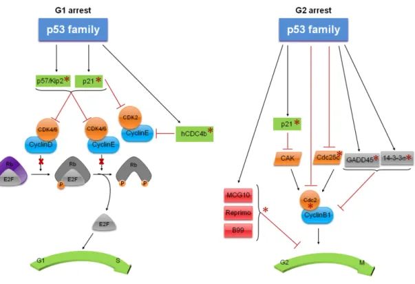
(27) [reviewed in (Wei et al., 2012)]. In fact, it was reported that ΔN isoforms can bind to p53REs activating specific target genes involved in cell cycle arrest and apoptosis (Dohn et al., 2001; Wu et al., 2003; Liu et al., 2004). Despite the involvement of p53 family proteins in different cellular processes, in this work a particular attention will be given to their roles on cell cycle, apoptosis and autophagy.. 1.1.2.1. Cell Cycle Cell cycle is a cellular process that is controlled by several mechanisms resulting in the transmission of the genetic information from one generation to the next. Cell cycle checkpoints (G1/S, S and G2/M) regulate the progression of cells through each stage of the cell cycle, in order to prevent the transmission of damaged genetic material to the daughter cells. These cell cycle checkpoints are regulated by a family of serine/threonine protein kinases, the cyclin-dependent kinases (CDK) (Fig. 2) [reviewed in (Vermeulen et al., 2003; Harms et al., 2004)]. The regulation of G1 phase is carried out by G1 cyclins D and E, and by CDKs 2, 4 and 6. In early G1 phase, the cyclin D forms a complex with CDKs 4 or 6, which can then target the retinoblastoma protein (Rb) for phosphorylation. Once phosphorylated, pRb binds and inhibits the E2F transcription factor. In late G1 phase, the cyclin E forms a complex with CDKs 4 or 6, which hyperphosphorylates pRB, releasing the E2F, which leads to the transcription of genes required for S-phase entry (Fig. 2). Under stress conditions, G1 arrest is induced through the inhibition of cyclin-CDK complexes by cyclindependent kinase inhibitors (CKIs), such as p21 and p57/Kip2. With the inhibition of these complexes, the function of E2F is compromised [reviewed in (Harms et al., 2004)]. In response to different stress signals, the p53 family proteins induce cell cycle arrest by inhibiting G1/S or G2/M transitions [reviewed in (Harms et al., 2004)]. Once activated, p53 family proteins induce the transcription of p21, a CDK inhibitor that binds to cyclins D/CDKs4/6 and E/CDKs4/6, preventing the G1/S transition. Additionally, p53 mediates the inhibition of cyclin E/CDK2 complexes by p21. In fact, to date, p21 is the only common target of the p53 family that leads to G1 arrest (el-Deiry et al., 1993; Lee and La Thangue, 1999; Dohn et al., 2001). The activity of cyclin E is also compromised through the induction of an F box protein and component of the SCF (Skp, Cullin, F-box containing complex) ubiquitin ligase complex, hCDC4b, which is a direct transcriptional target of p53 (Kimura et al., 2003). Moreover, it was described that the CDK inhibitor 7.
(28) p57/Kip2 is up-regulated by p63 and p73 leading to the induction of G1 arrest [reviewed in (Allocati et al., 2012)] (Fig. 2).. Figure 2. Cell cycle regulation by p53 family proteins. p53 family proteins induce cell cycle arrest by inhibiting G1/S or G2/M transitions. The asterisks refer to p53 family transcriptional activity [Adapted from (Harms et al., 2004)].. p53 family proteins regulate the G2/M transition by inhibiting the Cdc2, which needs to bind to cyclin B1 to be functional. Therefore, the repression of the cyclin B1/Cdc2 complex by p53 family proteins lead to a G2/M arrest (Taylor and Stark, 2001) [reviewed in (Allocati et al., 2012)]. Additionally, p53 family proteins regulate the G2/M checkpoint through modulation of the expression of multiple targets such as, Cdc25C, 14-3-3σ, p21, and GADD45 [reviewed in (Harms et al., 2004; Allocati et al., 2012)]. Both p53 and p73 induce G2/M arrest by repressing the mitosis promoting phosphatase, Cdc25C. This phosphatase removes the phosphate groups of Cdc2 leading to the cyclin B1/Cdc2 complex activation (Taylor and Stark, 2001) [reviewed in (Allocati et al., 2012)]. Besides Cdc25C, the CDK-activating kinase (CAK) adds phosphate groups to Cdc2 on specific points that are required for the activation of the cyclin B1/Cdc2 complex [reviewed in (Harms et al., 2004)]. p21 also participates in G2 arrest through the inhibition of the cyclin 8.
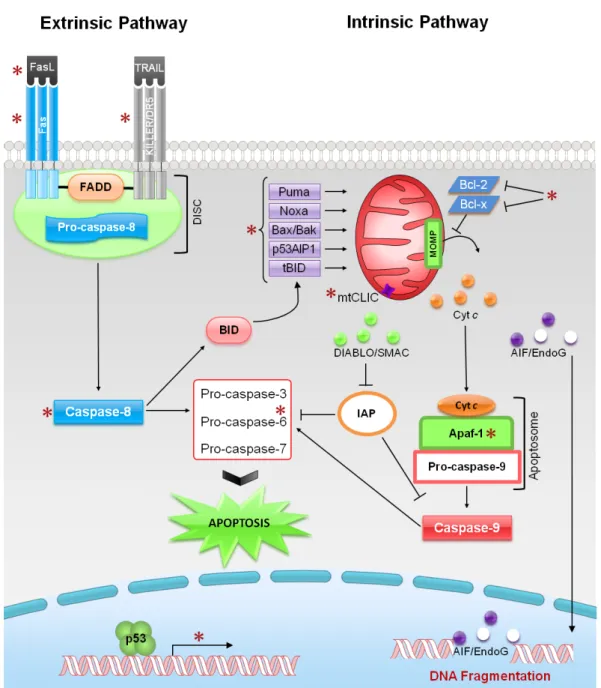
(29) B1/Cdc2 complex by CAK (Smits et al., 2000). The growth arrest and DNA damage inducible protein (GADD45) is a p53 target gene that binds to Cdc2, preventing its binding to cyclin B1, and, consequently, the formation of the protein complex necessary for mitotic entry, which results in G2 arrest (Zhan et al., 1999). The scaffold protein 14-3-3σ is involved in G2 arrest and is induced by both p53 and p73 and repressed by ΔNp63. In fact, this protein promotes cyclinB1/Cdc2 nuclear export. Through this mechanism, 14-33σ inhibits the cyclinB1/cdc2 complex by physically separating it from its target proteins (Hermeking et al., 1997). MCG10 (a RNA-binding protein), Reprimo (a highly glycosylated protein that is thought to play a role in cyclinB1/cdc2 localization) and B99 (a protein with microtubule localization) are proteins involved in G2 arrest which expression is induced by p53 (Zhu and Chen, 2000; Ohki et al., 2000; Utrera et al., 1998) (Fig. 2).. 1.1.2.2. Apoptosis Apoptosis is a type of cell death characterized by cell shrinkage, blebbing of plasma membrane, maintenance of organelle integrity, condensation and fragmentation of DNA. There are two main apoptotic pathways: the extrinsic (or death receptor pathway) and the intrinsic (or mitochondrial pathway) [reviewed in (Indran et al., 2011; Galluzzi et al., 2012)] (Fig. 3). The extrinsic pathway consists on the activation of cell death receptors that are located at the plasma membrane (Fig. 3). This apoptotic pathway can be activated by the binding of lethal ligands, such as FAS ligand (FASL), tumour necrosis factor α (TNFα) and TNF-related apoptosis inducing ligand (TRAIL), to different death receptors, such as FAS/CD95, TNFα receptor 1 (TNFR1) and TRAIL receptor (TRAILR)12, respectively. These receptors have an extracellular and an intracellular cytoplasmic (also known as death domain) domain. The death domain is responsible for the death signal transmission leading to the activation of the cell death receptors. The activation of these receptors leads to their trimerization and consequent clustering of the intracellular death domain, which subsequently recruits the protein adaptor FADD (Fas-associated protein with death domain). In turn, FADD recruits the initiator, pro-caspase-8 forming the deat-inducing signalling complex (DISC). The formation of the DISC complex results in the activation of pro-caspase-8 that, in turns, activates the executioner pro-caspases-3, -6 and -7, or mediates the proteolytic cleavage of BH3-interacting domain death agonist (BID), leading to the generation of a mitochondrion-permeabilizing fragment, called the truncated BID (tBID) [reviewed in (Harms et al., 2004; Indran et al., 2011; Galluzzi et al., 2012)]. 9.
(30) On the other hand, the intrinsic pathway can be activated by different stimuli leading to alterations in the inner mitochondrial membrane permeability (Fig. 3). These alterations lead to changes at the mitochondrial outer membrane permeabilization (MOMP), loss of mitochondrial transmembrane potential (∆. m). and to the release of pro-. apoptotic proteins [reviewed in (Indran et al., 2011)]. The pro-apoptotic proteins released are divided into two groups. The first one includes the cytochrome c (cyt c) and the direct IAP-binding protein with low isoelectric point (pI) (DIABLO; also kown as second mitochondria-derived activator of caspases, SMAC), which can induce apoptosis through a caspase-dependent manner. SMAC/DIABLO inhibits the anti-apoptotic function of several members of the inhibitor of apotosis (IAP) family, thereby activating the executioner caspases-3, -6, and -7, which will cleave nuclear and cytoskeletal structure proteins, leading to apoptosis [reviewed in (Harms et al., 2004; Elmore et al., 2007; Indran et al., 2011)]. The other group of pro-apoptotic proteins includes the apoptosis inducing factor (AIF) and the endonuclease G (EndoG), which relocate to the nucleus mediating a large-scale DNA fragmentation through a caspase-independent manner [reviewed in (Elmore et al., 2007; Indran et al., 2011, Galluzzi et al., 2012)]. This mitochondrial apoptotic pathway is controlled and regulated by Bcl-2 family members. This family consists of anti-apoptotic (e.g. Bcl-2 and Bcl-xL) and pro-apototic (e.g. Bax, Bak and Bid) members. In fact, these proteins dictate whether or not cells are committed to die. Moreover, it is well established that Bcl-2 family proteins play a central role in the regulation of cyt c release from mitochondria. For instance, Bcl-2 and Bcl-xL protect the membrane integrity by interacting with mitochondrial proteins that are able to form mitochondrial pores. Therefore, this interaction leads to the inhibition of cyt c release. On the contrary, Bax can homodimerize or heterodimerize with Bak or tBib, leading to the disruption of the mitochondrial membrane integrity through the formation of mitochondrial pores that then lead to the release of cyt c [reviewed in (Indran et al., 2011)]. Once released to the cytoplasm, cyt c binds to the apoptosis protease-activating factor 1 (Apaf1) and pro-caspase-9, forming the apoptosome. The apoptosome formation leads to procaspase-9 activation that, in turns, activates pro-caspases-3, -6 and -7, triggering apoptosis [reviewed in (Elmore et al., 2007; Indran et al., 2011)]. Bcl-2 family proteins include also the BH3-only proteins, such as Puma (p53 upregulated modulator of apoptosis) and Noxa. These pro-apoptotic proteins bind to the anti-apoptotic proteins blocking their effects at mitochondria [reviewed in (Elmore et al., 2007; Indran et al., 2011)].. 10.
(31) Figure 3. Intrinsic and extrinsic apoptotic pathways: regulation by p53. p53 up-regulates the expression of proteins involved in both intrinsic and extrinsic apoptotic pathways. The asterisks refer to p53 transcriptional targets [Adapted from (Indran et al., 2011)].. The p53 family proteins are able to induce apoptosis through both extrinsic and intrinsic pathways [reviewed in (Harms et al., 2004; Pietsch et al., 2008; Allocati et al., 2012)] (Fig. 3). In the extrinsic pathway, the p53 family proteins up-regulate the expression of the cell death receptor Fas/CD95 (Fukazawa et al., 1999; Gressner et al., 2005; Müller et al., 2005), of the TRAIL cell death receptors, p53 regulates KILLER/DR5 (Takimoto and El-Deiry, 2000) and p63/p73 regulate TRAIL-R1, TRAIL-R2 and TNFR1 11.
(32) (Gressner et al., 2005; Schuster et al., 2010), and of caspase-8 (Liedtke et al., 2003; Borrelli et al., 2009; Müller et al., 2005). In the intrinsic apoptotic pathway, p53 family proteins commonly up-regulate the pro-apoptotic proteins from of Bcl-2 family, such as Bax and Puma (Miyashita et al., 1994; Shimada et al., 1999; Nakano and Vousden, 2001; Pyati et al., 2011; Melino et al., 2004). Moreover, both p53 and p73 up-regulate Noxa (Oda et al., 2000a; Melino et al., 2002) and p53AIP1 (p53-apoptosis inducing protein 1), which localizes to the mitochondria where it interacts with Bcl-2 to facilitate the cyt c release and the consequent apoptosis induction (Oda et al., 2000b; Costanzo et al., 2002). Despite this, other apoptotic proteins exist which expression are only regulated by p53 [reviewed in (Harms et al., 2004; Elmore et al., 2007)]. For instance, p53 is able to promote apoptosis through transcriptional repression of anti-apoptotic members of the Bcl-2 family, namely Bcl-2 and Bcl-xL, which stabilize the MOMP (Sugars et al., 2001). Furthermore, p53 up-regulates Apaf-1, caspase-6 and Bid, which seems to function as a bridge between the extrinsic and intrinsic pathways (Fortin et al., 2001; MacLachlan and El-Deiry, 2002; Sax et al., 2002). After clivage by caspase-8, the tBid translocates to mitochondria participating in the intrinsic apopotic pathway (Sax et al., 2002). p53 also transcriptionally regulates the mitochondrial chloride intracellular channel (mtCLIC/CLIC4), an organellular chloride channel protein that reduces the MOMP (Fernandez-Salas et al., 2002). Besides its transcriptional function, accumulating data have shown that p53 can itself translocate to mitochondria, where it induces apoptosis through a transcriptionindependent mechanism [reviewed in (Chi, 2014)]. Briefly, in response to stress, p53 moves from the cytoplasm to mitochondria (Mihara et al., 2003), in which it forms a complex with Bcl-xL and Bcl-2 proteins. The formation of such complexes leads to the release of Bax and Bak, and to the consequent mitochondrial outer membrane permeabilization (MOMP) induction with cyt c release (Chipuk et al., 2004; Tomita et al., 2006).. 1.1.2.3. Autophagy Autophagy is a catabolic process that exerts an important role in the maintenance of cellular homeostasis. To ensure this, autophagy promotes the elimination of damaged or old organelles and degradation of long-lived proteins and protein aggregates. However, uncontrolled or excessive autophagy can lead to cell death [reviewed in (Yang and Klionsky, 2010; Napoli and Flores, 2013)]. Briefly, this catabolic process is characterized by the formation of autophagosomes, engulfing organelles and soluble factors and subsequently fusing with the lysosomes to generate the autolysosomes, where the 12.
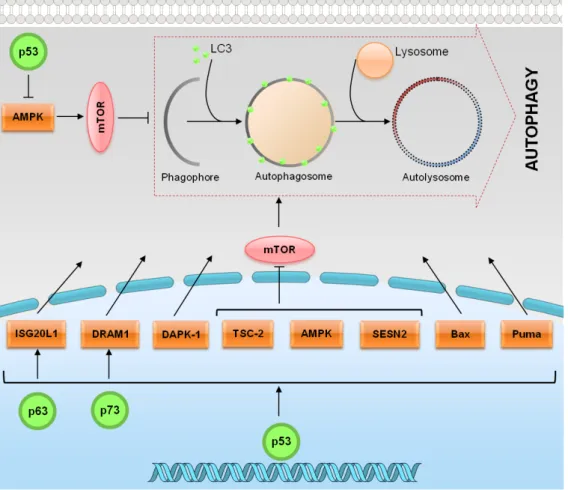
(33) degradation occurs [reviewed in (Napoli and Flores, 2013)]. Autophagy was initially identified in mammalians. However, the autophagy-related genes (ATGs) were first identified in yeast, and the majority of them have homologs in higher eukaryotes [reviewed in (Yang and Klionsky, 2010; Napoli and Flores, 2013)]. Most of the Atg proteins are recruited for the formation of the pre-autophagosomal structure (PAS). The formation of PAS is controlled by the mammalian target of rapamycin (mTOR) through two effector complexes: the protein kinase Atg1 (ULK1, ULK2 and ULK3 in mammals) and the lipid kinase Vps34 (hVps34 in humans). Additionally, two ubiquitylation-like conjugation systems, Atg5–Atg12–Atg16 and Atg8 (LC3 in mammals), also contribute to the formation of PAS. Autophagy induction is controlled by mTOR, which is a serine/threonine protein kinase that function as a central sensor of growth factors, nutrient signals and engery status [reviewed in (Yang and Klionsky, 2010)]. The p53 tumour suppressor protein is considered a master regulator of autophagy. Depending on its subcellular localization, p53 can activate or inhibit autophagy (Fig. 4) [reviewed in (Ryan, 2011)]. In response to stress signals, p53 is activated and, in the nucleus, it activates the adenosine monophosphate activated protein kinase (AMPK) pathway, resulting in the inhibition of the mTOR pathway and subsequent induction of autophagy (Feng et al., 2005). Besides AMPK, p53 transcriptionally regulates other proautophagic modulators such as death associated protein kinase 1 (DAPK-1), damageregulated autophagy modulator 1 (DRAM1), Bax, Puma, sestrin 2 (SESN2) and tuberous sclerosis protein 2 (TSC2) [reviewed in (Sui et al., 2011; Ryan, 2011)] (Fig. 4). In addition, it was demonstrated that several autophagy-related genes, such as Atg4, Atg5, Ulk and Atg7 and the two ubiquitylation-like conjugation systems Atg5–Atg12–Atg16 and Atg8 (LC3) are transcriptionally regulated by p53 (Kenzelmann Broz et al., 2013). Similarly to p53, p73 activates DRAM1. Different to the situation with p53, however, although p73 was also able to induce autophagy, this effect was DRAM-independent (Crighton et al., 2007). Additionally, the interferon stimulated exonuclease gene 20 kDa-like 1 (ISG20L1), which modulates genotoxic stress-induced autophagy, has been identified as a p53 family target gene (Eby et al., 2010). Although the molecular mechanism is still not fully understood, in the cytoplasm, p53 can inhibit autophagy through repression of the AMPK pathway, resulting in the activation of mTOR (Fig. 4) (Tasdemir et al., 2008).. 13.
(34) Figure 4. p53 family proteins distinctly regulate autophagy depending on its subcellular localization. In the nucleus, p53 family proteins induce the expression of proteins that activate autophagy. p53 may directly activate AMPK, TSC-2 and SESN2 and inhibit mTOR pathway, subsequently induce autophagy. p53 may also indirectly induce autophagy by binding to the promoter region of multiple genes that code for pro-autophagic modulators, such as DRAM1, DAPK-1, Bax and Puma. In the cytoplasm, p53 inhibits autophagy through repression of the AMPK pathway, resulting in the activation of mTOR [Adapted from (Ryan, 2011)].. 1.1.3. Therapeutic aplications As previously referred, the activation of p53, particularly of its capacity to induce an apoptotic cell death, has been considered an attractive strategy for anticancer treatment. In half of all human tumours retaining a wt p53 form, this may be achieved through inhibition of the p53 interaction with its major negative regulators, MDM2 and MDMX (also called MDM4).. 14.
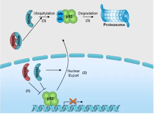
(35) 1.1.3.1. MDM2 and MDMX: Endogenous negative regulators The MDM2 protein was identified as a product that was amplified on double minute chromosomes in transformed mouse fibroblasts (Fakharzadeh et al., 1991). Latter, it was found that MDM2 was able to bind to p53 leading to the inhibition of its transcriptional activity (Momand et al., 1992; Oliner et al., 1993). Few years later, through a screening of a mouse cDNA expression library, MDMX, a homolog of MDM2, was discovered as a novel p53-interacting protein (Shvarts et al., 1996). Shvarts and colleagues (1996) demonstrated that similarly to MDM2, MDMX co-immunoprecipitates with p53 and its overexpression inhibites the p53 transcriptional activity. MDM2 and MDMX share some similarities in their structure (Fig. 5). Both proteins have a p53-binding domain at the Nterminal that is crucial for their binding to p53 and inhibition of its transcriptional activity. MDM2 and MDMX also have in common a zinc and a C-terminal RING finger domain. For MDM2, this latter domain is important for MDM2 homo-dimerization, MDM2-MDMX heterodimerization and E3 ubiquitin ligase activity. For MDMX, the RING finger domain is important for oligomerization and also gives to MDMX a minimal ubiquitin ligase activity. The major structural differences between these MDM proteins are found in the acidic domain, in the nuclear localization (NLS), and in the nuclear export signal domains (NES). For instance, MDMX has a shorter acidic domain compared to with MDM2, and lacks the NLS and NES domains (Fig. 5) [reviewed in (Waning et al., 2010). Regarding the MDM2 gene, its basal and inducible expressions are controlled by P1 and P2 promoters, respectively. The P2 promoter of the MDM2 gene is responsive to p53 induction [reviewed in (Manfredi, 2010)]. Although the obvious structural similarities between MDM2 and MDMX genes, the promoter region of MDMX gene remains not well characterized [reviewed in (Marine and Jochemsen, 2005)]. However, recently, Phillips and colleagues (2010) reported the existence of a p53-reponsive promoter (P2) in the first intron of the MDMX gene, thus establishing a negative regulatory feedback loop between p53 and MDMX.. Figure 5. Schematic representation of the major MDM2 and MDMX domains. [Adapted from (Lenos and Jochemsen, 2011)].. 15.
(36) Despite being structurally related proteins with the ability to inhibit the p53 transcriptional activity, evidence has emerged showing that these two proteins also play multifaceted and non-redundant roles in modulating p53 [reviewed in (Pei et al., 2012; Shadfan et al., 2012; Wade et al., 2013)]. Regarding the regulation of p53 by MDM2, both proteins form an autoregulatory feedback loop (Picksley and Lane, 1993; Wu et al., 1993). Once activated, p53 binds to the P2 promoter of the MDM2 gene inducing its transcription. In turn, MDM2 binds to the N-terminus of p53, via its N-terminal hydrophobic region (Böttger et al., 1999), and negatively regulates the p53 by three different mechanisms (Fig. 6): (1) inhibits the p53 transcriptional activity (Momand et al., 1992); (2) promotes the p53 nuclear export (Freedman et al., 1999), and (3) leads to p53 degradation via the ubiquitin–proteasome system (Haupt et al., 1997). In fact, MDM2 leads to p53 ubiquitination either in the form of a homodimer or of a or of a MDM2/MDMX heterodimer (Uldrijan et al., 2007; Dolezelova et al., 2012). Moreover, low MDM2 expression levels lead to the monoubiquitination of p53 resulting in its nuclear export and consequent transcription inhibition. Under these circumstances, monoubiquitylated p53 can localize to the mitochondria where it is deubiquitylated by the ubiquitin-specific protease-7 (USP7). Once deubiquitylated, p53 is able to induce apoptosis through a transcription-independent mechanism. Therefore, low levels of MDM2 are able to promote p53-mediated apoptosis (Marchenko et al., 2007). On the contrary, high MDM2 expression levels lead to the polyubiquitination of p53, which results in its proteasomal degradation [reviewed in (Nag et al., 2013)]. This degradation mechanism is responsible for the maintenance of low levels of p53 in unstressed cells (Lee and Gu, 2010). MDM2 regulates the p53 ubiquitination at lysine residues present in the C-terminal of p53. Additionally, these MDM2-targeted lysine residues can also be acetylated by p300/CREB-binding protein (CBP), a protein involved in p53 activation through acetylation (Lee and Gu, 2010). Acetylation of p53 leads to its activation and promotes its transcriptional activity (Tang et al., 2008). When ubiquitinated by MDM2, p53 is not able to be acetylated by p300/CBP, which leads to its proteassome degradation (Ito et al., 2001). It was also demonstrated that the p53 ubiquitination can be abolished due to the MDM2 acetylation by p300/CBP (Wang et al., 2004).. 16.
(37) Figure 6. Regulation of p53 activity and stability by MDM2 and MDMX. MDM proteins bind to p53 and inhibit its transcriptional activity (1). MDM2 promotes the p53 nuclear export (2) and, as an E3 ubiquitin ligase, the p53 ubiquitination and proteasomal degradation (3). Ub: ubiquitin. [Adapted from (Lenos and Jochemsen, 2011)].. Similarly to MDM2, MDMX also interacts with the p53 TAD through its p53-binding domain (Böttger et al., 1999), inhibiting the p53 transcriptional activity (Shvarts et al., 1996) (Fig. 6). In fact, the binding of MDMX blocks the p53 interaction with p300/CBP what leads to the inhibition of the p53 acetylation process (Sabbatini and McCormick, 2002). As referred before, since the lysine residues that are acetylated by p300/CBP are also targeted for ubiquitination by MDM2, MDMX may indirectly stimulate MDM2-mediated p53 ubiquitination by reducing the acetylation of those lysines [reviewed in (Lenos and and Jochemsen, 2011)]. Despite some ubiquitin ligase activity in vitro, contrary to MDM2, MDMX do not induce p53 nuclear export and degradation (Jackson and Berberich, 2000). In fact, in spite of the extensive homology of the RING finger domain of MDM2 and MDMX, MDMX has no detectable E3 ubiquitin ligase activity in vivo (Kawai et al., 2003; Meulmeester et al., 2003). Some studies showed that MDMX is not only a p53 inhibitor. In fact, under stress conditions, MDMX increases the p53 stability and promotes the p53 mitochondrial apoptosis [reviewed in (Mancini et al., 2010)]. In fact, under different DNA damages the 17.
Imagem
+7
Documentos relacionados