Aline Correa Abrahao(a) Beatriz Venturi Bonelli(b) Fábio Daumas Nunes(c) Eliane Pedra Dias(d) Márcia Grillo Cabral(a)
(a) Department of Oral Pathology and Oral
Diagnosis, Dental School, Federal University of Rio de Janeiro, Rio de Janeiro, RJ, Brazil.
(b) Oral Pathologist of the Air Force Central
Hospital, Rio de Janeiro, RJ, Brazil.
(c) Department of Oral Pathology, Dental
School, University of São Paulo, São Paulo, SP, Brazil.
(d) Department of Pathology, Medical School,
Fluminense Federal University, Niterói, RJ, Brazil.
Corresponding author:
Márcia Grillo Cabral
Federal University of Rio de Janeiro, School of Dentistry, Department of oral Pathology and Oral Diagnosis
Av. Professor Rodolpho Paulo Rocco, 325 - 1º andar - Cidade Universitária
Rio de Janeiro - RJ - Brazil CEP: 21941-913
E-mail: [email protected]
Received for publication on Aug 24, 2010 Accepted for publication on Nov 16, 2010
Immunohistochemical expression of
p53, p16 and hTERT in oral squamous
cell carcinoma and potentially
malignant disorders
Abstract: Oral carcinogenesis is a multi-step process. One possible step is the development of potentially malignant disorders known as leukopla-kia and erytroplaleukopla-kia. The objective of this study was to use immunohisto-chemistry to analyze the patterns of expression of the cell-cycle regulato-ry proteins p53 and p16INK4a in potentially malignant disorders (PMD) of the oral mucosa (with varying degrees of dysplasia) and in oral squamous cell carcinomas (OSCC) to correlate them with the expression of telomer-ase (hTERT). Fifteen PMD and 30 OSCC tissue samples were analyzed. Additionally, 5 cases of oral epithelial hyperplasia (OEH) were added to analyze clinically altered mucosa presenting as histological hyperplasia without dysplasia. p53 positivity was observed in 93.3% of PMD, in 63.3% of OSCC and in 80% of OEH. Although there was no correlation between p53 expression and the grade of dysplasia, all cases with severe dysplasia presented p53 suprabasal immunoexpression. p16INK4a expres-sion was observed in 26.7% of PMD, in 43.3% of OSCC and in 2 cases of OEH. The p16INK4a expression in OEH, PMD and OSCC was unable to differentiate non-dysplastic from dysplastic oral epithelium. hTERT positivity was observed in all samples of OEH and PMD and in 90% of OSCC. The high hTERT immunoexpression in all three lesions indicates that telomerase is present in clinically altered oral mucosa but does not differentiate hyperplastic from dysplastic oral epithelium. In PMD of the oral mucosa, the p53 immunoexpression changes according to the degree of dysplasia by mechanisms independent of p16INK4a and hTERT.
Descriptors: Mouth Neoplasms; Tumor Suppressor Protein p53; Cyclin-Dependent Kinase Inhibitor p16; Telomerase.
Introduction
immunohistochemi-cal staining has been considered to be predictive for malignant transformation and progression to OSCC.3 p53 expression in OSCC apparently does not correlate with differentiation grade but has been associated with patient outcome.4,5
The CDKN2A gene is inactivated in approxi-mately 70% of human cancers.6 Its codiied protein p16INK4a is a cell-cycle inhibitor that acts in the pRb-p16INK4a tumor suppressive pathway.6 Both PMD and OSCC have been linked to inactivation of the
CDKN2A gene by homozygous deletion.7
Other mechanisms may also contribute to dys-plastic cell clonal expansion and tumor progression, such as the ability of cells to regain the capacity to overcome growth arrest by rebuilding telomeric DNA.7,8 Telomeres are chromatin structures that cap the ends of eukaryotic chromosomes and ensure chromosome stability.8 At each DNA replication cycle, 30-150 base pairs of telomeric DNA are lost, driving cells into a metabolic state of irreversible growth arrest and replicative senescence.8 Telomer-ase is the enzyme in charge of rebuilding telomeres and is not expressed in normal somatic cells.7,8 The expression of telomerase in transformed oral epithe-lial cells could contribute to clonal expansion and to overcoming irreversible growth.8
Approximately 90% of primary human cancers show telomerase activity, evidenced by expression of the enzyme’s catalytic subunit, hTERT (human
telomerase reverse transcriptase), which is encoded by the TERT gene.9 However, the role of telomerase in oral carcinogenesis is unknown.10
The purpose of this study was to investigate p53, p16 INK4a and hTERT immunohistochemical expres-sion in PMD of the oral mucosa and OSCC and to evaluate correlations between their expression levels in these lesions.
Material and Methods
Tissue Samples
Fifty formalin-ixed, parafin-embedded biopsy specimens were retrieved from the Oral Pathology and Diagnosis Department of Federal University of Rio de Janeiro and from the Pathology Depart-ment of Fluminense Federal University archives. The samples used were 15 potentially malignant disor-ders (PMD) and 30 oral squamous cell carcinomas (OSCC). The lesions were classiied according to World Health Organization criteria.1,11 Five oral epi-thelial hyperplasia (OEH) samples were also added to the study to analyze hyperplastic oral mucosa without dysplasia. The PMD were clinically identi-ied as leukoplakia or erytroplakia and were graded according to the presence of epithelial dysplasia as mild, moderate or severe.1,11 The OSCC were classi-ied as well-differentiated (WD) or poorly differenti-ated (PD).1 Patients’ clinical data are summarized in Table 1.
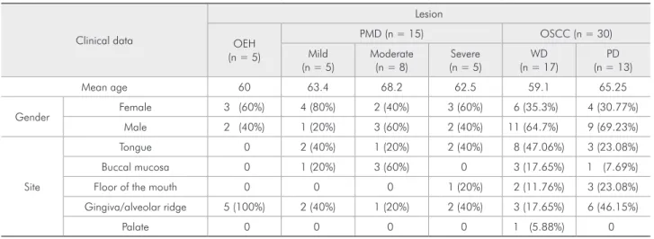
Table 1 - Patients’ clinical data.
Clinical data
Lesion
OEH (n = 5)
PMD (n = 15) OSCC (n = 30)
Mild (n = 5)
Moderate (n = 8)
Severe (n = 5)
WD (n = 17)
PD (n = 13)
Mean age 60 63.4 68.2 62.5 59.1 65.25
Gender Female 3 (60%) 4 (80%) 2 (40%) 3 (60%) 6 (35.3%) 4 (30.77%)
Male 2 (40%) 1 (20%) 3 (60%) 2 (40%) 11 (64.7%) 9 (69.23%)
Site
Tongue 0 2 (40%) 1 (20%) 2 (40%) 8 (47.06%) 3 (23.08%)
Buccal mucosa 0 1 (20%) 3 (60%) 0 3 (17.65%) 1 (7.69%)
Floor of the mouth 0 0 0 1 (20%) 2 (11.76%) 3 (23.08%)
Gingiva/alveolar ridge 5 (100%) 2 (40%) 1 (20%) 2 (40%) 3 (17.65%) 6 (46.15%)
Palate 0 0 0 0 1 (5.88%) 0
Immunohistochemistry
The streptavidin-biotin standard protocol was performed. Parafin-embedded tissues were cut into 3 µm thick sections, placed over slides, deparaf-inized in xylene and rehydrated in graded alcohol. Antigen retrieval was performed with Targetantigen retrieval solution pH 9 (Dako A/S, CA, USA) in a water bath, followed by incubation with 6% hydro-gen peroxide to quench endohydro-genous peroxidase. The sections were then incubated in blocking solution (3% bovine serum albumin) for 1 hour at room tem-perature, followed by primary antibody incubation, previously diluted in blocking solution. Anti-p53 (clone DO-7, 1:200 dilution – DAKO A/S, CA, USA) and anti-hTERT (Novocastra, clone 44F12, 1:75 dilution - Leica Microsystems, Berlin, Germany) an-tibodies were incubated for 30 minutes at room tem-perature, and the p16INK4a antibody (CINtecTM His-tology kit, clone E6H4, 1:250 dilution – DAKO A/S, CA, USA) was incubated overnight at 4°C. Sections were exposed to the LSABTM system (DAKO A/S, CA, USA), developed in diaminobenzidine (Dako A/S, CA, USA) and counterstained in Mayer’s he-matoxylin.
Positive immunohistochemistry expression of p53, p16INK4a and hTERT was deined by a nuclear, nuclear and cytoplasmic, and nuclear (evidenced by nucleolar positivity) staining pattern of epithelial cells, respectively. The results are expressed in both the number of positive cases and the percentage of immunostained cells after counting 100 cells in 10 consecutive high-power ields. p53 labeling in OEH and PMD was also evaluated as described
previous-ly by Cruz et al.:3 basal when conined to the basal layer; and suprabasal when both basal and supra-basal layers were positive.
Pearson correlation, Fisher’s exact test, Kruskall-Wallis and Mann Whitney tests were used for statis-tical analysis, and were performed with GraphPad Prism 5.00 (GraphPad Software, CA, USA). A p value less than 0.05 (p < 0.05) was considered statis-tically signiicant.
Results
The immunohistochemical results of p53, hTERT and p16INK4a in OEH, PMD and OSCC are summarized in Table 2. Positive p53 cases were a common event in all study groups. In 7 PMD and in the entire OEH group, a basal p53 staining pattern was found (Figure 1). An additional 7 cases of PMD showed a suprabasal p53 staining pattern (Figure 1). There was no statistical difference between dys-plasia degree and staining pattern in PMD samples. p53 positivity was also frequent in OSCC (Figure 2). No statistical difference was found between well-differentiated and poorly well-differentiated OSCC cases (p > 0.05).
Positive hTERT immunostained nuclei were ob-served through all epithelial cell layers in OEH and PMD (Figure 1). In the latter, no distinction was found between dysplasia grades and hTERT posi-tivity (p > 0.05). The majority of the OSCC cases were positive for hTERT staining (Figure 2), and no differences were found between WD and PD OSCC samples (p > 0.05).
Positive p16INK4a immunostaining in OEH and
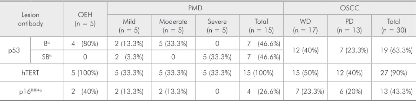
Table 2 - Distribution of positive cases in oral epithelial hyperplasia (OEH), potentially malignant disorders (PMD) and oral
squamous cell carcinoma (OSCC), in accordance to p53, p16 INK4a and hTERT.
Lesion antibody
OEH (n = 5)
PMD OSCC
Mild (n = 5)
Moderate (n = 5)
Severe (n = 5)
Total (n = 15)
WD (n = 17)
PD (n = 13)
Total (n = 30)
p53 B
a 4 (80%) 2 (13.3%) 5 (33.3%) 0 7 (46.6%)
12 (40%) 7 (23.3%) 19 (63.3%)
SBb 0 2 (3.3%) 0 5 (33.3%) 7 (46.6%)
hTERT 5 (100%) 5 (33.3%) 5 (33.3%) 5 (33.3%) 15 (100%) 15 (50%) 12 (40%) 27 (90%)
p16INK4a 2 (40%) 2 (13.3%) 2 (13.3%) 0 4 (26.6%) 7 (23.3%) 6 (20%) 13 (43.3%)
PMD showed stained cells arranged in clusters with skip areas in basal and suprabasal epithelial cell layers (Figure 1). No correlation between dysplasia grade and p16INK4a was observed (p > 0.05). In the OSCC group, positive p16INK4a cases were uncom-mon and two staining patterns were identiied. Ten cases presented positive cells organized in small iso-lated clusters, and the remaining three presented extensive sheets of immunostained cells (Figure 2). There was no correlation between p16INK4a immu-noexpression with WD or PD OSCC histological grades (p > 0.05).
In the PMD group, the immunohistochemical expression of p53 and p16INK4a was not correlated (p > 0.05). This study group presented higher means of hTERT positive cells (85.5) compared to p53 (35.6; p < 0.0001) and p16INK4a positive cells (5.5; p < 0.0001). The OSCC cases did not show correla-tion between the three antibodies (p > 0.05). How-ever, this group presented a signiicant difference when the means of p16INK4a positive cells (18.8) were compared to p53 (56.7; p < 0.001) and hTERT posi-tive cells (66.7; p < 0.0001).
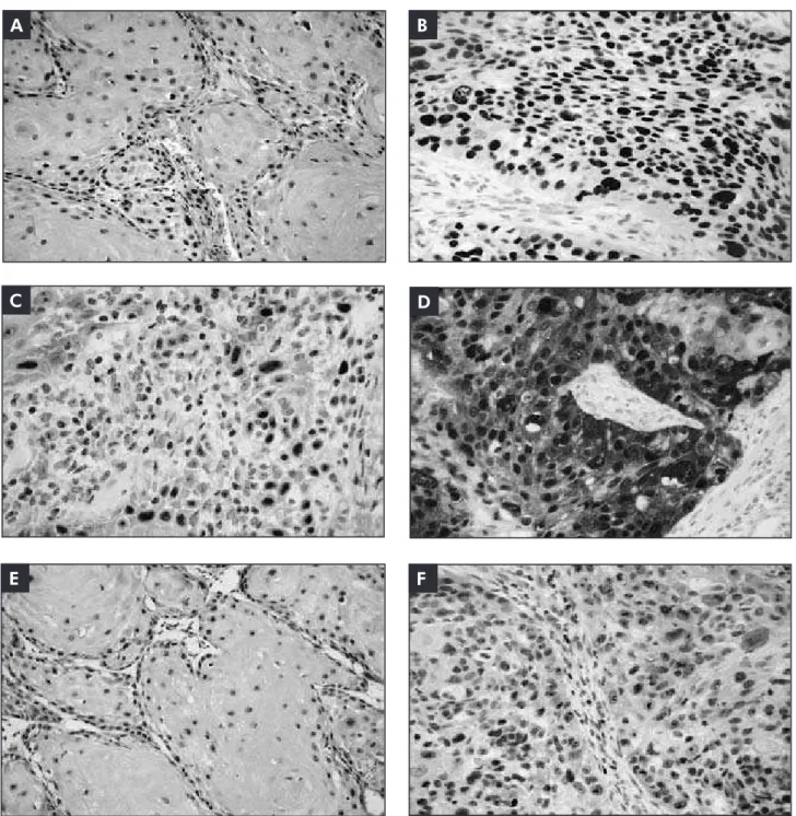
Figure 1 - Protein IHC
expression in PMD. A: p53
expression in basal epithelial
cell layer; B: p53 expression in
suprabasal epithelial cell layers;
C: p16INK4a expression in small
cell groups; D: hTERT expression
throughout epithelial cell layers. (SAB - original magnification 200x).
A
C
B
Discussion
Long-term retrospective reports have demon-strated that higher transformation rates to oral squamous cell carcinoma are not necessarily linked to severe grades of dysplasia.12,13 Also, reliable histo-logical grading systems and prognostic markers are lacking for OSCC.14
In normal oral epithelium, p53 is restricted to the proliferative basal cell layer.3 Overexpression of inactivated or mutated forms of p53 in oral epi-thelial dysplasia has been associated with high risk for transformation to early stage OSCC.4 Cruz et
al. showed that suprabasal p53 immunoexpression patterns are associated with high grades of
dyspla-A
D C
F B
E
Figure 2 - Protein IHC expression in OSCC. A and B: p53 in WD and PD cases; C: p16INK4a focal expression in a WD case;
sia and correlate with progress to oral squamous cell carcinoma.3 According to the authors, expression pattern should be considered a predictive marker for malignant transformation, although malignant transformation also occurs in the absence of supra-basal p53 staining or dysplastic changes.3 In this study, p53 suprabasal expression was found in 2 cases of mild dysplasia and in all 5 severe dyspla-sia samples. No statistical signiicance was found among histological grades, possibly due to the small number of cases studied. However, suprabasal p53 immunoexpression may be a useful tool for malig-nant transformation risk assessment of potentially malignant disorders independent of dysplasia grade. Further studies with follow up are necessary to con-irm this assumption.
p53 has been detected in a large percentage of OSCC cases by immunohistochemistry, relecting the altered status of this protein.15 More than 50% of our OSCC samples were p53 positive in accor-dance with the results described in the literature. Although we detected more cases of p53 positive WD-OSCC compared to PD-OSCC, this difference was not signiicant. Previous reports are inconclu-sive when relating p53 immunoexpression with the differentiation grade of OSCC.5,15
Inactivation of the p16INK4a gene is frequently identiied during early carcinogenesis. However, non-dysplastic mucosa and oral dysplastic lesions of-ten lack positive p16INK4a immunohistochemical ex-pression.16,17 Perhaps this lack of positive staining re-lects that in non-dysplastic oral epithelium, normal p16INK4a protein isbelow detection levels, whereas in epithelial dysplasia, the low expression is related to gene inactivation.12 The signiicance of p16INK4a im-munoexpression in OSCC is unknown but has been correlated with response to therapy, prognosis and tumor morphology.18 Among the studied OSCC, 18.8% were found positive for p16INK4a. The OEH and PMD cases were positive in scattered groups of cells. These results suggest that p16INK4a is unrelated to the degree of dysplasia, although the small num-ber of positive cases occurred in samples with mild and moderate dysplasia. These results may relect either the frequent genetic inactivation of p16INK4a in early phases of carcinogenesis or the p16INK4a
ex-pression in slow cycling progenitor cells.19
Angiero et al. demonstrated an increase in p16INK4a expression in higher grades of dysplasia and invasive OSCC.20 Likewise, Gologan et al. showed that p16INK4a immunoexpression was able to high-light dysplastic areas in oral epithelium.16 However, Bradley et al. showed a signiicant trend toward ab-sent expression of p16INK4a with increasing dysplasia severity.12 These different results are possibly due to the methodology or antibody used in the study. Oth-er aspects that may inluence immunohistochemical detection of p16INK4a in OSCC are related to etio-logical factors. Different cancer-causing agents may lead to p16INK4a gene inactivation as well as altered p53 and pRb tumor suppressive pathways.5,17 These changes may result in either loss or overexpression of p16INK4a in oral dysplasia and OSCC. HPV onco-genes are frequently found in oropharyngeal squa-mous cell carcinomas that display concomitant in-creased p16INK4a expression.17,21,22 According to Vidal and Gillison, patients with HPV-positive head and neck squamous cell carcinoma present better clini-cal outcomes compared to those with HPV-negative tumors.22 Our study shows thirteen OSCC with p16INK4a expression, and viral participation cannot be discarded. Other relevant etiopathological agents that may inluence p16INK4a expression are smoking and smoke-less tobacco use.21,23 The oral mucosa of smokers and lesions associated with smoke-less tobacco use express p16INK4a more frequently when compared to individuals that do not use tobacco.20,21
In oral carcinogenesis, telomerase expression may favor telomere stabilization and cell proliferation.
Telomerase activity is important in deregulated cell growth and escape from senescence, contrib-uting to clonal expansion of dysplastic cells that harbor p53 and p16INK4a abnormalities.19,24 Chen et
al. showed a correlation between cytoplasmic and nuclear hTERT expression in histologically differ-ent OSCC, suggesting that hTERT expression was a biomarker for this type of lesion.24 The authors reported that cytoplasmic hTERT was increased in dysplastic oral epithelium and OSCC compared to normal epithelium, whereas nuclear hTERT was decreased in OSCC.24 This work analyzed nuclear hTERT staining and found a higher hTERT expres-sion in WD-OSCC compared to PD-OSCC. The mean number of hTERT positive cells in the WD group had a tendency to be higher than in the PD group. It is possible that in PD-OSCC, other
mecha-nisms are involved in maintaining telomere length such as alternative lengthening of telomeres. Future studies are needed to help clarify the contribution of telomere lengthening and telomere-associated pro-teins in oral carcinogenesis.
Conclusion
p53, p16INK4a and hTERT are not associated with the grade of dysplasia in PMD of the oral mucosa or with the differentiation degree of OSCC. How-ever, suprabasal p53 immunoexpression was associ-ated with severe grades of dysplasia. The results also show that p16INK4a may not be useful for distinguish-ing hyperplastic oral epithelium from dysplastic oral epithelium. The intense hTERT expression in OEH, PMD and OSCC suggests that telomerase activity is involved in the development of hyperplastic and dys-plastic oral epithelium.
References
1. Barnes L, Eveson JW, Reichart P, Sidransky D. Editors. World Health Organization Classification of Tumours. Patholgy & Genetics of Head and Neck Tumours. Lyon: IARC Press; 2005. 430 p.
2. Mithani SK, Mydlarz WK, Grumbine FL, Smith IM, Califano JA. Molecular genetics of premalignant oral lesions. Oral Dis. 2007 Mar;13(2):126-33.
3. Cruz I, Napier SS, van der Waal I, Snijders PJ, Walboomers JM, Lamey PJ, et al. Suprabasal p53 immunoexpression is strongly associated with high grade dysplasia and risk for malignant transformation in potentially malignant oral lesions from Northern Ireland. J Clin Pathol. 2002 Feb;55(2):98-104. 4. Shah NG, Trivedi TI, Tankshali RA, Goswami JA, Shah
JS, Jetly DH, et al. Molecular alterations in oral carcino-genesis: significant risk predictors in malignant transforma-tion and tumor progression. Int J Biol Markers. 2007 Apr-Jun;22(2):132-43.
5. de Oliveira LR, Ribeiro-Silva A, Zucoloto S. Prognostic im-pact of p53 and p63 immunoexpression in oral squamous cell carcinoma. J Oral Pathol Med. 2007 Apr;36(4):191-7. 6. Nakahara Y, Shintani S, Mihara M, Kiyota A, Ueyama Y,
Matsumura T. Alterations of Rb, p16(INK4A) and cyclin D1 in the tumorigenesis of oral squamous cell carcinomas. Cancer Lett. 2000 Nov 10;160(1):3-8.
7. Freier K, Pungs S, Flechtenmacher C, Bosch FX, Lichter P, Joos S, et al. Frequent high telomerase reverse transcriptase
expression in primary oral squamous cell carcinoma. J Oral Pathol Med. 2007 May;36(5):267-72.
8. Gilley D, Tanaka H, Herbert BS. Telomere dysfunction in ag-ing and cancer. Int J Biochem Cell Biol. 2005 May;37(5):1000-13.
9. Masutomi K, Hahn WC. Telomerase and tumorigenesis. Can-cer Lett. 2003 May 15;194(2):163-72.
10. Luzar B, Poljak M, Marin IJ, Eberlinc A, Klopcic U, Gale N. Human telomerase catalytic subunit gene re-expression is an early event in oral carcinogenesis. Histopathology. 2004 Jul;45(1):13-9.
11. Warnakulasuriya S, Johnson NW, van der Waal I. Nomencla-ture and classification of potentially malignant disorders of the oral mucosa. J Oral Pathol Med. 2007 Nov;36(10):575-80. 12. Bradley KT, Budnick SD, Logani S. Immunohistochemical
detection of p16INK4a in dysplastic lesions of the oral cavity. Mod Pathol. 2006 Oct;19(10):1310-6.
13. Arduino PG, Surace A, Carbone M, Elia A, Massolini G, Gandolfo S, et al. Outcome of oral dysplasia: a retrospective hospital-based study of 207 patients with a long follow-up. J Oral Pathol Med. 2009 Jul;38(6):540-4.
14. Brennan M, Migliorati CA, Lockhart PB, Wray D, Al-Hashimi I, Axell T, et al. Management of oral epithelial dysplasia: a review. Oral Surg Oral Med Oral Pathol Oral Radiol Endod. 2007 Mar;103 Suppl:S19.e1-12.
16. Gologan O, Barnes EL, Hunt JL. Potential diagnostic use of p16INK4A, a new marker that correlates with dyspla-sia in oral squamoproliferative lesions. Am J Surg Pathol. 2005 Jun;29(6):792-6.
17. Karsai S, Abel U, Roesch-Ely M, Affolter A, Hofele C, Joos S,
et al. Comparison of p16(INK4a) expression with p53 altera-tions in head and neck cancer by tissue microarray analysis. J Pathol. 2007 Feb;211(3):314-22.
18. Muirhead DM, Hoffman HT, Robinson RA. Correlation of clinicopathological features with immunohistochemical expression of cell cycle regulatory proteins p16 and retino-blastoma: distinct association with keratinisation and dif-ferentiation in oral cavity squamous cell carcinoma. J Clin Pathol. 2006 Jul;59(7):711-5.
19. Rheinwald JG, Hahn WC, Ramsey MR, Wu JY, Guo Z, Tsao H, et al. A two-stage, p16(INK4A)- and p53-depen-dent keratinocyte senescence mechanism that limits replica-tive potential independent of telomere status. Mol Cell Biol. 2002 Jul;22(14):5157-72.
20. Angiero F, Berenzi A, Benetti A, Rossi E, Del Sordo R, Sidoni A, et al. Expression of p16, p53 and Ki-67 proteins in the pro-gression of epithelial dysplasia of the oral cavity. Anticancer Res. 2008 Sep-Oct;28(5A):2535-9.
21. Greer Jr RO, Meyers A, Said SM, Shroyer KR. Is p16(INK4a) protein expression in oral ST lesions a reliable precancerous marker? Int J Oral Maxillofac Surg. 2008 Sep;37(9):840-6. 22. Vidal L, Gillison ML. Human papillomavirus in HNSCC:
recognition of a distinct disease type. Hematol Oncol Clin North Am. 2008 Dec;22(6):1125-42, vii.
23. Tarakji B, Kujan O, Nassani MZ. An immunohistochemical study of the distribution of p 16 protein in oral mucosa in smokers, non-smokers and in frictional keratosis. Med Oral Patol Oral Cir Bucal. 2010 Sep 1;15(5):e681-4.