FACULDADE DE CIÊNCIAS
DEPARTAMENTO DE QUÍMICA E BIOQUÍMICA
CFTR Trafficking and Membrane Anchoring –
the role of cAMP signalling
Miguel Gonçalo de Oliveira Jones Ferrão Lobo
Mestrado em Bioquímica (Especialização em Bioquímica)
Dissertação orientada pelo Professor Doutor Carlos Miguel Farinha
Preface
In the 1990s life used to be simple. The entire family watched Formula 1 on Sunday afternoons and we still didn’t know what garbage recycling was. In an interesting analogy, biologists in the 90s thought of protein kinase A (PKA) as the only link between the second messenger cAMP and CFTR (first reported by Cheng et al. in 1991). Our data challenge this linear and somewhat simplistic view unveiling a novel relationship between CFTR and yet another cAMP effector, the exchange protein directly activated by cAMP (EPAC). We propose EPAC as a new player to the CFTR race circuit, a rally that begins in the endoplasmic reticulum, where the protein is translated, and finishes at the plasma membrane (PM), where the channel exerts its function. EPAC is involved in a number of cellular functions such as the regulation of cell-to-cell and cell-matrix adhesion and cytoskeleton rearrangements, processes that affect CFTR regulation. Here we present compelling evidence suggesting that cAMP (here replaced by an analogue that does not activate the PKA pathway) promotes the interaction of EPAC with CFTR with the aid of the protein adaptor NHERF1. The formation of this complex increases the stability of CFTR at the PM.
Using a multidisciplinary approach, we show that EPAC1 (the most abundant of the two EPAC isoforms known) and CFTR co-localize in cystic fibrosis bronchial epithelial cells (CFBE) – reporting this interaction in different airway epithelial cell lines that express endogenous EPAC1, namely A549, CFBE and Calu3. Cyclic AMP promotes EPAC translocation to the PM, in a CFTR-independent manner, as well as its interaction with CFTR. siRNAs or shRNAs against ezrin or NHERF1, proteins involved in the tethering of PKA in the proximity of CFTR, show that the latter (but not the previous) mediates the interaction between EPAC1 and CFTR. However, neither of these scaffolding proteins affects the translocation of EPAC1 to the PM. On the other hand, CFTR biotinylation at the PM shows that EPAC activation decreases CFTR endocytosis and promotes CFTR stabilization at the PM. Transient knockdown of EPAC1 has an opposite effect on CFTR stability. Additionally, EPAC activation does not affect CFTR total protein levels or the CFTR processing efficiency. Finally, the presence of the most common CFTR mutation, F508del, seems to affect levels of endogenous EPAC activation and EPAC-mediated cell adhesion.
In this way, a new link between CFTR regulation and cAMP was established. This novel player on the CFTR race circuit may contribute to a finer and more complex regulation of this channel. Definitely, life has just become more complicated.
i
Table of Contents
Acknowledgements ... v Abstract ... vii Resumo ... ix Abbreviations ... xiii 1. Introduction ... 11.1 Cystic Fibrosis - Overview ... 1
1.2 The CFTR gene and protein ... 1
1.3 CFTR folding and trafficking ... 4
1.4 CFTR anchoring at the plasma membrane ... 7
1.5 CFTR function ... 8
1.6 EPAC – Overview ... 11
1.7 EPAC protein and its activation ... 12
1.8 Spatial regulation of EPAC ... 14
1.9 Biological functions of EPAC ... 16
1.10 Objectives ... 18
2. Materials and Methods ... 21
2.1 Molecular Biology ... 21
2.1.1 Plasmid vectors and siRNAs ... 21
2.1.2 Transformation of competent bacteria ... 22
2.1.3 DNA extraction and quantification ... 22
2.1.4 Mutagenesis ... 23 2.1.5 DNA sequencing ... 23 2.2 Cell Culture ... 25 2.2.1 Cell types ... 25 2.2.1.1 CFBE cells ... 25 2.2.1.2 Calu3 cells ... 25 2.2.1.3 A549 cells ... 26 2.2.1.4 HEK293 cells ... 26 2.2.3 Transfections ... 26 2.2.4 Lentivirus infection ... 27
ii
2.2.4.1 Lentivirus production ... 27
2.2.4.2 Production of a stable cell line ... 27
2.3 Biochemical Analysis ... 28
2.3.1 Cell lysis and total protein quantification ... 28
2.3.2 Western blot ... 28
2.3.3 Immunoprecipitation ... 29
2.3.4 Rap1A activity assay ... 30
2.3.5 Biotinylation assay ... 31
2.3.6 Endocytosis assay ... 32
2.3.7 Cell fractionation ... 32
2.4 Live cell imaging ... 33
2.4.1 Measurements of cAMP levels and PKA or EPAC activity by fluorescence resonance energy transfer (FRET)... 33
2.4.2 Translocation of EPAC1 and co-localization studies ... 36
2.5 Functional Analysis ... 37
2.5.1 Adhesion assay ... 37
2.6 Statistical Analysis ... 37
3. Results ... 41
3.1 Effect of 007-AM treatment on EPAC1 ... 41
3.2 Co-localization and interaction between CFTR and EPAC1... 49
3.3 Effect of EPAC1 on CFTR stability ... 57
3.4 Effect of CFTR upon EPAC-induced cell adhesion ... 62
4. Discussion and Perspectives ... 65
4.1 Effect of 007-AM treatment on EPAC1 ... 66
4.2 Co-localization and interaction between CFTR and EPAC1... 67
4.3 Effect of EPAC1 on CFTR stability ... 70
4.4 Effect of CFTR upon EPAC-induced cell adhesion ... 72
4.5 Future Perspectives ... 73
References ... 75
Appendices ... 85
Plasmid maps ... 85
pcDNA3.1 (+/-) plasmid map ... 85
iii
pCAGGS plasmid map ... 86
pmCherry-C1 plasmid map ... 87
Forskolin standard curve for AKAR4 and camp FRET sensors ... 88
Preparation of GST-RalGDS-RBD-coupled beads for Rap1A activity assay ... 89
v Acknowledgements
First and foremost, I want to express my gratitude to my supervisor Prof. Carlos Farinha, for all the constant support, orientation and opportunities that without him wouldn’t have been possible. I really enjoyed to work with him and my time in the lab in Lisbon. I am also grateful to Prof. Margarida Amaral for welcoming me into her group and helping me when needed.
Special thanks are due to the many people in the group which always helped me. Ana Cachaço taught me how to dry my wet shoes during rainy days using a dryer. With Susana I learned how to evaluate the correct amount of butter on a toast. Zé taught me that many women are crazy, as I already thought. Ana Marta was my first bench partner, then replaced by a shorter but equally friendly and happy person, Sara Afonso. With both I learned how to keep my bench perfectly cleaned and organized. Inna, a special friend that probably works for DHL and taught me to ‘Do what I say and not what I do’. Without her, I would be more relaxed and with less work to do. Simão, the guy in the lab that uses the same clothes as me. Sara Canato has been proving that human batteries don’t run out. Veronica told me that the best way to go on holidays is if I got pregnant. Nikhil, who was my first roommate at an international meeting. Whaaaaatttt?? Ines taught me my first german word…
I also would like to express my gratitude to Prof. Manuela Zaccolo for the opportunity of working with her group in Oxford and also for all the support and constant availability. A really special thanks to Kostas for everything: help related to the lab, opinions regarding the scientific life, share of knowledge, rare meat and tzatziki. Big thanks to Stefania for support and guidance. I am also grateful to Caracoleta, who taught me some Italian words, Oliver (and his BMW), Marcela, Alex, Laura and Claudia.
vii Abstract
CFTR, Cystic Fibrosis Transmembrane Conductance Regulator, is a chloride channel expressed in the apical membrane of epithelial cells. The malfunction of this protein is responsible for cystic fibrosis, the most common lethal autosomal recessive disease in the Caucasian population. One of the numerous proteins that interacts with CFTR and also regulates the function of this channel through phosphorylation is protein kinase A (PKA). Because PKA activity is cAMP-dependent, this kinase has been recognized as the link between this second messenger and CFTR. However, PKA is not the only cAMP sensor within the cell.
EPAC, an exchange protein directly activated by cAMP, is another cAMP effector, involved in a number of cellular functions such as the regulation of cell-to-cell and cell-matrix adhesion, cytoskeleton rearrangements and cell polarization, processes that affect CFTR regulation and are defective in CF. The aim of this work was to characterize the interaction between CFTR and the most predominant isoform of EPAC, EPAC1, and to evaluate the impact of this cAMP sensor on CFTR biogenesis, trafficking and plasma membrane (PM) anchoring.
Using a multidisciplinary approach, our results show that EPAC1 and CFTR co-localize and co-immunoprecipitate in airway epithelial cells. The second messenger cAMP promotes EPAC1 translocation to the PM and its interaction with CFTR. The adaptor protein NHERF1, but not ezrin, mediates the interaction between EPAC1 and CFTR. Furthermore, EPAC activation does not affect CFTR total protein levels or CFTR processing efficiency but promotes CFTR stabilization at the PM. Additionally, the presence of the most common CFTR mutation, F508del, seems to affect EPAC activity and EPAC-mediated cell adhesion. Thus, this work provided an important characterization of a new CFTR interacting protein that links cAMP to cystic fibrosis modulation in a previously unreported mechanism.
ix Resumo
A Fibrose Quística (FQ), a doença recessiva autossómica letal mais comum na população caucasiana, é caracterizada por uma grave disfunção da função pulmonar causada pela obstrução das vias respiratórias devido à acumulação de muco e consequentes infecções bacterianas. A FQ é causada por mutações no gene que codifica para uma glicoproteína com 1480 resíduos de aminoácidos, designada CFTR (Cystic Fibrosis Transmembrane
Conductance Regulator). Esta proteína, presente na membrana apical de células
epiteliais, actua como um canal de cloreto. Até à data, foram identificadas cerca de 2000 mutações possíveis causadoras da doença, sendo a mais comum a delecção de três nucleótidos que correspondem ao resíduo de feninalanina na posição 508 da proteína (F508del).
Durante a sua síntese proteica, a proteína CFTR é inserida co-traducionalmente na membrana do retículo endoplasmático, onde adquire a sua conformação nativa, devido à acção de vários chaperones, e passa por um processo de glicosilação inicial. Daqui, é transportada para o Golgi onde os seus resíduos glicídicos são modificados, originando a sua forma madura. Posteriormente, é transportada em vesículas para a membrana plasmática (PM) onde exerce a sua função. Aqui, a proteína CFTR é endocitada sendo depois reciclada de volta para a PM ou enviada para degradação lisossomal. O equilíbrio entre estes processos é crucial para a quantidade de proteína presente na membrana.
Na PM, a proteína CFTR pode estar ancorada a filamentos de actina, o que contribui para a sua estabilidade membranar. A proteína CFTR, através da sua região C-terminal, interage com várias proteínas com um domínio PDZ, nomeadamente a proteína adaptadora NHERF1 (Na+/H+-exchanger regulatory factor isoform 1), que por sua vez
interage com proteínas do citoesqueleto, tal como a ezrina. Desta forma, o complexo CFTR-NHERF1-ezrina-actina contribui para a imobilização e ancoragem da proteína CFTR na PM, prevenindo a sua endocitose. Dessa forma, a ancoragem da CFTR na superfície celular é uma importante via a ser considerada em termos da intervenção terapêutica em doentes com FQ.
Uma vez na PM, a proteína CFTR está envolvida na regulação do transporte transepitelial de iões e água. A abertura do canal CFTR é regulada pela fosforilação catalisada pela proteína cinase A (PKA) em resposta a um aumento local dos níveis de AMP cíclico
x
(cAMP). A compartimentalização do cAMP depende da integridade do citoesqueleto e é essencial para um aumento da especificidade e eficiência da sinalização deste mensageiro secundário. Uma vez que a actividade da PKA é dependente dos níveis de cAMP, esta cinase tem sido reconhecida como a ligação entre a proteína CFTR e o cAMP. No entanto, a PKA não é o único sensor de cAMP que existe na célula; a proteína EPAC (exchange protein directly activated by cAMP) também actua como um efector do cAMP.
A proteína EPAC funciona como um factor de troca de nucleótidos de guanina para a Rap, uma pequena GTPase (enzima que hidrolisa GTP) da família Ras. Esta GTPase está activa quando ligada a GTP e inactiva quando ligada a GDP. Assim, a proteína EPAC promove a activação deste interruptor molecular, já que leva à ligação de GTP. Em resposta ao aumento dos níveis de cAMP, a proteína EPAC transloca do citosol para a PM, onde pode activar a Rap. A interacção com a PM pode ser feita de uma forma dependente de ácido fosfatídico ou através da proteína do citoesqueleto ezrina. Esta informação sugere que a proteína EPAC pode co-localizar, ou até mesmo interactuar, com a proteína CFTR, e, possivelmente, regular a sua função ou estabilidade membranar.
A proteína EPAC está envolvida em várias funções biológicas, principalmente devido às várias localizações sub-celulares que esta proteína pode ter e aos vários parceiros moleculares com que pode interagir. Nomeadamente, está envolvida na regulação da adesão célula-célula ou célula-matriz, organização do citoesqueleto ou polarização celular - processos que afectam a regulação da proteína CFTR, e que estão em geral alterados na FQ. Desta forma, o objectivo deste trabalho consistiu em caracterizar a interacção entre a proteína CFTR e a principal isoforma da proteína EPAC, a EPAC1, e avaliar o impacto deste sensor de cAMP na biogénese, tráfego e ancoragem da CFTR na PM.
O primeiro passo do trabalho consistiu em avaliar se o composto 007-AM de facto actua como um análogo do cAMP específico para a EPAC (e não PKA). Este estudo, bem como outros posteriores, foi realizado em células CFBE (Cystic Fibrosis Bronchial Epithelial) de forma a melhor mimetizar os processos que ocorrem no epitélio brônquico de pacientes com FQ. Outras linhas celulares de epitélio respiratório também usadas foram as linhas Calu3 (de glândula submucosa) e A549 (alveolar). A análise por FRET mostrou que o sensor dos níveis de cAMP baseado na estrutura da EPAC sofre grande alteração da
xi percentagem de FRET, o mesmo não se observando com o sensor da actividade da PKA – o que permitiu evidenciar a selectividade do 007-AM para a EPAC. A activação da proteína EPAC, em resultado do tratamento das células com 007-AM, promove a sua translocação para a PM, em células CFBE, de uma forma independente da proteína CFTR. Além disso, usando um sensor FRET da actividade da EPAC e um ensaio para a actividade da Rap1A (uma das isoformas da Rap), observou-se que células que expressam CFTR WT (wild-type) apresentaram um aumento de FRET na PM após tratamento com 007-AM, enquanto células que expressam CFTR F508del tendem a ter níveis mais elevados de Rap1A na forma activa. O tratamento com 007-AM promove também um aumento dos níveis de Rap1A activa em células que expressam CFTR WT mas não nas que expressam CFTR F508del.
Adicionalmente, o tratamento com 007-AM promoveu a adesão celular em células que expressam CFTR WT mas não nas células que expressam CFTR F508del. Isto sugere que, em células que expressam CFTR com esta mutação, a via EPAC-Rap não consegue exercer a sua função e, de forma a ultrapassar esta limitação, a activação endógena da EPAC se encontra aumentada. Consequentemente, uma activação adicional não é detectada, dados os elevados níveis basais de activação.
De seguida, a possível co-localização e interacção entre as proteínas CFTR e EPAC1 foi avaliada. Para isso, recorreu-se a microscopia confocal de fluorescência e a ensaios de co-imunoprecipitação. Observou-se que a EPAC1 co-localiza e interage com CFTR, quer na sua forma WT quer na sua forma mutada. Além disso, a activação da EPAC com 007-AM aumentou a interacção entre CFTR e EPAC1. No entanto, a activação da EPAC1 não afecta a razão de CFTR na forma madura e forma imatura (que permite avaliar a eficiência de processamento da CFTR). Estes dados sugerem que a EPAC não está envolvida na fase inicial da biogénese da CFTR (síntese, folding e processamento inicial).
Observámos também que a depleção de NHERF1 (usando siRNA ou shRNA), mas não de ezrina, impediu a interacção entre a CFTR e a EPAC1 mas sem afectar a localização sub-celular da última. Isto sugere que a interacção entre estas duas proteínas é mediada pela proteína adaptadora NHERF1.
xii
Finalmente, recorreu-se a ensaios de endocitose e biotinilação para avaliar o efeito da EPAC1 na estabilidade da CFTR na PM. Observou-se que a activação da EPAC decresce a quantidade de CFTR que é internalizada ao longo do tempo. Além disso, o tratamento com 007-AM levou a um aumento dos níveis de CFTR na PM enquanto a depleção da EPAC1 usando siRNA diminuiu estes níveis. Adicionalmente, a sobreexpressão de EPAC parece também promover a estabilidade da CFTR. Estas abordagens sugerem que a EPAC1 está envolvida na regulação da estabilidade da CFTR na PM.
Este efeito estabilizador permite também um aumento da quantidade de proteína CFTR F508del madura detectada após o tratamento simultâneo com o corrector de tráfego VX-809 e o agonista 007-AM.
Apesar dos avanços recentes no campo da FQ, ainda existem vários aspectos do tráfego e activação da CFTR por elucidar e parceiros moleculares por identificar. Desta forma, os resultados deste trabalho permitiram identificar uma nova ligação entre a proteína CFTR e a sinalização por cAMP. Este trabalho constitui assim uma importante caracterização de um novo interactor da CFTR, a proteína EPAC1, que liga o cAMP à modulação da FQ, por mecanismos até à data não descritos.
xiii Abbreviations
007 8-(4-Chlorophenylthio)-2'-O-methyladenosine-3',5'-cyclic monophosphate (8-pCPT-2’-O-Me-cAMP)
007-AM 8-(4-Chlorophenylthio)-2'-O-methyladenosine-3',5'-cyclic monophosphate acetoxymethyl ester (8-pCPT-2’-O-Me-cAMP-AM)
A549 Human alveolar epithelial cell line
ABC ATP-binding cassete
AC Adenylyl cyclase
AKAP A-kinase anchoring proteins AKAR4 A-kinase activity reporter 4
ASL Airway surface liquid
ATF Arginine-framed tripeptide
ATP Adenosine triphosphate
BSA Bovine serum albumin
CAAX Cystein-alyphatic-alyphatic-any aminoacid motif CaCC Calcium-activated chloride channel
cAMP Cyclic adenosine monophosphate Calu-3 Cancer lung 3 cell line
camps cAMP sensor
CDC25-HD Cell division cycle 25 homology domain
CF Cystic fibrosis
CFBE41o- / CFBE Cystic fibrosis bronchial epithelial cell line
CFP Cyan fluorescent protein
CFTR Cystic fibrosis transmembrane conductance regulator CNB Cyclic nucleotide–binding domain
COP-II Coating protein II C-terminal Carboxyl-terminal
DAPI 4',6-diamidino-2-phenylindole
DEP Disheveled, Egl-10, pleckstrin domain DNA-PK DNA damage–responsive kinase
DMSO Dimethylsulfoxide
E3KARP NHE3 kinase A regulatory protein EBP50 Ezrin-binding protein
xiv
EDEM ER degradation-enhancing α-mannosidase-like protein EDTA Ethylenediaminetetraacetic acid
EMEM Eagle’s minimal essential medium ENaC Epithelial sodium channel
EPAC/REPAC Exchange protein directly activated by cAMP
ER Endoplasmic reticulum
ERAD Endoplasmic reticulum-associated protein degradation ERM Ezrin/radixin/moesin
ERQC Endoplasmic reticulum quality control
ESI-09 3-[5-(tert.-Butyl)isoxazol-3-yl]-2-[2-(3-chlorophenyl)hydrazono]-3-oxopropanenitrile
F-actin Filamentous actin
FBS Fetal bovine serum
FLIM Fluorescence lifetime imaging
FRET Fluorescence resonance energy transfer
Frsk Forskolin
GAP GTPase-activating protein
GAPDH Glyceraldehyde 3-phosphate dehydrogenase
GDP Guanosine diphosphate
GEF Guanine nucleotide exchange factor
GFP Green fluorescent protein
GRASP Golgi reassembly stacking protein
GST Glutathione S-transferase
GTP Guanosine triphosphate
GTPase Guanosine triphosphate hydrolase HBSS Hank's Balanced Salt Solution HEK Human embryonic kidney cell line
HRP Horseradish peroxidase
Hsc70/Hdj2 Heat shock cognate 70kDa protein/human DnaJ homolog 2 Hsp70/Hdj-1 Heat shock protein 70kDa protein/human DnaJ homolog 2 Hsp90 Heat shock protein 90kDa protein
HUVEC Human umbilical vein endothelial cell line IBMX 3-isobutyl-1-methylxanthine
xv
IP Immunoprecipitation
IPTG Isopropyl β-D-1-thiogalactopyranoside
LB Luria broth
MAP1 Microtubule-associated protein 1
MSD Membrane-spanning domain
NBD Nucleotide binding domain
NHERF Na+/H+-Exchanger Regulatory Factor N-terminal Amino-terminal
ORCC Outwardly rectifying chloride channel
P60 Plates with 60mm of diameter
P100 Plates with 100mm of diameter PAGE Poliacrylamide gel electrophoresis
PBS Phosphate buffer saline
PBS-T Phosphate buffer saline with tween PCR Polymerase chain reaction
PDE Phosphodiesterase
PDE4D3 Phosphodiesterase 4D3
PDZ Post synaptic density protein (PSD95), Drosophila disc large tumor suppressor (Dlg1), and zonula occludens-1 protein (zo-1)
Pen Penicilin
PKA Protein kinase A
PM Plasma membrane
PP2A Protein phosphatase-2A
PPase Protein phosphatase
PVDF Polivinylidene difluoride
RA Ras-association domain
RanBP2 Ran-binding protein 2
RBD Rap-binding domain
RD/R domain Regulatory domain
REM Ras exchange motif domain
RFP Red fluorescent protein
RFU Relative fluorescence units
RNA Ribonucleic acid
xvi
ROMK Renal outer medullary potassium channel R-ras Ras-like small GTPase
RT Room temperature
SBT Spectral bleed-through
SDS Sodium dodecylsulphate
SEM Standard error of the mean value
shRNA Small hairpin RNA
siRNA Small interfering RNA
Strep Streptomycin
TBS Tris buffer saline
TBS-T Tris buffer saline with tween
TM Transmembrane segments
TNF-R1 Tumor Necrosis Factor Receptor 1 Tris Tris(hydroxymethyl)aminomethane UGGT UDPglycoprotein glucosyltransferase YFP Yellow fluorescent protein
WB Western Blot
WCL Whole cell lysates
Chapter I
1 1. Introduction
1.1 Cystic Fibrosis - Overview
Cystic Fibrosis (CF) is the most common potentially lethal autosomal recessive disorder in Caucasians, affecting about 1 in 2500-6000 newborns. Remarkably, the heterozygote frequency is about 1 in 25 individuals1. CF is clinically characterized by chronic lung disease, which is the main cause of morbidity and mortality. Airway obstruction by thick mucus and chronic infection by Pseudomonas aeruginosa eventually lead to loss of pulmonary function2. Other CF symptoms include pancreatic dysfunction, elevated salt concentration in sweat and male infertility3.
1.2 The CFTR gene and protein
In 1989, the gene responsible for CF was identified to be located at the long arm of chromosome 7 (7q.31.2) and named Cystic Fibrosis Transmembrane Conductance Regulator (CFTR)4. This gene encodes for a cyclic AMP (cAMP)-regulated chloride channel expressed in a number of epithelial tissues. It was found to harbor mutations in all CF patients analyzed, the most common of which being a deletion of three nucleotides encoding for the phenylalanine residue in position 508 in the protein (F508del)5. This mutation is present in 90% of CF patients in at least one of their two mutated CFTR alleles.
Gene sequence and direct mutation analysis were turning points in the history of CF and opened new opportunities for molecular and cellular studies in this field. To date, ~2000 CFTR mutations have been described, most of which cause CF6. Between these gene defects and the ultimate clinical phenotype of respiratory insufficiency, a series of events that have termed the ‘CF pathogenesis cascade’, sequentially occurs (Figure 1.1)2. To better understand the diversity of mechanisms through which single mutations cause CF, CFTR mutants are grouped into functional classes, according to their specific effects upon CFTR biosynthesis and function – and this allows the design of ‘mutation-specific therapy’7. Class I mutations abolish protein production. Class II mutations result in
defective protein processing (F508del mutation belongs to this class). Class III proteins are defective in the regulation of the channel. Class IV mutations are characterized by
2
defective ion conductance. Class V mutations result in decreased protein synthesis. Class VI mutations decrease CFTR stability at the cell surface2.
Figure 1.1: Mechanism of CF dysfunction. (a) The CF pathogenesis cascade, from the primary
CFTR gene defect to lung deficiency. (b) Classes of defects in the CFTR gene include the absence of synthesis (class I); defective protein maturation and premature degradation (class II); altered regulation, such as diminished ATP binding and hydrolysis (class III); defective chloride conductance or channel gating (class IV); reduced amount of CFTR transcripts/protein due to promoter or splicing abnormality (class V); and accelerated turnover from the cell surface (class VI)2, 3.
3 Currently, search of new potential therapeutic targets and pharmacological therapies to rescue the molecular defects responsible for CF are the most important topics of disease-related research2. In 2012, Kalydeco (developed as VX-770) was approved by the Food and Drugs Administration (USA), and later by the European Medicines Agency, as the first drug targeting the molecular basis of the disease (first mutation-specific therapy). This compound, that corrects the gating defect caused by several mutations on CFTR, has been approved initially for patients carrying the G551D gating mutation8 and more recently for other Class III/IV mutations. Ongoing clinical trials are exploring a combination of VX-770 with others compounds, namely VX-809, that corrects the trafficking defect of CFTR with other mutations, such as F508del9. Thus, approaches aimed at correcting the basic CF defect still hold promise for completely curing the disease.
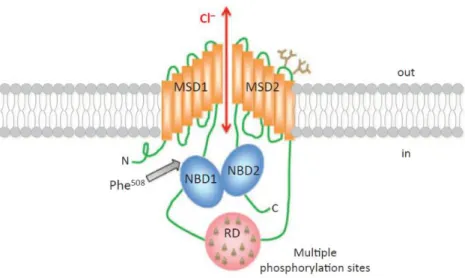
The CFTR gene directs the synthesis of transcripts of about 6.5kb (after splicing) and encodes for an integral membrane glycoprotein composed of 1480 amino acids4, 10. The CFTR protein functions as a chloride channel predominantly expressed at the apical membrane of epithelial cells lining the target organs: lung, pancreas, intestine and sweat gland2. As a member of the ATP-binding cassette (ABC) transporter superfamily, CFTR has a symmetrical multi-domain structure, consisting of two membrane spanning domains (MSD1 and MSD2), each composed of six transmembrane segments (TM1-TM12) which form the pore through which anions pass, and two nucleotide-binding domains (NBD1, which harbors F508del mutation, and NBD2)5. Both NBDs bind and hydrolyze ATP, regulating the channel opening11. CFTR structure also contains a central and highly charged regulatory (R) domain, absent from all other ABC transporters, with multiple phosphorylation consensus sites (Figure 1.2)10, 12. The phosphorylation status of this domain may regulate the dimerization of the NBDs domains13. CFTR C-terminus, well known for accommodating a PSD95, Dlg1, ZO-1 (PDZ)-binding motif, is also involved in several protein interactions14.
4
Figure 1.2: CFTR structure at the plasma membrane. MSD, membrane spanning domains; RD,
regulatory domain (contains multiple phosphorylation sites); NDB, nucleotide-binding domain (NDB1 harbors the most frequent CF-causing mutation, F508del)10.
1.3 CFTR folding and trafficking
Although CFTR folding is a highly regulated process, because CFTR is such a large multidomain membrane glycoprotein, proper folding is difficult to achieve. Hereupon, during CFTR biogenesis, only 20-40% of the newly synthesized protein escapes the endoplasmic reticulum (ER) quality control (ERQC), undergo complex glycosylation in the Golgi complex and acquire the native conformation. The remaining protein is tagged for degradation by the ubiquitin-proteasome pathway15, 16. Thus, the coordinated folding of CFTR and its individual domains occur through an iterative process in three different cellular compartments: ER membrane and lumen, as well as cytosol17.
Similarly to what happens with other multidomain glycoproteins, CFTR is co-translationally inserted into the ER membrane and simultaneously N-linked to glycosyl groups. Additionally, as a membrane protein, CFTR follows the general route of the secretory pathway to reach the plasma membrane (PM), i.e. through the Golgi10.
In this way, while CFTR is being translated in a cytosolic ribosome, an ER targeting motif is recognized by the signal recognition particle, causing it to be targeted to the ER surface and co-translationally inserted into the ER membrane through the Sec61 translocon
5 complex17. While the nascent polypeptide chain is being inserted in the ER membrane, the exposed parts in the cytosol associate with the cytosolic molecular chaperones Hsp90, Hsc70/Hdj-2, and Hsp70/Hdj-1 (with this forming a first checkpoint in the ER quality control (ERQC) for CFTR; Figure 1.3)10, 18. Immediately after insertion into the ER membrane, the newly synthesized CFTR polypeptide chain emerging into the ER lumen is core-glycosylated at Asn894 and Asn900. CFTR N-glycosylation, which occurs by the addition of a glycoconjugate with 14 osidic residues, plays a pivotal role in its folding, sorting and trafficking10, 18. This glycoconjugate is processed by glucosidases generating a glycosylated intermediate recognizable by luminal calnexin and calreticulin chaperones that allow the folding to progress (second checkpoint)15, 19. After these two initial ER folding checkpoints, CFTR conformation is assessed at a third checkpoint. This is a retention mechanism that recognizes arginine-framed tripeptide (ATFs) motifs at the ER exit sites. From this point, in the ER, there are two different pathways:
-If the protein didn’t attain the proper folding, it is recognized by UDPglycoprotein glucosyltransferase (UGGT), which re-glycosylates CFTR. Hereupon, a new round of chaperone binding, de-glucosylation and proofreading begins. If the protein undergoes too many of these rounds, eventually it becomes subject to the ubiquitination-dependent endoplasmic-reticulum-associated protein degradation (ERAD). After being marked by the ER degradation-enhancing α-mannosidase-like protein (EDEM) for degradation, CFTR is retro-translocated to the cytoplasm and degraded by the ubiquitin-proteasome pathway10,
15. Most of the CFTR bearing the F508del mutation, owing to its incorrect folding, is
retained and degraded at the ER due to the ERQC system10;
-If the protein is folded correctly, it proceeds to the secretory pathway. CFTR is released from the ER, loaded into COPII-coated vesicles that traffic to the Golgi, where its glycan moieties undergo further processing. The fully glycosylated CFTR is then incorporated into secretory vesicles and delivered to the apical membrane, exposing its glycans to the extracellular space and exerting its function10. More recently, ER stress was reported to induce CFTR trafficking from the ER to the apical membrane through a Golgi-independent GRASP-dependent unconventional secretion pathway20.
During the maturation process, CFTR undergoes several glycosylation steps, first in the ER and then in the Golgi apparatus. For CFTR, glycosylation at the ER creates the core-glycosylated 135-140kDa (immature) form of the protein (also called band B). From the ER, wild-type (WT) CFTR traffics through the Golgi complex, where it is processed by
6
multiple Golgi glycosyltransferases and glycosidases, creating the fully 150-160kDa mature form of CFTR (also termed band C)10, 16.
Figure 1.3: Checkpoints for the ER quality control of CFTR. At the first checkpoint, Hsc70 and
Hsp70 interact with the cytosolic domains of nascent CFTR to assess its conformation – this is the major mechanism for retaining and discarding F508del-CFTR. At the second checkpoint, WT-CFTR proceeds through the folding pathway through interaction of its N-glycosyl residues with calnexin. After the third checkpoint (retention mechanism that recognizes arginine-framed tripeptide (AFT) motifs), correctly folded CFTR exits the ER, proceeding through the secretory pathway. Ub, ubiquitin10.
Additionally, after being delivered to the PM, CFTR can be subjected to endocytosis. CFTR is internalized into early endosomes where it can either return to the PM, through Rab11/MyoVb-driven recycling endosomes, or diverted from the recycling pathway into late endosomes, via Rab7, followed by lysosomal degradation21-23. As CFTR internalization at the cell surface is a rapid process compared to CFTR biosynthesis and maturation, the recycling of internalized channels is considered to be a key process in maintaining a functional pool of CFTR at the PM10.
7 1.4 CFTR anchoring at the plasma membrane
Recycling of internalized CFTR to the PM has been considered to be the main mechanism for sustaining a functional pool of CFTR at the cell surface, albeit some evidence suggest that up to 50% of surface CFTR exists in an immobile pool, tethered to filamentous actin (F-actin), in airway epithelial cells10, 24. Because the CFTR C terminus (amino acids residues DTRL) forms a consensus PDZ binding domain (C-terminal X-[S/T]-X-[V/I/L]), it can bind to several proteins that contain these PDZ domains24, 25. One of these C-terminus-interactors is the PDZ adaptor protein Na+/H+-exchanger regulatory factor isoform-1 (NHERF-1, also known as EBP50, ezrin-binding protein, 50kDa)26-28. NHERF-1 anchors CFTR at the PM to the actin cytoskeleton and is also important to target exosome- and endosome associated CFTR to the apical membrane of polarized epithelial cells. The first involves the interaction of CFTR-bound NHERF-1 with the ezrin/radixin/moesin (ERM) family protein ezrin, locking CFTR in an immobile and actin-tethered complex that prevents its endocytosis (Figure 1.4)10, 26. NHERF1 increases the chloride channel activity of CFTR. It has also been suggested that NHERF1 may induce CFTR dimerization, facilitating CFTR intermolecular interactions, which alter channel conformation and activity. However, the role of PDZ-domain proteins in the formation of CFTR dimers is controversial26.
NHERF1 role upon CFTR stabilization involves also interaction with small GTPases (guanosine triphosphate hydrolase) of the Rho family. These GTPases, found in all eukaryotic organisms, are divided into three subfamilies, grouped according to their functional and structural similarity to their three founding members, RhoA, Rac1, and Cdc4210. The members of the Rho family are key regulators of actin cytoskeleton dynamics, cell polarity and membrane trafficking through their modulation of F-actin remodeling10, 29, 30. Consistently, NHERF1 overexpression stimulates the activation of endogenous RhoA and RhoA-activated kinase (ROCK), thus leading to reorganization of the actin cytoskeleton and stabilization of the multiprotein complex F508del-CFTR– NHERF1–ezrin–actin at the apical PM31. Therefore, CFTR surface anchoring and retention
8
Figure 1.4: CFTR anchoring at the plasma membrane. Several proteins interact directly or
indirectly with CFTR, including protein phosphatase-2A (PP2A), Na+/H+ exchanger regulatory factor isoform-1 (NHERF1), protein kinase A (PKA) and ezrin, forming a multiprotein complex that stabilizes CFTR at the plasma membrane. ERM, ezrin, radixin, moesin binding domain; NBD, nucleotide-binding domain; R, regulatory domainAdapted from 26.
1.5 CFTR function
CFTR functions mainly as a chloride channel, playing a fundamental role in fluid and electrolyte transport across the epithelial cells and regulation of the airway surface liquid composition. However, CFTR function is not restricted to being a chloride channel, being also permeable to other anions, namely bicarbonate, thus providing an important role as a regulator of transmembrane pH gradients32, 33. CFTR is also implicated in the control of extracellular reactive oxidative species balance, as a result of glutathione transport34, 35. Therefore, because this channel regulates transepithelial salt and water movement, it coordinates epithelial hydration and efficient mucociliary clearance. This is also achieved through the regulation of other PM channels, namely the epithelial sodium channel (ENaC), calcium-activated chloride channels (CaCCs), outwardly rectifying chloride channels (ORCCs) and renal outer medullary potassium channel (ROMK)36-39. Contrary to other ABC transporters, CFTR is incapable of actively driving ion transport against a gradient; instead, it functions as a passive channel that allows bidirectional flow of ions14.
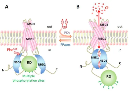
9 CFTR chloride channel gating is tightly regulated by the balance of kinases and phosphatases activity within the cell and by the cellular levels of ATP and cAMP. The MSDs contribute to the formation of the chloride pore and NBDs bind and hydrolyze ATP to regulate the channel gating. Activation of the cAMP-dependent protein kinase A (PKA) drives the phosphorylation of multiple serine/threonine residues within the R domain, required to channel activation. Finally, protein phosphatases dephosphorylate the R domain and return the channel to its quiescent state (Figure 1.5)12.
Figure 1.5: Regulation of the CFTR chloride channel opening. Schematic model showing the
regulation of CFTR through cAMP-dependent phosphorylation of the R domain (RD) and ATP binding and hydrolysis at the NBDs. In and Out denote the intra- and extracellular sides of the cell membrane. MSD, membrane-spanning domain; NBD, nucleotide-binding domain; P, phosphorylation of the R domain; PKA, protein kinase A; PPase, protein phosphatase40.
10
This PKA-mediated activation of CFTR requires integrity of the actin cytoskeleton and compartmentalized cAMP within the cell41. It has been reported that the cAMP increase required for PKA-mediated activation of CFTR must be specifically localized near the membrane, in the subcortical compartment31, 41. Additionally, this compartmentalization is dependent on the integrity of the subcortical cytoskeleton and on the presence of adenylate cyclases (AC; responsible for the production of cAMP) and A-kinase anchoring proteins (AKAPs), such as ezrin. AKAPs are scaffold proteins, anchoring PKA, phosphatases, phosphodiesterases and other signaling proteins in the vicinity of CFTR. The formation of multiple protein macromolecular complexes in subcellular microdomains increases the specificity and efficiency of signaling towards CFTR, allowing a cAMP-dependent control of chloride efflux14, 41-44. Although it is well-established that cAMP plays a crucial role in CFTR regulation, PKA is not the only cAMP sensor within the cell.
11 1.6 EPAC – Overview
Since the discovery that cAMP activates the phosphorylating enzyme PKA, the cAMP messenger system has been shown to regulate cAMP production by heteromeric guanine nucleotide–binding proteins (G proteins), subsequent binding of cAMP to PKA, and consequent phosphorylation of PKA substrates45, 46. cAMP is a second messenger that has a role in many physiological processes ranging from the regulation of heart rhythm, insulin secretion or neurotransmitter release47, 48. Initially, PKA was considered to be the essential effector molecule mediating many of these physiological effects initiated by receptors coupled to generation of cAMP45, 46, 49. However, PKA has not been clearly linked to all these processes45. A database screen conducted to explain the insensitivity of cAMP-induced activation of the small GTPase Rap1 to inhibitors of PKA, in 1998, led to the identification of EPAC, an exchange protein directly activated by cAMP (also known as cAMP-GEF), heralding the age of signal transduction47, 48, 50. This approach identified independently two proteins containing a cyclic nucleotide-binding (CNB) domain: EPAC1 and EPAC2 (or RAPGEF3 and 4, respectively). Another protein highly homologous to EPAC with activity toward Rap, known as REPAC (or RAPGEF5), was later identified; however, this protein lacks a cAMP-binding domain51.
EPAC proteins function as guanine nucleotide exchange factors (GEFs) for both Rap1 and Rap251. Rap is a small Ras-like GTPase that was first identified as a protein that could suppress the oncogenic transformation of cells by Ras. It belongs to the Ras superfamily of small G proteins, specifically to the Rap subfamily, which cycle between an inactive guanosine diphosphate (GDP)-bound state and an active guanosine triphosphate (GTP)-bound state. Thus, small G-proteins function as simple molecular switches with an inactive GDP-bound and active GTP-bound conformation47, 50, 52. GEFs catalyze the exchange of GDP for GTP and thereby the activation of the G protein, whereas GTPase-activating proteins (GAPs) enhance GTP hydrolysis. Several other GEFs for Rap, in addition to EPAC, have been identified. These GEFs connect different inputs to Rap activation and are associated to distinct functions of Rap. The most intriguing RapGEF, however, is EPAC, because this GEF represents a novel target for cAMP, independent from the classical target protein PKA51. Rap, whose activation by cAMP occurs independently of PKA, is activated by several extracellular stimuli and is involved in an immense diversity of cellular processes such as cell proliferation and adhesion, actin cytoskeleton dynamics, cell polarity, exocytosis, membrane protein recycling and cell differentiation50, 54-61.
12
EPAC1 and EPAC2 are present in most tissues, although with different expression levels: EPAC1 is highly abundant in blood vessels, skeletal muscles, central nervous system, kidney, adipose tissue, ovary and uterus; EPAC2 is mostly expressed in the central nervous system, pancreas and adrenal gland45, 50, 62. REPAC is strongly expressed in the human brain63.
1.7 EPAC protein and its activation
EPAC1 and EPAC2 are multidomain proteins that consist of an N-terminal regulatory region and a C-terminal catalytic region (Figure 1.6a). The regulatory region has one (EPAC1) or two (EPAC2) CNB domains and a DEP (Dishevelled, Egl-10, and Pleckstrin) domain. The catalytic region harbors the CDC25-homology domain (CDC25-HD) for enzymatically exchange activity, which is stabilized by a Ras exchange motif (REM) domain. There is a Ras-association (RA) domain between these domains. The regulatory region has an autoinhibitory function that is relieved by binding of cAMP (Figure 1.6b)47, 48,
51, 64, 65. In the inactive conformation, the CNB domains sterically prevent Rap binding to
the catalytic site. As already known for EPAC2, an ionic interaction between the C-terminal CNB (CNB-B) domain and the CDC25 domain dynamically stabilizes this conformation. Upon binding of cAMP, a subtle change within the CNB-B domain allows the regulatory region to move away from the catalytic region. This position is stabilized by interactions between cAMP, CNB-B and REM domains47, 66, 67.
13
Figure 1.6: Structure of EPAC. (a) Schematic representation of the domain architecture of both
EPAC proteins. The regulatory region contains one or two CNB (cyclic nucleotide–binding) domains and a DEP (Disheveled, Egl-10, and Pleckstrin) domain. The catalytical region harbors the CDC25-homology domain (CDC25-HD), the Ras exchange motif (REM) domain and a Ras-association (RA) domain. (b) Crystal structure model of both inactive and active EPAC2. For simplicity, only the catalytic region and the CNB-B domain (indicated with a dotted line in panel a) are shown47.
Hormonal stimulation of many heterotrimeric G-protein-coupled receptors frequently results in the activation of AC leading to an increase in cAMP levels. cAMP can be then degraded by phosphodiesterases (PDE). In the past, the discovery and analysis of cAMP-mediated events has been greatly facilitated by specific compounds that modulate intracellular cAMP levels. The most commonly used ones are forskolin, a natural compound that directly activates ACs, and a variety of PDE inhibitors, such as 3-isobutyl-1-methylxanthine (IBMX), both added to further enhance cAMP levels47, 48, 68. During the development of EPAC-selective analogs of cAMP, it was noticed that EPAC proteins lack the glutamate residue that, in PKA and cAMP-gated ion channels, interacts with the ribose
A
14
of cAMP. Taking advantage of this knowledge, 8-pCPT-2’-O-Me-cAMP (also known as 007) was developed as a selective agonist for EPAC, being ten times more efficient than cAMP in activating EPAC1. To increase the relatively low membrane permeability of this compound, an acetoxymethyl (AM)-ester was introduced to mask the negatively charged phosphate group, originating 007-AM. This modification allows exceptional cell permeability being intracellularly removed by esterases to generate 00747, 69-71. Despite 007-AM also activating EPAC2, it was reported recently that sulfonylurea selectively activates EPAC2 isoform, but not the closely related EPAC1, further establishing a new class of isoform-selective enzyme activators48, 72.
1.8 Spatial regulation of EPAC
Although cAMP can rapidly diffuse within the cytosol, it becomes unevenly distributed and concentrated in local microdomains. Thus, cAMP-elevating hormones do not induce homogenous increases of cAMP within the cell. The molecular basis of this cAMP signaling compartmentalization involves its synthesis by membrane-associated AC, subsequent diffusion through the cell and local cAMP degradation by PDEs. As a result, these cAMP mediators generate cAMP gradients within the cell47, 73. In addition to cAMP compartmentalization, cAMP effectors are also spatially regulated by binding to scaffolding proteins, as has been previously shown for PKA (section 1.5). AKAPs target PKA to distinct subcellular locations and mediate the assembly of large signaling complexes, linking PKA to specific cellular functions. In a similar way, EPAC proteins are spatially regulated by different anchoring mechanisms47.
In response to the cAMP-induced conformational change, EPAC1 is targeted to the PM. This is essential for the ability of EPAC1 to induce its downstream effectors, like Rap, at the PM74. The DEP domain is required for this translocation and it has been shown to tether EPAC1 to phosphatydic acid (PA) at the membrane75. EPAC1 is also tethered to the PM by an additional mechanism, through phosphorylated (active) ERM scaffolding proteins76. Thus, EPAC1 can be recruited to PM by two different pathways: one is through PA and is cAMP- and DEP-dependent; the other way is through ERM proteins and is cAMP-independent and N-terminal-region-dependent75, 76. Radixin has also been reported to bind EPAC1 and PKA simultaneously, bringing these two cAMP sensors together and creating a functional cAMP-sensing compartment for efficient signal transduction77. All
15 these data suggest that EPAC1 might co-localize or even interact with CFTR, which argues for a possible function of this GEF in CFTR function regulation or membrane stability. Additionally, EPAC1 is activated by stimuli that result in increased subcortical cAMP, the same stimuli that induce PKA-mediated activation of CFTR, which further supports this hypothesis.
EPAC2 is targeted to the PM after binding to activated Ras proteins, via its RA domain and independently of its conformational state78.
Several other subcellular localizations have been reported for EPAC1, some of which link EPAC1 to specific cellular processes (Figure 1.7). Nuclear EPAC1 was found to regulate the DNA damage–responsive kinase (DNA-PK) within the nucleus79. EPAC1 is also
targeted to microtubules, in interphase or mitotic cells, and this interaction is probably required for the role of EPAC in microtubule polymerization. EPAC1 has also been observed in other localizations that may be associated with distinct functions, such as centrosomes, mitochondria, the nuclear pore complex and the apical membrane of renal epithelial cells47, 52. The multidomain structure of EPAC indicates that it may have multiple binding partners, and indeed numerous interacting proteins have already been described for both EPAC1 and EPAC2. Alternative splicing may further add another layer of complexity to the spatial regulation of EPAC47.
Figure 1.7: Spatial regulation of EPAC1. (a) Spatial regulation of the EPAC1, showing how the
diverse EPAC1-interacting proteins (blue) affect the localization of this GEF. ERM, Ezrin, Radixin, Moesin; MAP1, microtubule-associated protein 1; PDE4D3, phosphodiesterase 4D3; RanBP2, Ran-binding protein 2Adapted from 52.
16
1.9 Biological functions of EPAC
The major catalytic function of EPAC is the guanine nucleotide exchange of Rap1 and Rap2, with EPAC thus controling the Rap-mediated processes downstream of cAMP. However, several other GEFs or GAPs can regulate Rap activity and Rap1 and Rap2 might not mediate all effects of cAMP-EPAC (Figure 1.8). It has been reported that EPAC1 also activates R-ras, a Ras-like small GTPase implicated in the control of integrin-mediated cell adhesion. Undoubtedly, the EPAC-selective cAMP analog 007 has helped to highlight a role for EPAC and Rap in a wide range of biological processes, ranging from exocytosis of insulin in the β-cells of the pancreas to the regulation of calcium currents in cardiomyocytes, permeability of the vascular endothelium, ionic transport across intestinal cells and tissue fibrosis. Most of these processes are also modulated by signaling via the cAMP effector PKA, proving the interconnectivity between both cAMP pathways47, 48, 80-83. Indeed, both cAMP targets are often associated with the same biological process, in which they fulfill either opposite or synergistic effects. This dual control may enhance the dynamic range of cAMP signaling, as PKA-mediated events are proposed to occur at much lower cAMP levels than the activation of EPAC84. For increased complexity, the spatial localization of EPAC is also a crucial determinant of its function48. Thus, it is anticipated that the study of EPAC as a cAMP-signaling mediator is a starting field with a lot more to be discovered, in the same way that cAMP has been a major hit of research since its discovery more than 50 years ago.
17
Figure 1.8: Interconnectivity between the EPAC- and PKA-signaling pathway. After stimulation of a
receptor (R) coupled to a stimulatory G protein (Gs), adenylate cyclase (AC) is activated, promoting cAMP synthesis and consequent activation of both EPAC and PKA. Both proteins are involved in the regulation of cell adhesion, cell–cell junction formation, secretion and ion channels. Inhibitory G proteins may also inhibit AC activity. PDE, Phosphodiesterase; GEF, guanine-nucleotide-exchange factors; GAP, GTPase-activating protein48.
Although a potential connection between CFTR and EPAC has never been reported, this cAMP effector is involved, as already mentioned, in the regulation of to-cell and cell-matrix adhesion, cytoskeleton rearrangements and cell polarization, processes that affect CFTR regulation41, 85, 86. Thus, more detailed knowledge about a potential relationship between CFTR and EPAC is needed, as EPAC may be a new link between CFTR and cAMP.
18
1.10 Objectives
Here, we proposed to study the role of a novel cAMP signalling pathway in the regulation of CFTR at the plasma membrane in order to better understand the mechanisms by which the protein is anchored at its cellular location and ultimately how can this be modulated for the benefit of CF patients.
Specifically, the aim of the present work is to study the interaction between CFTR and EPAC1, the predominant isoform of EPAC in cAMP signaling, and to evaluate the impact of this cAMP effector on CFTR biogenesis, trafficking and PM anchoring. In order to achieve this goal, we propose to:
1- Validate 007-AM as a specific cAMP analogue for EPAC in Cystic Fibrosis Bronchial Epithelial cells (CFBE) by fluorescence resonance energy transfer (FRET) and co-immunoprecipitation (co-IP);
2- Validate cAMP-induced translocation of EPAC in CFBE cells by confocal live-cell imaging and cell fractionation;
3- Evaluate co-localization and interaction between CFTR and EPAC1 (and the effect of 007-AM) by confocal live-cell imaging and co-IP;
4- Characterize the molecular linkers involved in the CFTR and EPAC1 interaction and the role of PDZ binding domain on CFTR-EPAC1 interaction;
5- Assess the effect of EPAC1 on CFTR PM stability using endocytosis and biotinylation assays;
6- Assess the dependence of EPAC-induced cell adhesion dependence on the expression of CFTR.
Chapter II
21 2. Materials and Methods
2.1 Molecular Biology
2.1.1 Plasmid vectors and siRNAs
All plasmids and siRNAs (small interfering RNA) used in this work are listed on the following tables (Table 2.1 and 2.2, respectively). Maps for all vectors are available in
Appendices Plasmid maps.
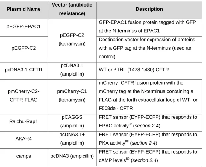
Table 2.1: List of plasmids used and their description.
Plasmid Name Vector (antibiotic
resistance) Description
pEGFP-EPAC1
pEGFP-C2 (kanamycin)
GFP-EPAC1 fusion protein tagged with GFP at the N-terminus of EPAC1
pEGFP-C2
Destination vector for expression of proteins with a GFP tag at the N-terminus (used as control) pcDNA3.1-CFTR pcDNA3.1 (ampicillin) WT or ∆TRL (1478-1480) CFTR pmCherry-C2-CFTR-FLAG pmCherry-C1 (kanamycin)
mCherry- CFTR fusion protein with the mCherry tag at the N-terminus containing a FLAG at the forth extracellular loop of WT- or F508del- CFTR
Raichu-Rap1 pCAGGS
(ampicillin)
FRET sensor (EYFP-ECFP) that responds to EPAC activity87 (section 2.4)
AKAR4 pcDNA3.1+
(ampicillin)
FRET sensor (EYFP-ECFP) that responds to PKA activity88 (section 2.4)
camps pcDNA3 (ampicillin) FRET sensor (EYFP-ECFP) that responds to cAMP levels89 (section 2.4)
22
Table 2.2: List of siRNA used.
siRNA Name Brand (Reference)
Silencer® Select Negative Control No. 2 siRNA Life Technologies (4390846) Silencer® Select siRNA RAPGEF3 Life Technologies (4392420) siGENOME SMART pool non-targeting siRNA Thermo Scientific (D-001206-13) siGENOME SMART pool human EZR Thermo Scientific (EG:7430) siGENOME SMART pool human SLC9A3R1 Thermo Scientific (EG:9368)
2.1.2 Transformation of competent bacteria
The bacterial strains used for cloning and DNA amplification were: One Shot TOP10 (Invitrogen) or 2); XL1-Blue competent cells (Stratagene). Preparation of competent bacteria was done in-house according to the technique described by Hanrahan90.
Bacteria were transformed by incubating a 200µL aliquot of competent bacteria with DNA (at least, 100ng of ligation products or 5ng of purified plasmids) for 30 min in ice. Afterwards, the mixture was submitted to heat-shock (90 sec at 42ºC), incubated 2 min on ice followed by incubation in LB media without antibiotic for 45 min at 37ºC at 220rpm, in order to allow antibiotic resistance to be expressed. After pelleting the bacteria (3000g for 5 min), the supernatant was discarded and the pellet was resuspended in the remaining medium. This suspension was then plated into LB-agar supplemented with appropriate concentration of the selected antibiotic and plates were incubated overnight at 37ºC. Transformed bacterial colonies were then grown in LB medium supplemented with the appropriate antibiotic and plasmid DNA was extracted. Clones were stored in liquid LB medium supplemented with 15% (w/v) glycerol (Sigma-Aldrich) at -20ºC.
2.1.3 DNA extraction and quantification
Plasmid DNA was purified, at small or large scale, with QIAGENMiniprep kit (QIAGEN) or QIAGENMaxiprep kit (QIAGEN), respectively, accordingly with manufacturer’s guidelines. DNA concentration was determined by measurement of the absorbance at 260nm using a Nanodrop 2000 spectrophotometer (Thermo Scientific) and its purity was evaluated by assessment of the A260/A280 ratio. Only DNA with a ratio above 1.8 was considered pure
23
2.1.4 Mutagenesis
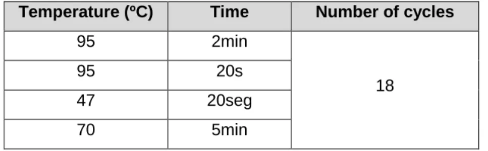
The TRL deletion was performed on pcDNA3.1-WT-CFTR using the KOD Hot Start DNA Polymerase (Novagene) with a pair of complementary custom designed mutagenic primers (Thermo Electron Corporation): delTRL_FW (forward primer, sequence 5’- GAAGAGGTGCAAGATTAGAGAGCAGCATAAATG-3’) and delTRL_REV (reverse primer, sequence 5’-CATTTATGCTGCTCTCTAATCTTGCACCTCTTC-3’). Primers were designed with the software BioEdit (http://www.mbio.ncsu.edu/bioedit/bioedit.html). The PCR program used for the mutagenesis reaction is displayed in the following table.
Table 2.3: PCR program for the mutagenesis reaction.
Temperature (ºC) Time Number of cycles
95 2min
18
95 20s
47 20seg
70 5min
After confirming the DNA amplification by agarose gel electrophoresis, the PCR products were incubated for 1h with DpnI (Invitrogen), a restriction enzyme that specifically hydrolyzes methylated and semi-methylated DNA, thus degrading the non-amplified template DNA. After bacteria transformation (section 2.1.2) and DNA extraction (section
2.1.3), the presence of the mutation was confirmed by DNA sequencing (section 2.1.5).
2.1.5 DNA sequencing
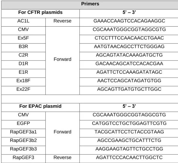
The sequencing reactions were outsourced to StabVida. Primers used for CFTR and EPAC plasmids are listed below (Table 2.4). All sequencing primers were obtained through StabVida. The resulting sequences were analyzed using the BioEdit software by comparison with a reference sequence.
24
Table 2.4: Primers used for sequencing.
Primers
For CFTR plasmids 5’ – 3’
AC1L Reverse GAAACCAAGTCCACAGAAGGC
CMV Forward CGCAAATGGGCGGTAGGCGTG Ex5F CTCCTTTCCAACAACCTGAAC B3R AATGTAACAGCCTTCTGGGAG C2R AGCAGTATACAAAGATGCTG D1R GACAACAGCATCCACACGAA E1R AGATTCTCCAAAGATATAGC Ex18F AACTCCAGCATAGATGTGG Ex22F AGCAGTTGATGTGCTTGGC
For EPAC plasmid 5’ – 3’
CMV Forward CGCAAATGGGCGGTAGGCGTG EGFP CATGGTCCTGCTGGAGTTCGTG RapGEF3a1 TACGCATTCCTCTACCGTAAG RapGEF3b2 AGCCGAAGCTGCATTTCTG RapGEF3b3 AAGGAAGTAGTTCTGCCTGG
25 2.2 Cell Culture
Cells were cultured in the appropriate medium in plastic flasks or plates, in an incubator with a humidified atmosphere of 5% CO2 at 37ºC. For cell detachment from the plastic
surface, cells were washed with in PBS and then incubated with trypsin (Life Technologies). Cell lines were stored frozen at -80ºC or in liquid nitrogen in 40% (v/v) of the appropriated cell medium, 50% (v/v) FBS and 10% (v/v) DMSO (Sigma-Aldrich). To minimize cell damage, freezing was performed in a way that allows a cooling speed around 1ºC/min. After thawing, cells were washed and seeded with the indicated medium for each cell line (see below). To eradicate/prevent mycoplasma contamination, cells were treated occasionally with Myco4 (AppliChem). To assess cell contamination, a PCR test for Mycoplasma was performed.
2.2.1 Cell types 2.2.1.1 CFBE cells
Cystic Fibrosis Bronchial Epithelial (CFBE41o-) cells91, from now on simply referred as CFBE, stably overexpressing either WT- or F508del-CFTR92 or without expressing CFTR (parental CFBE) were cultured in Eagle’s minimal essential medium (EMEM; Life Technologies) supplemented with 10% fetal bovine serum (FBS; Life Technologies), 2mM L-glutamine (Invitrogen). When appropriate/needed, medium was also supplemented with an antibiotic mixture of penicillin (Pen; 100U/mL; Life Technologies) and streptomycin (Strep; 100µg/mL; Life Technologies) and with puromycin (2.5µg/mL; Life Technologies).
2.2.1.2 Calu3 cells
Non-transduced Calu393 cells (Calu3-WT) or expressing a control (Calu3 shControl), Ezrin (Calu3 shEzrin) or NHERF1 shRNA (Calu3 shNHERF1) were generated in this work (section 2.2.4) and used for further analysis. Cells were cultured in DMEM-F12 (Lonza) media supplemented with 10% FBS and with or without puromycin (5µg/mL).
26
2.2.1.3 A549 cells
A549 cells overexpressing mCherry-WT- or mCherry-F508del-CFTR94 under control of a tet-on promoter were cultured in DMEM (Lonza) media supplemented with 10% FBS and puromycin (2.5µg/mL). CFTR expression was induced with 10 µg/mL of doxycycline and 1mM of sodium butyrate for 24h.
2.2.1.4 HEK293 cells
Human Embryonic Kidney (HEK293) cells95 were cultured in DMEM media supplemented with 10% FBS.
2.2.3 Transfections
Cells were transiently transfected with plasmid DNA or siRNA (section 2.1.1) using Lipofectamine2000 (Life Technologies), a cationic liposome formulation that forms DNA complexes able to fuse with cell membrane, according to the manufacture’s guidelines96.
Usually, for one 6 well-plate, 750µL of OptiMEM (Life Technologies) were mixed with 36µL of Lipofectamine and other 750µL of OptiMEM were mixed with 12µg of plasmid DNA or 1µL of 20µM siRNA and incubated at RT (room temperature) for 5min. After that, the second mixture was added to the first one and incubated 30min at RT. 250µL of this final mixture was added to each well with 60-90% confluent cultured cells.
27
2.2.4 Lentivirus infection 2.2.4.1 Lentivirus production
HEK293 cells were transiently transfected with a mixture of the packaging (pCMV-dR8.74psPAX2), envelop (pMD2.G) and hairpin-pLKO.1 vector (Empty vector control, SHC001; NHERF1, SHCLND SLC9A3R1; Ezrin, SHCLND EZR; Sigma-Aldrich) plasmids using X-tremeGENE9 DNA transfection reagent (LifeScience), according to the manufacturer’s guidelines, and incubated at 37ºC. Because lentivirus start to appear in the media supernatant ~22h post-transfection, 18h after incubation at 37ºC, the transfection reagent medium was removed and replaced with high-BSA growth media (DMEM supplemented with 10%iFBS (inactivated FBS), 1.1g/100mL BSA (Bovine serum albumin) and 1x Pen/Strep). The next day, the media was harvested and stored at -20ºC. The cells were incubated again with high-BSA growth media and the medium was harvested one last time 24h later.
2.2.4.2 Production of a stable cell line
After washing 70% confluent Calu3-WT cells with Hank's Balanced Salt Solution (HBSS; Life Technologies), EMEM medium supplemented with 10% (v/v) FBS, polybrene (8µg/mL) and 5-20% (v/v) lentivirus-containing medium (section 2.2.4.1) was added to the cells. After centrifugation (2200rpm) at 37ºC for 30min, the cell plates were incubated at 37ºC for 24h. The medium was then replaced by EMEM supplemented with 10% (v/v) FBS and puromycin (5µg/mL), which allows the selection of cells that were efficiently infected. This medium change was repeated for, at least, 5 consecutive days.
28
2.3 Biochemical Analysis
2.3.1 Cell lysis and total protein quantification
For protein extraction, cells were washed 3 times with cold PBS and lysed with sample buffer (1.5% (w/v) SDS; 5% (v/v) glycerol; 0.01% (w/v) bromophenol blue; 0.05mM dithiotreitol; 0.095M Tris pH 6.8). DNA was sheared by enzymatic action of 5-25U/mL of benzonase (Sigma-Aldrich) in the presence of MgCl2 2.5mM.
Total protein quantification was performed using Bradford method. 1mL of Bradford reagent (Sigma Aldrich) was added to 2µL or 5µL of sample. A standard curve was performed by adding 1mL of Bradford solution to increasing concentrations of BSA (Sigma Aldrich) standards. Afterwards, the absorbance was measure at 595nm and data analysis was performed.
2.3.2 Western blot
Protein extracts were separated by SDS-polyacrylamide gel electrophoresis (PAGE) on 7.5, 8, 10 or 12.5%T (w/v) separating and 4%T stacking gels at 100-150 V. Subsequently, proteins were transfered onto Immobilon polivinylidene difluoride (PVDF) membranes (Millipore) at 400 mA for 1.5h. After blocking with 5 % (w/v) skimmed milk in phosphate-buffered saline (PBS: NaCl 137mM; KCl 2.7mM; KH2PO4 1.5mM; Na2HPO4 6.5mM, pH
7.4) containing 0.1% (v/v)-Tween (PBS-T) or Tris-buffered saline with 0.5% Tween 20 (TBS-T; Alfa Aesar) for 1h at RT, membranes were incubated overnight with primary antibodies (Table 2.5) at 4ºC. After three 10 min washes with PBS-T/TBS-T, the membranes were incubated 1h at RT with horseradish peroxidase-conjugated secondary antibody (see antibodies on Table 2.5) and washed as above. All antibodies were diluted in blocking solution. Chemiluminescent detection was performed using: Chemidoc XRS+ analyser (BioRad) and the signal was developed with the ImmunStar Western C Chemiluminescence kit (BioRad) or using Compact X4 Automatic Processor (Xograph) and the signal was developed using ECL Western Blotting Substrate (Thermo Scientific). The quantification of band volume was performed using the Image Lab software (BioRad) or ImageJ (http://imagej.nih.gov/ij/). In order to re-incubate with another primary antibody, the membranes were washed at least 5 times for 10 min with PBS-T or TBS-T, or washed 1h with PLUS Western Blot Stripping Buffer (46430, Thermo Scientific). After this washing step, the membranes were blocked again for 1h at RT.