Amoxicillin-loaded lipid nanoparticles against
Helicobacter pylori infections
Ana Rita Oliveira Macedo Pinto
Master Thesis for the degree in Master of Science in Bioengineering –
Specialization in Molecular Biotechnology
Supervisor:
Prof. PhD Salette Reis
UCIBIO/REQUIMTE, Departamento de Ciências Químicas, Faculdade de
Farmácia, Universidade do Porto
Co-supervisors:
PhD Cláudia Nunes
UCIBIO/REQUIMTE, Departamento de Ciências Químicas, Faculdade de
Farmácia, Universidade do Porto
MSc Daniela Lopes
UCIBIO/REQUIMTE, Departamento de Ciências Químicas, Faculdade de
Farmácia, Universidade do Porto
i
Abstract
Nowadays, the increasing antibiotic resistance verified at a worldwide level lead to the failure of many treatments available for infectious diseases, such as Helicobacter pylori. This bacterium affects around 50% of the world population and is classified as human carcinogenic by the World Health Organization. Unfortunately, the efficacy of the standard triple therapy against this infection has been declined, mainly due to the increasing number of emergent antibiotic resistant bacterial strains. However, other factors are associated with the lack of efficacy, namely the degradation of antibiotics under acidic conditions, their low diffusion across the mucus layer, their insufficient residence time in the stomach, among others. Therefore, lipid nanoparticles emerged as a promising biocompatible drug delivery strategy. They can improve drugs pharmacokinetic properties, leading to an improvement of the efficacy and a decrease in the incidence of side effects. Further, the risk of development of bacterial resistance against antibiotics-loaded nanoparticles is reduced.
In this work, amoxicillin-loaded solid lipid nanoparticles (SLNs) were developed to protect amoxicillin against acidic degradation and to enhance its absorption, with the aim to increase the bioavailability. Preliminary studies were performed in order to select the most adequate lipid and preparation method of the lipid nanoparticles. From these screenings, cetyl palmitate was selected as the solid lipid and double emulsion technique was chosen as the most suitable preparation technique. Further, SLNs composed of cetyl palmitate, linolenic acid, dioleoylphosphatidylethanolamine (DOPE), Tween®80 and amoxicillin were optimized by Box-Behnken design. The optimal formulation had a low polydispersity index around 0.137 with a mean size diameter around 200 nm and a zeta potential superior to |30| mV. Additionally, the loading capacity was satisfactory high, being around 7%. The optimal formulation was stable in a suspension form at 4ºC during at least 2 months, while in the lyophilized SLNs amoxicillin degradation was visible after 1 month. In vitro release studies revealed an initial burst release (less than 30%) followed by an increased release at the simulated mucosa medium (pH 5). Then, a sustained and controlled phase was observed at physiologic pH 7.4. Methylthiazolydiphenyl-tetratozium bromide (MTT) assay revealed that the optimal SLNs suspension does not have cytotoxic effects in both L929 and MKN28 cell lines. Additionally, other three formulations with decreasing complexity compared with the abovementioned nanoparticles composition were prepared for future evaluation of the most important parameters involved in the nanoparticles-bacteria interactions. These formulations were also characterized and evaluated in in vitro release and cell viability studies, with similar results comparative to the optimal formulation.
Keywords: antimicrobial resistance, Helicobacter pylori infections, amoxicillin, lipid nanoparticles, oral
administration, lipid nanoparticles preparation techniques, nanoparticles characterization, Box-Behnken design, MTT assay.
ii
iii
Acknowledgments
I’m deeply grateful to Prof. Dr. Salette Reis for the great opportunity to work in such a fantastic research group and for all the guidance, concern and enthusiasm.
To Dr. Cláudia Nunes, thank you for your positive energy and enthusiastic way to see every result. Thank you also for your support, guidance and for the good moments.
To Daniela Lopes, thank you for everything you did during this project and for always being by my side. Thank you for your patience, I know that sometimes I was annoying and had some crazy ideas of doing many things at the same time. You never said no to anything, even when it seemed impossible. I think we were bought super women these last months.
I’m am grateful to Dr. Sofia Lima for all the guidance, knowledge and patience during the cellular assays.
I also want to thank all the people from the lab for making my days more fun. Thank you all for the good moments that I will take with me. A special thanks to my “roomie” Andreia for always being available when I needed something and to care about me.
To my friends “Nenas” for the friendship, care and for making my life better. You made my last 5 years wonderful. I will miss our adventures and maybe the days we passed like crazy studying in “Biblioteca da Maia”.
To Helena, thank you for your “eternal” friendship, since I was 6 years old you walked by my side.
To Thomas, thank you for your support and for listening to me. I know sometimes I was annoying but you were always patient. Thank you also for making me see that work is not everything that matters.
Last but not least, to my parents, brother and grandmother. Thank you for understanding my late arrivals for dinner and the enthusiasm you showed when I talked about my project and my days in the lab. Thank you for all your support.
iv
v
Table of Contents
ABSTRACT ...i
ACKNOWLEDGMENTS ... iii
TABLE OF CONTENTS ... v
LIST OF FIGURES ... ix
LIST OF TABLES ... xi
LIST OF APPENDICES ... xiii
LIST OF ABBREVIATIONS ... xv
CHAPTER 1 – INTRODUCTION ... 1
1.1
Introducing Infectious Diseases ... 1
1.2
Nanoparticles As Drug Delivery Systems ... 2
1.2.1 INTRODUCING NANOTECHNOLOGY ... 2
1.2.2 LIPID NANOPARTICLES (LNPS) ... 4
1.2.2.1 Solid Lipid Nanoparticles ... 5
1.2.2.2 Nanostructured Lipid Carriers ... 6
1.2.2.3 LNPs Synthesis Techniques ... 7
1.3
Experimental Design And Optimization Of Formulations ... 9
1.3.1 BOX-BEHNKEN DESIGN (BBD) ... 10
1.4
Lipid Nanoparticles For Oral Administration ... 11
1.5
Helicobacter pilory Infections: A Step Towards Innovation Is Needed ... 13
1.6
Aim and Strategy ... 15
vi
2.1
Materials ... 19
2.2
Amoxicillin Stability Studies ... 19
2.3
Screening of Lipids ... 20
2.4
LNPs Preparation ... 20
2.4.1 MODIFIED FREE ORGANIC-SOLVENT EMULSIFICATION/SONICATION METHOD ... 20
2.4.2 DOUBLE EMULSION METHOD ... 21
2.5
Lyophilization ... 22
2.6
LNPs Characterization ... 22
2.6.1 PARTICLE SIZE MEASUREMENTS ... 22
2.6.2 THE ZETA POTENTIAL MEASUREMENTS ... 23
2.6.3 AMOXICILLIN ENCAPSULATION EFFICIENCY ... 23
2.6.4 AMOXICILLIN LOADING CAPACITY ... 24
2.6.5 TRANSMISSION ELECTRON MICROSCOPY ... 24
2.6.6 DIFFERENTIAL SCANNING CALORIMETRY ANALYSIS ... 25
2.7
Experimental design and optimization of formulations ... 26
2.8
Stability Studies ... 28
2.9
In vitro Amoxicillin Release Study ... 28
2.10
In vitro Cell Viability Studies ... 29
2.11
Statistical Analysis ... 30
CHAPTER 3 - RESULTS AND DISCUSSION ... 31
3.1
Amoxicillin Stability Studies ... 31
3.2
Lipid Screening ... 32
3.3
Methods Screening ... 34
3.4
Experimental Design and Optimization of Formulations ... 36
3.4.1 EFFECT OF FORMULATION VARIABLES ON PARTICLE SIZE ... 40
3.4.2 EFFECT OF FORMULATION VARIABLES ON PDI ... 41
3.4.3 EFFECT OF FORMULATION VARIABLES ON LC ... 42
vii
3.5
LNPs Characterization ... 44
3.5.1 LYOPHILIZED SLNS ... 46
3.5.1.1 Transmission Electron Microscopy ... 46
3.5.1.2 Differential Scanning Calorimetry Analysis ... 47
3.5.1.3 Stability Studies ... 50
3.5.2 SLNS SUSPENSIONS ... 54
3.5.2.1 Transmission Electron Microscopy ... 54
3.5.2.2 Stability Studies ... 56
3.6
In vitro Amoxicillin Release Study ... 59
3.7
In vitro Cell Viability Studies ... 61
CHAPTER 4 - CONCLUSIONS ... 65
CHAPTER 5 - FUTURE WORK ... 67
REFERENCES ... 69
APPENDICES ... I
viii
ix
List of Figures
Figure 1.1: SLNs crystalline matrix and different types of NLCs matrices. ... 6
Figure 1.2: Cubic representation of the BBD for the optimization of three variables. ... 10
Figure 1.3: Schematic representation of the H. pylori infection. ... 14
Figure 1.4: Schematic representation of the composition of the ideal formulation. ... 16
Figure 1.5: Schematic representation of the three formulations (F1, F2 and F3) that will be studied in this work. ... 17
Figure 2.1: Scheme of the modified free organic-solvent emulsification/sonication method. 21 Figure 2.2: Scheme of the double emulsion method. ... 22
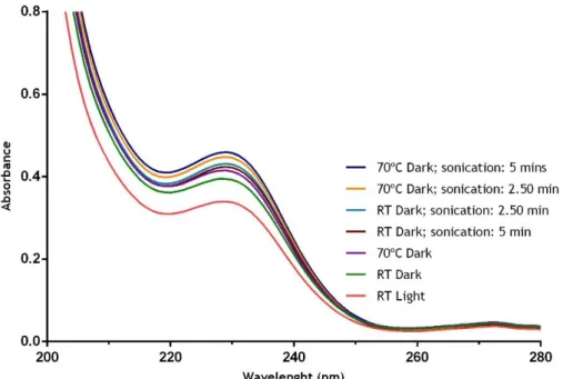
Figure 3.1: UV-Vis analysis of different conditions, viz. light (light/dark), temperature (RT/70ºC) and sonication (2.50/5 minutes), to perform AMX stability studies. RT, Room temperature. ... 32
Figure 3.2: Physical stability of the formulations obtained by modified emulsification/sonication method over time (0 (T0) and 1 (T1) weeks). ... 34
Figure 3.3: Physical stability of the formulations obtained by double emulsion method over time (0 (T0) and 1 (T1) weeks). ... 35
Figure 3.4: EE and LC of the formulations obtained by the modified emulsification/sonication and the double emulsion methods. ... 35
Figure 3.5: In vitro AMX release in three simulated conditions to mimic oral administration at 37°C: stomach lumen (pH 1.2), mucus layer (pH 5) and physiological medium (pH 7.4). ... 36
Figure 3.6: Observed vs. predicted values plot obtained for particle size using the quadratic model (R2=0.99667). Predicted values and observed values are represented in nm. ... 38
Figure 3.7: Observed vs. predicted values plot obtained for PDI using the quadratic model (R2=0.9238). ... 39
Figure 3.8: Observed vs. predicted values plot obtained for LC using the quadratic model (R2=0.99999). ... 39
Figure 3.9: Response surface plots of variances in particles size by changing (A) AMX mass and lipid mass, (B) AMX mass and Tween®80 concentration and (C) lipid mass and Tween®80 concentration... 41
Figure 3.10: Response surface plots of variances in PDI by changing (A) AMX mass and lipid mass, (B) AMX mass and Tween®80 concentration and (C) lipid mass and Tween®80 concentration... 42
Figure 3.11: Response surface plots of variances in LC by changing (A) AMX mass and lipid mass, (B) AMX mass and Tween®80 concentration and (C) lipid mass and Tween®80 concentration. ... 43
x
Figure 3.12: Schematic representation and composition of the four formulations (F1, F2, F3
and F4) studied in this work. ... 45
Figure 3.13: P4 formulation synthetized by double emulsion technique. ... 45 Figure 3.14: Schematic representation of all formulations studied in this work and their
different conditions of storage. ... 46
Figure 3.15: TEM images of the lyophilized SLNs. ... 47 Figure 3.16: Differential scanning calorimetry thermograms of: (A) F4 SLNs, (B) P4 SLNs, (C) F4
mixture and (D) P4 mixture. ... 50
Figure 3.17: Evaluation of the particle size of the lyophilized formulations over time (0 (T0),
1 (T1), 2 (T2) and 4 (T4) weeks). ... 51
Figure 3.18: Evaluation of the PDI of the lyophilized formulations over time (0 (T0), 1 (T1), 2
(T2) and 4 (T4) weeks). ... 52
Figure 3.19: Evaluation of the zeta potential of the lyophilized formulations over time (0 (T0),
1 (T1), 2 (T2) and 4 (T4) weeks). ... 52
Figure 3.20: Evaluation of the LC of the lyophilized formulations over time (0 (T0), 1 (T1), 2
(T2) and 4 (T4) weeks). ... 53
Figure 3.21: Lyophilized SLNs synthetized by double emulsion technique, after 1 month of
storage at RT: A) P3 formulation (placebo), B) F3 formulation. ... 54
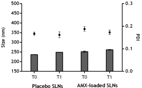
Figure 3.22: TEM images of the SLNs suspensions. ... 55 Figure 3.23: Evaluation of the particle size of the SLNs suspensions over time (0 (T0), 1 (T1),
2 (T2), 4 (T4) and 8 (T8) weeks). ... 56
Figure 3.24: Evaluation of the PDI of the SLNs suspensions over time (0 (T0), 1 (T1), 2 (T2), 4
(T4) and 8 (T8) weeks). ... 57
Figure 3.25: Evaluation of the zeta potential of the SLNs suspensions over time (0 (T0), 1 (T1),
2 (T2), 4 (T4) and 8 (T8) weeks). ... 58
Figure 3.26: Evaluation of the LC of the SLNs suspensions over time (0 (T0), 1 (T1), 2 (T2), 4
(T4) and 8 (T8) weeks). ... 58
Figure 3.27: SLNs suspensions synthetized by double emulsion technique, after 1 month of
storage at 4ºC: A) P3 formulation (placebo), B) F3 formulation. ... 59
Figure 3.28: In vitro AMX release profiles from lyophilized SLNs (F1, F2, F3 and F4) and free
AMX, simulating the gastrointestinal transit conditions, at body temperature (37ºC). ... 60
Figure 3.29: In vitro AMX release profiles from SLNs suspensions (F1, F2, F3 and F4) and free
AMX, simulating the gastrointestinal transit conditions, at body temperature (37ºC). ... 61
Figure 3.30: L929 cell viability assessed by MTT assay as a function of the different formulations
and concentrations of SLNs tested (0, 0.5, 1 and 2 mg/mL). ... 62
Figure 3.31: MKN28 cell viability assessed by MTT assay as a function of the different
xi
List of Tables
Table 2.1: Scheme of the 7 different conditions of light, temperature and sonication tested for
AMX stability assessment. ... 20
Table 2.2: Mixtures compositions studied by DSC technique. ... 25
Table 2.3: Independent variables and their levels in the BBD. ... 26
Table 2.4: Formulations studied by the BBD. ... 27
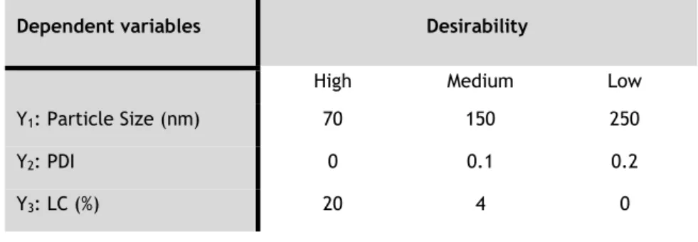
Table 2.5: Dependent variables and their desirability levels in the BBD. ... 28
Table 3.1: Composition and characterization of the NPs composed of the three most promising lipids (Gelucire 43/01, Cetyl palmitate, Compritol 888 ATO). ... 33
Table 3.2: Composition and characterization of the formulations analysed in preliminary experiments. ... 37
Table 3.3: Formulations studied by the BBD and the corresponding particle size (Y1), PDI (Y2) and LC (Y3). ... 38
Table 3.4: Regression analysis for particle size (Y1), PDI (Y2) and LC (Y3). ... 40
Table 3.5: Composition and characterization of the optimal formulation obtained by BBD, with the predicted and experimental values for particle size, PDI and LC. ... 44
Table 3.6: DSC parameters of bulk cetyl palmitate, placebos and AMX-loaded SLNs and the corresponding bulk mixtures: melting temperatures, enthalpy and RI. ... 49
xii
xiii
List of Appendices
Appendix 1: Various SLNs and NLCs formulations studied for oral delivery of drugs/compounds.
... I
Appendix 2: DSC thermogram of the bulk cetyl palmitate. ... III Appendix 3: DSC thermograms of: (A) F1 SLNs, (B) P1 SLNs, (C) F1 mixture and (D) P1 mixture.
... III
Appendix 4: DSC thermograms of: (A) F2 SLNs, (B) P2 SLNs, (C) F2 mixture and (D) P2 mixture.
... IV
Appendix 5: DSC thermograms of: (A) F3 SLNs, (B) P3 SLNs, (C) F3 mixture and (D) P3 mixture.
... IV
Appendix 6: Evaluation of the particle size of the ressuspended formulations stored at RT over
time (0 (T0), 1 (T1) and 2 (T2) weeks). ... V
Appendix 7: Evaluation of the PDI of the ressuspended formulations stored at RT over time (0
(T0), 1 (T1) and 2 (T2) weeks). Bars represent the PDI (left Y axis). ... V
Appendix 8: Evaluation of the zeta potential of the ressuspended formulations stored at RT
over time (0 (T0), 1 (T1) and 2 (T2) weeks). ... VI
Appendix 9: Evaluation of the LC of the ressuspended formulations stored at RT over time (0
(T0), 1 (T1) and 2 (T2) weeks). Bars represent the LC (left Y axis). ... VI
Appendix 10: Evaluation of the particle size of the ressuspended formulations stored at 4ºC
over time (0 (T0), 1 (T1) and 2 (T2) weeks). ... VII
Appendix 11: Evaluation of the PDI of the ressuspended formulations stored at 4ºC over time
(0 (T0), 1 (T1) and 2 (T2) weeks). ... VII
Appendix 12: Evaluation of the zeta potential of the ressuspended formulations stored at 4ºC
over time (0 (T0), 1 (T1) and 2 (T2) weeks). ... VIII
Appendix 13: Evaluation of the LC of the ressuspended formulations stored at 4ºC over time (0
xiv
xv
List of Abbreviations
AMR Antimicrobial Resistance AMX Amoxicillin
ANOVA Analysis of variance BBD Box-Behnken design CSK CSKSSDYQC peptide DLS Dynamic Light Scattering
DMEM Dulbecco’s Modified Eagle’s Medium DMSO Dimethyl Sulfoxide
DNA Deoxyribonucleic acid
DOPE Dioleoylphosphatidylethanolamine DSC Differential Scanning Calorimetry EDTA Ethylenediaminetetraacetic acid EE Encapsulation Efficiency
ELS Electrophoretic Light Scattering FBS Fetal Bovine Serum
FDA Food and Drug Administration GRAS Generally Recognized As Safe H. pylori Helicobacter pylori
HPH High-Pressure Homogenization
IARC International Agency for Research on Cancer IRQ IRQRRRR peptide
IUPAC International Union of Pure and Applied Chemistry LC Loading Capacity
LNPs Lipid Nanoparticles
MTT Methylthiazolydiphenyl-tetratozium Bromide NLCs Nanostructured Lipid Carriers
NPs Nanoparticles
OVAT One-Variable-At-a-Time PBS Phosphate Buffered Saline PDI Polydispersity Index PE Phosphatidylethanolamine PLX Polymixin B Sulphate PVA Polyvinyl Alcohol Pen Strep Penicillin- Streptomycin
xvi RI Recrystallization Index
RPMI Roswell Park Memorial Institute RT Room Temperature
SA Sodium Alginate sCT Salmon calcitonin SD Standard Deviation SLNs Solid Lipid Nanoparticles SPC Soybean Phosphatidylcholine TEM Transmission Electron Microscopy TMC N-trimethyl chitosan
1
Chapter 1 – Introduction
1.1 I
NTRODUCING
I
NFECTIOUS
D
ISEASES
At the beginning of the 20th century, infectious diseases were the leading cause of death at
a worldwide level [1]. However, since the introduction of penicillin in the 1940s the field of antibiotics emerged [2]. Consequently, various natural products capable of killing bacteria were discovered, which led to a significant decrease in morbidity and mortality resultant from infectious diseases [2]. Nevertheless, we are currently living in a new era, driven by the development of antimicrobial resistance (AMR) [3]. In the United States, AMR is estimated to be responsible for the addition of $20 billion to the total health care costs and $35 billion in costs to society [4]. AMR is also a concern in the European Union, since it is estimated that the phenomenon causes 25 000 deaths and costs more than US$1.5 billion per year [5]. These costs include both healthcare expenses and losses in productivity [5].
AMR is a natural evolutionary process in bacteria, since antimicrobial resistance genes existed long before the therapeutic application of antibiotics [4]. The occurrence of mutations is highly probable in an infection cycle due to the large number of bacteria and the high intrinsic rate of mutation [6]. If one of the mutations confers resistance to an administered antibiotic, a natural selection occurs, where all the sensitive bacteria are killed, while the bacteria with the mutation grow [6]. Due to the wide use of antimicrobial agents at a global level, there is a preferential selection for bacteria that are able to express resistance genes against antibiotics [4]. Thus, AMR has been rapidly evolving and spreading, leading to no assurance that new antimicrobial drugs can timeously respond to the increasing rates of bacterial resistance [2]. Thus, AMR is becoming a worldwide threat to the efficient treatment of infectious diseases [7].
Besides AMR, other drawbacks of the current antimicrobial therapies lead to unsuccessful eradication of pathogens. Intrinsic factors of antimicrobial agents can also compromise their therapeutic effects [8]. These factors include poor cellular penetration, limited intracellular retention, inefficient subcellular distribution and decreased intracellular activity [8]. Antibiotics are usually administered by oral or intravenous routes [2]. Oral administration is the most convenient and safest route for drug administration [9]. This route is cost-effective, presents less complications and it is less intrusive than intravenous administration, increasing therapeutic compliance [9, 10]. In addition, prolonged intravenous drug administration can cause local and vascular infection and patient discomfort [9, 10]. However, both routes present a major drawback characterized by systemic side effects, due to nonspecific drug distribution [2]. Oral administration is also associated with a poor bioavailability, due to poor solubility and/or poor permeability of drugs [9]. The acidic pH of the stomach, the contact with enzymes and biliary salts can also reduce drugs’ bioavailability through their degradation [9, 11].
2
Further, the hepatic first-pass metabolism contributes to reduce the concentration of drugs in the systemic circulation, since during this phenomenon drug metabolism occurs [9, 12]. Consequently, infection conditions usually require high-doses of antibiotics in order to achieve an adequate concentration in serum, which commonly lead to adverse side effects [13]. However, even applying an aggressive antibiotic treatment, the complete eradication of the infection can be difficult to achieve [13].
The increasing of antibiotic resistance, the loss of antibiotic efficacy and the lack of new antibiotics in the market have become a worldwide challenge to provide adequate treatment for infectious diseases [7, 14]. Thus, novel effective therapies are necessary to overcome these limitations.
1.2 N
ANOPARTICLES
A
S
D
RUG
D
ELIVERY
S
YSTEMS
1.2.1 I
NTRODUCINGN
ANOTECHNOLOGYNanotechnology is a multidisciplinary branch of science that integrates the fields of medicine, chemistry, biology and engineering [15]. According to International Union of Pure and Applied Chemistry (IUPAC) definition, nanoparticles (NPs) are structures of any shape with an equivalent diameter of approximately 1 to 100 nm [16]. However, the definition of nanomaterial is controversial with some definitions supporting that particles of dimensions greater than 100 nm might also have physicochemical properties that are characteristic to the nanoscale [17]. Hence, it is important that the accepted definition of NPs is not restricted to the size per se, but also to the physicochemical properties such as charge, shape and surface characteristics [17]. Herein, in this thesis, the term nanoparticle will be applied for particles in the range of 1-999 nm. Due to their nanoscale, these materials have novel physicochemical and biological features with potential for biosciences applications [18]. The high surface area-to-volume ratio of nanostructures is responsible for their different behaviour from the bulk material [19]. Consequently, nanomaterials have mechanical, electrical, chemical, optical and magnetic properties that can be advantageous in the design of drug delivery systems [19]. In the past few decades, NPs emerged as a promising drug delivery strategy to overcome AMR [4]. To design an ideal nanocarrier some characteristics must be considered. Nanocarriers must be biocompatible, biodegradable, nontoxic and be able to target the infection sites [14]. In addition, they must have a high drug payload and prevent drug release before reaching the target [14]. Thus, therapeutic concentrations at these sites can be achieved, reducing side effects related to non-targeted drug release [14].
NPs have been explored in the treatment of infectious diseases since they improve the antibacterial activity of antibiotics on several bacteria [20]. This activity is dependent on the size, shape, morphology, surface modifications and stability of NPs [20]. Nonetheless, some NPs have intrinsic antibacterial activity, such as silver NPs [21]. NPs are able to protect drugs from the acidic conditions of the stomach, improve their half-life and maintain a sustained drug
3
release at the target site [22]. These factors lead to higher concentrations of antibacterial agents near the bacterium, even with lower doses [22]. Hence, these drug delivery systems can minimise side effects and improve the therapeutic efficacy, decreasing the potential development of bacterial resistance [22]. Moreover, many types of lipophilic and water-soluble antibiotics can be carried at the core or at the surface of NPs by chemical conjugation, physical encapsulation or surface adsorption [23, 24]. However, covalent linking is the only technique that allows a precise control of the amount of drug loaded into the nanoparticle [18]. Through these methods, NPs are capable of loading multiple antimicrobial agents within their structure [4]. As a consequence, antimicrobial resistance mechanisms are unlikely to be developed against these systems, since it requires multiple simultaneous gene mutations in the microbial DNA [4].
Antimicrobial NPs improve drugs pharmacokinetics and their therapeutic index, including serum solubility and stability [2, 24]. Further, they prolong drugs half-life in systemic circulation, promoting a sustained and controlled release of the drug and a uniform distribution at the infection site [2, 24]. The target can be achieved using a passive or active strategies [4]. The passive targeting results from enhanced vascular permeability at the target infection site, if associated to inflammation, which allows a selective extravasation and retention of NPs [4]. On the other hand, in active targeting, the NPs are functionalized with specific ligands (e.g. antibodies, peptides, folic acids, among others) that recognise and bind to receptors at the target cells [4, 18]. For instance, drug delivery systems can be conjugated with antibodies against a specific antigen selectively found at the surface of the target microorganism [18]. The targeting can also be achieved through an activation upon an external stimuli, producing physicochemical changes that lead to drugs release at the target site [25]. These stimuli include magnetic and electric fields, ultrasounds, temperature, enzymatic activity, pH, ionic strength and redox potential [25]. Among these stimuli, pH stimuli has been widely researched [25]. One of its applications consist in the improvement of oral drug delivery systems [25]. For instance, Lin et al. (2009) formulated heparin-chitosan NPs, self-assembled by mixing heparin and chitosan at pH 1.2-2.5 to apply in the treatment of Helicobacter pylori (H. pylori) infections [26]. The system was stable at the gastric lumen (pH 1.2-2.5) due to electrostatic interactions within the structures [26]. After, the NPs infiltrated into the mucus layer (pH 4.5-7.0) and contacted with H. pylori along the gastric epithelium (pH~7.4) [26]. At the site of infection, the deprotonation of chitosan occurred, which weakened electrostatic interactions and led to system instability and disintegration [26]. As a consequence, the drug was released, acting locally on the bacteria at a bactericidal concentration [25, 26].
After drug-loaded NPs efficiently reach the infected tissue, they can deliver their payloads into target cells through three different pathways: adsorption, contact release or endocytosis [8]. In the last pathway, nanocarriers penetrate the target cells and release the antibiotics in the internal medium, avoiding resistant mechanisms related with activation of efflux systems and decreasing of membrane permeability [22, 24]. Due to these target delivery strategies, it is expected to reach a higher dose of antibiotic at the infection site with a lower administration
4
frequency and dose [4]. Consequently, systemic adverse effects of therapy are minimized, which improves patient compliance to the treatment [2, 4].
Currently, a number of nanoparticle-based drug delivery systems have been approved for clinical use in a variety of diseases, while many other nanoparticle formulations are under clinical trials [24]. In January 2012, 67 nanodevices were identified in the market, from which 33 were associated to nanotherapeutics [27]. According to the BCC Research report, the market value of the worldwide nanomedicine industry was $214.2 billion in 2013 and $248.3 billion in 2014. By the year of 2019, it is projected that the global market of nanomedicine reach $528 billion [28]. The progress of nanomedicine market over the past few years is a consequence of the increasing investment of companies on nanotechnology [2]. Therefore, this phenomenon is changing the paradigm of current treatments against many diseases, including infectious diseases [2]. However, the clinical nanoparticle-based drug delivery systems safety profiles must be considered upon a long-term exposure, being this parameter the most concerning in the use of nanotechnology for medical applications [2].
1.2.2 L
IPIDN
ANOPARTICLES(LNP
S)
In the last decades, many colloidal drug carriers have been studied to improve drugs solubility by oral administration [29]. These carriers include micelles, nanoemulsions, nanosuspensions, polymeric NPs and liposomes [29]. For the majority of these carriers, a low cost large-scale production method is currently not available [29]. Nevertheless, LNPs adopted the best features of other colloidal systems and emerged as a strategy to overcome their limitations [29]. These NPs are usually composed of a matrix of physiological or physiologically related lipids characterized by their versatility, biocompatibility and biodegradability [29-31]. Lipids are natural materials that can be degraded by natural processes, such as enzymatic activity [31]. Due to these processes, complex lipids are easily and completely degraded in the human body [31]. Therefore, the excipients that compose the matrix of LNPs are generally recognized as safe (GRAS) for oral and topical administration [32].
LNPs exhibit other outstanding advantages when compared with other colloidal drug delivery systems, including improved kinetic stability, drug solubility and controlled drug release [30, 31]. Besides, LNPs production is cost-effective, being easy to scale up according to the production process, which increases the researchers’ interest on these nanocarriers [30, 32].
Formulations for oral administration are possible as aqueous dispersion or alternatively transformed into tablets, pellets, capsules or powders in cachets [30, 32]. These formulations can protect encapsulated drugs from the environment of gastrointestinal tract and their nanoparticulate state promotes the uptake by M cells of Peyer’s patches [30]. Consequently, LNPs enables the system to bypass the effect of first-pass metabolism, through lymphatic absorption [30, 33].
Different types of LNPs have been engineered, such as solid lipid nanoparticles (SLNs) and nanostructured lipid carriers (NLCs) [34]. These NPs demonstrated high drug loading for
5
hydrophilic and lipophilic drugs and long-term shelf stability [29]. Despite the increasing interest in the field of LNPs, a low number of formulations are currently in the market [32]. Consequently, more investment on LNPs investigation is required [32].
1.2.2.1 S
OLIDL
IPIDN
ANOPARTICLESSLNs were introduced in 1991 as an alternative to colloidal drug carriers [35]. SLNs are nanospheres with a matrix composed of lipids in a solid state at both room and body temperatures and with a mean particle size from 50 to 1000 nm (Figure 1.1) [32]. Fatty acids, waxes, monoglycerides, diglycerides and triglycerides are widely used to construct the rigid core of SLNs [34]. To stabilize the solid matrix, various non-toxic surfactants can be added during SLNs preparation, such as poloxamers, polysorbates or polyvinyl alcohol (PVA) [14, 34, 36]. From these, PVA is the most commonly used due to its physical and chemical properties, biocompatibility and low acute oral toxicity [36, 37]. Besides, PVA is approved by Food and Drug Administration (FDA) for use in medical and food applications [36, 37].
SLNs combine advantages of other drug delivery systems, while overcome some of their limitations [38]. These drug delivery systems are characterized by a good physical stability, biodegradability and they are able to incorporate both lipophilic and hydrophilic drugs, depending on the preparation method [14, 18]. Due to their solid core identical to polymeric NPs, SLNs provide simultaneously a protection of the encapsulated drug and its controlled delivery [32, 38]. Furthermore, these LNPs are easily produced at an industrial scale being possible to avoid the use of organic solvents [29]. Many drug delivery systems based on SLNs have been prepared for various administration routes, showing improved bioavailability and targeted delivery of antimicrobial drugs [2, 38].
Besides the advantages, several drawbacks are still associated with the use of SLNs as drug delivery systems. The loading capacity (LC) of SLNs is determined by the crystalline structure of the solid core [29]. In fact, the drug is incorporated in the spaces between the fatty acid chains, the lipid layers or in the amorphous clusters in crystal imperfections [29]. SLNs composed of a highly purified lipid can form a perfect crystalline lattice, which reduces the available spaces to accommodate the drug [29]. Consequently, these carriers have a limited drug loading capacity [29, 34]. Other disadvantages of SLNs are associated with a relatively high water content (70-99.9%) and with storage conditions. More specifically, during the storage, the degree of order of the solid matrix is increased, which contributes to the formation of a perfect crystalline lattice [29, 31]. Therefore, the number of imperfections in the crystal lattice is reduced, leading to an early drug release [29, 31]. Moreover, gelation phenomenon and the increase of the particle size may occur with storage time [29, 39].
SLNs constitute an attractive drug delivery system due to their advantages when compared with other drug carrier systems [40]. However, due to the drawbacks associated with these systems, a second generation of LNPs emerged: nanostructured lipid carriers [32].
6
1.2.2.2 N
ANOSTRUCTUREDL
IPIDC
ARRIERSNLCs were developed to overcome the potential limitations of SLNs. The second generation of LNPs are composed of a lipid matrix with both solid and liquid lipids [34]. This matrix has a melting point depression when compared with the pure solid lipid matrix of SLNs, being however solid at body temperature [30]. Besides, NLCs matrices are less-ordered and have imperfections between the liquid lipid and the solid lipid, providing more spaces to incorporate drugs (Figure
1.1) [32, 34]. In NLCs the drug can be accommodated between the fatty acid chains, lipid layers
and imperfections [31]. Consequently, using NLCs, drug loading capacity is enhanced, while drug release during storage is minimized [29, 34]. Besides, these nanocarriers are less susceptible to gelation during both preparation and storage processes, having a lower water content when compared to SLNs [31, 34].
According to the preparation method and the lipid composition, there are three types of NLCs: imperfect type, multiple type and amorphous type [30, 40]. The imperfect type NLCs is obtained when small amounts of liquid lipids are added to the solid matrix, which leads to imperfections in the structure [30, 40]. Therefore, these imperfections create free spaces for drug accommodation in amorphous clusters (Figure 1.1, I) [40]. Multiple type NLCs are prepared by mixing higher amounts of oil with the solid lipid [38]. In this phenomenon, the solubility of oil molecules in the solid lipid is exceeded, which leads to a phase separation [38, 40]. Consequently, oily nanocompartments are formed in the solid matrix [38, 40]. This type of NLCs can be advantageous, since the solubility of many lipophilic drugs in oils is higher than in a solid lipid [38, 40]. Hence, the existence of oily nanocompartments improves drug solubility, while the solid lipid matrix prevents degradation and drug release during storage (Figure 1.1,
II) [38, 40]. Finally, the amorphous type of NLCs is composed of a structureless solid amorphous
matrix [40]. This amorphous matrix is obtained by mixing solid lipids with specific lipids (e.g. Miglyol®812 or isopropylmyristate) [40]. Due to the amorphous state of the matrix in this type of NLCs, crystallization process upon cooling is avoided [38, 40]. Thus, drug release during storage time is minimized (Figure 1.1, III) [40].
Figure 1.1: SLNs crystalline matrix and different types of NLCs matrices. I – Imperfect type NLCs, II – Multiple type NLCs, III – Amorphous type NLCs. Adapted from [41].
7
1.2.2.3 LNP
SS
YNTHESIST
ECHNIQUESVarious synthesis techniques can be used for the production of LNPs. Some important parameters must be considered in the selection of the most suitable technique for LNPs preparation, such as the physicochemical properties and the stability of the drug to be incorporated [42]. Besides, the desirable particle characteristics and stability should also be considered [42].
High-Pressure Homogenization (HPH) is a suitable technique for preparation of LNPs [38].
This technique can be performed at high temperatures (hot HPH) or at temperatures below room temperature (RT) (cold HPH) [38]. In hot HPH, both the lipid and the drug are melted at approximately 5-10ºC above the lipid melting point [38]. After, the homogeneous dispersion is mixed with an aqueous surfactant solution prepared at the same temperature [38]. Consequently, a hot pre-emulsion is formed by high speed stirring [29]. The hot pre-emulsion is processed in a high-pressure homogenizer at the same temperature until a nanoemulsion is obtained [29]. The lipid droplets of the nanoemulsion recrystallize into LNPs upon cooling down at RT [29]. The cold HPH is similar to hot HPH, since both the lipid and drug are melted [29, 38]. However, in cold HPH the cooling down process occurs rapidly using liquid nitrogen or dry ice, which leads to the formation of solid lipid microparticles [29, 38]. These microparticles are then suspended in a cold aqueous surfactant and homogenized at/or below RT [29]. Through this process, LNPs are produced [29]. Cold HPH is suitable for hydrophilic or thermo-labile drugs, since avoids drug degradation at high temperatures and drug distribution into aqueous phase during homogenization [29]. HPH technique has been widely used for LNPs production since it is easy to scale up, avoids organic solvents and has a short production time [30, 31]. However, the use of high operating temperatures and cavitation forces can cause thermodynamic and mechanic stress [30]. In order to overcome these disadvantages and to encapsulate drugs with a wide variety of physicochemical features, new production methods for LNPs preparation have been extensively investigated [30].
Microemulsion technique was developed and optimized by Gasco and his co-workers and
has been adapted and/or modified by other researchers [38]. In this method, the solid lipid is melted and the drug dispersed in the melted lipid [29]. After, an aqueous solution, containing both the surfactant and co-surfactants, is prepared at the same temperature and added to the melted lipid [29]. The mixture is subjected to stirring, which leads to the formation of a microemulsion [38]. Subsequently, it is dispersed in cold water (2-10ºC) using stirring [29]. Consequently, the microemulsion breaks into ultrafine nanoemulsion droplets that crystallize to form LNPs [29]. This technique can be used at an industrial scale [29]. However, the removal of the surplus water, usually using ultrafiltration or lyophilisation, is the main concern of this technique [29]. The high concentration of surfactant and co-surfactants required in this method is also a disadvantage, since it is less desirable for regulatory purposes and applications [29, 38].
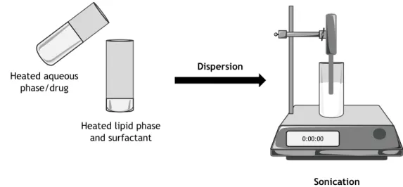
In the emulsification-sonification method, both lipid and drug are melted at approximately 5-10ºC above the lipid melting point [29]. After, an aqueous surfactant solution at the same
8
temperature is added to the homogenously dispersed mixture by a high shear mixing device [29]. The resultant emulsion is then ultrasonicated until a nanoemulsion is obtained and, afterward, cooled down at RT, forming LNPs [29]. However, the possible contamination of the emulsion by probe sonicator during sonication process is a concern in the use of the method [29].
In solvent emulsification-evaporation, the lipid is dissolved in a water-immiscible organic solvent (e.g. chloroform, cyclohexane) [29]. After, the mixture is emulsified in an aqueous phase containing surfactants under continuous stirring [29]. The solvent is evaporated during emulsification at a reduced pressure, which leads to lipid precipitation into NPs [29]. This method can be performed at RT, avoiding heat during the preparation [29, 38]. Consequently, solvent emulsification-evaporation technique is suitable for thermo-labile drugs [29]. However, the main concerns are associated with possible solvent residues in the final dispersion and the production of a very diluted dispersion due to the limited solubility of the lipid in the organic solvent [29, 38]. Consequently, the dispersion needs to be concentrated by ultrafiltration or evaporation [29].
In solvent diffusion technique, partially water-miscible organic solvents (e.g. benzyl alcohol, ethyl formate) are used [29]. In this method, the organic solvents are mutually saturated with water to ensure initial thermodynamic equilibrium of both lipids [29]. The oil-in-water emulsion is passed into water under continuous stirring, which leads to solidification of the dispersed phase, forming LNPs due to diffusion of the organic solvent [29]. However, similarly to the microemulsion technique, the dispersion is very diluted [29]. Consequently, it needs to be concentrated by ultrafiltration or lyophilisation [29]. Besides, the use of organic solvents that can remain in the final preparation is also a concern of this technique [29].
The solvent injection technique is similar to the solvent diffusion method [29]. Lipids are dissolved in a miscible solvent (e.g. acetone, isopropanol and methanol) or water-miscible solvent mixture [29]. Then, using an injection needle, the mixture is quickly injected into an aqueous solution of surfactants [29]. This preparation method is fast and easy to handle [29]. Besides, it does not require technically sophisticated equipment [29]. However, the main concern is associated with the use of organic solvents [29].
The double emulsion technique was first described by Cortesi et al. (2002) [43]. This method is based on the solvent emulsification-evaporation technique and is suitable for loading hydrophilic drugs into LNPs [29]. During LNPs preparation, both the drug and the stabilizer are encapsulated in the inner aqueous phase of the water-in-oil-in-water double emulsion [29]. The stabilizer is required to prevent drug partition to the external water phase during solvent evaporation [29, 38]. In the double emulsion procedure, an aqueous solution of drug is emulsified in molten lipid to obtain a primary water-oil emulsion, stabilized by the addition of emulsifiers to the aqueous phase [42]. Then, the primary emulsion is dispersed in a second aqueous solution containing stabilizers, under constant stirring [42]. Thus, a water-oil-water double emulsion is generated [42]. The formulations obtained through this method are usually named as “lipospheres” due to their larger particle size when compared with SLNs [29].
9
In the high speed stirring and/or ultrasonication technique, LNPs are produced by spray congealing [38]. After, these particles are subjected to high speed stirring or sonication, which leads to the formation of lipid nanopellets [38]. LNPs preparation by this method is easy and does not require technical and sophisticated equipment [38]. The main concern is associated to a broader particle size distribution, which fluctuates into the micrometer range due to the high speed stirring process [38]. Consequently, this phenomenon can lead to physical instabilities during storage [38]. Besides, the possible metal contamination due to ultrasonication is also a disadvantage of the technique [38].
1.3 E
XPERIMENTAL
D
ESIGN
A
ND
O
PTIMIZATION
O
F
F
ORMULATIONS
In the development of drug delivery systems, experimental design and optimization are crucial tools to facilitate the definition of the formulation composition and the manufacturing process [44, 45].
The traditional optimization of an experimental response involves monitoring the influence of one-variable-at-a-time (OVAT), while the remaining variables are kept at a constant level [44, 46]. However, this approach does not guarantee the optimum composition, since the interactive effect of one or more variables on others is not considered [44, 46]. Consequently, the final formulation obtained applying OVAT methodology is satisfactory, but mostly sub-optimal [44]. Besides, it is costly and time-consuming once an increased number of experiments are required, leading to a higher consumption of reagents and materials [46, 47].
Currently, to overcome the inconveniences of the traditional approach, multivariate statistic techniques have been widely applied to optimize the design of formulations [46-48]. These strategies involve designs for which the levels of all variables are changed simultaneously, allowing the assessment of interaction effects among the variables in study [48]. Consequently, a multivariate approach requires fewer experiments when compared to univariate approaches, leading to a lower reagent consumption and considerably less laboratorial work [48]. One of the most relevant multivariate techniques is the response surface methodology [46]. This approach consists in mathematical and statistical methods, based on linear or square polynomial equations [46]. Hence, the behaviour of an experimental data set is mathematically described in order to perform statistical previsions [46]. In an experimental procedure, several variables or factors may influence the final formulation [45]. Therefore, an initial screening is performed to select the independent variables and interactions that have a significant influence on the system, leading to a delimitation of the experimental region [46]. Consequently, a suitable design can be performed accordingly to the stablished by the previous screening [46]. The resultant data is then processed through mathematic-statistical treatment using a polynomial function [46]. Therefore, the optimum value for each variable is determined [46].
10
1.3.1 B
OX-B
EHNKEND
ESIGN(BBD)
In a BBD the levels of independent variables must be adjusted at three levels (-1, 0, +1) with equally spaced intervals between the levels [46]. In this model, computer optimization processes and a mathematical function of response surface methodology evaluate the effect of the levels of independent variables on the responses [49].
The BBD is a very useful approach when several variables are simultaneously studied in an optimization process [49]. This approach exhibits outstanding advantages when compared with full three-level factorial designs, since it requires fewer runs, is more efficient and has lower costs for a large number of variables [46, 47]. Moreover, in BBD the combinations for which all factors are simultaneously at their lowest or highest are rejected [48]. Therefore, experiments performed under extreme conditions are avoided [48]. The BBD is being widely used to evaluate the effect of different variables on SLNs and NLCs formulations [47, 50]. One interesting application of optimized formulations obtained by this methodology is to improve bioavailability of oral drug delivery systems using NPs [49].
Usually, BBD applied to LNPs production requires approximately 15 to 17 runs for three or four dependent variables [49, 51, 52]. In this methodology, independent variables are selected to investigate their influence in the behaviour of dependent variables previously fixed [49, 51]. The most common dependent variables reported in studies for synthesis of SLNs and NLCs are the particle size, encapsulation efficiency (EE) and LC [49, 51]. However, other dependent variables such as zeta potential [53], turbidity [52] and polydispersity index (PDI) [50] have also been reported. On the other hand, drug to lipid ratio [51], surfactant concentration and homogenization speed [51] are the independent variables usually selected for LNPs preparation. Nonetheless, lipid concentration [52], homogenization time [53] and surfactant/lipid molar ratio [54] are also investigated for optimization of LNPs.
BBD is commonly represented by a multidimensional cube with a variable number of points. For instance, in a BBD involving 15 experimental runs, three replicated central points are used to determinate the experimental error and the precision of the design [55]. The remaining runs are associated to points located at the edges of the cube (Figure 1.2) [50].
11
1.4 L
IPID
N
ANOPARTICLES
F
OR
O
RAL
A
DMINISTRATION
As abovementioned, the oral delivery is the most cost-effective route and simultaneously the most comfortable to the patient [9, 10]. However, due to the extreme and severe conditions of the gastrointestinal tract, SLNs and NLCs have been widely studied to improve drugs pharmacokinetics and to treat a wide range of diseases (Appendix 1). For that purpose, oral absorption is a crucial step. Thus, Zhang et al. (2012) studied the absorption of SLNs both in stomach and intestine [56]. In this work, oral candesartan cilexetil-loaded SLNs were formulated by a film-homogenization technique [56]. They verified that absorption of the resultant SLNs occurred mainly in the intestine via internalization into the enterocytes [56]. Besides, the developed SLNs were absorbed more rapidly than the free drug [56]. In order to investigate cellular uptake, internalization pathways and transcytosis routes of both SLNs and NLCs, Neves and her colleagues (2016) used Caco-2 cell monolayers as an intestinal model [57]. From this study, the authors concluded that LNPs internalization occurs manly through a clathrin-mediated endocytosis [57]. In addition, both SLNs and NLCs crossed the intestinal barrier predominantly by a transcellular route [57]. The internalization across the intestinal barrier can also be promoted by specific ligands, such as was performed by Fan et al. (2014) [58]. They used salmon calcitonin (sCT)-loaded SLNs modified with CSKSSDYQC (CSK) peptide or IRQRRRR (IRQ) peptide [58]. Both peptides showed to improve SLNs cellular uptake using Caco-2/HT29-MTX co-cultured cells [58]. Moreover, it was observed that SLNs modified with CSK and IRQ were internalized by clathrin- and caveolae-dependent endocytosis [58]. These results are in accordance with the mechanisms of SLNs internalization at intestinal level described by Neves and her colleagues [57, 58].
An interesting advantage of LNPs is their ability to be modified in order to improve their physicochemical stability for oral administration. For instance, Ramalingam et al. (2016) developed a nontoxic surface modified formulation of resveratrol-loaded SLNs using N-trimethyl chitosan (TMC) graft palmitic acid [59]. The modified NPs showed a sustained release and enhanced bioavailability of resveratrol due to the mucoadhesive and high absorption properties of TMC [59]. The relative bioavailability of the drug was found to be 3.8-fold higher from SLNs than free drug [59]. Pandita et al. (2014) were also focused on the development of a SLN-based delivery system to encapsulate resveratrol [60]. In this study, the system was composed of a lipid matrix of stearic acid coated with poloxamer 188 and lecithin [60]. The resultant SLNs showed a sustained drug release and a significant 8.035-fold improvement in oral bioavailability as compared to resveratrol suspension [60]. Another example of a compound used to functionalize LNPs in order to enhance drugs bioavailability is biotin. Zhou et al. (2015) studied the effect of biotin in oridonin-loaded NLCs [61]. Biotin-modified NLCs showed a relative bioavailability of 171.01%, while non-modified NLCs had a relative bioavailability of 143.48% [61]. Additionally, no significant differences in release were observed between non-modified NLCs and biotin-modified NLCs [61].
12
In order to improve the optimization of LNPs for oral delivery, the abovementioned experimental design, BBD, has been applied. More specifically, Varshosaz et al. (2010) used the BBD in the optimization of buspirone HCL-loaded SLNs prepared by emulsification-evaporation and sonification method [49]. A preliminary screening was performed to identify the independent variables, namely lipid type, surfactant percentage, speed of homogenizer and acetone:dichloromethane ratio, which significantly affected particles size [49]. These variables were considered in the 17 experimental runs of the BBD to obtain a formulation with a maximum level of LC and minimum levels of both particle size and zeta potential [49]. The optimized formulation revealed an enhanced oral bioavailability of the drug in 2.53-fold compared to that of the drug solution [49]. In a study performed by Neupane and co-workers (2014), decitabine-loaded NLCs were also optimized using BBD [62]. In this process, 17 experimental runs were obtained to investigate the effect of independent variables in particle size, PDI and EE [62]. The selected independent variables were lipid and surfactant concentrations and the number of homogenization cycles [62]. According to the study, the desired formulation should have a particle size in a range of 100-200 nm, the minimum PDI and the maximum EE [62]. The optimal formulation showed more affinity towards tumour cells and increased cytotoxicity activity against cancer cells than the plain drug [62].
Regarding the application of LNPs for oral administration, it can be very extensive. For example, LNPs have been applied to load nutraceutics. Luan and co-workers prepared and optimized baicalin-loaded NLCs [63]. The NPs showed a biphasic drug release pattern with an initial burst release and a sustained release afterwards [63]. Thus, a significant enhancement of bioavailability was observed [63]. Zhuang et al. (2010) optimized vinpocetine-loaded NLCs to obtain the most favourable physicochemical characteristics [64]. The optimized formulation showed a sustained release profile without an observable burst-release effect [64]. Furthermore, in vivo studies revealed that the relative bioavailability of the resultant formulation was 322% in comparison with the free drug [64]. Anticancer delivery systems based on NLCs have also been studied by many research teams around the globe. For instance, How and co-workers (2013) developed a stable anticancer tamoxifen-loaded NLCs formulation [65]. In in vitro studies it was verified that encapsulation in NLCs preserved the cytotoxicity of the drug against tumour cancer cell lines [65]. LNPs for oral administration have also been studied for administration of hypolipidemic drugs, such as lovastatin and simvastatin. Chen et al. (2010) investigated the effect of the surfactants myverol and soybean phosphatidylcholine (SPC) in lovastatin-loaded NLCs [66]. They concluded that NLCs with myverol were more stable in the gastric environment and showed significantly higher bioavailability than NLCs containing SPC [66]. However, SPC system exhibited slower release [66]. In a study performed by Tiwari and Pathak (2011), optimized simvastatin-loaded NLCs showed a significantly higher EE and improved gastrointestinal absorption when compared with simvastatin-loaded SLNs [67]. As a consequence, in in vivo pharmacokinetic studies, NLCs revealed 2.29 folds increase in bioavailability as compared to SLNs [67].
13
LNPs loading antimicrobial agents have been extensively studied. A more detailed description of the oral administration of LNPs to combat infectious diseases was reviewed in a book chapter (Rita M. Pinto, Daniela Lopes, Cláudia Nunes, Bruno Sarmento, Salette Reis. Oral administration of lipid-based delivery systems to combat infectious diseases. Accepted at Nanoparticles in the Life Sciences and Biomedicine, Pan Stanford Publishing). In particular, antibiotics-loaded LNPs were proved as a successful delivery system with increased antibacterial efficiency. For instance, Xie et al. (2011) formulated ofloxacin-loaded SLNs using palmitic acid as the lipid matrix and PVA as emulsifier by hot homogenization and ultrasonication method [36]. The formulation exhibited an initial fast release of the drug followed by a slow and sustained phase, which contributed to maintain the effective therapeutic drug concentrations and increased bioavailability [36]. Consequently, the NPs developed in this study demonstrated in vitro and in vivo antibacterial efficacy against Staphylococcus aureus [36]. A similar study was performed by Dong et al. (2011) where norfloxacin was loaded into SLNs for oral administration [68]. The resultant SLNs demonstrated an effective antibacterial activity while no cytotoxic effects were detected [68]. More recently, the polymixin B sulphate (PLX) was also studied [69]. However, the salt form of PLX is a hydrophilic antibiotic with a cationic charge, which is an obstacle for an efficient loading into SLNs [69]. Thus, Severino et al. (2015) developed a novel lipid-polymer hybrid SLNs-based system to encapsulate the drug [69]. For that purpose, the hydrophilicity of the drug was decreased via previous cross-linking with the anionic polymer sodium alginate (SA), in a SA/PLX ratio (1:1) [69]. The resultant SLNs revealed an improved LC [69].
In the previously described studies, both SLNs and NLCs formulations showed an enhanced bioavailability and pharmacological activity of the loaded drug, requiring less frequency of dosage. Although SLNs and NLCs only recently emerged as drug delivery systems, many researchers have developed formulations for oral delivery that showed potential for further clinical applications.
1.5 H
ELICOBACTER PILORY
I
NFECTIONS
:
A
S
TEP
T
OWARDS
I
NNOVATION
I
S
N
EEDED
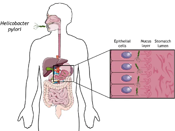
H. pylori is a gram-negative bacterium, usually characterized by a spiral-shaped form that can be converted in coccoid cells under a hostile environment [70]. This bacterium is a human pathogen able to resist to the acidic pH (1 or 2) of the stomach lumen and to penetrate within the mucosa (Figure 1.3) [22, 70]. Thus, the bacteria colonize the region between the mucus layer and epithelial cells, where the pH is near to neutral (pH 4-6) [22, 70]. The colonization process occurs due to several virulence factors, including bacteria flagella that allow bacteria mobility and adhesins responsible for the adherence to the mucosa and to epithelial cells [22, 70]. Other factors, such as urease production, phospholipase secretion and cytotoxic production, are crucial for the bacteria pathogenesis [70].
14
Figure 1.3: Schematic representation of the H. pylori infection. The bacterium is acquired by oral ingestion and colonizes the region between the mucus layer (pH 5) and epithelial cells (pH 7). Adapted from [22].
Nowadays, H. pylori bacteria have a huge impact worldwide, affecting around 50% of the global population [70]. However, the infection incidence is variable among countries and groups of people, reaching over 80% of the middle-aged adults in developing countries [71]. Several conditions are responsible for these variations among prevalence rates, including race/ethnic, aging and socioeconomic conditions [70]. The bacteria are acquired by oral ingestion and usually an asymptomatic colonization occurs [70, 71]. In fact, only approximately 20% of the infected patients develop chronic gastritis and peptic ulcer [22]. A more severe condition caused by the persistent colonization of H. pylori can be developed, namely gastric cancer [22, 70]. Due to this clinical outcome, the bacteria are classified as a human carcinogen (Group 1) according to the International Agency for Research on Cancer (IARC) and the World Health Organization (WHO) [22]. Although without a clear explanation, H. pylori have also been associated to other diseases, such as idiopathic thrombocytopenic purpura, iron deficiency anaemia, ischemic heart disease, stroke, Parkinson’s diseases and Alzheimer’s disease [22].
Currently, a standard triple therapy combining a proton pump inhibitor and two to three antibiotics is used against H. pylori infections, according to International Guidelines [70, 72]. The therapy is usually applied twice a day, for about 7 to 14 days [70]. The most common antibiotics used in this treatment are amoxicillin (AMX), clarithromycin and metronidazole or tinidazole [70]. Nonetheless, the efficacy of this therapy has been declining over the last years, especially when applied just for 7 days [73]. The decrease in the number of successfully treated
15
infections is mainly a consequence of the increasing number of emergent antibiotic resistant H. pylori bacteria strains [73]. Thus, the standard 7-day triple-therapy is recommended only for the cases where clarithromycin resistance is lower than 15-20%, while a higher antibiotic resistance rate requires a 14-day regime [73]. Nonetheless, the successful cases of H. pylori eradication using the prolonged therapy are far from the desirable, with a rate of eradication of 70% in non-ulcer dyspepsia patients and 81.7% in peptic ulcer patients [73].
Besides the intrinsic H. pylori antimicrobial resistance, other factors compromise the success of the triple therapy. The acidic pH of the stomach presents a huge drawback for the oral administration of both AMX and clarithromycin, since these antibiotics are easily degraded in the stomach conditions [22]. Thus, lower amount of effective drug is maintained in vivo [22]. Furthermore and as a consequence of the gastric transit, antibiotics remain in the stomach for an insufficient time, hindering their diffusion across the mucus layer [22, 70]. This lead to a lower concentration of drug that reaches the bacteria, located between the mucus layer and the epithelial cells [22, 70]. To overcome this limitation, higher and multiple doses are required, which may lead to discomfortable side effects, such as nausea, vomiting, abdominal pain, among others [22, 70]. The frequency of side effects combined with the duration of the therapy are the main reasons for a lack of patient compliance [22]. Thus, the discontinuation of the therapy commonly leads to an incomplete eradication of the bacteria and to potential development of antibiotic resistance [70, 74]. In the last years, the resistance prevalence of H. pylori to metronidazole has increased in around 40% and 90% in developed and developing countries, respectively [75].
In an attempt to overcome the drug resistance verified for the triple therapy, several regimens have been tested [75]. For instance, the efficacy of new antibiotics or new antibiotic combinations was evaluated for the eradication of H. pylori [73]. Nevertheless, the therapies failed mainly due to the high costs associated and the adverse effects that lead to a poor patient compliance, among other factors [75]. Moreover, the development of vaccines to prevent H. pylori infections is still in an experimental phase [73].
Besides some approaches are emerging, the lack of a suitable therapy is still a concern. Thus, a step towards innovation in anti-H. pylori therapies is urgently needed, which lead to the aim of the current project.
1.6 A
IM AND
S
TRATEGY
As previously mentioned, AMX is one of the most commonly antibiotics used in the standard triple therapy against H. pylori infections [70]. Usually this antibiotic is orally administered to eradicate the bacteria [76]. Nevertheless, some limitations are associated to this administration route for AMX. For instance, Lozniewski et al. (1999) reported that AMX is unstable at the normal gastric pH (1 to 2), which may be a consequence of hydrolytic degradation of AMX [76]. Therefore, a delivery system able to protect the drug from the acidic environment and to release it near the bacterium (almost neutral pH) would be advantageous
16
to avoid the drug’s degradation [4]. Furthermore, and taking into account the advantages already mentioned regarding the oral administration of LNPs, they have a huge potential as AMX delivery systems, since they are able to protect it from the acidic gastric environment [22]. Moreover, AMX is considered a low permeability drug according to the Biopharmaceutics Classification System, with low affinity to lipid phases [77, 78]. This limitation leads to a poor absorption through the gastric mucus layer and, consequently, an enhanced intestinal absorption and first-pass metabolism. Due to the lipid composition of LNPs, they already proved their usefulness in improving drugs bioavailability, by enhancing their absorption. Hence, they can be a promising strategy to deliver low permeability drugs.
In this context, the present project consists in the development of an innovative AMX delivery system based on LNPs. The main purpose of this system is to improve the efficiency of the treatment for H. pylori infections, improving the residence time of AMX in stomach, protecting it from degradation, enhancing their absorption and, ultimately, increasing antibiotic concentration at the target site [22]. To achieve this purpose, AMX-loaded LNPs were developed. The ideal NPs are composed of the solid lipid, chosen by a lipid screening, Tween®80, linolenic acid and dioleoylphosphatidylethanolamine (DOPE) (Figure 1.4). According to literature, linolenic acid is a fatty acid that has been recently highlighted due to its antibacterial activity against all strains of H. pylori bacteria, including the ones resistant to metronidazole [75]. Thus, in the current work, linolenic acid was encapsulated with AMX, as an adjuvant to the therapy. On the other hand, DOPE was selected for functionalization and also as a surfactant, since it is an unsaturated phosphatidylethanolamine (PE) [22]. Lingwood and his colleagues (1992) observed that PE, which is a predominant lipid in the antrum of the human stomach, is a receptor for H. pylori adhesion [79, 80]. Considering the interaction between PE and the bacterium, it is possible to develop anti-adhesion drug delivery systems based on PE, acting simultaneously as an active targeting [22, 80]. In the present project, DOPE was used at the surface of the NPs to target and block the bacteria adhesion to the gastric mucosa.
Figure 1.4: Schematic representation of the composition of the ideal formulation.
Besides, other three formulations (F1, F2, F3) (Figure 1.5) with decreasing degrees of complexity were characterized. The main goal of studying these formulations is to have a full physicochemical and morphologic evaluation of the NPs to be applied in future studies of