Dany da Silva
Mestrado em Biologia Celular e Molecular
Departamento de Biologia 2019
Orientador
Doutor Jorge Manuel Moreira Gonçalves,
Professor Catedrático, Faculdade de Farmácia
Coorientador
Doutor Armando Jorge Gomes Teixeira,
Professor Auxiliar, Faculdade de Ciências
Adrenergic
contribution
to the stress
induced
FCUP
Adrenergic contribution to the stress induced carcinogenesis
II
Todas as correções determinadas pelo júri, e só essas, foram efetuadas. O Presidente do Júri,
Dany da Silva
Faculdade de Ciências da Universidade do Porto
Rua do Campo Alegre 1021/1055, 4169-007 Porto, Portugal Email: up201708266@fc.up.pt
Doutor Jorge Manuel Moreira Gonçalves, Professor Catedrático Faculdade de Farmácia da Universidade do Porto
Rua Jorge de Viterbo Ferreira 228, 4050-313 Porto Email: jgoncalves@ff.up.pt
Doutor Armando Jorge Gomes Teixeira, Professor Auxiliar Faculdade de Ciências da Universidade do Porto
Rua do Campo Alegre 1021/1055, 4169-007 Porto, Portugal Email: agteixei@fc.up.pt
FCUP
Adrenergic contribution to the stress induced carcinogenesis
IV
I, Dany Silva, student number 201708266, currently undertaking a Cell and Molecular Master’s Degree, edition 2018/2019, hereby declare that this project was written by myself and using my own words, not contemplating quotations from published and unpublished sources indicated and acknowledged as such. I am aware of any consequence as a result of plagiarism.
Porto, 19 January 2019 Dany Silva
Agradecimentos
Em primeiro lugar, quero agradecer ao meu orientador Prof. Jorge Gonçalves, não só pela oportunidade que ele me deu mas também pelo seu esforço, apoio e partilha constante dos seus conhecimentos científicos. Ao Prof. Jorge Teixeira por ter aceitado a orientação deste projeto.
Agradeço também à Prof. Paula Fresco, por todo o seu tempo disponibilizado, pela sua simpatia e por todo o seu apoio e que me deu durante este percurso. À Prof. Clara Quintas, pela sua disponibilidade e simpatia, e pela preciosa ajuda que me deu durante a realização deste trabalho.
Um agradecimento especial à Mónica e à Céu, pela ajuda constante, pela paciência, companhia, amor e apoio.
A todos os meu amigos que me acompanharam durante a realização deste trabalho, em especial, à Filipa e à Salomé.
Por fim, gostaria de agradecer toda a minha família, por sempre ouvirem as minhas preocupações, e por todas as palavras de apoio que me deram.
FCUP
Adrenergic contribution to the stress induced carcinogenesis
VI
Resumo
O cancro da mama é a neoplasia maligna mais frequente em mulheres. Vários estudos demonstraram que o stress pode favorecer o aumento da incidência e mortalidade associado ao cancro. As catecolaminas (noradrenalina e adrenalina), liberadas sob condições de stress, podem ser responsáveis por este aumento de cancro associado ao stress. Estes mensageiros exercem os seus efeitos através de uma família de GPCR, denominada receptores adrenérgicos, apresentando três subfamílias distintas de recetores: α1, α2- e β-adrenoreceptores. Destes, os receptores β-adrenégicos parecem ser os mais envolvidos no cancro.
O presente trabalho teve como objetivo investigar os efeitos mediados pelos receptores β-adrenégicos em duas linhas celulares diferentes, as células MCF-10A, uma linha celular não tumorigénica e as células MCF-7, uma linha celular tumorigénica.
Tal como o stress, a menopausa também parece ser um fator de risco no desenvolvimento do cancro da mama. Durante a menopausa, em paralelo com a diminuição dos níveis de estrogênio, existe também um aumento na liberação de catecolaminas. Sabendo o impacto dos estrogénios no desenvolvimento e progressão do cancro da mama, neste estudo também foi investigado a influência dos estrogénios (17β-estradiol; principal forma ativa dos estrogénios) nos efeitos mediados pelos receptores β-adrenégicos.
Os resultados obtidos nestre trabalho apoiam a hipótese que as catecolaminas podem promover a tumorigénese sob condicções de stress ou em condições em que existe níveis elevados de catecolaminas plasmáticas, como o observado durante a menopausa. Esta conclusão é suportada pela observação de que a isoprenalina, um agonista dos receptores β-adrenégicos, aumentou o potencial tumorigénico das células MCF-10A, evidenciado pelo aumento da viabilidade celular, formação de colónias e capacidade de produzir adrenalina. O efeito da isoprenalina na indução de tumorigénese foi menos evidente nas células MCF-7, onde apenas foi observado um aumento na proliferação celular.
O estrogénio 17β-estradiol preveniu os efeitos tumorigénicos causados pela ativação dos receptores β-adrenégicos nas células não-tumorigénicas MCF-10A, efeito este mediado pelo receptor GPER. No entanto, observou-se ainda que, o 17β-estradiol, em células tumorigénicas, induziu um aumento da proliferação celular, o que sugere que, nas mulheres com elevado risco de já possuirem células tumorigénicas (como as células MCF-7), a administração de estrógenos poderá causar um aumento na incidência de cancro da mama em vez de ocorrer uma prevenção deste tipo de cancro.
Este estudo também mostrou que o propranolol, um antagonista dos receptores β-adrenérgicos, causou uma inibição acentuada da viabilidade / proliferação celular nas células MCF-7, não causando efeitos nas células MCF-10A. O efeito seletivo do propranolol nas células tumorigénicas poderá dever-se a uma influência na degradação de material obtido por autofagia (ou por processos idênticos). Esta ação seletiva do propranolol nas células tumorigénicas pode ser particularmente útil como uma nova intervenção farmacológica no cancro da mama.
Palavras-chave: cancro da mama, stress, catecolaminas, receptores adrenérgicos,
FCUP
Adrenergic contribution to the stress induced carcinogenesis
VIII
Abstract
Breast cancer is the most frequent malignancy in women. Several reports demonstrated that stress may favour the increase of cancer incidence and mortality. Catecholamines (noradrenaline and adrenaline) are released under stress conditions and may be responsible for this increase in cancer associated with stress. These messengers exert their effects through a family of GPCR, called adrenergic receptors (AR) which present three distinct subfamilies: α1, α2- and β-adrenoceptors. From these, the β-adrenoceptors (β-AR) appear to be most involved in cancer.
The present work aimed at investigate the β-AR mediated effects in two different cell lines, the MCF-10A cells, a non-tumorigenic cell line and the MCF-7 cells, a tumorigenic cell line.
As stress, menopause also seems to be a risk factor for breast cancer. During menopause there is also an increase in the release of catecholamines which is parallel with a decrease in oestrogen levels. Knowing the impact that oestrogens also has in the development and progression of breast cancer, the influence of 17β-estradiol (the main active from of oestrogens) in this pathway was also investigated.
The results presented here support the view that catecholamines may promote tumorigenesis under stress or under other conditions where high levels of plasmatic catecholamines are reached, such as those observed during menopause. This conclusion was supported by the observation that isoprenaline, a β-AR agonist, increased the tumorigenic potential of the non-tumorigenic MCF-10A cells, revealed by increased cell viability, colony formation and capacity to produce adrenaline. The effect of isoprenaline in inducing tumorigenesis was less evident in MCF-7 cells, where only an increase in cell proliferation was observed.
The oestrogen, 17β-estradiol prevented the tumorigenic effects caused by β-AR activation in non-tumorigenic MCF-10A cells, an effect mediated by the GPER. However, it was further observed that 17β-estradiol per se, was capable to induce cell proliferation in tumorigenic cells, suggesting that the administration of oestrogens in a population of women with high probability to already have tumorigenic cells (like the MCF-7 cells), an increase in incidence of breast cancer rather than a prevention of cancer may be observed.
This study also showed that propranolol, a β-AR antagonist, caused a marked inhibition of MCF-7 cell viability/proliferation while it did not affect MCF-10A cells. These selective effects in tumorigenic cells seem to be due to an influence in the degradation of material obtained by autophagy (or any identical processes). This selective action of propranolol in tumorigenic cells may be particularly useful as a new pharmacological intervention in breast cancer.
FCUP
Adrenergic contribution to the stress induced carcinogenesis
X
Index of Contents
AGRADECIMENTOS ... V RESUMO ... VI ABSTRACT ... VIII INDEX OF CONTENTS ... X INDEX OF FIGURES ... XIII INDEX OF TABLES... XVII ABBREVIATIONS ... XVIIII. CHAPTER: INTRODUCTION ... 1
1. INTRODUCTION ... 2
1.1. Cancer ... 2
1.2. Breast cancer ... 2
1.3. Breast structure and the development of cancer ... 5
1.4. Risk factors for breast cancer: the role of stress ... 7
1.5. Adrenergic stimulation and cancer ... 11
1.6. Oestrogens, adrenergic stimulation and cancer ... 13
II. CHAPTER: AIM... 15
2. AIM ... 16
III. CHAPTER: MATERIAL AND METHODS ... 17
3. MATERIAL AND METHODS ... 18
3.1. Chemicals ... 18
3.4. Cell treatment ... 19
3.5. MTT reduction assay ... 20
3.6. Kinetic Label-Free Proliferation Assay ... 21
3.7. Colony formation assay ... 21
3.8. Protein expression by Western blot ... 22
3.9. Detection of catecholamines by HPLC-ECD ... 23
3.10. Morphological transformation assay ... 23
3.11. Monodansylcadaverine and acridine orange staining ... 24
3.12. Data and statistical analysis ... 24
IV. CHAPTER: RESULTS ... 25
4. RESULTS ... 26
4.1. Effects of the β-AR activation in cell viability of the human breast non-tumorigenic cell line (MCF-10A) ... 26
4.2. Influence of 17β-estradiol in the β-AR-mediated effects in MCF-10A cell viability ... 28
4.3. Effects of the β-AR activation and the influence of 17β-estradiol in the clonogenicity of the MCF-10A cells ... 30
4.4. Effects of the β-AR activation on the expression pattern of the TH enzyme in MCF-10A cells ... 31
4.5. HPLC-ECD detection of catecholamines in MCF-10A cells ... 32
4.6. Effects of the β-AR activation in MCF-10A neoplastic transformation ... 33
4.7. Effects of the β-AR activation in cell viability of the human breast tumorigenic cell line (MCF-7) ... 34
4.8. Influence of 17β-estradiol on the β-AR-mediated effects, in MCF-7 cell viability ... 36
4.9. Effects of the β-AR activation in MCF-7 cell proliferation ... 39
4.10. Influence of 17β-estradiol on the β-AR-mediated effects, in MCF-7 cell proliferation ... 41
4.11. Influence of membrane cholesterol in the β-AR effects in MCF-7 breast cancer cells ... 44
4.12. Influence of propranolol in MCF-7 cells morphology ... 48
4.12.1. Influence of propranolol in MCF-7 vacuole formation ... 48
4.12.2. Influence of autophagy inhibition on the effects caused by propranolol……… ... 50
FCUP
Adrenergic contribution to the stress induced carcinogenesis
XII
V. CHAPTER: DISCUSSION ... 51
5. DISCUSSION ... 52
VI. CHAPTER: CONCLUSION ... 61
6. CONCLUSION ... 62
REFERENCES ... 62
Index of Figures
Figure 1. Worldwide incidence (A) and mortality (B) of cancer in females in 2018 ... 4 Figure 2. Scheme depicting the structure of human breast and its association with the
formation of several diseases, including tumours. ... 5
Figure 3. Pathway for the biosynthesis of catecholamines. ... 9 Figure 4. ANS fight-or-flight stress responses releases catecholamines into tumor
microenvironment.. ... 12
Figure 5. Effects of the β-AR agonist isoprenaline on MCF-10A cell viability, after 24h of
incubation. ... 27
Figure 6. Effects of the β-AR antagonist propranolol, alone (A) or in combination with
isoprenaline (B), on MCF-10A cell viability, after 24h of incubation ... 27
Figure 7. Effects of the ER agonist 17β-estradiol, alone (A), or in combination with
isoprenaline (B), on MCF-10A cell viability, after 24h of incubation ... 28
Figure 8. Effects of 4-hydroxytamoxifen (4-OHT) and the combine exposure of the β-AR
agonist isoprenaline with increasing concentrations of 4-OHT, on MCF-10A cell viability, after 24h of incubation. ... 29
Figure 9. Influence of 17β-estradiol on the effects caused by the β-AR agonist
isoprenaline in MCF-10A clonogenicity, after 7 days of treatment ... 31
Figure 10. Expression pattern of TH enzyme in MCF-10A cells, in the absence and in
the presence of isoprenaline ... 32
Figure 11. Effects of the β-AR agonist isoprenaline on MCF-7 cell viability, after 24h (A)
FCUP
Adrenergic contribution to the stress induced carcinogenesis
XIV
Figure 12. Effects of the β-AR antagonist propranolol, on MCF-7 cell viability after 24h
(A) or 72h (B) incubation. ... 35
Figure 13. Interaction between the β-AR agonist, isoprenaline, with the β-AR antagonist,
propranolol (A) or with the β2-AR inverse agonist, ICI 118551 (B) and their effects on MCF-7 cell viability after 24h of incubation ... 36
Figure 14. Effects of the ER agonist, 17β-estradiol, on MCF-7 cell viability after 24h (A)
or 72h (B) of incubation. ... 37
Figure 15. Effects of the combined exposure of the β-AR agonist, isoprenaline with
increasing concentrations of the ER-agonist, 17β-estradiol on MCF-7 cell viability, after 24h (A) or 72h (B) of incubation ... 37
Figure 16. Effects of the combined exposure of the β-AR antagonist, propranolol with
increasing concentrations of the ER-agonist, 17β-estradiol on MCF-7 cell viability, after 24h (A) or 72h (B) of incubation. ... 38
Figure 17. Influence of 17β-estradiol in the effects caused by the β-AR, antagonist,
propranolol (100 µM), on MCF-7 cell viability, after 24h (A) or 72h (B) of incubation ... 39
Figure 18. Effects of the β-AR agonist, isoprenaline on MCF-7 cell proliferation, after
24h (A) or 72h (B) incubation ... 40
Figure 19. Effects of the β-AR antagonist, propranolol, in MCF-7 cell proliferation, after
24h (A) or 72h (B) of incubation.. ... 40
Figure 20. Effects of the ER agonist, 17β-estradiol, on MCF-7 cell proliferation after 24h
(A) or 72h (B) incubation. ... 41
Figure 21. Effects of the combine exposure of the β-AR agonist, isoprenaline with
increasing concentrations of the ER-agonist, 17β-estradiol in MCF-7 cell proliferation, after 24h (A) or 72h (B) of incubation. ... 42
Figure 22. Effects of the combine exposure of the β-AR antagonist, propranolol with
increasing concentrations of 17β-estradiol on MCF-7 cell proliferation, after 24h (A) or 72h (B) of incubation. ... 43
Figure 23. Effects of the combined exposure of the ER-agonist, 17β-estradiol, and the
β-AR antagonist, propranolol (30 µM – C; 100 µM – A; B), on MCF-7 cell proliferation, after 24h (A) or 72h (B and C) incubation. ... 43
Figure 24. Effects of methyl-β-cyclodextrin, in MCF-7 cell viability, after 72h of
incubation.. ... 45
Figure 25. Influence of methyl-β-cyclodextrin (M-βCD; 1 mM), on the effect caused by
the β-AR agonist isoprenaline on MCF-7 cell viability, after 72h of incubation. ... 45
Figure 26. Influence of methyl-β-cyclodextrin (1 mM), on the effect caused by the
ER-agonist, 17β-estradiol, on MCF-7 cell viability, after 72h of incubation. ... 45
Figure 27. Effects of methyl-β-cyclodextrin, on MCF-7 cell proliferation after 24 (A) or
72h (B) incubation.. ... 46
Figure 28. Influence of methyl-β-cyclodextrin (1 mM), on the effect caused by the β-AR
agonist, isoprenaline, on MCF-7 cell proliferation after 24 (A) or 72h (B) of incubation.. ... 47
Figure 29. Influence of methyl-β-cyclodextrin (1 mM), on the effect caused by the
ER-agonist, 17β-estradiol in MCF-7 cell proliferation, after 24 (A) or 72h (B) of incubation. ... 47
Figure 30. Effect of propranolol (100 µM) in MCF-7 vacuole formation ... 49 Figure 31. Influence of 3´MA on the effect caused by propranolol (100 µM). ... 50
FCUP
Adrenergic contribution to the stress induced carcinogenesis
XVI
Figure 32. Effects of the β-AR agonist, isoprenaline, or the ER agonist, 17β-estradiol,
on MCF-10A cell viability (A), after 72h of incubation ... 77
Figure 33. Effects of propranolol (100 µM) on MCF-7 cell morphology and in the number
of cells. ... 78
Figure 34. Effects of the β-AR agonist, isoprenaline, on MCF-10A neoplastic
Index of Tables
Table 1. Classification of breast cancer into several subtypes based on the molecular
profiles. ... 7
Table 2. Main transduction mechanisms for the ARs subtypes ... 10 Table 3.Concentration of catecholamines using the conditioned medium of MCF-10A
FCUP
Adrenergic contribution to the stress induced carcinogenesis
XVIII
Abbreviations
3´MA 3´ methyladenine
ANS Autonomic nervous system
AR Adrenergic receptors
AVOs Acidic vesicular organelles
BARK β-adrenergic receptor kinase
CNS Central nervous system
CTR Control
DMEM/F12 Dulbecco’s Modified Eagle’s Medium/ F-12
DMEM-HG Dulbecco’s Modified Eagle’s Medium – high
glucose
EGF Epidermal growth factor
EGFR Epidermal Growth Factor Receptor
EPAC Exchange protein activated by adenylyl cyclase
ER Oestrogen receptor
FAK Focal adhesion kinase
FBS Fetal bovine serum
GPER G-protein-coupled oestrogen receptor 1
HER2 Human epidermal growth factor receptor 2;
HPLC-ECD High-performance liquid chromatography
with electrochemical detection
ISO Isoprenaline
MDC Monodansylcadaverine
MTT 3-(4,5-dimethylthiazol-2-yl)-2,5-diphenyl
tetrazolium bromide
M-βCD Methyl-β-cyclodextrin
PKA Protein kinase A
PMA Phorbol 12-myristate 13-acetate
PNMT Phenylethanolamine N-methyltransferase
PR Progesterone receptor
SERM Selective oestrogen receptor modulator
TH Tyrosine hydroxylase
1. Introduction
1.1. Cancer
Cancers results from an abnormal and uncontrolled cell division. They differ from benign tumours from their ability to invade surrounding tissues and to originate secondary tumours at distant locations (Hanahan and Weinberg, 2011).
Most cancers fall into one of three main groups: carcinomas, sarcomas and leukemias, according to the type of cell from which they arise. From these, nearly 90% of human cancers, are carcinomas, malignancies of epithelial cells (Ding et al., 2013).
The first step in the development of cancer is the initiation phase. According to the genetic hypothesis for the origin of cancer, it involves the alterations or mutations of genes (e.g. in pro-oncogenic genes such as RAS and MYC or in tumor suppressor genes such as BRCA1, BRCA2 and TP53), which can lead to dysregulation of biochemical signalling pathways associated with cellular proliferation, survival and differentiation (Finver et al., 1988; Baker et al., 1990; Downward, 2003; Friedenson, 2007). These alterations may arise spontaneously or be induced by exposure to non-genotoxic and genotoxic carcinogens, such as radiation, hormones, chemicals, infections and hypoxia (Benigni et al., 2013; Cuninghame et al., 2014). While genotoxic carcinogens directly interact with DNA (as either parent chemicals or reactive metabolites) and induces DNA damage, non-genotoxic carcinogens have been associated, for instance, with changes in oxidative stress, modulation of metabolizing enzymes and disruption of the balance between proliferation and apoptosis (Benigni et al., 2013; Lee et al., 2013; Cuninghame et al., 2014).
The following steps of cancer formation involves i) the accumulation of actively proliferating preneoplastic cells (hyperplasia) ii) phenotypic changes (dysplasia) and further genotypic changes in cells, allowing for an increase in invasive and metastatic potential and iii) metastasis, which involves the spreading of cancer cells from the primary site to other parts of the body through the bloodstream or the lymph system (Lodish H, 2000).
1.2. Breast cancer
Breast cancer is, by far, the most common cancer in women (Bray et al., 2018). Estimations for 2018 showed that this disease accounted for almost one in four cancer
cases among women, with more than two million new cases worldwide (Figure 1A). It represents 15% of the overall female cancer associated mortality (Figure 1B), being the deadliest type of cancer, in women (Bray et al., 2018).
The incidence of breast cancer is globally increasing: 1.7 million cases in 2012 vs 2.1 million cases in 2018, showing a 25% increase, 5% per year (Ferlay et al., 2015; Bray et al., 2018). Most of the occurrences take place in developed countries where the higher incidence rates are associated with a higher prevalence of known risk factors for the development of this disease (Bray et al., 2018).
For many less developed countries, the incidence rates for breast cancer are also expected to increase, as a result of a more ‘‘westernised’’ lifestyle, coupled to a longer life expectancy (Forjaz de Lacerda et al., 2018).
In spite of the higher incidence of breast cancer cases in developed countries, the mortality is similar to that observed in developing countries (Figure 1C, red bars) (Bray et al., 2018). This was mainly achieved due to crucial improvements in diagnosis techniques (e.g. mammography) and therapies (e.g. anti-estrogenic therapies, chemotherapeutic agents, monoclonal antibodies), available in developed countries (Shulman et al., 2010; Bray et al., 2018). In fact, in these countries, the five-year survival rate (% of people with cancer in a treatment group who is alive five years after the diagnostic or treatment) of early stage breast cancer can reach values up to 80 to 90% (Ferlay et al., 2015).
Figure 1. Worldwide incidence (A) and mortality (B) of cancer in females in 2018. C) Region-specific incidence and
mortality age-standardized rates for cancers of the female breast in 2018 (Bray et al., 2018)
In Portugal, breast cancer incidence is similar to that observed in other developed countries. However, the mortality rate in Portugal is one of the lowest observed in Europe (16.6 per 100.000 women vs 21.8 per 100.000 women in Europe) (Ferlay et al., 2018). According to the projections published by Forjaz de Lacerda et al. (2018), in Portugal, the incidence of breast cancer is expected to continue to increase, especially in the North, where, in 2025, the incidence rate is expected to be approximately 1.2 to 1.3 times higher among northern women (268.5 per 100 000 woman-years), compared to the South (230.8) and the Centre (204.1), respectively. Contributions for this increase in incidence rates may be associated with a higher socio-economic status, which is known to be associated with the developing of breast cancer and to the adoption of a westernized lifestyle among Portuguese women, who are now more likely to have lower parity, delay childbearing and have increased usage of contraceptive methods (Parkin et al., 2001; Marques-Vidal et al., 2012; Forjaz de Lacerda et al., 2018).
1.3. Breast
structure and the development of cancer
Breast malignancies arise from epithelial cells of the glandular milk ducts or lobules of the breast, mainly in the terminal duct lobular units (Harris et al., 2012).
The structure of the human breast and its association with the formation of several diseases, including tumours is shown in Figure 2. The glandular tissue consists of a branching ductual-lobular system (Geddes, 2007). It is made of 12–20 lobes, which are drained by collecting ducts that converge at the nipple in a radial arrangement (Wellings et al., 1975; Geddes, 2007). Each of the lobes are further divided in lobules, which in turn is made up of acini. The acini and the terminal duct form the secretory units of the mammary gland, the terminal duct lobular units, the main structures where breast carcinomas arise (Harris et al., 2012).
All these structures are also surrounded by the stroma, composed of varying amounts of fat, connective tissue, blood vessels, nerves and lymphatics (Harris et al., 2012).
Figure 2. Scheme depicting the structure of human breast and its association with the formation of several diseases, including tumours (Koeppen, 2008).
If cancerous cells are restricted to the mammary ductal–lobular system, tumours are classified as benign in situ carcinomas. If the neoplastic cells rupture the basal membrane (membrane that separate the epithelium from the stroma) and invade into the adjacent stroma, the tumour becomes malignant (Weigelt and Bissell, 2008).
Malignant breast cancers are very heterogeneous, exhibiting a wide range of morphological types, molecular profiles and clinical behaviours (Kleihues and Cavenee, 2000). Based on morphological characteristics (e.g. growth pattern, cytology, differentiation pattern), invasive breast cancers can be classified into at least 21 different subtypes, each of one showing particular prognostic and clinical characteristics (Kleihues and Cavenee, 2000; Weigelt and Bissell, 2008).
Oestrogens are steroid hormones and critical regulators of the normal breast development and function (Ascenzi et al., 2006). The effects of oestrogens are driven by the three known oestrogen receptors (ER): two are nuclear hormone receptor (ERα, ERβ) while the other one is a seven trans-membrane domain G-protein-coupled oestrogen receptor 1 (GPER). The ERα is the main receptor involved in the carcinogenic action of oestrogen (Lipovka and Konhilas, 2016), regulating cell proliferation, cancer metastasis and the anti-apoptotic effects of oestrogen (Lipovka and Konhilas, 2016). It is well established that factors known to increased exposure to oestrogens over a woman’s lifespan which occurs with early menarche, late menopause, hormone replacement therapy and obesity represents an increased risk for the development of breast cancer (Collaborative Group on Hormonal Factors in Breast, 2012; Dall and Britt, 2017).
Breast cancers are classified according to the ER molecular profiles into two major groups: ER positive and ER negative tumours (Perou et al., 2000; Cancer Genome Atlas, 2012). These two major groups can be further divided in five subgroups based on HER2 expression and other molecular characteristics (Perou et al., 2000; Cancer Genome Atlas, 2012). Subtypes based on this classification, as well as incidence rates and some therapeutic approaches, are summarised in Table 1.
Table 1. Classification of breast cancer into several subtypes based on the molecular profiles.
HER2, human epidermal growth factor receptor 2; PR, progesterone receptor; EGFR- Epidermal Growth Factor Receptor. From Holliday and Speirs (2011); Kittaneh et al. (2013); Dias et al. (2017)
Breast cancers responsive to ER are the most common type of breast cancer diagnosed (Lumachi et al., 2013), which reveals the role of oestrogens as one of the key factors in the initiation and progression of breast cancer.
The ER status is used in clinical practice to predict the response to adjuvant anti-estrogenic therapies (Harris et al., 2007). The development of therapies to target ER and its signalling pathway, such as the selective oestrogen receptor modulator (SERM), tamoxifen, or aromatase inhibitors not only led to a dramatic increase in the survival rate - 91% at 5 years post diagnosis and 80% at 15 years (Nasrazadani et al., 2018), but was shown to be highly effective in preventing ER+ breast cancers in high-risk women (Richardson et al., 2007; Vogel et al., 2010).
1.4. Risk factors for breast cancer: the role of stress
Aside from reproductive factors (e.g. early menarche, late menopause, low parity) breast cancer may be also associated with various others risk factors that may initiate or modify the process of neoplastic transformation of breast cells. Some of these factors may include family susceptibility (BCRA1; BCRA2 mutations), race (higher risk in Caucasian women), higher body mass index and behavioural factors, such as exposure to psychological stress (Friedenson, 2007; Chida et al., 2008; Gnerlich et al., 2011; Marques-Vidal et al., 2012).
Exposure to stress has long been associated with a negative impact on human health, especially if the stress is experienced chronically. Prolonged exposure to stress is known
Subtypes Imunoprofile Clinical characterisitcs Prevalence
Luminal A ER+, PR+/-, HER2- , Low Ki67 Usually chemotherapy responsive 40%
Luminal B ER+, PR+/-, HER2+, High Ki67
Usually endocrine responsive, variable to chemotherapy.
20%
Basal ER-, PR-, HER2-EGFR+, High Ki67,
Endocrine nonresponsive, usually chemotherapy responsive
15%
Claudin-low
ER-, PR-, HER2-
E-cadherin, claudin-3, claudinin-4 and 7 low
Intermediate response to chemotherapy 10%
HER2 ER-, PR-, HER2+
Trastuzumab responsive, chemotherapy responsive
to lead to maladaptive responses in various body organs and to pathophysiological mechanisms such as cardiovascular and autoimmune diseases, psychological disturbances and metabolic dysregulations (Dimsdale, 2008; Mariotti, 2015; Morey et al., 2015; Yaribeygi et al., 2017).
Stress is a complex process involving environmental and psychosocial factors which activates a cascade of information-processing pathways both in the central nervous system (CNS) and peripheral nervous system (Thaker et al., 2007).
To cope with stress situations, humans, like other organisms, activates mainly, two hormonal pathways: 1) the hypothalamic – pituitary – adrenal axis, resulting, mainly, in the released of glucocorticoids from the adrenal cortex and through 2) the sympathetic division of the autonomic nervous system (ANS), leading to an increase in circulating catecholamines (adrenaline and noradrenaline) (Goldstein, 2003; Zhou et al., 2016b; McEwen, 2017).
The efferent pathways of the ANS consist of two kinds of neurons that transmit impulses from the CNS to the peripheral target tissues. The preganglionic neuron originates in the CNS. The axon of this neuron travels to an autonomic ganglion, where it synapses with a postganglionic neuron by releasing acetylcholine and leading to a released of noradrenaline. Alternatively, acetylcholine from preganglionic neuron can also directly stimulate the adrenal medulla (acting as a post-ganglionic neuron) to induce the release of noradrenaline and adrenaline into the bloodstream (Fink, 2000).
The ANS response towards as a stressor occurs rapidly (Fink, 2000). It is mediated primarily by the sympathetic adrenomedullary system, where it requires the activation of the CNS from sympathetic neurons and release of catecholamines, and the subsequent adrenergic stimulation of the target organs (Thaker et al., 2007).
In general, adrenergic receptors activation leads to the effects commonly seen in the “fight-or-flight” response: pupil dilation, increased sweating, mobilization of energy by increasing glycogenolysis and lipolysis, increased heart rate and increased blood pressure (Tank and Lee Wong, 2015; Rabasa and Dickson, 2016; Zhou et al., 2016b). This response is considered to be an important survival mechanism, enabling a quick reaction to life-threatening situations (Rabasa and Dickson, 2016).
The synthesis of catecholamines occurs mainly in the brain, adrenal medulla and in some sympathetic nerve fibers (Wong et al., 2007; Daubner et al., 2011). Figure 3 describes the steps leading to the conversion of the amino acid L-tyrosine into noradrenaline and adrenaline.
Figure 3 Pathway for the biosynthesis of catecholamines. The first reaction involves the conversion of L-tyrosine into
L-DOPA by the tyrosine hydroxylase (TH) enzyme. L-DOPA is then converted into dopamine by the enzyme DOPA decarboxylase. Subsequently, dopamine can be converted into noradrenaline by the enzyme dopamine β-hydroxylase. The enzyme phenylethanolamine N-methyltransferase (PNMT), with S-adenosyl-L-methionine as cofactor can convert noradrenaline into adrenaline (Daubner et al., 2011)..
Adrenaline and noradrenaline exert their effects on peripheral tissues via interaction with a class of G protein-coupled receptors, called adrenoreceptors or adrenergic receptors (AR) (Alexander et al., 2017). This class can be subdivided into three pharmacological types: α1; α2;and β-adrenoreceptors (β-AR), which can be further classified in several subtypes (α1A, α1B, α1D; α2A, α2B, α2C; β1, β2, β3, respectively), each of one presenting
different affinity for their natural ligands (noradrenaline and adrenaline) (Alexander et al., 2017).
Coupling to G proteins is assumed to be the main transduction system of ARs, although some cross-talk may occur with other transduction systems (Tilley, 2011; Song et al., 2018). Table 2 shows the main transduction pathways for the several subtypes of ARs.
Table 2. Main transduction mechanisms for the ARs subtypes (Alexander et al., 2017)
Over the last decades, successive studies have addressed the relationship between stress and its related messengers, namely catecholamines, and the development of various types of cancers, and identified psychosocial factors including stress, chronic depression and lack of social support as risk factors in cancer progression (Spiegel, 1994; Spiegel and Giese-Davis, 2003; Moreno-Smith et al., 2010; Campbell et al., 2012; Mariotti, 2015).
By using meta-analytic methods, Chida et al. (2008) further confirmed that stress-related psychosocial factors were associated with adverse effects on cancer incidence and that stressful life experiences were related to poor cancer survival and higher mortality (Chida et al., 2008). In line with these observations, increased in catecholamine levels in individuals who experience acute or chronic stress was reported (Rupp and Jacob, 1995; Schmidt and Kraft, 1996; Tang et al., 2013; Bastos et al., 2018), which further supports the association between stress and its adrenergic messengers (noradrenaline and adrenaline) and cancer progression.
Ars Primary transduction mechanisms Secondary transduction mechanisms α1A, α1B, α1D Gq/G11 family: Phospholipase C stimulation Calcium channel Phospholipase A2 stimulation Phospholipase D stimulation α2A, α2B, α2C Gi/Go family:
Adenylate cyclase inhibition Potassium channel Calcium channel
Gs family:
Adenylate cyclase stimulation
β1
Gs family:
Adenylate cyclase stimulation
Gi/Go family:
Guanylate cyclase stimulation β2
Gs family:
Adenylate cyclase stimulation
Gi/Go family:
Guanylate cyclase stimulation
β3
Gs family:
Adenylate cyclase stimulation
Gi/Go family:
Guanylate cyclase stimulation Adenylate cyclase inhibition
In addition to the adrenaline and noradrenaline originated from the adrenal gland or from the adrenergic sympathetic nerve fibres, it was also found that some cancer cells may also be able to produce catecholamines (Wong et al., 2007; Shi et al., 2011). This appear to be the case of breast cancer cells (HER-2 overexpressing cells), where it was found that the β-adrenoceptor, in cooperation with the HER2 receptor, induces an autocrine adrenaline release and a strong mitogenic effect in these cells (Shi et al., 2011). The impact of catecholamines production within the tumor and their putative involvement in the control of tumorigenesis and immunosuppression of the tumor microenvironment are poorly understood.
1.5. Adrenergic stimulation and cancer
In addition to the classical role of adrenaline and noradrenaline in the “fight or flight” response, several studies showed that catecholamines have a role in promoting the progression of cancer (Shi et al., 2011; Coelho et al., 2017; Cui et al., 2019) which may be particularly relevant in conditions where a persistent adrenergic stimulus occurs due to the long-lasting elevation of plasma catecholamines such as in chronic stress. Pre-clinical studies (in vitro and in vivo studies), showed that stress-activated pathways can lead to tumour proliferation, resistance to apoptosis, invasion, metastasis, increased angiogenesis and alteration in the immune responses and in the stroma microenvironment in several types of cancer, further supporting the link between stress and tumorigenesis (Moreno-Smith et al., 2010; Cole and Sood, 2012).
The effects of the adrenergic system in cancer appear to be mostly dependent on the β-AR (Coelho et al., 2017), where the presence of the β1-AR and β2-AR subtypes have been virtually described in all cancer types (Cole and Sood, 2012).
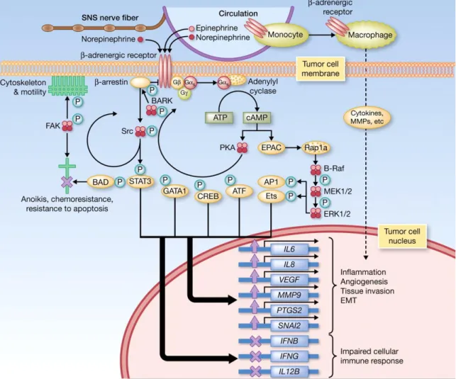
The first evidence for the role of β-AR, in cancer was found in a human lung adenocarcinoma cell line, in which, isoprenaline, a β-adrenergic agonist, increased proliferation of these cells (Schuller and Cole, 1989). Successive studies have then shown that activation of β-AR cause pro-tumorigenic effects (increased proliferation, invasion and angiogenesis, resistance towards apoptosis and impaired cellular immune response) in different types of cancer cell lines/tumours, including breast cancer (Schuller, 2010; Madden et al., 2011; Cole and Sood, 2012; Coelho et al., 2017). Putative transduction mechanisms that couple activation of ARs to cellular pathways relevant for carcinogenesis have been put forward and are depicted in Figure 4. The model proposes that the transient increase of intracellular cAMP triggered by Gs
activation, activates two major biochemical effector systems: the protein kinase A (PKA) and the exchange protein activated by adenylyl cyclase (EPAC), leading to the activation of several transcription factors and important pathways in cancer (Cole and Sood, 2012; Coelho et al., 2017).
Figure 4. ANS fight-or-flight stress responses releases catecholamines into tumor microenvironment.
Catecholamines bind to β-AR, resulting in the activation of PKA which phosphorylate multiple target proteins. This led to the activation of several transcription factors (e.g. transcription factors of the CREB/ATF and GATA families, β-adrenergic receptor kinase (BARK), STAT3, and the focal adhesion kinase (FAK)), leading to changes in cell trafficking and motility, as well as cellular resistance to apoptosis. The second biochemical effector system activated by the β-AR is the activation of EPAC by cAMP, resulting in the activation of the mitogen-activated protein kinase signalling pathway and downstream effects on diverse cellular processes. The general pattern of transcriptional responses induced by β-adrenergic signalling includes upregulated expression of metastasis-associated genes involved in inflammation, angiogenesis, tissue invasion, and epithelial-mesenchymal transition, and downregulated expression of genes facilitating antitumor immune responses (Cole and Sood, 2012).
A link between activation of AR and cancer is also corroborated by studies showing the impact of the clinical use of β-blockers in cancer incidence and mortality. Propranolol, a β-AR antagonist widely used to treat conditions such as hypertension, angina pectoris
and arrhythmias was associated with a significant reduction in tumour recurrence, longer disease-free intervals, reduced risk of metastasis and reduced risk of cancer death (e.g. breast, melanoma, ovarian, prostate cancer) (Powe et al., 2010; Barron et al., 2011; Childers et al., 2015). Moreover, emerging evidence reported that propranolol may have various mechanisms of action that can be particularly relevant in cancer treatment: it has anti-proliferative and anti-metastatic effects and it is able to modulate the tumour stroma (e.g. immune cells, cancer-associated fibroblast) (Campbell et al., 2012; Zhou et al., 2016b; Shaashua et al., 2017; Ashrafi et al., 2018; Chen et al., 2018; Nagaraja et al., 2018). All these characteristics make propranolol a potential candidate for drug repurposing in cancer.
1.6. Oestrogens, adrenergic stimulation and cancer
Chronic stress is not the only condition in which a sustained release of adrenergic messengers occur. Evidence exist supporting that the same may occur during menopause, particularly during hot flashes events (Kronenberg et al., 1984; Freedman, 2014). Although the endocrine physiology of the hot flashes is still not well understood, it is known to be mediated by an interrelation between adrenergic signalling and the decrease in oestrogen levels, observed during the menopause (Freedman, 2014). A relationship between the occurrence of hot flashes and an increased incidence of breast cancer is still debatable (Huang et al., 2011; Chlebowski et al., 2018). A large-scale study has found that women presenting persistent vasomotor symptoms, which includes hot flashes and night sweats, are more likely to be diagnosed with breast cancer (Chlebowski et al., 2018) what brings additional evidence for the existence of a link between the hormonal disturbances underlying menopause (namely increase in catecholamine release and decrease of oestrogens) and the development of cancer. It should be kept in mind that a putative interaction between adrenergic signalling and oestrogen would not be exclusive of tumorigenesis. A relationship between oestrogens and adrenergic signalling is observed in the normal mammary gland where the β-AR levels fluctuate with the estrous cycle, being in a higher number during the proestrous and estrous cycle (Marchetti and Labrie, 1990). Interestingly, this association can also be found in mammary tumors where treatment of ovariectomized rats with 17β-estradiol caused a highly significant increase in β-AR levels and tumor growth (Marchetti et al., 1991).
Further studies are needed to clarify this relationship between oestrogens and adrenergic signalling. A better understanding of the mechanisms underlying the β-adrenergic influence in cancer and the influence of oestrogens in this pathway may offer new opportunities to identify possible pharmacological targets to treat breast cancer.
2. Aim
The present work aimed at further investigate the role of adrenergic signalling in tumorigenesis in breast cancer cells and how it can be modified by oestrogens and by alterations in cell metabolism that occurs during tumorigenesis. This study will focus on the effects mediated by β-AR in two different breast cell lines, in the MCF-10A cells, a non-tumorigenic cell line and in the MCF-7 cells, a tumorigenic cell line.
For these purposes, the effects of agonists and antagonists of β-ARs (isoprenaline and propranolol, respectively) in cellular viability/proliferation, a critical component in the carcinogenesis cascade were studied. Potential differences in adrenergic signalling in non-tumorigenic and tumorigenic cells were investigated in order to bring new contributions to better understand the claimed clinical efficacy of β-blockers, such as propranolol, in cancer, in particular breast cancer.
Knowing the impact that oestrogens also have in the development and progression of breast cancer and their relationship with the adrenergic stimulation, the influence of 17β-estradiol (the most potent and prevalent oestrogen) in this pathway and on the β-AR signalling was also investigated in order to better understand whether the adrenergic signalling is influenced by oestrogens and, if so, if this influence is altered along the tumorigenesis process.
3. Material and methods
3.1. Chemicals
Fetal bovine serum (FBS) and Dulbecco’s Modified Eagle’s Medium – high glucose (DMEM-HG), supplemented L-Glutamine were obtained from Biochrom (Biotecnómica, São Mamede, Portugal). 4-Hydroxytamoxifen and phorbol myristate acetate were obtained from Tocris Bioscience (Biogen, Madrid, Spain). Goat anti-rabbit IgG conjugated with horseradish peroxidase (sc-2004) was from Santa Cruz Biotechnology Inc. (Frilabo, Maia, Porto, Portugal). Monoclonal anti-tyrosine hydroxylase antibody (ab137869) and the polyclonal anti-tyrosine hydroxylase antibody (ab112) were obtained from Abcam (Cambridge, UK). Trypsin/EDTA was obtained from Invitrogen (Alfagene, Carcavelos, Portugal). L-glutamine was purchase from Gibco (Biotecnómica, São Mamede, Portugal). Dulbecco’s Modified Eagle’s Medium/F-12 (DMEM/F12), (-)-Isoprenaline, (+)- propranolol, 17β-estradiol, methyl-β-cyclodextrin, penicillin/streptomycin mixture (10 000 U.mL-1/10 mg.mL-1, respectively), 3-(4,5-dimethylthiazol-2-yl)-2,5-diphenyl tetrazolium bromide (MTT), dimethyl sulfoxide (DMSO), crystal violet solution, hydrocortisone, epidermal growth factor, human insulin, acridine orange, 3-methyladenine (3´MA), sodium pyruvate, Hoescht 33258 and monodansylcadaverine (MDC) were purchased from Sigma-Aldrich (Sigma-Aldrich-Química S.A., Sintra, Portugal).
3.2. Cells and culture conditions
The experimental work described in this dissertation was performed using two different cell lines, MCF-7, a tumorigenic ER+ breast cancer cell line and MCF-10A, a non-tumorigenic ER- cell line. Both cells lines were purchased from ATCC ®. All experiments were carried out in batches of cells with passages number lower than 35 in MCF-10A and lower than 50 in MCF-7 to avoid genotypic/phenotypic changes. The MCF-7 cells were routinely cultured in T25 cm2 cell culture flask with DMEM-HG medium supplemented with 10% heat-inactivated FBS and 1% of a mixture of penicillin/streptomycin (10000 U.mL-1/10 mg.mL-1, respectively). MCF-10A cells were cultured in T75 cm2 cell culture flaks with DMEM/F12 medium supplemented with sodium bicarbonate (1.2 g.L-1), stable L-glutamine (2 mM), epidermal growth factor (EGF; 20 ng.mL-1), human insulin (3.5 µg.mL-1), hydrocortisone (0.5 µg.mL-1), 10% heat-inactivated FBS, 1% of a mixture of penicillin/streptomycin (10000 U.mL-1/10 mg.mL-1,
respectively) and pH adjusted to 7.4. Both cells lines were maintained at 37ºC, in a humidified atmosphere to avoid medium evaporation, and with 5% CO2 to allow optimum pH during culture. For cell culture maintenance, cells were split twice a week and kept below 90% confluence. For this purpose, culture medium was removed, and cells were subsequently washed with 100 mM PBS. Then, cells were treated with a trypsin solution (0.25% trypsin; 0.025% EDTA in PBS) and incubated for six minutes, at 37ºC, to allow the detachment of the cells. Cells were then resuspended in complete medium and allowed to attach in new cell culture flaks.
3.3. Determination of the optimal percentage of serum
Serum is a key component for growing and maintaining cells in culture. It is an important source of growth factors, proteins, hormones, minerals and adhesion factors which facilitates cell survival and growth (Van der Valk et al., 2010).
In order to study the effects of the treatments in MCF-7 and in MCF-10A cells, it was necessary to perform a dose-response curve by increasing the concentrations of FBS for each cell line. This allowed to i) decrease the influence of the FBS (it imposes strong mitogenic effects on cells) on the effects caused by each treatment and ii) to distinguish cell survival from cell proliferation in functional studies by selecting a serum concentration that did not affect cell viability. In MCF-10A, EGF and the human insulin supplements were also removed since they also induced a strong mitogenic effect in these cells.
The functional assays, in MCF-7 breast cancer cells, were performed in the absence of FBS, since MCF-7 cells continue to proliferate in the absence of FBS.
For the MCF-10A breast non-tumorigenic cells, the concentration of 4% FBS was chosen for the functional assays, since it was the only concentration of serum that induced a similar homogenous response through each experimental timepoint.
3.4. Cell treatment
Prior to every experiment, the MCF-7 and the MCF-10A cells were centrifuged at 1500 rpm, for five minutes at 20ºC, and viable cells were counted using the trypan blue dye exclusion method. MCF-7 and MCF-10A cells were then prepared using an initial cell density of 2.3 × 10⁴ cells.mL-1 and 4.0 × 10⁴ cells.mL-1, respectively, in complete medium
and incubated for 24h. After this period, MCF-7 cells were additionally incubated in serum-free medium for 24h to starve the cells from hormones and growth factors. Cells were then treated with increasing concentrations of the non-selective ER agonist, 17β-estradiol (1 - 100 nM), the non-selective β-AR agonist, isoprenaline (0.1 – 10 µM), the non-selective β-AR antagonist, propranolol (1 µM – 100 µM) or with the β2-AR inverse agonist, ICI 118,551 (10 µM), alone or in combination. In some experiments, MCF-7 were also treated with a cholesterol depletion agent, methyl-β-cyclodextrin (1 - 1000 µM), alone or in combination with the compounds mention above. MCF-10A were also treated with 4-hydroxyamoxifen (1 -10000 nM), alone or in combination with isoprenaline. The concentrations for the agonists/antagonists were chosen based on the different affinities towards its target receptors (available in: International Union of Basic and Clinical
Pharmacology/The British Pharmacological Society
http://www.guidetopharmacology.org). Stock solutions of the compounds tested were prepared in DMSO or in water and freshly diluted to the final concentration in medium (DMEM-HG in MCF-7 cells and DMEM/F12 supplemented with 4% FBS in MCF-10A cells), immediately before the experiment. The appropriate control/solvent experiments were carried out using cells exposed to DMSO (0.1%). Cells were then incubated at 37ºC, up to 72h depending on the experimental assay.
3.5. MTT reduction assay
The MTT reduction assay was carried out in order to evaluate cell viability, after exposure to the compounds described before. This assay consists on the endocytosis of and subsequent reduction of the MTT reagent (yellow colored) into insoluble formazans crystals (purpled colored), that can be quantified by spectrophotometry. This step is exclusively carried out by living cells through NAD(P)H-dependent cellular oxidoreductase enzymes. Since inviable cells lose the ability to convert MTT into formazan crystals, this method allows for the quantification of viable cells.
This assay was carried out in the 10A non-tumorigenic breast cells and in the MCF-7 breast cancer cells. To perform this assay, cells were seeded in 96-well plates (200 µL per well). After drug treatment, cells were incubated for 24, 48 and 72h. At the end of each timepoint, cell culture medium was removed and the MTT reagent (0.5 mg.mL-1 in PBS, 100 µL) was added to the plates and incubated for three hours, protected from light. At the end of this incubation period, the MTT solution was removed and 100 µL DMSO were added to each well to dissolve the formazan crystals formed. Thereafter, absorbance was measured at 570 nm in an automated microplate reader (Sinergy HT,
Biotek Instruments Inc, Vermont, USA). Results are expressed as percentage of the respective control. All conditions were performed in parallel triplicates or sextuplicates.
3.6. Kinetic Label-Free Proliferation Assay
To characterize cell proliferation upon exposure to treatments, the number of cells was monitored using an automated label-free cell counting, as previously described (Clayton, 2017). This method uses defocus high contrast brightfield images, which causes a cell mediated-refraction of the light in order to produce a bright spot. The Gen5 Image Analysis Software can then accurately count the bright spots, enabling long-term proliferation studies in a label free manner.
This assay was initially carried out in the MCF-10A and in MCF-7 cells. Nevertheless, for the MCF-10A cells it was not possible to accurately quantify the number of cells at every time point due to their slow growing abilities, and due to the experimental methodology performed (the MCF-10A were cultured in 4% serum DMEM/F12 without supplements, which lead to some cell death).
To perform this assay, cells were seeded in 96-well plates (200 µL per well). After drugs treatment, four high contrast brightfield images, at the center of the well, were captured (Lionheart FX microscope, BioTek Instruments, Winooski, USA), for each time point (T0; T24, T48; T72h): two focused images were used for reference, and the other two defocused images for cell counting. The Gen5 Image Analysis Software was then used to perform the stitching and kinetic alignment of the two defocused images and to do the image pre-processing in order to obtain the best possible enhancement of contrast, reducing each cell to a single bright spot. Object masking thresholds were then set to identify each cell for counting. Plates were maintained at 37 ºC inside the Lionheart FX microscope chamber throughout the imaging steps. Results for the number of cells obtained at each time point were then normalized to the cell number achieved at T0, and the results were expressed as percentage of the respective control, for each time point. All conditions were performed in parallel, in triplicates or sextuplicates.
3.7. Colony formation assay
The colony formation assay was performed as described previously (Soares et al., 2014) with some modifications. MCF-10A cells were seeded in 12-well plates with an initial density of 1000 cells per well in complete medium and allowed to attach for 24h.
Thereafter, medium was replaced with fresh complete medium, in the absence or in the presence of the drugs. After 72h incubation, cells were re-treated with the appropriate drugs and allowed to continually grow for a total of seven days. In the end of the experiment, colonies were fixed with 4% (w/v) paraformaldehyde in PBS (five minutes), stained with crystal violet (0.5% v/v, in distilled water) for five minutes and rinsed twice with distilled water. Representative images of the colonies were taken using a digital camera. Afterwards, crystal violet was eluted with glacial acetic acid (10%) and absorbance at 600 nm was measured in an automated microplate reader (Sinergy HT, Biotek Instruments Inc, Vermont, USA). All the conditions were performed in parallel and in triplicates.
3.8. Protein expression by Western blot
To carry out this assay, MCF-10A cells were seeded in petri dishes of 60.1 cm2 (10 mL per dish). After 24h incubation with the treatment, cell culture medium was removed, and cells were rinsed with ice-cold PBS. Total cell protein were extracted by adding 300 μL of lysis buffer with protease inhibitors 5 mL RIPA, 25 μL Na3VO4 200 mM, 25 μL NaF 1 mM, 100 μL PMSF 100 mM, 1 μL aprotinin (10 mg.mL-1) and 1 μL of leupeptin (10 mg.mL -1). Ceramic beads of 1.4 mm diameter were added each tube, and the samples were then subjected to two cycles of 15 seconds at 5800 rpm in the Precellys Evolution Homogenizer to enhance cell disruption (Bertin Instruments, France). Afterwards, the lysates were incubated on ice for one hour and then centrifuged at 20000 g for 45 minutes, at 4°C. To ensure the loading of the same amount of protein for all the samples, the total protein concentration in the supernatant was determined using the Bradford method (10 μL of each sample diluted 1:10 + 200 μL dye reagent diluted 1:5 and filtered 0,2 μm), using bovine-albumin as standard. Absorbance were measured at 570 nm in an automated microplate reader (Sinergy HT, Biotek Instruments Inc, Vermont, USA). Afterwards, equal amounts of protein (20 μg) were heat-denatured through boiling at 70ºC for ten minutes in 6X sample buffer (0.35 M Tris-HCl pH 6.8, 4% sodium dodecyl sulfate (SDS), 30% glycerol, 9.3% dithiothreitol and 0.01% bromophenol blue). Proteins were subjected to a 10% SDS-PAGE (polyacrylamide gel electrophoresis) at 125 V for one hour with running buffer (25 mM Tris Base, 250 mM glycine, 0.1% SDS).
Proteins were next electrotransferred from gels onto pure nitrocellulose membranes at 25 V and 2.5 A, for three minutes using the Trans-Blot turbo Transfer System (Bio-Rad). Next, membranes were incubated under gentle agitation for one hour at room
temperature with 5% bovine-albumin in PBST (0.1% Tween 20 in PBS pH 7.4, filtered) in order to block additional reactive sites that might exist on the nitrocellulose membrane. Immunoblotting was then carried out overnight, at 4ºC, by probing the membranes with the primary antibody rabbit anti-tyrosine hydroxylase (ab137869- 1:5000; ab112- 1:500). By the end of this incubation period, nitrocellulose membranes were washed with PBST and incubated for one hour at room temperature, under gentle shaking, with the secondary antibody anti-rabbit IgG conjugated to horseradish peroxidase (1:5000). The immunocomplexes formed were detected using an enhanced chemiluminescence system and a Novex ECL detection kit. The immunoblots were scanned using the BioRad ChemiDoc MP Imaging System and images were captured using Image Lab software.
3.9. Detection of catecholamines by HPLC-ECD
HPLC with electrochemical detection (HPLC-ECD) was used in order to evaluate the presence of catecholamines in MCF-10A and in MCF-7 cells.
To carry out this assay, both cell lines were seeded in petri dishes of 60.1 cm2 (10 mL per dish). Upon 24h incubation in the absence or in the presence of isoprenaline (in 0% serum to minimize the influence of FBS), supernatants were collected and perchloric acid 2 M (1:10) was added. The samples were filtered (Spin X filters, Costar) using centrifugation (12000 g for five minutes) and frozen (-20ºC) until analysis. HPLC-ECD analysis were performed at the Laboratory of Clinical Chemistry of the Centro Hospitalar do Porto - Hospital de Santo António, with the collaboration of Dr. Henrique Reguengo. The concentration of catecholamines in the supernatants were calculated using noradrenaline and adrenaline standard solutions.
.
3.10. Morphological transformation assay
To evaluate MCF-10A cell neoplastic transformation, a morphological transformation assay was carried out. MCF-10A cells were seeded in 24-well plates with an initial density of 1 × 10⁵ cells per well in complete medium and allowed to reach confluency. Thereafter, medium was replaced with fresh complete medium, in the absence or in the presence of drugs (isoprenaline or PMA). Then, cells were incubated continually for one month. Cells were refed two times per week in the absence or in the present of drugs, and analysed for the presence of transformed foci, using a phase contrast microscope.
Occasionally, images were also taken with the Lionheart FX microscope. At the end of the experiment, cells were also stained with Hoescht 33258 (5 µg.mL-1) to increase the detailed microscopic visualization and examination of foci.
3.11. Monodansylcadaverine and acridine orange staining
Acridine Orange staining was performed as described (Evangelatov et al., 2016). MCF-7 cells were seeded in 96-well plates (200 µL per well). After drugs treatments, MCF-MCF-7 cells were stained with 5 μg.mL-1 of acridine orange in serum free-medium for 15 min at 37ºC, washed, and analysed on the Lionheart FX microscope. Data were analysed using Gen5 Image Analysis Software.To visualized autophagic structures, the monodansylcadaverine (MDC; 50 µM)marker was used. Cells were treated as for acridine orange staining. Images were taken using the Lionheart FX microscope.
3.12. Data and statistical analysis
Data were firstly tested for normality (Shapiro-Wilk test) and homogeneity of variance (Levene’s test), using the GraphPad Prism X V software package. Differences between treatments and controls were compared using one-way analysis of variance (ANOVA), followed by the post-hoc multiple comparisons Dunnett’s t test, whenever applicable. In assays assessing the combination effects, differences between treatments were assessed using ANOVA, followed by post-hoc multiple comparisons Tukey’s t test, whenever applicable or using a Student´s t test. A value of p˂0.05 was considered to denote significant statistical differences.
4. Results
4.1.
Effects of the β-AR activation in cell viability of the
human breast non-tumorigenic cell line (MCF-10A)
To clarify the effects of the β-AR activation, a set of experiments was firstly performed to characterise the effects of the adrenergic ligands, on the viability of MCF-10A cells, using the MTT assay.
Isoprenaline and propranolol, a β-AR agonist and antagonist, respectively, were used to evaluate the effects of the β-AR activation in MCF-10A cell viability. Isoprenaline is a potent and full β-AR agonist: pKi values: i) β1-AR: 6.6 - 7.0; ii) β2-AR: 6.4 and iii) β3-AR: 5.1 - 6.2 whereas propranolol is a β-AR antagonist: pKi values: i) β2-AR: 9.1 – 9.5 and ii) β3-AR: 6.3 - 7.2.
The effects of the adrenergic ligands in MCF-10A cell viability are shown in Figure 5. After 24h incubation, the β-AR agonist isoprenaline (0.1 – 10 µM) increased viability, of MCF-10A cells and the maximal increase was observed in cells treated with 0.1 µM isoprenaline (Figure 5). However, when cells were incubated with isoprenaline for 72h those effects were no longer observed: isoprenaline (0.1 – 10 µM) failed to alter viability of MCF-10A cells (Figure 32A, Annex).
The β-AR antagonist, propranolol (1 – 100 µM), per se, had no effect in MCF-10A cell viability, after a 24h incubation period (Figure 6A). Moreover, when combined with isoprenaline (0.1 µM), propranolol prevented the increase in MCF-10A cell viability caused by isoprenaline (Figure 6B).
Figure 5. Effects of the β-AR agonist isoprenaline on MCF-10A cell viability, after 24h of incubation. Upon seeding,
MCF-10A cells were incubated for 24h and subsequently treated with the indicated drugs for further 24h (T0). To evaluate cell viability, the MTT assay was used. A detailed description of the method can be found in the Materials and Methods section. Results were expressed as percentage of control and then as a difference between the effect caused by isoprenaline and the effect caused by the control. Results are presented as mean ± SEM, from 6-8 independent experiments. Significant differences from control: * p<0.05; ** p<0.01; one-way analysis of variance (ANOVA), followed by post-hoc multi-comparisons Dunnet´s t test.
Figure 6. Effects of the β-AR antagonist propranolol, alone (A) or in combination with isoprenaline (B), on MCF-10A cell viability, after 24h of incubation. Upon seeding, MCF-MCF-10A cells were incubated for 24h and subsequently
treated with the indicated drugs (T0) for further 24h. To measure the cell viability, the MTT assay was used. A detailed description of the method can be found in the Materials and Methods section. Results are expressed as percentage of control in panel A and as a difference between the effect caused by isoprenaline 0.1 µM and the effect caused by the combined exposure of propranolol (1 and 100 µM) + isoprenaline in panel B. Results are presented as mean ± SEM, from 6-8 independent experiments.
4.2. Influence of 17β-estradiol in the β-AR-mediated effects in
MCF-10A cell viability
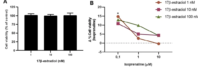
To evaluate the influence of oestrogens in the effects mediated by the β-AR, 17β-estradiol (a full ER agonist; pKi values: i) ER-α: 9.8, ii) ER-β: 9.3, iii) GPER: 8.2 – 8.5) was used.
The effect of estradiol on MCF-10A cells viability is shown in Figure 7A: 17β-estradiol (1 – 100 nM), per se, did not changed cell viability either when cells were incubated for 24h (Figure 7A) or for 72h (Figure 32B, Annex).
The effects caused by exposure to 17β-estradiol and isoprenaline were only evaluated at 24h exposure, as no effects were observed for both drugs at 72h incubation (Figure
32, Annex).
As shown in Figure 7B, treatment of MCF-10A cells with 17β-estradiol, inhibited the increase in cell viability caused by isoprenaline. This inhibition was more evident for cells treated with 0.1 µM isoprenaline (Figure 7B) since it was at this concentration that isoprenaline induced a maximum increase in MCF-10A cell viability (Figure 5A).
Figure 7. Effects of the ER agonist 17β-estradiol, alone (A), or in combination with isoprenaline (B), on MCF-10A cell viability, after 24h of incubation. Upon seeding, MCF-10A cells were incubated for 24h for adherence and
subsequently treated with the indicated drugs (T0) for additional 24h. To evaluate cell viability, the MTT assay was used. A detailed description of the method can be found in the Materials and Methods section. Results are expressed as percentage of control in panel A and as a difference between the effect caused by isoprenaline and the effect caused by the combined exposure of 17β-estradiol (1 – 100 nM) plus isoprenaline in panel B. Results are presented as mean ± SEM, from 6-8 independent experiments. Significant differences from isoprenaline 0.1 µM: * p<0.05; one-way analysis of variance (ANOVA), followed by post-hoc multi-comparisons Dunnet´s t test.