Universidade do Minho
Escola de Ciências da Saúde
Eduardo Manuel Loureiro de Campos
T
HE ROLE OFAP2γ
TRANSCRIPTION FACTOR INTHE MODULATION OF ADULT GLUTAMATERGIC NEUROGENESIS IN DEPRESSION
O
PAPEL DO FATOR DE TRANSCRIÇÃOAP2γ
NAMODULAÇÃO DA NEUROGÉNESE GLUTAMATÉRGICA ADULTA EM DEPRESSÃO
Dissertação de Mestrado Mestrado em Ciências da Saúde
Trabalho efetuado sob a orientação da: Doutora Luísa Alexandra Meireles Pinto
DECLARAÇÃO
Nome: Eduardo Manuel Loureiro de Campos
Endereço eletrónico: [email protected] Telefone: 914267312
Número do Cartão de Cidadão: 13644316
Título da dissertação:
The role of AP2γ transcription factor in the modulation of adult glutamatergic neurogenesis in depression
Orientador:
Doutora Luísa Alexandra Meireles Pinto
Ano de conclusão: 2016
Designação do Mestrado: Ciências da Saúde
É AUTORIZADA A REPRODUÇÃO INTEGRAL DESTA DISSERTAÇÃO APENAS PARA EFEITOS DE INVESTIGAÇÃO, MEDIANTE DECLARAÇÃO ESCRITA DO INTERESSADO, QUE A TAL SE COMPROMETE.
Universidade do Minho, 30 de Setembro de 2016 Assinatura:
v
AGRADECIMENTOS
Estas breves páginas de agradecimentos, não me permitem mostrar a gratidão que tenho a todas as pessoas que ao logo do meu Mestrado em Ciências da Saúde e do meu percurso, me ajudaram e influenciaram no dever de cumprir os meus objetivos tanto pessoais como académicos. O esforço aqui depositado não mais é, senão o resultado da amizade e apoio de todos a quem abaixo deixo algumas breves palavras, mas com um sincero e profundo sentimento de reconhecido agradecimento.
As minhas primeiras palavras de agradecimento vão para a minha orientadora Luísa Pinto. Agradeço-te todo o apoio demonstrado, pela facilidade na partilha de conhecimentos, pelas oportunidades oferecidas e por me teres proporcionado todas as ferramentas necessárias para o desenrolar de todo este trabalho. Um sincero obrigado, por mesmo em férias e sobrecarregada de trabalho me teres sempre ajudado nesta reta final do meu mestrado.
Ao Nuno Dinis Alves, por tanto me teres ensinado. Por teres partilhado comigo longas horas de bancada e de biotério sem nunca nada me cobrar. Pelos conselhos e 1001 ajudas que me deste no desenrolar desta tese. Tenho a perfeita noção, que sem o teu apoio todo este trabalho apresentado nesta dissertação teria sido bem mais complicado. Obrigado.
Ao resto do meu grupo de trabalho, Patrícia, Rita, Joana e António por todo o apoio, pela discussão de resultados, pelos momentos de entreajuda, descontração e bom ambiente de trabalho.
À Catarina Ferreira e Joana Silva por se terem disponibilizado para me ensinar e ajudar sempre que fosse preciso em diferentes protocolos e técnicas.
Aos meus caros colegas de mestrado, pelas partilhas e companheirismo nestes últimos dois anos de trabalho e estudo.
Expresso também a minha gratidão a todos os NERDs, pelo apoio, sugestões partilhadas e auxílio prestado no desenrolar deste trabalho. Agradeço também a este domínio, à ECS e a todo ICVS por ter contribuído para o meu amadurecimento académico e científico cruciais para as minhas próximas etapas.
vi
Aos meus amigos de sempre, Ana Coelho, Adrien, André e Tiago, obrigado pela amizade, por me apoiarem e tanto ouvirem falar do meu trabalho e do ICVS.
À Cláudia, obrigado por tornares a casa de Braga num lar. Pelas conversas e jantares, pela boa disposição e por tanto me aturares todos os dias.
A toda a minha família, em especial os meus Pais, um enorme obrigado por todo o carinho que em mim depositam, por tanto confiarem e apoiarem-me incondicionalmente em todas as minhas etapas pessoais e académicas. A vós, meus alicerces, devo tudo o que sou, e espero um dia poder-vos recompensar de todos os modos e feitios. Muito obrigado por me inspirarem a muito trabalhar para atingir todos os meus objetivos.
As minhas últimas palavras de agradecimento, mas não de todo menos importantes, vão para minha cara-metade. Bárbara, muito obrigado! Obrigado por simplesmente teres aparecido na minha vida e a teres virado do avesso. Se não levar mais nada, levo-te a ti! Obrigado por acreditares mais em mim do que eu próprio. Obrigado por estares sempre em meu auxílio, pela paciência, carinho, companheirismo, amizade e todo o amor que me dás. Obrigado por veres em mim mais do que um homem com sonhos e devaneios. Também eu espero que o futuro nos sorria muito.
vii
ABSTRACT
Major depressive disorder (MDD) is a multidimensional psychiatric disease, considered by the World Health Organization as one of the leading causes of disability. Despite the importance of this disease in modern societies and the large investment of resources already made in its study, the processes underlying its pathophysiology remain poorly understood. Several hypotheses have been proposed to clarify the neurobiological mechanisms underlying this psychiatric disorder, being the link between adult hippocampal neurogenesis and MDD a central topic in the past decades. Previous studies have identified AP2γ as a key regulator of adult hippocampal neurogenesis in mice, being expressed in a subpopulation of adult transient amplifying progenitors, and acting as a regulator of basal progenitors, promoting proliferation and glutamatergic neuronal differentiation. Thus, we wanted to further explore the impact of AP2γ in brain neurophysiology and behavior during development and at adult stages, dissecting also its mechanisms both in healthy and depressive states. With this study, we were able to understand the impact of AP2γ in post-natal development and during juvenile age, through the AP2γ constitutive knockout (KO) model. In the developmental milestones assessment we did not find any major impairment in the behavioral performance of AP2γ KO mice, since all parameters analyzed, including the ones where we found differences, were within the typical range for appearance of the developmental milestones. However, in the juvenile behavior assessment and in the hippocampal glutamatergic neurogenesis process, impairments were found, since AP2γ KO mice showed anxious-like behavior and decreased proliferation of immature neurons. To study the impact of modulating the transcription factor AP2γ in depression we exposed both constitutive and conditional KO animal models to a chronic stress protocol, which efficiently induced core depressive-like symptoms. Through the conditional AP2γ KO mice, we were able to elucidate the impact of deleting AP2γ on behavior and neurogenesis in depressive-like conditions specifically in adult age, without the interference of potential functions of the gene during early development that may appear in the constitutive AP2γ model. Through a multidimensional behavioral analysis, we observed that both models presented similar results in the three most affected behavioral dimensions in depression, namely anxiety, mood and cognition. Regarding anxiety and mood no major differences were found between genotypes in both animal models. Moreover, AP2γ KO mice presented cognitive deficits in basal conditions, but when exposed to chronic mild stress no detrimental effects of deletion of the gene were observed. In this work, we also identified, through a broad analysis of the dentate gyrus neurogenic niche, alterations of epigenetic regulators in the AP2γ constitutive KO mice after uCMS exposure. The reported results not only support the involvement of AP2γ in the transcriptional network that modulates the juvenile and adult neurogenic process, but also highlight the potential of this molecule as a future therapeutical tool in neuropsychiatric disorders, in which neurogenesis is impaired.
ix
RESUMO
O transtorno depressivo persistente é uma doença psiquiátrica multidimensional, considerada pela Organização Mundial de Saúde como uma das principais causas de incapacidade. Apesar da importância desta doença na sociedade moderna, e do largo investimento de recursos já feitos no seu estudo, os processos subjacentes à sua patofisiologia continuam pouco percebidos. Várias hipóteses foram propostas para clarificar os mecanismos neurobiológicos implícitos nesta doença psiquiátrica, tendo sido o vínculo entre a neurogénese hipocampal adulta e a depressão um tópico central nas décadas passadas. Estudos anteriores identificaram o AP2γ como um regulador chave da neurogénese hipocampal adulta em ratinhos, sendo expresso numa subpopulação de células progenitoras de rápida amplificação adultas, e atuando como regulador de progenitores basais, promovendo a proliferação e a diferenciação neuronal glutamatérgica. Deste modo, propusemos continuar a explorar o impacto do AP2γ na neurofisiologia cerebral e no comportamento, durante a fase de desenvolvimento e na idade adulta, procurando entender também os seus mecanismos tanto no estado saudável como em depressão. Com este trabalho, fomos capazes de entender o impacto do AP2γ no desenvolvimento pós-natal e em idade juvenil, a partir do modelo animal de deleção constitutiva do AP2γ. Na avaliação dos marcos de desenvolvimento, não encontramos nenhuma alteração no desempenho comportamental nos animais com deleção de AP2γ, visto que todos os parâmetros analisados, incluindo os que encontramos alguma diferença, se encontravam dentro dos intervalos típicos de aparecimento dos marcos de desenvolvimento. Contudo, na avaliação do comportamento juvenil e no processo de neurogénese glutamatérgica hipocampal observamos défices, visto que os animais com deleção de AP2γ apresentaram comportamento ansioso e uma diminuição da proliferação de neurónios imaturos. Para estudar o impacto da modulação do fator de transcrição AP2γ em depressão expusemos tanto o modelo animal com deleção constitutiva bem como o modelo animal condicional do gene a um protocolo de stress crónico, o qual eficientemente induziu sintomas primários de depressão. Através do modelo animal condicional do AP2γ, conseguimos compreender o impacto da deleção do AP2γ na modulação do comportamento e neurogénese em condições depressivas especificamente em idade adulta, sem interferência das potenciais funções do gene durante o período de desenvolvimento dos animais, que poderão surgir no modelo animal constitutivo do AP2γ. Através, de uma análise comportamental multidimensional, observamos que ambos os modelos apresentaram resultados similares nas dimensões comportamentais mais afetadas na depressão, nomeadamente a ansiedade, o humor e a cognição. Relativamente à ansiedade e ao humor não encontramos grandes diferenças entre genótipos em ambos os modelos animais. Além disso, os modelos animais de deleção do AP2γ apresentaram défices cognitivos em condições basais, mas após exposição ao stress crónico não foram observados os efeitos prejudiciais da deleção do gene. Neste trabalho, também identificamos, através de uma análise abrangente do nicho neurogénico girus denteado, um reguladores epigenéticos alterados no animal constitutivo do AP2γ. Os resultados apresentados não só suportam o envolvimento do AP2γ na rede transcripcional responsável pela modulação do processo neurogénico juvenil e adulto, como também destacam o potencial desta molécula em abordagens terapêuticas futuras em doenças neuropsiquiátricas, nas quais a neurogénese se encontra afetada.
xi
TABLE OF CONTENTS
Agradecimentos ... v Abstract ... vii Resumo ... ix Table of Contents ... xi List of Abreviations ... xvList of Figures ... xix
List of Tables ... xxi
Chapter 1 - Introduction ... 1
1.1. Depression ... 3
1.1.1. State of the art ... 3
1.1.2. Modeling depression in animal models ... 5
1.2. Adult neurogenesis in the mammalian brain: genetic and epigenetic modulation of the hippocampal neurogenic process ... 6
1.2.1. Neurogenesis in the adult mammalian brain ... 6
1.2.2. The hippocampal neurogenic niche: an overview of the adult hippocampal neurogenic process ... 9
1.2.3. Transcriptional network underlying hippocampal neurogenesis: a focus on the transcription factor activating protein 2 gamma (AP2γ) ... 11
1.2.4. Epigenetic regulation of adult hippocampal neurogenesis: DNA methylation and DNA demethylation as epigenetic choreographers ... 15
1.3. Implications of adult hippocampal neurogenesis deregulation in the etiopathogenesis of depression ... 19
1.3.1. Adult hippocampal neuroplasticity on the pathophysiology of depression ... 19
1.3.2. Transcriptional and epigenetic deregulation of adult hippocampal neurogenesis as a possible precipitator of depression ... 20
Chapter 2 - Research Objectives ... 25
Chapter 3 - Materials and Methods ... 29
xii
3.2. Genotyping ... 32
3.3. In vivo tamoxifen injections ... 34
3.4. Unpredictable chronic mild stress protocol ... 34
3.5. Serum corticosterone measurements ... 35
3.6. Behavioral tests ... 35
3.6.1. Developmental milestones ... 35
3.6.2. Elevated-plus maze ... 41
3.6.3. Open Field test ... 41
3.6.4. Forced Swimming test ... 42
3.6.5. Tail suspension test ... 42
3.6.6. Sucrose splash test ... 42
3.6.7. Cognitive function assessment ... 43
3.7. BrdU labeling in vivo ... 45
3.8. Fixation of mice brains and tissue processing ... 45
3.9. Immunostainings ... 46
3.10. Western blot ... 47
3.11. Quantitative real-time PCR ... 47
3.11.1. RNA extraction ... 48
3.11.2. cDNA transformation and quantitative Real-time PCR ... 48
3.12. Data analysis ... 49
3.13. Experimental design ... 51
Chapter 4 - Results ... 53
4.1. Functional impact of the transcription factor AP2γ in the neurodevelopment ... 55
4.1.1. Developmental milestones assessment ... 55
4.1.2. The transcription factor AP2γ impact on behavior of juvenile mice ... 58
4.1.3. The transcription factor AP2γ impact on hippocampal glutamatergic neurogenesis in juvenile mice ... 60
4.2. Functional impact of the transcription factor AP2γ in Depression ... 62
4.2.1. Validation of the uCMS model of depression ... 62
4.2.2. Assessment of the behavior dimensions affected by the uCMS protocol ... 65 4.3. AP2γ modulatory mechanisms of adult hippocampal neurogenesis in depressive-like animals 75
xiii
4.3.1. AP2γ impact on the protein levels of different transcription factors involved in the adult
hippocampal neurogenic process ... 75
4.3.2. AP2γ transcription factor impact on epigenetic regulators of adult hippocampal glutamatergic neurogenesis ... 79
5.1. Role of the transcription factor AP2γ during postnatal development ... 85
5.2. The role of AP2γ in juvenile mice ... 86
5.2.1. Impact of AP2γ on behavioral performance of juvenile mice ... 86
5.2.2. AP2γ modulation of hippocampal proliferation and neurogenesis in juvenile mice ... 88
5.3. Functional impact of the transcription factor AP2γ in depression ... 89
5.3.1. The uCMS protocol as a model of depression ... 89
5.3.2. Role of AP2γ in the modulation of behavioral domains affected by depression ... 91
5.3.3. The modulatory action of AP2γ in the hippocampal neurogenic niche ... 93
5.4. AP2γ impacts on epigenetic regulators of adult hippocampal glutamatergic neurogenesis – A preliminary perspective ... 94
5.5. AP2γ transcription factor – An integrated perspective ... 96
Chapter 6 – Concluding Remarks ... 99
Chapter 7 – References ... 103
Chapter 8 - Annexes ... 117
xv
LIST OF ABREVIATIONS
# 5caC - 5-carboxylcytosine 5fC - 5-formylcytosine 5hmC - 5-hydroxymethylcytosine 5mC - 5-methylcytosine A ACTH - Adrenocorticotropic AD - Alzheimer’s disease ADs - Antidepressant drugsANPs - Amplifying neural progenitor cells AP2γ - Activating protein 2 gamma
B
BDNF - Brain-derived neurotrophic factor BrdU - 5-bromo-2-deoxyuridine
C
CBP - cyclic AMP-response element binding protein
CFC - Contextua fear conditioning cm - centimeter
CMS - Chronic mild stress CNS - Central nervous system coREST - REST corepressor
CREB - cAMP response element binding CSF - Cerebrospinal fluid
CUS - Chronic unpredictable stress
D DAPI - 4-6-diamidino-2-phenylindole DCX - Doublecortin DG - Dentate gyrus DNMTs - DNA methyltransferases E E - Embryonic day
ELISA - enzyme-linked immunossorbant assay EPM - Elevated-plus maze
ESCs - Embryonic stem cells
F
FBS - fetal bovine serum FGF - Fibroblast growth factor FOX - Forkhead box protein FST - Forced swimming test
G
GADD45β - DNA-damage-inducible protein 45β GCL - Granular cell layer
GFAP - Glial fibrillary acidic protein
H
h - hours
hA - Horizontal astrocytes HDAC - HDAC inhibitor HDAC - Histone deacetylases HPA - Hypothalamic-pituitary axis
xvi I i.p. - Intraperitoneally K kDa - Kilodalton KO - Knockout L
IncRNAs - Long non-coding RNAs
M
M - Molar mA - milliamp
MDB1 - Methyl binding protein 1 MDD - Major depressive disorder MeCP2 - Methly-CpG- binding protein mg - Milligram
mg/kg - Milligram per kilo mg/ml - Milligram per milliliter min - minutes
miRNAs - MicroRNA ml - Milliliter
mRNA - messenger RNA MWM - Morris water maze
N
NaCl - Sodium Chloride NeuN - Neuronal nuclei
NeuroD - Neurogenic differentiation Ngn2 - Neurogenin 2
NOR - Novel object recognition NPCs - Neural progenitor cells
NSCs - Neural stem cells
O
OB - Olfactory bulb OF - Open field
P
Pax6 - Paired box gene 6 PBS - Phosphate buffered saline
PBS-T - Phosphate buffered saline-Triton PCR - Polymerase chain reaction PFA - Paraformaldehyde
PFC - Prefrontal cortex PND - Postnatal day
Prox1 - Prospero homeobox 1
PSA-NCAM - Polysialylated-neural cell adhesion molecule
Q
QNPs - Quiescent neural progenitors
qRT-PCR - Quantitative real time - Polymerase chain reaction
R
rA - Radial astrocytes
REST - Repression element 1 silencing transcription factor
RMS - Rostral migratory stream rpm - Rotations per minute RT - Room temperature
RT-PCR - Reverse transcriptase - Polymerase chain reaction
xvii S
S - seconds
SEM - Standard error of the mean SEZ - Subependymal zone
SGZ - Subgranular zone SOX2 - SRY-related HMG box 2
T
TET - Ten-eleven translocation TGFβ - Transforming growth factor β TST - Tail suspension test
TM - Melting temperature Tbr - T-box brain protein
TAPs - Transient amplifying progenitor Trx6 - Trithorax protein
U
uCMS - Unpredictable chronic mild stress
V
VPA - Valproic acid
W
xix
LIST OF FIGURES
Figure 1. Neurogenesis is a process not an isolated event. ... 7
Figure 2. The two neurogenic niches in the adult mammalian brain. ... 8
Figure 3. Developmental stages in the adult hippocampal neurogenic process. ... 11
Figure 4. Transcriptional factors involved in the regulation of the adult hippocampal neurogenic process ... 15
Figure 5. Epigenetic regulators of the adult hippocampal neurogenic process. ... 17
Figure 6. Impact of hippocampal glutamatergic neurogenesis in depression. ... 21
Figure 7. Representation of the 6-kb genomic fragment of AP2γ. ... 33
Figure 8. Schematic representation of the CFC protocol. ... 45
Figure 9. Experimental design ... 51
Figure 10. The constitutive deletion of one allele of AP2γ impact on the milestones development and maturity. ... 57
Figure 11. The impact of the constitutive deletion of one allele of AP2γ in the emotional and mood behavioral dimension of juvenile mice. ... 60
Figure 12. Impact of the constitutive deletion of one allele of AP2γ in the post-natal hippocampal glutamatergic neurogenesis of juvenile animals. ... 62
Figure 13. Validation of the uCMS model of depression in the constitutive AP2γ KO animal model. .... 63
Figure 14. Validation of the uCMS model of depression in the conditional AP2γ KO animal model. ... 64
Figure 15. Impact of the constitutive deletion of one allele of AP2γ in the anxiety and mood behavioral dimensions of depression. ... 67
Figure 16. Impact of the conditional deletion of one allele of AP2γ in the anxiety and mood behavioral dimensions of depression. ... 69
Figure 17. Impact of the constitutive deletion of one allele of AP2γ in the cognitive behavioral dimension of depression. ... 73
Figure 18. Impact of the conditional deletion of AP2γ in the cognitive behavioral dimension of depression. ... 74
Figure 19. Western blot analysis of Sox2, AP2γ Pax6 and Tbr2 in adult hippocampal dentate gyrus of the constitutive AP2γ KO animals. ... 77
Figure 20. Western blot analysis of AP2γ, Pax6, DCX and Tbr2 in adult hippocampal dentate gyrus of the conditional AP2γ KO animals. ... 78
xx
Figure 21. Epigenetic modulators involved in DNA methylation in the hippocampal DG of AP2γ constitutive KO depressed-like animals. ... 80 Figure 22. Epigenetic modulators involved in DNA demethylation in the hippocampal DG of AP2γ constitutive KO depressed-like animals. ... 82
xxi
LIST OF TABLES
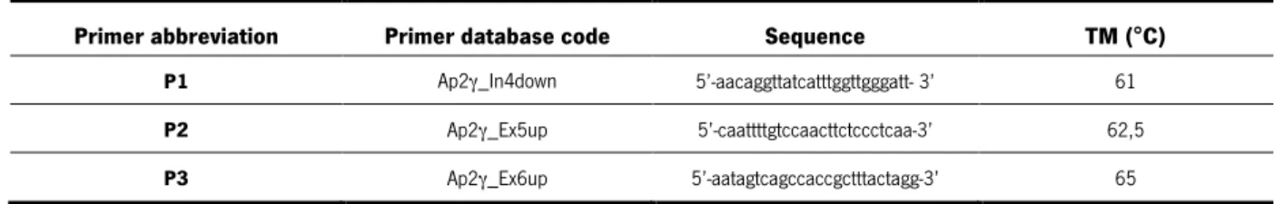
Table 1. List of PCR primers sequence needed to genotype AP2γ gene and respective melting
temperature (TM) ... 32
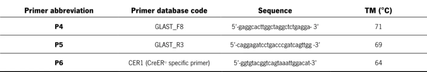
Table 2. List of PCR primers sequence needed to genotype GLAST: CreERT2 mice and respective melting temperature (TM) ... 33
Table 3. Primer mix composition and reaction conditions ... 33
Table 4. Data sheet for developmental milestones ... 36
Table 5. Adaptation of time latency registered for each test into dichotomic scores ... 38
Table 6. Summary of each test analyzed and its evaluated dimension ... 40
Table 7. List of primary antibodies ... 46
Table 8. List of secondary antibodies ... 46
Table 9. Solutions compositions ... 46
Table 10. cDNA mix composition ... 48
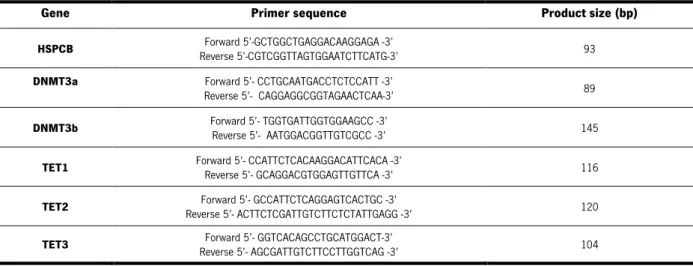
Table 11. List of PCR primers used to amplify genes associated with epigenetic mechanisms ... 49
Table 12. PCR mix composition and reaction conditions ... 49
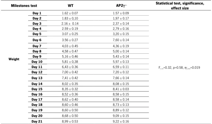
Table 13. Summary of body weight and anogenital distance throughout the 21 days of the milestones protocol and eye opening day ... 55
Table 14. Summary of the average days of mature response obtained in each genotype during the neurobiological reflexes milestones assessment. ... 58
Table 15. Statistical analysis of the juvenile behavior tests. ... 59
Table 16. Statistical analysis of the post-natal glutamatergic neurogenesis of juvenile animals. ... 61
Table 17. Statistical analysis of the parameters used to validate the uCMS protocol in the constitutive KO animal model. ... 64
Table 18. Statistical analysis of the parameters used to validate the uCMS protocol in the conditional KO animal model. ... 64
Table 19. Statistical analysis of the behavioral tests used to evaluate anxiety-like behavior and mood of the constitutive AP2γ KO animal model. ... 68
Table 20. Statistical analysis of the behavioral tests used to evaluate anxiety and mood dimension of the conditional AP2γ KO animal model. ... 70
Table 21. Statistical analysis of the behavioral tests used to evaluate the cognitive dimension of the constitutive AP2γ KO animal model. ... 72
xxii
Table 22. Statistical analysis of the behavioral tests used to evaluate the cognitive dimension of the conditional AP2γ KO animal model. ... 75 Table 23. Statistical analysis of the western blots performed in the hippocampal dentate gyrus of AP2γ constitutive KO animals. ... 77 Table 24. Statistical analysis of the western blots performed in the hippocampal dentate gyrus of AP2γ conditional KO animals. ... 78 Table 25. Statistical analysis of DNMTs gene expression quantification in the dorsal and ventral DG of the constitutive AP2γ KO animal model. ... 80 Table 26. Statistical analysis of the TET genes expression quantification in the dorsal and ventral DG of the constitutive AP2γ KO animal model. ... 81
1
CHAPTER 1 - INTRODUCTION
CHAPTER 1
INTRODUCTION
3
1) INTRODUCTION
1.1. Depression
1.1.1. State of the art
Major depressive disorder (MDD) is considered by the World Health Organization (WHO) as one of the world leading causes of disability, since it is estimated that around 350 million people worldwide are affected with this psychiatric disorder. It is the most disabling medical condition, in terms of years lost due to disability, and it is foreseen that by 2030 depression will be the major contributor to the global illness burden (Willner et al. 2013). Patients suffering from this disorder usually display a loss of interest for experiencing pleasurable activities (anhedonia), changes in appetite and sleep pattern, abnormal sadness states, high levels of anxiety, lack of energy and ultimately suicidal ideation. Moreover, rather than low self-esteem, depressive patients present a deeply negative view of the world and the future, showing also deficits of attention, interpretation and memory (Mathews and MacLeod 2005; Willner et al. 2013; Bergstrom and Meacham 2016).
Despite the importance of this multidimensional psychiatric disease in modern societies and the large investment of resources already made to look for efficient treatment, the processes underlying its pathophysiology remain poorly understood. It is accepted that this complex disorder involves gene-environment interactions, but the genetic and gene-environmental subtracts are largely unknown. Even though there is little knowledge regarding the real causes for the precipitation of MDD, vulnerability or predisposition to develop depression may occur throughout lifetime due to negative environmental stimuli. It is recognized that harmful early life experiences, such as inadequate familial relations, increase the risk for a depressive episode (Willner et al. 2013; Slavich and Irwin 2014). Also, it is consensually accepted that there is a familial predisposition to inherit this disorder through “stress-provoking” genes passing on across generations and providing vulnerability to develop MDD (Slavich and Irwin 2014). However, such genetic transmission pattern, which does not follow the Mendelian laws, is highly complex, and even with the evolution of technical means and the human genome sequencing, it has not been clarified yet. These scientific difficulties to understand the mechanisms underlying depression, can be explained by the multiple neurological systems that are likely involved in the etiopathogenesis of depression (Marsden 2013). Due to the lack of knowledge in this scientific field, there are still many unmet medical needs to address.
4
Looking for a way to successfully revert and treat major depression, several hypotheses have been proposed to clarify the neurobiological mechanisms underlying the onset, maintenance and recovery from this psychiatric disorder: the neurochemical and neurotrophin hypotheses; involvement of cytoquines and inflammatory agents; glutamate excitotoxicity; altered HPA axis; the phase-shift; and the neurogenic hypothesis (Bessa et al. 2009a; Hasler 2010). Depression has a great impact on the central nervous system (CNS) inducing structural and neuroplasticity alterations in brain regions such as the prefrontal cortex (PFC), the amygdala, the ventral striatum (including the nucleus accumbens), and the hippocampus (Pittenger and Duman 2008). Indeed, in the past three decades, a vast number of studies have revealed that during a depressive episode, it is possible to observe cell loss and neuronal atrophy in the hippocampus, which is a brain area with relevant roles both in adult brain neuroplasticity and behavioral control (Pittenger and Duman 2008; Serafini 2012; Mateus-Pinheiro et al. 2013). Several mechanisms were proposed to explain this cell loss and neuronal atrophy, among which we could find the glucocorticoid and glutamate toxicity for both glia and neurons (Duman 2009; Kudryashova 2015), the decreased neurotrophic factors expression (Castren et al. 2007) and also the reduced neuronal plasticity (dendritic arborization atrophy and neurogenesis reduction in the hippocampal neurogenic niche) (Bessa et al. 2009a) in animal models of depression. The potential link between adult neurogenesis and MDD has drawn some attention in the past decades (WuiMan et al. 2013). Although being a controversial topic, the so called neurogenic hypothesis of depression, has brought different relevant questions that challenge the classical conceptions regarding depression, and addresses the neurogenic process as a key pathological player and therapeutical target in stress-related disorders (Eisch and Petrik 2012).
Assuring the implication of adult neurogenesis in depression will support the need to better understand the adult hippocampal neurogenic niche not only in physiological but also pathological conditions. Such knowledge will possibly lead to additional therapeutical approaches by artificially regulating the endogenous neural progenitors pool, in order to sustain hippocampal neurogenesis, and counteract the inhibitory effects induced by depression. This goal could be achievable through genetic or epigenetic regulation of the adult hippocampal neurogenesis process. For this, research should focus in finding key transcriptional factors and epigenetic modulatory molecules that regulate hippocampal neurogenesis, and thus present strong therapeutical potential. Altogether, it may be relevant to bring new insights on the transcriptional network and the epigenetic mechanisms underlying adult neurogenesis in the healthy and “diseased” brain, to fully understand this highly complex neurobiological process, and its potential role as a therapeutical solution for depression.
5
1.1.2. Modeling depression in animal models
Modeling of human neuropsychiatric disorders, such as depression, in animals is extremely challenging given the subjective nature of many key symptoms, the lack of objective tests, and the poor knowledge regarding the onset, maintenance and recovery from such diseases (Nestler and Hyman 2010). Nonetheless, a lot of effort has been made in order to construct animal models and protocols to understand the pathophysiology of depression and develop different modulatory drugs with therapeutic actions.
Knowledge of the etiopathogenesis of depression has progressed substantially in the last years, in part due to studies in animals models (Patricio et al. 2013). The validity of an animal model for formulation of hypotheses and for the development of novel therapeutic strategies encompasses: the use of known etiological factors (etiological validity), it must mimic the behavioral and neurological symptoms observed in human disease (face validity) and importantly, it must respond to clinically effective treatments (predicted validity) (Berton et al. 2012; Patricio et al. 2013). Although selected depressive symptoms may be irreproducible in animals, such as suicidal ideation, a number of models exhibit considerable construct validity when targeting other clinical phenotypes of depression. One of the most important advances in understanding psychiatric disorders, like depression, has been the development of mice with altered expression of specific targets, being it a receptor, transporter, enzyme or signal transduction molecule (Tecott and Wehner 2001; Cryan and Mombereau 2004). These new tools have the potential to verify novel targets for antidepressant activity for which few established pharmacological tools exist. Moreover, these genetically altered mice will enable better testing of the validity of current molecular theories of depression (Cryan and Mombereau 2004). Although there are a large number of mice strains that have been generated with a phenotype that has been interpreted as being related to depression or antidepressant action, there are three most recommend mice strains to use in a depression study: C57Bl/6J, SV/129 and BALB/c mice (Bergner et al. 2016).
There are several models of depression described in the literature: chronic unpredictable stress (CUS), unpredictable chronic mild stress (uCMS), social stress, early life stress, learned helplessness, fear conditioning and olfactory bulbectomy (Duman 2010; Patricio et al. 2013). Despite none of these models can fully recapitulate the complexity and heterogeneity of the human disease, they are considered robust approaches to study depression. However, the uCMS protocol (Willner et al. 2013), based in the principles of the CMS and CUS protocols was proven to be a more robust approach to model the human depression at the lab. In this model, after exposure to chronic mild stressors implemented in an unpredictable way, stressed animals present depressive-like symptoms such as anhedonia, anxiety and
6
cognitive deficits, showing in this way impairments in all three behavioral dimensions known to be affected in humans with depression (Bessa et al. 2009a; Mateus-Pinheiro et al. 2013). Moreover, these stressed animals show impaired neuroplasticity, and compromised regulation of the corticosterone levels, another well-known molecular phenotypes of depression and other stress-related disorders (Mateus-Pinheiro et al. 2013; Patricio et al. 2015). Although the uCMS was first described using a rat animal model, this model of depression induction has also been validated in mice (Surget and Belzung 2009), maintaining its translational value as it induces some core alterations that are similar to those observed in depressed patients (Sibille et al. 2009; Nollet et al. 2013).
In sum, although the currently available rodent models have significant limitations, ranging from weak validation to poor predictive power for drug efficacy in human disease, they have been a powerful tool to investigate the pathophysiology of depression. Further understanding the mechanisms underlying the pathophysiology of depression is of the upmost importance to allow improvement of the experimental animal models and lead to more complete and targeted depressive studies.
1.2. Adult neurogenesis in the mammalian brain: genetic and epigenetic modulation of the hippocampal neurogenic process
1.2.1. Neurogenesis in the adult mammalian brain
The discovery of neurogenesis in the adult mammalian brain, overturned the long-held dogma that the adult central nervous system (CNS) was immutable, and had no capacity for generating new cells (Altman and Das 1965; Deng et al. 2010). Although the emergence of adult neurogenesis as a research field in neuroscience has brought much excitement, there was a lot of reluctance manifested towards the first reports in this area (Egeland et al. 2015). Despite the initial skepticism, it is now well established that new neurons are continuously generated, differentiated and integrated in the preexisting brain neuronal networks (Doetsch et al. 1999; Gage 2002; Deng et al. 2010). Adult neurogenesis is not a single isolated event, is thus a complex process, involving a wide range of highly regulated steps, starting with the proliferation of neural stem cells (NSCs) that will then divide to give rise to transient amplifying progenitors (TAPs) which will be responsible for the rapid expansion of the multipotent progenitor cells pool. TAPs will then differentiate in immature cells, committed to a neuronal phenotype (neuroblasts), that will undergo morphological and physiological maturation with acquisition of neuronal characteristics, and will finally become functionally integrated in the pre-existing network (Figure 1) (Balu and Lucki 2009).
7
In the adult brain there are specific areas where neurogenesis persists throughout life, known as neurogenic niches (Urban and Guillemot 2014). Such spatially defined brain regions where neurogenesis occurs display the presence of immature NSCs from which new neurons can develop, and a permissive microenvironment rich in cell-extrinsic factors needed to favor the generation of new cells (Urban and Guillemot 2014).
Although being a controversial topic, there are two consensual neurogenic brain regions broadly recognized in the adult mammal brain: the subependymal zone (SEZ) lining the lateral ventricles and the subgranular zone (SGZ) of the hippocampal dentate gyrus (DG) (Figure 2) (Zhao et al. 2008; Balu and Lucki 2009; Urban and Guillemot 2014). These two neurogenic niches are largely responsible for the formation of distinct types of neurons. In the SEZ the precursor cells are mostly found in the temporal walls of the lateral ventricles. Here, newly-born precursor cells generate neuroblasts that migrate along the rostral migratory stream (RMS), reaching the olfactory bulb (OB). At the OB, neuroblasts differentiate and mature, becoming largely GABAergic granule, and periglomerular inhibitory interneurons (Chumley et al. 2007; Belenguer et al. 2016). In the DG, the precursor cell population resides throughout the SGZ with specific gradients (Silva et al. 2006). After being formed in the SGZ, the newly-born neuronal cells – neuroblasts, become committed to a neuronal lineage and migrate into the granular cell layer (GCL), where they fully differentiate into excitatory glutamatergic granule neurons (Brill et al. 2009). In addition to these two consensually recognized neurogenic niches,
Figure 1. Neurogenesis is a process not an isolated event.
Adult neurogenesis is regulated at many different stages of cell development. Here the term neurogenesis comprises all necessary steps, starting with the division of a NSC and resulting in the existence of a functionally fully integrated newborn neuron. Interestingly, a high percentage of the newborn neurons die before becoming fully integrated in the network. Gliogenesis also happen at a lower percentage in the adult neurogenic niches. Newborn glial cells are thought to be generated from the same progenitor cells that give rise to neurons. Adapted from (Kempermann, 2011).
8
some research groups have presented evidence that neurogenesis can occur in other brain regions like the striatum (Luzzati et al. 2006; Inta et al. 2016), the cortex (Kodama et al. 2004; Ohira et al. 2010), the amygdala (Goncalves et al. 2008) and the hypothalamus (Fowler et al. 2002; Kokoeva et al. 2005). However, these results are quite controversial and further studies are needed to ashore that these neurogenic niches have indeed NSCs and a permissive microenvironment to allow the formation of new functional neurons.
Despite the increasingly intense research, a great number of questions regarding the adult neurogenesis process remain to be answered and understood. It is unquestionably recognized that in the healthy adult mammalian brain new neurons can be generated, but its functional relevance remains to be fully comprehended. While this singularity is confined to a few privileged brain regions, the generation of new neurons in the post-natal brain represents a new dimension of plasticity, impacting both directly and indirectly on neuronal remodeling and repair. This promising therapeutical target, for a wide range of neuropathological contexts, is one of the main reasons why this field is so interesting.
Figure 2. The two neurogenic niches in the adult mammalian brain.
Two regions of the adult mammalian brain are broadly recognized to be neurogenic under physiological conditions: (SEZ) Precursor cells residing in the walls of the lateral ventricles give rise to interneurons that integrate into the olfactory bulb. (SGZ) Neurogenesis in the adult hippocampal dentate gyrus generates new excitatory granule cells throughout life. These two processes of adult neurogenesis originate from different precursor cell populations, are independently regulated, and serve entirely different regions (Kempermann 2011). Abbreviations: CSF - Cerebral spinal fluid.
9
In the healthy brain, the functional importance of adult neurogenesis has been differently associated between the two most commonly accepted neurogenic niches. While the SEZ has already been associated with olfactory discrimination (Moreno et al. 2009), hippocampal neurogenesis has been related with memory, learning, pattern separation and even emotional behavior (Deng et al. 2010). Somehow the functional importance of adult neurogenesis in the SEZ has not yet been highly associated with a wide range of neuropathological conditions. However, there are already some studies reporting impairments in this specific neurogenic niche in neurodegenerative conditions, such as Alzheimer’s disease (AD) (Curtis et al. 2007). Abnormal alterations in the hippocampal neurogenesis have been associated to a variety of pathologies, including neuropsychiatric disorders (Chambers 2013; Schoenfeld and Cameron 2015; Kang et al. 2016). Numerous research groups are trying to unveil the biological mechanisms underlying these disorders, also allowing us to better understand the process of hippocampal neurogenesis.
1.2.2. The hippocampal neurogenic niche: an overview of the adult hippocampal neurogenic process
In the adult mammalian brain, hippocampal neurogenesis produces new excitatory granule cells in the DG, in a highly regulated and complex process, that can be divided into four major steps: (1) the precursor cell phase, that comprises the proliferation of NSCs and the expansion of the precursor cell pool; (2) the early survival phase, during which the majority of the newborn cells are eliminated even before they make synaptic contacts with reach their target regions; (3) the post-mitotic maturation phase where it is possible to observe the functional integration of newly-born neurons into pre-established neural networks; and (4) the late survival phase, in which the establishment of new synapses with pre-existing surrounding cells is completed and a final selection occurs based on the newborn neurons functionality (Kempermann et al. 2004; Balu and Lucki 2009; Nicola et al. 2015) (Figure 3). Interestingly, this post-natal neurogenesis mimics the embryonic neurogenic process, but differing in the fact that, in the adult brain, the newborn neurons are generated in an already mature microenvironment, and as such they have to integrate the pre-existing neuronal circuits.
The SGZ of the hippocampal DG, contains a heterogeneous progenitor cell population with distinct degrees of stemness, that can be identified by a specific group of molecules, expressed by each cell type. These different molecules (presented in Figure 2) are strongly associated with the different phases of the adult hippocampal neurogenesis process. The type-1 progenitor cells, also known as quiescent neural progenitor (QNPs) cells and NSCs, are believed to be multipotent stem cells with
10
unlimited self-renewal capacities. These cells have astroglial and radial glia-like properties that can be further distinguishable into two classes: horizontal astrocytes (hA) and radial astrocytes (rA). The asymmetrical division of type-1 progenitor cells give rise to two consecutive stages of transient amplifying neural progenitor cells (ANPs): type-2a progenitor cells, followed by the type-2b progenitor cells. The main differences between these ANPs are their proliferative potential and its increasing stage of differentiation. It is in this phase of the neurogenic process that emerges a neuronal or non-neuronal lineage commitment, being for this reason, a decisive checkpoint in the determination of the neural progenitors’ cell-fate. Different reports have demonstrated that these ANPs are highly mitotic cells with symmetric divisions (Doetsch et al. 1999; Encinas et al. 2006). But at some point they exit the cell cycle and enter into a postmitotic stage in which they give rise to neuroblasts (also known as type-3 progenitor cells) and establish network connections with the pre-existing neural circuits (Kempermann et al. 2004). These last cells are intermediate progenitors in the formation of new granule neurons, expressing the microtubule associated protein doublecortin (DCX) that will be crucial, to the maturation and migration of the newly-born cells into its final location in the GCL (Balu and Lucki 2009; Nicola et al. 2015). Here, they fully mature and integrate the pre-existing neural-circuits, elongating their axons and establishing new functional connections. It is currently assumed that the interval that takes to a newly-born cell to become a fully maturated and integrated granular neuron is typically referred to be approximately 4 to 5 weeks (Zhao et al. 2006; Zhao et al. 2008). Nevertheless, there are some authors who claim that the complete period of adult neurogenesis can take as much as 7 weeks, as this is the time needed by the new neurons to be electrophysiologically indistinguishable from the remaining pre-existing neuronal cells (Ambrogini et al. 2004).
Breaking down adult hippocampal neurogenesis in these few accessible phases opens up a new view on how neuronal development occurs under the condition of the adult hippocampus (Kempermann et al. 2004). The hippocampal neurogenic niche turns out to be a fined tuned complex process with many developmental steps sensitive to different regulatory influences. These regulatory mechanisms are still to be fully understood, but in the past years, several efforts have been made to comprehend the complex transcriptional and epigenetic orchestration of adult hippocampal neurogenesis.
11
1.2.3. Transcriptional network underlying hippocampal neurogenesis: a focus on the transcription factor activating protein 2 gamma (AP2γ)
In the past years, there has been a great effort in the field to understand the transcriptional regulators involved in the hippocampal glutamatergic neurogenesis process, both in early developmental stages and also during adulthood. Complementing the current knowledge regarding the transcriptional network responsible for post-natal neurogenesis is highly relevant, so that new regulatory molecules could be used in repair and therapeutical strategies for neurological diseases. During cortical development, regulation of glutamatergic neurogenesis is controlled by a set of transcriptional factors including Pax6, Ngn2, Tbr2, NeuroD and Tbr1 (Englund et al. 2005). Interestingly, it was found that during post-natal glutamatergic neurogenesis, interneurons recapitulate this transcriptional sequence (Sox2àPax6àNgn2àTbr2àNeuroDàTbr1) that hallmarks the embryonic glutamatergic neurogenic process in the developing cerebral cortex (Brill et al. 2009).
Transcription factors such as Sox2, Pax6, Tbr2, Ngn2 and NeuroD have already been proved not only to participate in this transcriptional sequence, but to have key modulatory actions on the Figure 3. Developmental stages in the adult hippocampal neurogenic process.
Neuronal development in the adult DG encompasses several highly regulated steps. This process begins with the division of neural stem cells (NSCs), also known as quiescent neural progenitors (QNPs or type 1 progenitors), giving rise to amplifying neural progenitors (ANPs or type 2 progenitors). ANPs start to exhibit the first signs of cell-lineage commitment and eventually exit the mitotic phase to become neuroblasts (type 3 progenitors). Then, the neuroblasts will differentiate and migrate towards its final destination where they will integrate and maturate into fully mature granular neurons, establishing synapses within the pre-existing circuits. Each cell type can be distinctively identified by cellular and neuronal markers, some of which are indicated in the figure. It is currently accepted that the entire process of adult glutamatergic neurogenesis takes around 47 weeks. Abbreviations: GFAP -Glial fibrillary acidic protein; DCX - Doublecortin; PSA-NCAM - Polysialylated-neural cell adhesion molecule; NeuN - Neuronal Nuclei.
12
glutamatergic neurogenic process. Sox2 transcription factor is also essential for pluripotency of the epiblast, embryonic stem cells (ESCs) and reprogrammed induced pluripotent stem cells. Moreover, Sox2 is expressed at early stages of CNS development and during post-natal neurogenesis by type 1 and type 2a cells. Previous studies have already reported that mutations and targeted ablation of this transcription factor leads to a reduced number of type 1 cells and decreased proliferation and glutamatergic neurogenesis in the SGZ of the hippocampal DG (Favaro et al. 2009). The same output was provided by animals with targeted deficiencies in Pax6, which has key modulatory actions in brain development and post-natal neurogenesis, being involved in the control of cell proliferation and neuronal fate determination (Maekawa et al. 2005). Furthermore, reduced expression of the transcription factor Tbr2 in the SGZ of the DG have already been showed to cause impairments in glutamatergic neurogenesis, since its highly involved in the coordination and regulation of the TAPs (Hodge et al. 2008). Likewise, studies with deletion and overexpression of Ngn2 and NeuroD, which are involved in granule neuroblasts production and neuronal fate specification, respectively, revealed the key actions of these transcripts in the modulation of the glutamatergic neurogenic process (Gao et al. 2009; Roybon et al. 2009).
Activating protein 2 gamma (AP2γ) is a transcription factor that integrates the transcriptional network regulating the glutamatergic neurogenic process, acting as a downstream target for Pax6, and being involved in the regulation of basal progenitors determinants, such as, Tbr2 and NeuroD (Figure 4). Importantly, AP2γ is critical for the specification of glutamatergic neocortical neurons and their progenitors (Pinto et al. 2009).
The AP2γ gene is part of the AP2 transcription factor family, which in mammals, comprises 5 members, AP2α, AP2β, AP2γ, AP2δ and AP2ε, all sharing common structural and functional features (Bosher et al. 1996; Oulad-Abdelghani et al. 1996; Moser et al. 1997; Zhao et al. 2001; Eckert et al. 2005). AP2 proteins have a conserved transcriptional activator domain at the amino-terminal end, acting as homo- or heterodimers, and their dimerization-binding mechanisms are mediated by a basic helix-span-helix motif (Pinto 2008). Furthermore, both dimerization as well as the basic domain are essential for DNA-binding. These family of transcription factors was also identified in chicken, Xenopus and bony fish (Eckert et al. 2005). The poor similarity between these homologs, their paralogs in Drosophila and Caenorhabditis, and the inexistence of AP2 transcription factors in yeast, is suggestive of a late emergence of these transcription factors in evolution and its predominance in vertebrate species (Eckert et al. 2005).
13
Generally, these AP2 proteins are recognized to be involved in various systems and biological processes (such as, cell proliferation, cell adhesion, developmental morphogenesis, tumor progression and cell fate determination), through the regulation of a large number of target genes with different biological functions (Batsche et al. 1998; Ebert et al. 1998; Maconochie et al. 1999). The different functions of these proteins seems to be largely dependent on their interaction partners in the spatially and locally defined system where they act. Many proteins are known to physically interact with this family of proteins, and therefore are influenced with their presence.
Throughout the developmental phases, AP2 family of transcription factors are often co-expressed, and their proteins seem to have, at least, partially redundant functions. However, different phenotypes are obtained with the deletion of a specific AP2 gene, and there is no resembles between the mutant of another member of this family (Pinto 2008). For instance, selective loss of AP2α leads to severe malfunctions in craniofacial features (both skeletal and epidermal tissue), first appearing at embryonic day (E) 9.5, which was supposed to be due to a significant increase in apoptosis of migratory neural crest cells at E9 (Schorle et al. 1996). In the knockout (KO) mice for AP2β evident kidney abnormalities are found, during the embryonic development. At E16.5 the tubuli and collecting ducts undergo cystic transformation due to cell-autonomous apoptosis of renal epithelia (Moser et al. 1997). Both of these different phenotypes, resulting from the deletion of AP2α and AP2β, are lethal.
In mouse, AP2γ (or Tcfap2c or Tfap2c according to the mouse genome informatics database) is expressed during developmental stages, both in central and peripheral nervous system, as well in the adult mouse forebrain (Pinto et al. 2009). In the developing mouse embryo, expression of AP2γ was early detected in all trophoblast cells at day 3.5 (E3.5), and its expression is maintained in all trophoblast cell lineages, with higher expression levels laterally and rostrally, following the gradient of neurogenesis (Werling and Schorle 2002; Eckert et al. 2005). Expression levels further increase to mid-neurogenesis (E14) in the progenitor layer, declining from then on (Eckert et al. 2005). AP2γ protein is also expressed in a subset of apical ventricular zone progenitors including the population that starts to express Tbr2 (Pinto et al. 2009). This protein is present in numerous regions of the adult mouse brain, specifically in the GCL of the adult cerebellum and in the white matter of the forebrain (Pinto et al. 2009). AP2γ mRNA is highly expressed in both of the referred neurogenic niches (SGZ of the DG and SEZ of the lateral ventricles), and also highly expressed in the RMS, in the GCL, glomerular layer and mitral cell layer of the OB (Pinto, 2008; Mateus-Pinheiro et al. 2016). In the SEZ, AP2γ is expressed in a group of bromodeoxyuridine (BrdU)-retaining stem cells, suggesting that the involvement of this
14
transcription factor might not be restricted to primordial developmental stages, and that can be involved in adult glutamatergic neurogenesis (Pinto, 2008).
All of the above observations strongly suggest that AP2γ functional role is not restricted to primordial developmental stages, being also involved in the modulation of the adult glutamatergic neurogenesis. Therefore, characterizing this transcription factor is crucial to understand its functional relevance in the regulation of the adult hippocampal neurogenic process. Recently published findings from our group, showed AP2γ transcription factor as a positive regulator of adult neurogenesis in the hippocampal DG, as its overexpression increments the generation of new neurons in this region, and its deletion, both in vitro and in vivo, results in a marked reduction of the neuroblasts population (Mateus-Pinheiro et al. 2016). Mechanistically, AP2γ acts as an effector of Sox2 and Pax6 in the promotion of Tbr2 expression in hippocampal progenitor cells. AP2γ expression produces a net effect in Tbr2 protein levels within the hippocampal DG (decreasing significantly when deleting AP2γ), suggesting that AP2γ regulates post-natal glutamatergic neurogenesis by mobilizing TAPs, rather than interfering with the NSCs pool. The presence of an alternative regulatory pathway using AP2γ as an intermediate transcription regulator, in parallel with direct regulation of Tbr2 by Pax6, suggest that AP2γ function may allow a fine-tuning of the neurogenic process, by either rapidly expanding or restricting the TAPs pool.
In summary, AP2γ is an important modulator of the adult hippocampal neurogenic process, and as such this transcription factor may be a promising target to use for novel therapeutical tools in pathological conditions in which neurogenesis is affected.
15
1.2.4. Epigenetic regulation of adult hippocampal neurogenesis: DNA methylation and DNA demethylation as epigenetic choreographers
The concept of epigenetics was first introduced almost a century ago to describe the molecular events that are involved in early embryonic development (Yao et al. 2016). Epigenetics is now widely accepted as the interface between genes and the environment, using for this, cellular processes that do not change the genomic sequence, but have the ability to elicit relatively persistent biological effects (Ma et al. 2010). Several mechanisms have been hardly associated with changes in gene expression, that do not arise from alterations in DNA sequence, among which we can find: DNA methylation, DNA demethylation, histone modifications, chromatin remodeling and regulation mediated by non-coding RNAs, including microRNAs (miRNAs) and long non-coding RNAs (lncRNAs) (Yao et al. 2016). Despite being a relatively recent concept in the neuroscience field, different epigenetic mechanisms have been linked with pivotal roles in different stages of neurogenesis. The participation of these epigenetic mechanisms in the regulation of NSCs proliferation, fate specification and differentiation, is now Figure 4. Transcriptional factors involved in the regulation of the adult hippocampal neurogenic process The different neuronal developmental steps in the adult hippocampal neurogenic process are largely associated with the expression of different transcriptional factors. Sox2 is a transcription factor that controls the development of the nervous system from its earliest stages, being highly expressed by NSCs. Pax6 is highly involved in the brain development being highly expressed by type 1 and type 2a cells. AP2γ transcription factor, besides having key roles in developmental stages, integrates this transcriptional network, acting as a downstream target of Pax6 and having key modulatory actions upon Trb2 and NeuroD. Ngn2 transcription factor is expressed by type 2a cells, whereas Tbr2 and NeuroD are expressed by type 2b and type 3 cells, having key modulatory actions upon the neurogenic process. Tbr1 is expressed by immature neurons and granule cells, displaying important roles also in the cortical formation. Prox1 is also expressed in immature granular cells with specific roles in cell development. CREB and REST transcription factors are expressed by both immature and mature granule neurons, being involved in neuronal survival, fate choice and differentiation.
16
becoming to be recognized as fundamental for the balanced production of new neuronal and glial cells, needed for the homeostatic brain function (Mateus-Pinheiro et al. 2011). Cumulative evidence now suggests that epigenetic dysregulation also plays an important part in neurodegenerative disorders, and more interestingly, in psychiatric disorders (Yao et al. 2016).
Epigenetic mechanisms are becoming gradually accepted to play dynamic roles in adult neurogenesis. Notably, the intracellular epigenetic program regulating adult neurogenesis is suggested to be quite similar to the epigenetic modulation occurring during the embryonic developmental neurogenesis, but is also determined by new extrinsic physiological and environmental stimulus that allow the alignment of neurogenesis with the external needs (Ninkovic and Gotz 2007). Even though epigenetics in the field of adult neurogenesis is still in its nascent stage, a global picture of the epigenetic involvement on this field begins to emerge (Figure 5).
Although there are different epigenetic mechanisms playing important and specific roles in adult hippocampal neurogenesis, in this work we mainly focused in two: DNA methylation and DNA demethylation.
1.2.4.1. DNA methylation
DNA methylation involves the chemical covalent addition of a methyl group to the fifth carbon in the cytosine pyrimidine ring: that is, the production of 5-methylcytosine (5mC). Usually, studies of DNA methylation have focused on regions that enclose a high frequency of CG dinucleotides, which are commonly known as CpG islands (Montalban-Loro et al. 2015; Yao et al. 2016). In most mammalian, CpG islands are hypomethylated, which ensures genomic stability, imprinted gene silencing and X-inactivation. Interestingly, it was found that the majority of the dynamic DNA methylation in neurons does not occur at CpG islands and instead takes place in regions low in CpG densities (Yao et al. 2016). After the DNA methylation marks are established, a group of methyl-CpG-binding proteins behave as readers to interpret 5mC signal and mediate its function. Methyl-CpG-binding domain protein 1 (MBD1) occupies and protects the methylation of the promoter for basic fibroblast growth factor 2 (FGF2), which generates growth factors essential for the neural development (Yao et al. 2016). The depletion of MBD1 impairs adult hippocampal neurogenesis and genomic stability, due to a hypomethylation and depression of FGF2 in NSCs, resulting in this way in the failure of these cells to differentiate (Zhao et al. 2003; Li et al. 2009). Also involved in DNA methylation reading are many transcription factors with specific binding to methylated and unmethylated DNA motifs of distinct sequences (Hu et al. 2013). Thus, in contrast to the prevalent idea that 5mC nucleotides diminishes
17
transcription factor binding, DNA methylation increases the variety of binding sites for transcription factors highly known to be involved in the regulation of neurogenesis (Yao et al. 2016). However, there is the need to understand these binding sites specificities and their effect on gene expression during neurogenesis.
DNA methylation is catalyzed by a family of DNA methyltransferases (DNMTs) that are responsible for maintaining or producing 5mCs on the genome. There are two types of methylation reactions, both mediated by DNMTs. The de novo methylation, which is catalyzed by DNMT3a and DNMT3b, is highly important for embryogenesis, neural development and the establishment of methylation patterns (Montalban-Loro et al. 2015). The other methylation reaction is promoted by the action of DNMT1, and is responsible to copy the existing methylation patterns during DNA replication for inheritance. DNMT1 is abundantly expressed in the embryonic, perinatal, and adult CNS in both dividing NSCs and mature neurons, where it maintains DNA methylation state, whereas, DNMT3a and DNMT3b are highly expressed in postnatal NSCs and are required for neurogenesis and neuronal maturation (Montalban-Loro et al. 2015; Yao et al. 2016). A mutation in any of the three major DNMTs genes in mice leads to severe developmental abnormalities and embryonic, or early postnatal lethality
Figure 5. Epigenetic regulators of the adult hippocampal neurogenic process.
The adult hippocampal neurogenic process is exposed to a complex epigenetic regulation, with important functional implications. Distinct types of regulators have been identified and associated with different epigenetic mechanisms. Epigenetic regulators such as PcG protein and MDB1 are involved in the regulation of the initial steps of neurogenesis, contributing to NSCs self-renewal and maintenance. The transcriptional activation of specific genes by TrxG proteins, together with the action of chromatin remodeling complexes such as REST/CoREST complex and its molecular partners will allow the progenitor cells to exit the proliferation cycle and become committed to a neural cell lineage. The action of regulators like MeCP2, will contribute to post-mitotic neuronal differentiation and maturation. Some epigenetic regulators like HDACs, Hats and DNMTs are involved in several regulatory mechanisms of the adult neurogenic process, integrating several regulatory complexes involved in the transcriptional activation of pro-neurogenic genes.
18
(Li et al. 1992; Okano et al. 1999). In mice, a deficiency for DNMT1 leads to deficits of neuronal function and lethality in neural progenitors at embryonic stages (Montalban-Loro et al. 2015). In vitro, it was possible to see that the depletion of DNMT3a leads to gene silencing, and loss of DNMT3b promotes a deficient NSCs differentiation instead of proliferation (Martins-Taylor et al. 2012). Although at this point, there are some evidences regarding the functions of DNMTs in the neurogenic process, further studies are required to comprehend their genomic targets and their context-dependent roles.
1.2.4.2. DNA demethylation
DNA methylation marks are reversible through both passive replication dependent demethylation and active demethylation. Involved in the active demethylation process are the ten-eleven translocation (TET) family of methylcytosine oxygenases, that in mammals comprise 3 members: TET1, TET2 and TET3. These enzymes promote an active DNA demethylation through the oxidation of 5mC into the recently characterized epigenetic marker 5-hydroxymethylcytosine (5hmC) (Tahiliani et al. 2009). In further studies it was revealed that the TET enzymes could further oxidize 5hmC to 5-formylcytosine (5fC) and then to 5-carboxylcytosine (5caC) (Yao et al. 2016).
The notion of DNA demethylation as a neurogenic choreographer emerged by the finding that growth arrest and DNA-damage-inducible protein 45β (GADD45β) promotes adult hippocampal neurogenesis (Ma et al. 2009; Yao et al. 2016). The GADD45 family members are highly associated with active DNA demethylation in different systems (Rai et al. 2008; Yao et al. 2016). In the embryonic and adult brain, GADD45β protein enhances promoter DNA demethylation and the expression of several genes, such as brain-derived neurotrophic factor (BDNF) and fibroblast growth factor 1 (Fgf1), in glutamatergic neurons, which in turn promotes the proliferation of NSCs and generation of new neurons in the hippocampal DG (Yao et al. 2016). Genome-wide profiling revealed that 5hmC is relatively abundant in the mouse ESCs, in the early developing embryo and also in the adult brain (Montalban-Loro et al. 2015). During embryonic neurogenesis, 5hmC accumulates, as NSCs give riseto mature neurons, and its overall level continues to increase during ageing. By contrast, the differentiation of ESCs caused a reduction of 5hmC expression. Interestingly, the acquisition of 5hmC in several developmentally activated genes does not coincide with the demethylation of 5mC, confirming that 5hmC itself can serve as an epigenetic marker (Yao et al. 2016).
Until this moment, studies regarding the function of TET proteins in the brain are mostly focused on TET1, but it is already known that the different isoforms have preferences for distinct genomic sites to demethylate, suggesting in this way, that these proteins have independent but
19
interactive roles in neurogenesis (Santiago et al. 2014; Yao et al. 2016). TET1 was considered a key regulator for the progenitor cell pool in the hippocampal DG, since the KO mice for this gene exhibit a decreased number of NSCs in the adult SGZ, and these progenitor cells showed decreased proliferation capacities when isolated and grown as neurospheres (Huang et al. 2014). Regarding the function of both TET2 and TET3 in neurogenesis and in neuronal differentiation little is know, apart from some evidences showing that neuronal differentiation is accompanied by an upregulation of these proteins (Hahn et al. 2013). However, some insights regarding the role of TET3 in neurogenesis have emerged lately, through the depletion of this protein in Xenopus laevis embryos in which it was possible to see a repression of many key developmental genes also involved in neurogenesis, such as Pax6, Ngn2 and Sox2 (Yao et al. 2016).
Although there is some progress in the elucidation of the DNA demethylation role in the neurogenic process, there are still a lot of blank spaces. Importantly, we still need to understand how this epigenetic mechanism is influencing the adult neurogenic process, and its interactions with other epigenetic intervenients regulating adult hippocampal neurogenesis.
1.3. Implications of adult hippocampal neurogenesis deregulation in the etiopathogenesis of depression
1.3.1. Adult hippocampal neuroplasticity on the pathophysiology of depression Adult hippocampal neurogenesis represents a crucial form of neuroplasticity in the hippocampal formation, which is a brain structure deeply involved in various neuropsychiatric disorders (Kang et al. 2016). Deregulation of adult hippocampal neuroplasticity is currently accepted to be involved in the pathophysiology of several neuropsychiatric diseases (Balu and Lucki 2009). As highlighted in the first part of this thesis, possibly one of the most striking findings in this scientific field was the discovery of adult neurogenesis imbalances involvement in depression, leading to the so called “neurogenic hypothesis of major depression” (Kempermann et al. 2008). This was the first theory connecting adult neurogenesis imbalances to this psychiatric disorder, and even more important, the first cellular hypothesis of depression (Kempermann et al. 2008). It postulates that impairments on the production of new neurons and reduced neuroplasticity may be related to depressive-like behaviors, based primarily on findings that stress inhibits adult hippocampal neurogenesis, and causes dendritic atrophy (Schoenfeld and Cameron 2015). This hypothesis is being highly supported by different evidences linking reduced neurogenesis to depressive-behavior, and by the observed pro-neurogenic