UNIVERSIDADE DO ALGARVE
FACULDADE DE CIÊNCIAS DO MAR E DO AMBIENTE
Devdopmental ostcology of thc gilthcad sca bream {Sparus aurata, L.) W \. • ... í ^ • i /■' •■ ••• v '.y íí-grí; ' —; ■ . . : J. f > I 1 ; -v. -..yy. " ... -TSÍSí: ..yyÇÍSií;. ly
Dissertação para a obtenção do grau de doutor em Biologia especialidade de Fisiologia Animal
oi W ° J = Ç a í S 3 fD ?■ r B S . O" fS õ" a vQ O TESES SD
Manuel Almeida dos Ramos Faustino
UNIVERSIDADE DO ALGARVE
FACULD/ADE DE CIÊNCIAS DO MAR E DO AMBIENTE
Dcvclopmcntal osteology of the gilthead sca brcam
{Sparus aurafa, L.) ^ W.y. ú •: -} -> < . • >• y . v- A-.X V..- , . ^ , ^ ! I f-í V : , . AlI i ^ \ 1: v v \ miv.,;:. nA\ v\V ^ " r^vweeoweeíctóf • ■ ■ '''■•
Dissertação para a obtenção do grau de doutor em Biologia especialidade de Fisiologia Animal
Orientadora: Profa Doutora Deborah Mary Power
Manuel Almeida dos Ramos Faustino
2-}- 05 o2 iU3 i
O conteúdo desta dissertação é da exclusiva responsabilidade do autor
(MaynÁA>eX/Alm^dx^cíoyKcuno^J:(MA^Íyu>') mfaust@ualg.pt
À Linda
Aos meus filhos, Joana, André e Alexandre
AGRADECIMENTOS
A realização deste trabalho só foi possível mediante o apoio e colaboração de vóirias pessoas e entidades, às quais gostaria de deixar registado o meu sincero reconhecimento.
À Profa Doutora Deborah Power, orientadora deste projecto e principal responsável por me ter envolvido nesta "aventura", pela disponibilidade, permanente incentivo e partilha de conhecimentos.
Ao Prof. Doutor Adelino Canário, pela amizade e constante disponibilidade demonstrada.
Ao Dr. Pedro Pousão, pelas facilidades concedidas na utilização dos tanques de cultivo nas instalações do IPIMAR (Olhão).
Ao Dr. Pedro Guerreiro pela ajuda na manutenção das culturas.
Aos colegas do "antiquíssimo" G 22 (actual grupo de Endocrinologia Molecular e Comparada) e aos que depois foram chegando, não apenas pelo excelente ambiente de trabalho proporcionado, mas sobretudo pelo companheirismo, espírito de entreajuda e amizade.
Ao Doutor Eduardo Cavaco, pela criteriosa leitura desta dissertação e valiosas sugestões.
Aos colegas do Conselho Pedagógico do Liceu de Faro (actual Escola Secundária João de Deus), cujo parecer foi decisivo para a concessão da equiparação a bolseiro e sem a qual este projecto teria sido de difícil conclusão.
Ao Departamento de Gestão de Recursos Educativos do Ministério da Educação, pela concessão da equiparação a bolseiro, a qual ajudou a tornar viável a realização deste projecto.
À Fundação para a Ciência e Tecnologia (Projecto PRAXIS XXI - 2/2.1/BIA 469/94) e à União Europeia (Projecto FAIR - CT96 - 1422) que, em parte, ajudaram a financiar este projecto.
Aos meus pais, pelo permanente incentivo para fazer mais e melhor.
A Linda, ao André e ao Alexandre, cúmplices indispensáveis nesta "aventura", pela dedicação, amor e carinho.
SUMMARY
The gilthcad sea bream, Sparus aurafa (family Sparidac) is one of the most frequently used species in the aquaculture of Southern Europe. Several studies have been made about the biology and rearing of this species, however little is known concerning the development of the skeletal system.
The normal developmental process of ali skeleton (viscerocranial region, vertebral column, pectoral, caudal, pelvic, dorsal and anal fins and associated structures), in sea bream, is described and characteriscd in detail using the Alcian blue/Alizarin red staining and alkaline phosphatase technique. At hatching sea bream has no cartilaginous or bony structures. The first elements to appear are those related with feeding and manoeuvrability (sclerotic cartilage, Jaws, cartilaginous suspensorium, cartilaginous hyoid arch and cartilaginous branchial archcs), i.e. structures of head skeleton, which permits the opening and closing of the mouth and components of the pectoral fins (cleithrum, and cartilaginous coracoid-scapula) which are essentials for both propulsion and swimming activity. The early formation of these skleletal structures seems to be related with their fundamental importance for larval survival. Dorsal and anal fins and associated structures are the last elements to appear and develop.
Comparing the overall development of the skeletal system of sea bream with other teleosts it is shown that this process is strongly conscrved. There is a remarkable similar number of structures and their order of appearance during development has been conserved.
The overall development of the skeleton in sea bream appears to support the theory of saltatory ontogeny which prediets that development does not occur at a constant velocity but is a saltatory process with boundaries arising between progressive changes in form and function.
Ossified structures are classified according to their origin: cartilage replacement bonés , which has a cartilaginous precursor and dermal bonés which develop within or just beneath the skin without any cartilaginous precursor. The ontogeny of different regional structures revealed that generally, the dermal bones ossify before the cartilage replacement bones.
One of the principal problems that affect the aquaculture of fish is the appearance of specimens with skeletal anomalies which reduce the larval quality and subsequently the final price. Thus, the frequency and nature of anomalies and meristic variations in sea bream are studied and characterised. A detailed analysis is given and show that two regions are most susceptible, the body axis and caudal fin complex. Curiously the anomalies occur predominantly in cartilage replacement bones and it is surprising that this fact has not previously been recorded, which suggests that a new approach is required when these alterations are being considered in fish.
In conclusion, the present thesis provides the basis, about sea bream, for: i) future comparative morphogenetic and phylogcnetic studies: ii) defining índices of quality:
iii) further improvement of larval production and, probably, a better ecological understanding of this species.
RESUMO
A dourada, Sparus aurafa (família Sparidae) é uma das espécies mais utilizada na aquacultura do sul da Europa. Diversos estudos têm sido feitos sobre a biologia e cultura desta espécie, no entanto até hoje pouco se conhece sobre o desenvolvimento do seu sistema esquelético.
O normal desenvolvimento de todo o esqueleto (região viscerocranial, coluna vertebral, barbatanas peitorais, caudal, pélvicas, dorsal e anal e estruturas associadas) da dourada, é descrito e caracterizado detalhadamente com a utilização da coloração por azul de AIcian/vermelho de Alizarin e da técnica da fosfatasc alcalina. Ao eclodir os exemplares de dourada não apresentam qualquer estrutura cartilagínea ou óssea. Os primeiros elementos a surgir são os que estão relacionados com a alimentação e a mobilidade (esclerótica cartilagínea, mandíbulas, suspensório cartilagíneo, arco hióide cartilagíneo e arcos branquiais cartilagíneos), quer dizer, estruturas da cabeça que permitem abrir e fechar a boca e componentes das barbatanas peitorais (cleitrum e coracóide-escápula cartilagíneo) os quais são essenciais para a propulsão e para a actividade natatória. A precoce formação destas estruturas do esqueleto parecem estar relacionadas com a sua importância fundamental para a sobrevivência das larvas. As barbatanas dorsal e anal, bem como as estruturas associadas são os últimos elementos a surgir e a desenvolver-se.
A comparação entre o desenvolvimento do esqueleto da dourada e outros teleósteos mostra que este é um processo fortemente conservado. Existe uma notável semelhança entre o número de estruturas e a sua ordem de aparecimento, durante o desenvolvimento, tem sido conservada.
O desenvolvimento global do esqueleto, em dourada, parece estar de acordo com a teoria da ontogenia saltatória, a qual postula que o desenvolvimento não é um processo que ocorre a velocidade constante mas sim saltatório, em que surgem fronteiras entre mudanças sucessivas de forma e função.
As estruturas ósseas são classificadas de acordo com a respectiva origem: ossos de substituição, os quais têm um precursor cartilagíneo e ossos dérmicos os quais se desenvolvem no interior do tecido conjuntivo. A ontogenia das diferentes estruturas regionais demonstram que geralmente os ossos dérmicos são os primeiros a ossif içar.
Um dos principais problemas que afecta a aquacultura cm peixes é o aparecimento de exemplares com anomalias do esqueleto, as quais reduzem a qualidade das larvas e consequentemente o preço final. Desta forma, a frequência e a natureza das anomalias e variações mcrísticas, em dourada, são estudadas. E fornecida uma análise detalhada, vcrificando-se que são duas as zonas mais afectadas: o eixo longitudinal do corpo e o complexo caudal. Curiosamente é nos ossos de substituição que as anomalias são mais frequentes, sendo surpreendente que este facto não tenha ainda sido objecto de qualquer referência. Isto leva a que, neste âmbito c em peixes, uma nova c diferente abordagem deva ser feita.
Em conclusão, a presente tese fornece informação essencial, acerca da dourada, para:
i) futuros estudos comparativos, tanto morfogcnéticos como filogenéticos; ii) definir índices de qualidade;
iii) posteriores melhoramentos da produção das larvas c provavelmente para um melhor conhecimento ecológico da espécie.
CONTENTS
page
Summary ^
Resumo vi
CHARTER 1 General introduction 1 5keletal framework 4 Sfrucfura! charac teriza fion 6 A quo tic organisms 11 Endocrine contra! o f ca lei um me tabolism 12 Oe vehpmen ta! on togeny 14 Charactcrization of the species used in the
study 15 Morphobgy 15 Oistribution 16 Biohgy 17 The scope of this thesis 21
CHARTER 2 Dcvclopmcnt of ostcological structurcs in the sca bream {Sparus aurata): vertebral column
and caudal fin complcx 33
CHARTER 3 Devclopmcnt of the pcctoral, pclvic, dorsal and
CHARTER 4 Ostcologic dcvclopmcnt of thc visccrocranial skeleton in sca brcam {Sparus aurata): alternativc ossifícatíon stratcgics in teleost fish
CHARTER 5 Charactcrization and frequcncy of the principal skeletol alterations in culturcd sca brcam
{Sparus aurata) 137
CHARTER 6 Final discussion 165 Future pcrspcc+ivcs 177 QuoUfy índices in aquacultura 177 The endócrina sysfem 179 Cellular ar acellular bane? 179 Incidence of anomalias and fype of bone 180 Morphome frics 180 Phyhgeny 180
CHARTER 1
Chapfer 1
GENERAL INTRODUCTION
Fishes constitute slightly more than one-half of the total number of approximately 48.170 recognized living vertebrate species (Nelson, 1994). Besides their important role in the diet of many people, they represent an important element in the economy of many nations. In 1994 the world aquaculture production of shellfish and finfish reached more than 18.5 million tonnes. Fish production accounted for 13 million tonnes of this total, although surprisingly marine fish only accounted for 3.4 % (FAO, 1996).
It is commom knowledge that teleost larvae produced in hatcheries are frequently of variable quality. The assessment of larval quality is based on a relatively limited number of índices that include alterations in externai morphology, such as the appearance and alteration in pigmentation of flatfish (Gartner, 1986), opercular deficiency, osteological and morphological deformities and failure to form a fuctional swimbladder (Paperna, 1978; Kitajima et aí, 1981; Barahona-Fernandes, 1982; Chatain & Ounais-Guschmann, 1990; Daoulas et aí. 1991; Rodger <& Murphy, 1991; Andrades et aí. 1996; Hilomen-Garcia, 1997). Ali these deformities are common in sea bass and sea bream and are directly related to reduced growth and a higher predisposition to disease and stress with consequent high mortality rates. Ali these aspects directly affect the public image of aquaculture and may decrease the commercial value of reared fish. Within this context characterisation of the development of the skeleton, meristic variation and identification of abnormalities become an important starting point for the development of tools to estimate quality of hatchery
General infroducfion
reared larvae and fish. Morcover, it represents the first step to understanding how, why and when deviation from the normal skeletal plan may occur during development.
Skeletal framework
Fishes, amphibians, reptiles, birds and mammals are included in the subphylum Vertebrata (Craniata), phylum Chordata (Nelson, 1994). Organisms are classified as Chordates if they possess a notochord, pharyngeal slits, a dorsal tubular nerve cord and an endostyle (Walker á Liem, 1994). The main characteristic of vertebrates is the presence of a cartilaginous or bony endoskeleton. The cranium protects a well developed brain which together with the spinal cord and nerves forms a sensory system and the vertebral column that replaces the notochord in the adult stage. The vertebrates are distinguished from the other chordates by two principal factors: i) the duplication of the Hox gene complex (Marx, 1992; Holland, 1997) and ii) the neural crest, an embryonic tissue from which the first vertebrate skeletal tissue appears to have arisen (e.g., probably dermal bonés, teeth, anterior neurocranium and visceral arches) (Noden, 1983; Langille A Hall, 1988; Thorogood, 1988; Nelson, 1994).
The differing physical and chemical properties of water and air have affected the evolution of the vertebrates. In tetrapods (four-footed) such as amphibians, reptiles, birds and mammals, a range of changes occured in ali organ systems as an adaptation to emergence onto land and the diverse environments encountered. For example the fish-amphibian transition was much more than a transition from water to land. It was a transition from fins to feet that took
Chapfer 1
place in the water (Edwards, 1989). Reptiles were the first tetrapods fully adapted to terrestrial habitats and amniotic eggs represented significant adaptations to the dry conditions on land. Birds and mammals, which are among the dominant groups of animais on land, were derived from reptiles and are endothermic tetrapods. Table I summarize some of the major functional differences between fishes and tetrapods (Walker A Liem, 1994; Kardong, 1997; Pough et aí, 1999).
Table I. Principal characteristics of fishes and tetrapods. * Presence of fins
* Epidermis, without keratine, is alive and me+abolically active * Car+ilaginous or bony endoskeleton
* Numerous bonés make up the cranium FISHES
* Heart with 1 atrium * Single circulation
* Nasal sac typically does not open directly into the mouth * Poikilotherms
* 2 pairs of pentadactyl (five-digit) limbs
* Mostly (including adult terrestrial amphibians) present epidermis with keratin and with the superficial layer of dead and avascular cells
* Endoskeleton well ossified
TETRAPODS * Reduced number of the cranium bonés * Adults with lungs
* 2 atrium heart (left and right) * Double circulation
* Nasal sac open directly into the mouth * Endotherms
General infroducfion
Bone, cartilage and other structural materiais forms the skeletal framework of vertebrates. The skeleton maintains body shape and together with the muscular system supports and permits movement of the body. In addition to its structural function, calcified skeletal tissues may be of considerable importance as a reservoir of essential ions such as calcium and phosphate both of which are fundamental in numerous biochemical processes (for example muscle contraction, storage and release of energy, Walker A Liem, 1994). The buffering nature of water and its density have allowed the evolution of a variety of body forms with associated muscular and skeletal alterations. The skeletal modifications and adaptations that evolved in different fish species is one of the traditional methods of identifying and classifying fish into distinct groups and is the basis of taxonomic classification. Two main groups of fish can be identified if endoskeleton type is considered, the Chondrichthyes (sharks, chimaeras, rays, skates) and the Osteichthyes. The former group has a persistent cartilaginous skeleton and the latter group has a bony skeleton that results from replacement of embryonic cartilage with bone.
Structural chara c teriza tion
Cartilage development is one of the earliest morphogenetic steps in the skeletogenesis. The overall process consists of a highly coordinated and orchestrated series of events involving the commitment and differentiation of mesenchymal cells to chondrocytes (Fig. 1). Cartilage includes only two cell types; (i) the chondrocytes, that morphologically are round cells surrounded by an extracellular matrix and within the matrix lacunae and (ii) the perichondral cells in its enveloping mesenchyme. Extracellular matrix, which is synthetised and
Chapfer 1
secreted by chondrocytes, contains predominantly type II collagen, proteoglycans and glycoproteins (Bertin, 1958; Cormack, 1984; Reddi, 1994; Junqueira et aí, 1995).
Figure 1. Diagram of chondrocytc genesis.
Proteoglycans (chondroitin sulfate and keratan sulfate) are associated with molecules of hyaluronic acid and the latter complex binds to and interaets with type II collagen (Fig. 2). Furthermore chondronectin (a glycoprotein) binds specifically to type II collagen and proteoglycans to promote the aderence of chondrocytes to the matrix (Cormack, 1984; Junqueira et aí, 1995).
undifferentiated
mesenchymal cell chondroblast chondrocytes
hyalur^"^ —''A
core protein
chondroitin sulphate type II collagen fibrils
keratan sulphate
link protein
ôenera! infroducfion
Cortilage grows by two independent mcchanisms, that may occur separately or simultaneously: appositional growth results from the recruitment of fibroblasts from the perichondrium that covers the cartilage and interstitial growth that results from the mitotic division of chondrocytes. Cartilage lacks blood vessels (is avascular) and transfer of nutrients and oxygen to this tissue occurs by diffusion from the nearby capillary bed of the perichondrium (Cormack, 1984; Junqueira etoí, 1995). As mentioned before, perichondral cells participate in the proliferation and differentiation of chondrocytes, however the precise molecular mechanism by which this occurs is largely unknow (Long A Linsenmayer, 1998).
Endochondral bone formation in vertebrates requires precise coordination between proliferation and differentiation of the participating chondrocytes. Endochondral bone formation during vertebrate embryogenesis is a highly regulated process. During this process, young chondrocytes initially undergo rapid proliferation. After proliferation mature chondrocytes produces large amounts of extracellular matrix and subsequently become hypertrophic. The composition and the properties of the cartilage matrix in the hypertrophic zone changes and allows the invasion of blood vessels from the perichondrium and finally the replacement of the cartilage matrix by bone (Reddi, 1981; Vortkamp et ai, 1996).
Bone like cartilage, is a specialized connective tissue that arises from mesenchyme (Fig. 3). Bone is surrounded externally by a membranous periosteum and in terrestrial vertebrates is typically made up of three components:
(i) bone cells - osteoblasts, that synthesize the organic matrix, osteocytes, that are important for deposition and resorption of bone and osteoclasts that participate in the bone resorption;
(ii) extracellular organic material or organic matrix, which consist of a
Chapfer 1
network of type I collagen, proteoglycans and glycoproteins;
(iii) extracellular mineral material, that is predominantly constituted by crystals of hydroxyapatite (calcium and phosphate salts).
0 ■
undifferen+ia+cd
mesenchymal cell osteoblast osteocyte
o \
i m %
hematopoietic
stem ccll monoeyte osteoclast
Figure 3. Scheme with the genesis of bony cells.
Bone in terrestrial vertebrates is a highly vascular tissue permeated throughout by blood capillaries and the exchange of gas and metabolites between osteocytes and capillaries depends upon the communication through innumerable tiny canais, the canaliculi, that perforate the matrix (Cormack, 1984; Francillon-Vieillot et ai, 1990; Junqueira et aí. 1995; de Ricqlès et aí. 1991).
Osteoblasts are cuboid-shapped cells that secrete the organic matrix (Junqueira et aí, 1995). They are located at the surface of the bone tissue and are in contact with neighboring osteoblasts. The secretion of the organic matrix components occurs at the cell surface, which is in contact with the older bone matrix. Subsequently, calcium salts are deposited in the newly formed matrix
General infroducfion
and this process is called bone apposition. Osteoblasts are very rich in alkaline phosphatase. This enzyme participates in the mineralization process and due to this fact is frequently used as an índice of bone formation and matrix mineralization (Doty Schofield, 1976; Ingleton et al, 1983; Gruber et aí, 1988; Rodan, 1992; Komaki et aí, 1996).
Osteocytes arise from osteoblasts, they are typical bone cells and occupy lacunae in the matrix which develop as the osteoblasts synthetize new bone. Osteocytes have numerous interconnecting cytoplasmatic projections that provide communication across the bone and are involved in secretion and resorption of bone matrix (Cormack, 1984; Rodan, 1992; Junqueira et aí., 1995).
Osteoclasts are multinucleated giant cells directly implicated in bone resorption, they secrete proteases and acids that dissolves the matrix and release bone mineral (Roodman, 1996). Bone resorption is restricted to a specialized surface of the osteoclast membrane that appears as a ruffled border. The interface between the ruffled border of the osteoclast and the bone is known as the "sealing zone" (clear zone) and provides a micro- environment favourable for bone resorption (Cormack, 1984; Rodan, 1992; Junqueira et aí., 1995). In this zone the acidic pH solubilizes the bone mineral and the lysosomal enzymes digest the matrix (Baron et aí., 1985; Junqueira et aí., 1995). Some substances thought to be involved in osteoclast function have been identified and include, for example, integrin receptors that bind to extracellular matrix proteins and vesicular proton ATPase, both of which are abundant in the ruffled border and are necessary for acidification of the resorption space (Rodan, 1992). Resorption by osteoclasts can be determined by measuring tartrate-resistant acid phosphatase activity, which serve as an index of osteoclast activity and bone resorption (Barka A Anderson, 1962; Gruber et al, 1988; Inoue et aí., 1995; Roodman, 1996).
Chapfer 1
The major component of bone mineral is hydroxyapatite a crystalline calcium phosphate matrix. Hydroxyapatite crystals surrounded by proteoglycan aggregates lie along the collagen fibrils. Glycoproteins like sialoprotein and osteocalcin due to their composition have an elevated potential for calcium binding and may be responsible for promoting calcification of bone matrix (Junqueira et aí, 1995).
The interaction between the different bone cells types allow bone to repeatedly and continuously undergoe remodelling, i.e. submitted to resorption and deposition processes (Francillon-Vieillot et al, 1990; de Ricqlès et aí, 1991)
in response to a range of factors.
Aquatic organisms
The structure of bone in aquatic organisms differs from that characterised in terrestrial organisms, for example, the Harversian system characteristic of humans, is practically never encountered in lower vertebrates, especially in bony fishes. Bone in teleost fishes may be cellular as in Aconthopagrus oustralis, Pagrus auratus and Rhabdosargus sarba (Hughes et aí, 1994) or acellular (lacking osteocytes) as in Oryzias íatipes (Ekanayake à Hall, 1987), Aphanius mento (Stibane, 1992), Hemichromis bimacuíatus (Sire & Huysseune, 1993), Oncorhynchus mykiss (Takagi & Kaneko, 1995), Oreochromis nibticus (Witten, 1997). In Osteichthyes, the histological feature of skeletal tissues in bony fish, during normal development, cover a broad spectrum in which cartilage and bone represent the two extremes (Bertin, 1958; Huysseune á Verraes, 1986; AAeunier <& Huysseune, 1992).
General infroducfion
this mecms that the skeleton must be adapted to undergo almost unlimited growth. Osteogenesis can occur during the life cycle, being interrupted by certain physiological events, such as lack of food (Bertin, 1958), development of the gonads, reproduction or senescence (Balon, 1986; Jobling, 1995; Kamler, 1995). Two main mechanisms for the formation of calcified tissue exist and according to the mechanism used bone may be classified as, i) dermal or membrane bone and ii) cartilage replacement bone. In dermal bone there is direct deposition of bone in connective tissue and in cartilage replacement bone the cartilaginous matrix is progressively replaced by bone (Cormack, 1984; Junqueira et ai, 1995).
Endocrine confro! of cotei um metabolism
Calcium (Ca 2+) is an element of primordial importance in numerous physiological functions of the vertebrates. Besides its role in bone growth, cellular replication or as a second messenger (Hadley, 1992) it is also involved in the regulation of muscle contraction (Raven â Johnson, 1999).
The exchange of calcium between plasma and bone tissue is a complex process and numerous questions remain about the way in which this occurs.
Terrestrial vertebrates take up calcium from the diet, a doubtful and fickle source of calcium. The episodic supply of calcium may have been the driving force for the evolution of bone as a calcium reservoir. This also required the development of a complex system of bone cells and blood vessels to allow both deposition and mobilization of bone minerais. The existence of a calcium reservoir in terrestrial vertebrates allows them to survive in periods of food deprivation or high calcium demands such as during growth or reproduction
Chapfer 1
(Wendelaar Bonga A Pang, 1992). In terrestrial vertebrates the main hormones that regulate calcium leveis in plasma are parathyroid hormone (PTH) synthesized in parathyroid glands and 1,25-dihydroxyvitamin Ds, both are hypercalcemic and a hypocalcemic factor, calcitonin (CL), synthesized by "C" cells in the thyroid (mammals) (Hadley, 1992; Wendelaar Bonga Pang, 1992). Other hormones such as prolactin (PRL), growth hormones (CH) and estrogens are also involved in this metabolic process but perform minor roles (Wendelaar Bonga Pang, 1992).
Calcium regulation in fishes differs from terrestrial vertebrates in terms of environmental availability of calcium. In addition to dietary calcium, fishes have continuous access to calcium present in water. Control of calcium homeostasis in fish is a process with some indications but with reduced certainties.
The endocrine control of calcium metabolism in fish is also proposed to be a consequence of the interaction of hyper- and hypocalcemic hormones. However, the hormones involved in calcium regulation in fish differ from those in terrestrial vertebrates. Fish lack a parathyroid gland and little evidence for the existence of this hormone in fish exists. However, the related hormone, parathyroid hormone gene-related peptide (PTHrP), is expressed in the pituitary of both elasmobranchs (Ingleton et aí, 1995) and teleosts (Danks et aí, 1993) and the gene has recently been cloned in Fugu rubripes and Sparus aurata (Flanagan et aí, 2000; Power et aí, 2000) although its function has yet to be determined.
Although 1,25-dihydroxyvitamin Ds is known to be hypercalcemic the status of 1,25-dihydroxyvitamin D3 in calcium regulation of fishes is still enigmatic. In fishes CT is produced in the ultimobranchial bodies and although it has been reported to promote bone formation by osteoclasts (Wendelaar Bonga
General infroducfion
Lammers, 1982) its function is still poorly understood. Stanniocalcin (STC) is an important hypocalcemic factor in fish (Hazon A Balment, 1997), it is uniquc for holosts and teleosts and is produced by the corpuscles of Stannius (Pang, 1974; Wendelaar Bonga â Pang, 1992). It is proposed to reduce active calcium uptake at the gills (Verbost et al, 1993), intestine (Sundell et aí, 1992) and probably also in the kidney (Flick et al, 1996).
Deveíopmentaí ontogeny
In agreement with Balon (1986) the life cycle of fishes, from fertilisation to death, can be divided into five principal periods:
Embryonic períod - begin with fertilization of the eggs and is characterised by endogenous feeding;
Larval períod - initiated wi+h the transition to the exogenous feeding:
Juveníle períod - start as soon as the full differentiation of fins and the regression or replacement of larval organs. This period is characterised by fast growth;
Adult períod - begin with the sexual maturation and the production of the first gametes. As the available resources are usually utilised in the development of gonads and reproduction the somatic growth is less than in Juvenile period. In the adult period is also clearly visible the changes in externai morphology and colour;
Senescence períod - the growth is very slow or probably inexistent. This period has a conspicuous decreases in the fertile gametes produced and can last from few days or weeks to severa 1 years.
Chapfer 1
The main factors that control growth are growth hormones secreted by the pituitary gland and steroid hormones from the gonads. However a range of environmental factors such as water temperatura, salinity, competition, food availability and photoperiod can interact with each other to influence growth
rates.
CHARACTERIZATION OF THE SPECIES USED IN THE STUDY
Morphobgy
The gilthead sea bream {5parus auratd) is a teleost fish that belongs to the order Perciformes, which is the largest order of Vertebrates and the most diverse of ali fish ordens. The sea bream is a member of the Sparidae family that includes 29 genera and about 100 species (Nelson, 1994).
Sparidae are characterised by the presence of a single dorsal fin, usually composed of 10-13 spines and 10-15 soft rays, an anal fin with 3 spines and 8-14 soft rays, maxilla covered by a sheath when the mouth is closed, six branchiostegal rays and 24 vertebrae (Nelson, 1994). Jaw teeth are generally well developed and differentiated into conical (canine-like) or flat (incisor-like) teeth in front and rounded, molar-like teeth laterally. They possess a single continuous lateral line, long and pointed pectoral fins, pelvic fins below or just behind pectoral fin bases, with 1 spine and 5 soft rays and the caudal fin is forked (Bauchot A Hureau, 1986).
General infroducfion
Division - Teleostei; Subdivision - Euteleostei; Superordem - Acanthopterygii; Series - Percomorpha; Order - Perciformes; Suborder - Percoidei; Superfamily - Percoidea; Family - Sparidae; Genus - Sparus-, Species - Sparus aurata {L.)
Sparidae represent an important group within world fish culture, representing together with Serranidae, 23% of total marine finfish production (New, 1991). Within this group of commercially important species, sea bream production has the highest growth-rate and at present the highest commercial value and occupies a central position in southern European fish farm production. It has a long history of cultivation along the Mediterranean coast, initially under semi-intensive production and more recently in intensive regimes and within Portugal it is one of the main cultivated species (Direcção Geral das Pescas e Aquicultura, 1990-1995, 1997-1999; FAO, 1996). Between 1990 and 1998 Portuguese aquacultura production of sea bream increased from 105 to 1221 tonnes and in 1998 it accounted for 613 tonnes of the farmed fish produced in the Algarve (Direcção Geral das Pescas e Aquicultura, 1990-1995,1997-1999).
Oisfribufion
Sparidae are marine fish (very rarely brackish and freshwater) that have a wide geographical distribution being found in the Atlantic, Indian and Pacific oceans.
Sea bream, as mentioned above, is an important aquacultura species in southern Europa and the wild fish inhabits the Mediterranean, the east Atlantic from Great Britain to the Canary and Cape Verde Islands; it has also occasionally been found in the Black Sea (Whitehead et aí, 1986; Fisher et aí., 1987). This
species has a wide ecological range and it may inhabit open coastal marine environments or lagoonar systems with highly variable salinity.
Biohgy
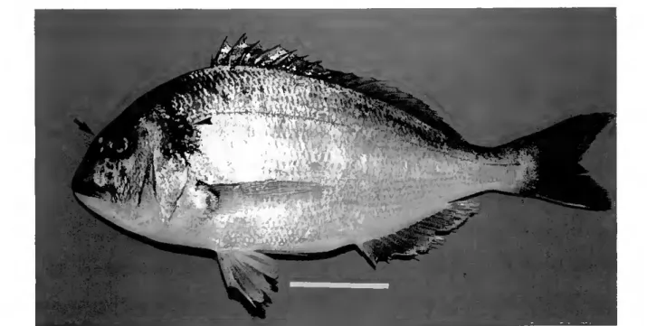
Sea bream has a characteristic appearance, it has a silvery grey colouration with a darkly pigmented zone at the origin of the lateral line, and it is ovoid in shape (Fig. 4); (Whitehead et ai, 1986; Fisher et aí, 1987). It's common name, gilthead sea bream, is derived from the presence in adults of a golden curved bar across the forehead that is bordered by two dark zones (Fig. 4). It is essencially a carnivorous species (Whitehead et aí, 1986; Fisher et aí., 1987), commonly feeding on molluscs, crustaceans, little fishes and sporadically it may also feed on algae (Quero, 1984), polichaets and insects (Arias, 1980).
- \Wk ■■ s m JlWÁ-Í ' ■fi m m ■'.is mm szmi V. i mmms
Figure 4. Photograph showing a left lateral view of an adult sea bream. The darker zone at the origin of the lateral line is indicated by an arrowhead and the golden coloured bar across the forehead is indicated by an arrow. Scale bar indicate 5.0 cm.
General infroducfion
Adult sca bream reach 30 - 35 cm in length and ocassionally may exceed 70 cm. Sea brcam is a protandrous hermaphroditic species (D'Ancona, 1941; Pasquali, 1941; Zoahar et ai, 1978). During the first year of their life ali fish are male and under certain environmental conditions the same may occur in the second year. From the first or second year some males undergo a sex change to female (Zoahar et ai, 1978". Zoahar et ai, 1984). In the Algarve spawning generally occurs from November to February, although manipulation of temperature and photoperiod can change the spawning season.
Embryonic development may also be affected by externai factor, such as salinity (Zohar, 1984), photoperiod (Tandler <& Helps, 1985) and water temperature (Villani, 1976; Zaki, 1984; Polo et aí, 1991; our observations). At 12 0C and 17 0C hatching occurred, respectively, at 72 hours post-fertilization (hpf) and 48-50 hpf (Villani, 1976); at 16 0C it occurs at between 65 and 70 hpf (Zaki, 1984) while at 28 0C it occurs after only 24 hpf (Polo et aí, 1991) and at 19 0C, the optimal rearing temperature, hatching starts at 40 hpf (our observations). Table I and Figure 5 summarizes the main developmental steps of sea bream from fertilization to hatching at 19 0C (our observations).
Typically fertilised sea bream eggs are transparent with a single oil globule in the yolk of approximately 1 mm diameter and at hatching sea bream embryos are optically clear, with few melanophores and a primordial finfold around the trunk in the median position (Kiriakos et aí, 1994; our observations). Their length ranges from 2.0 mm total length (tip of snout to posterior tip of finfold) (Zaki, 1984) to 2.7 mm notochord length (tip of snout to tip of notochord on small larvae prior to flexure) (our observations) and the yolk sac is oval and measures 1.1 mm in length (our observations). The mouth and jaws are absent and the eyes are unpigmented. The auditory vesicles are clearly visible as is the notochord and the segments (our observations).
Chapfer 1
Tablc I. Summary of devclopmcnt of sea bream at 19 0C.
hour post-fertilization Dcscription of stage
0 Fertilization
0.3 Beginning of swelling
3 Cleavage (16 cells)
9 Blastula (early epiboly)
15 Oastrula (50% epiboly)
19 Segmentation (4 somites)
26 Segmentation (16 somites)
40 Hatching (free embryo)
84 Free embryo (yolk absorption - 50%)
120 Larvae (exogenous feeding)
d) e)
O o O
Q
g) h)
©
Figure 5. Embryonic and larval sea bream development. (a) 0.3 hours post-fertilization (bpf), beginning of swelling; (b) 3 hpf, 16 blastomeres; (c) 9 hpf, blastula (early epiboly): (d) 15 hpf, gastrula (50% epiboly); (e) 19 hpf, segmentation (4 somites); (f) 26 hpf, segmentation (16 somites): (g) 40 hpf, hatching (free embryo); (h) 84 hpf, free embryo (yolk absorp+ion - 50%); (i) 120 hpf, larvae (exogenous feeding). Scale bar indicate 1.0 mm.
General infroducfion
Few hours (3-4 h) after hatching thc free embryo (yolk-sac larvae) body is fully straightened and the larvae quickly move to the water surface. During the first 3-4 days after hatching the free embryo is dependent for nutrient on the yolk-sac reserves. The transition to exogenous feeding occurs at about 4 days after hatching (our observation) and is a criticai step; failure at this step is frequently responsible for the high mortalities observed in hatcheries (Lumare A Villani, 1970; Person-Le Ruyet A Verillaud, 1980).
At hatching sea bream has no cartilaginous or bony structures (Faustino A Power, 1998, 1999, 2001). This mean that from this time to exogenous feeding a range of cartilaginous or bony elements must develop for survival. The first elements to appear are those which are related with feeding and manoeuvrability, l.e. structures of head skeleton which permits opening and closing of the mouth and components of the pectoral fin which are essentials for propulsion and increase the swimming activity. After these important events the osteological development continues with the appearance of the remaining viscerocranial elements, vertebral column and associated elements and the others fin structures (Faustino A Power, 1998,1999, 2001).
Several reports about sea bream egg and larval development and nutritional requirements have appeared (Villani, 1976; Person-Le Ruyet A Verillaud, 1980; Carnus A Koutsikopoulos, 1984; Kadmon ef aí, 1985; Tandler et aí, 1989).
The understanding and characterisation of sea bream development is essential if a rational approach is to be taken to improvement of larval quality. Improvements in quality of larvae will have important implications for the
performance of subsequent rearing stages.
Chapfer 1
THE SCOPE OF THIS THESIS
Numerous studies of cartilage and/or bony skeletal development in wild or reared teleosts exist in thc literature (Kendall, 1972; Houde á Potthoff, 1976; Potthoff et aí, 1984; Potthoff et aí, 1986; Matsuoka, 1987; Collette  Gillis, 1992; Watson Walker, 1992; Balart, 1995; Adriaens A Verraes, 1997; Hoshino A Amaoka, 1998; Vandewalle et aí, 1999), however the objective of such studies has generally been to characterise fish larvae in the wild and thus most of them give an incompleta report of skeletal ontogeny because, i) generally they describe the development of only part of the skeleton, or ii) they refer only to very early stages or iii) are limited only to the structures that persist in the juvenile or adult (Lau A Shafland, 1982; Potthoff et aí., 1984; Potthoff et aí,
1986; Collette A Gillis, 1992; Balart, 1995; Suda, 1996; Voskoboinikova, 1998). In recent years as a consequence of the importance of Sparidae to aquacultura, some studies about skeletal development have been reported in the literature. However, generally the studies lack details or fail to cover ali of the structures that make up thc skeleton (Matsuoka, 1985, 1987; Koumoundouros et aí., 2000). In sea bream the only studies that exist are those that describe the abnormalities observed in operculum and caudal fin complex (Kiriakos et aí., 1994; Koumoundouros et aí, 1997a, b).
The study and ontogcnic characterization of skeletal structures provida essential knowledge for further improvement of larval production and, probably, a better ecological understanding of this species. Moreovcr, characterization of the pattern of skeletal ontogeny is an important step for the identification of factors that can interfere with normal skeletal development (Kwain, 1975; Somasundaram et aí., 1984; Bcngtsson et aí., 1988; Wicgand et aí., 1989;
General infroducfion
Campbell, 1995; Toften A Jobling, 1996; Lien et aí, 1997; von Westernhagen Dethlefsen, 1997) and for understanding the alterations in the normal developmental process that may give rise to abnormalities.
The scope of the present thesis therefore, was to investigate the ontogeny of the skeleton in the sea bream. In Chapter 2 the normal development of cartilaginous structures in the vertebral column and caudal fin complex and the timing of their transformation into bone is described.
The developmental patterns of cartilage and bone in the remaining fins are characterized in Chapter 3. The results are compared with the development of these structures in related species. The sequential appearance of these fins was also analysed in relation to the need for more complex movements as the larvae grow and need to avoid predators and capture prey.
In Chapter 4 a precise account of the ontogeny of ossification of the viscerocranial skeleton is given. The evolutionary importance of the viscerocranial skeleton is considered and a hypothesis about the timing of ossification is proposed after cross species comparison of this structure.
In addition the origin of ossified tissue, cartilage replacement bone or dermal bone, is indicated for ali the structures analysed (Chapters 2, 3 and 4).
In Chapter 5 the frequency of meristic variations and abnormalities were studied and a detailed analysis of abnormalities is presented. Two regions of sea bream are most susceptible to abnormalities, the body axis and the caudal fin complex, both structures which appear early in the ontogeny suggesting that many of the anomalies apparent in the juveniles probably have their onset in early development.
Chapter 1
REFERENCES
Adriaens, D. A Verraes, W. (1997). The ontogeny of chondrocranium in Ciariasgariepinus. trends in siluroids. Journal of Fish Biology^O, 1221-1257.
Andrades, J. A., Becerra, J. A Fernández-Llebrez, P. (1996). Skeletal deformities in larval, Juvenile and adult stages of cultured gilthead sea bream {Sparus aurafa L.). Aquaculfure
141,1-11.
Arias, A. (1980). Crescimiento, regimen alimen+ario y reproducción de la dorada {Sparus aurafa L.) y dei robalo {Dicenfrarchus labrax L.) en los esteros de Cadiz. Invesfigación Pesquera 44, 59-83.
Balart, E. F. (1995). Development of the vertebral column, fins and fin supports in the japanese anchovy, Engrauhs Japonicus (Clupeiformes: Engraulididade). Bulletin of Marine Science 56, 495-522.
Balon, E. K. (1986). Saltatory ontogeny and evolution. Biology fórum. Ri vista di Biologia 79, 151- 190.
Barahona-Fernandes, M. H. (1982). Body deformation in hatchery reared European sea bass {Dicenfrarchus labrax, L). Types, prevalence and effect on fish survival. Jorna! of Fish BiohgyZl, 239-249.
Barka, T. A Anderson, P. J. (1962). Histochemical methods for acid phosphatase using hexazonium pararosanilin as coupler. Journal of Hisfochemisfry and Cyfochemisfry 10, 741-753.
Baron, R., Neff, L, Louvard, D. A. A Courtoy, P. J. (1985). Celi mediated extracellular acidification and bone resorption: evidence for a low pH in resorbing lacunae and localization of a 100 kD lysosomal membrane protein at the osteoclast ruffled border. Journal o f Celi Biology 101,2210-2222.
Bauchot, M-L. A Hureau, J-C. (1986). Sparidae. In Fishes of fhe Norfh-easfern Aflanfic and fhe Mediferranean, Vol. II, (Whitehead, P. J., Bauchot, M-L., Hureau, J-C., Nielson, J. A Tortonese, E., eds), pp. 513-1007, Paris, Unesco.
Bengtsson, A., Bengtsson, B. E. A Lithner, G. (1988). Vertebral defects in fourhorn sculpin, Myoxocepha/us quadricornis L., exposed to heavy metal pollution in the Culf of Bothnia. Journal of Fish Biology ZZ. 517-529.
Bertin, L. (1958). Tissus squelettiques. In Traifé de Zoo/ogie. Anafomie, Sysfémafique, Bio/ogie, Vol. 13, (Grasse, P. P., ed.), pp. 532-550. Paris, Masson.
Campbell, W. B. (1995). Genetic variation of vertebral fusion patterns in coho salmon. Journal of Fish Biology 4b, 717-720.
General infroducfion
Carnus, P. A Koutsikopoulos, C. (1984). Incubation expérimentale et développement embryonnaire de la Daurade royale, Sparus aurafa (L), à différentes températures. Aquaculfure 42, 117-128.
Chatain, B. A Ounais-Guschemann, N. (1990). Improved rate of initial swimbladder inflation in intensively reared Sparus aurafa. Aquaculfure SA, 345-353.
Collette, B. B. A Cillis, &. B. (1992). Morphology, systcma+ics, and biology of the double-lined mackerels (Gramtnaforcynus, Scombridae). Fishery Bullefin U.S. 90,13-53.
Cormack, D. H. (1984). Infroducfion fo Hisfohgy, Ist edn. Philadelphia: JB Lippincott.
D^ncona, U. (1941). Ulteriori osservazione e considerazioni sullermafroditismo e il differenziamen+o sessuale dellorata {Sparus aurafus L.). Pub/icazioni delia Sfazione Zoo lógica di Napoli 18, 313-336.
Danks, J. A., Devlin, A. J., Ho, P. M. W., Diefenbach-Jagger, H., Power, D. M., Canario, A., Martin, T. J. A Ingleton, P. M. (1993). Parathyroid hormone-related protein is a factor in normal fish pituitary. General Comparafive Endocrinohgy 92, 201-212.
Daoulas, C., Economou, A. N. A Bantavas, I. (1991). Osteological abnormalities in laboratory reared sea bass {Oicenfrarchus labrax) fingerlings. Aquaculfure 91.169-180.
Direcção Cerai das Pescas e Aquicultura (1990). Recursos de Pesca, Série Estatística, Vol. 4A-B, Lisboa.
Direcção Cerai das Pescas e Aquicultura (1991). Recursos de Pesca, Série Estatística, Vol. 5A-B, Lisboa.
Direcção Cerai das Pescas e Aquicultura (1992). Recursos de Pesca, Série Estatística, Vol. 6A-B, Lisboa.
Direcção Cerai das Pescas e Aquicultura (1993). Recursos de Pesca, Série Estatística, Vol. 7A-B, Lisboa.
Direcção Cerai das Pescas e Aquicultura (1994). Recursos de Pesca, Série Estatística, Vol. 8A-B, Lisboa.
Direcção Cerai das Pescas e Aquicultura (1995). Recursos de Pesca, Série Estatística, Vol. 9A-B, Lisboa.
Direcção Cerai das Pescas e Aquicultura (1997). Recursos de Pesca, Série Estatística, Vol. 11A-B, Lisboa.
Direcção Cerai das Pescas e Aquicultura (1998). Recursos de Pesca, Série Estatística, Vol. 12A-B, Lisboa.
Chapter 1
Direcção Geral das Pescas e Aquicultura (1999). Recursos de Pesca, Série Estatística, Vol. 13A-B, Lisboa.
Doty, S. B. <& Schofield, B. H. (1976). Enzyme histochemistry of bone and cartilage cells. Progress in Histochemistry and Cytochemistry 8,1-38.
Edwards, J. L. (1989). Two perspectives on the evolution of the tetrapod limb. The Amencan ZoologistZ9, 235-254.
Ekanayake, S. A Hall, B. K. (1987). The development of acellularity of the vertebral bone of the japanese medaka, Oryzias tatipes (Teleostei; Cyprinidontidae). Journal of Morphoíogy
193, 253-261.
FAO (1996). Aquacultura production statistics 1985-1994. FAO Fisheries Circular No. 815, Rev. 8. Rome: FAO.
Faustino, M. A Power, D. M. (1998). Development of osteological structures in the sea bream {Sparus auratd)'. vertebral column and caudal-fin complex. Journal of Fish Biology 52,11- 22.
Faustino, AA. A Power, D. AA. (1999). Development of the pectoral, pelvic, dorsal and anal fins in cultured sea bream. Journal o f Fish Biology^, 1094-1110.
Faustino, AA. A Power, D. AA. (2001). Osteologic development of the viscerocranial skcleton in sea bream: alternative ossification strategies in teleost fish. Journal of Fish Biology 58, 537-572.
Fisher, W., Bauchot, AA. L. A Scheneider, AA. (1987). Fiches FAO didentification des espècespour le besoin de la pêche (Revisi on 1). Mediterrannée et Mer Noir. Zone de pêche 37, Vol II - Vertébrés, pp. 761-1530. Rome: FAO.
Flanagan, J. A., Power, D. AA., Bendell. L. A., Guerreiro, P. AA., Fuentes, J., Clark, AA. S., Canano, A. AA. V., Danks, J. A. A Brown, B. L. (2000). Cloning of the cDNA for sea bream {Sparus auratd) parathyroid hormone-related protein. General and Comparativa Endocrinohgy 118, 373-382.
Flick, G., Klaren, P. H. AA., Schoenmakers, Th. J. AA., Bijvelds, AA. J. C., Verbost, P. AA. A Wendelaar Bonga, S. E. (1996). Celular calcium transport in fish: unique and universal mechanisms. PhysiologicalZoo/ogy 69, 403-417.
Francillon-Vieillot, H., de Buffrenil, V., Castanet, J., Geraudie, J., AAeunier, F. J., Sire, J-Y., Zylberberg, L. A de Ricqlès, A. (1990). AAicrostructure and mineralization of vertebrate skeletal tissues. In 5keletal biomineralization: Patterns, processes and evolucionary
trends. Vol. I, (Cárter, J. G., ed.), pp. 471-530. New York: Van Nostrand Reinhold.
Gartner, J. V. (1986). Observations on anomalous conditions in some flatfish (Piseis: Pleuronectiformes), with a new record of partial albinism. Environmenta! Biology of
General infroducfion
Gruber, H. E., Marshall, 6. J., Nolasco, L. M., Kirchen, M. E. & Rimoin, D. L (1988). Alkaline and acid phospha+ase demonstration in human bone and cartilage: effects of fixation interval and methacryla+e embedments. Sfain Technology (&, 299-306.
Hadley, M. E. (1992). Endocrino/ogy, 3rd edn. New Jersey: Prentice-Hall, Inc.
Hazon, N. á Balment, R. J. (1997). Endocrinology. In The Physiology of Fishes, 2nd edn., (Evans, D. H. ed.), pp. 441-463. Boca Raton: CRC Press.
Hilomen-Oarcia, G. V. (1997). Morphological abnormalities in hatchery-bred milkfish {Chanos chanosforsskdi) fry and Juveniles. Aquaculfure 152, 155-166.
Holland, P. W. H. (1997). Vertebrate evolution: Something fishy about /-/avgenes. Currenf Bio/ogy 7,570-572.
Hoshino, K. <& Amaoka, K. (1998). Osteology of the flounder, Tephrinecfes sinensis (Lacepède) (Teleostei: Pleuronectiformes), with comments on its relationships. Ichfhyological Research 45, 69-77.
Houde, E. D. á Potthoff, T. (1976). Egg and larval development of the sea bream Archosargus rhotnboidalis (Linnaeus): Pisces, Sparidae. Bullefin of Marine Science 26, 506-529.
Hughes, D. R., Basset, J. R. &. Moffat, L. A. (1994). Histological identification of osteocytes in the allegedly acellular bone of the sea breams Acanfhopagrus australis, Pagrus aurafus and Rhabdosargus sarba (Sparidae, Perciformes, Teleostei). Anatomy and Embryo/ogy 190,163-179.
Huysseune, A. <& Verraes, W. (1986). Chondroid bone on the upper pharyngeal Jaws and neurocranial base in the adult fish AsfatofUapia elegans. The American Journal of Anatomy 177, 527-535.
Ingleton, P. M., Saitens, P. V., Coulton, L. A. á Russel, R. G. G. (1983). An ultrastructural study of alkaline phosphatase in a transplantable rat osteogenic sarcoma. Me fabo He Bone bisease <£ ReíatedResearch 5, 23-31.
Ingleton, P. M., Hazon, N., Ho, P. M. W., Martin, T. J. á Danks, J. A. (1995). Immunodetection of parathyroid hormone-related protein in plasma and tissues of an elasmobranch Scyhorhinus can icu la. General Compara tive Endocrinology 9%, 211-218.
Inoue, H., Tanaka, N. A Uchiyama, C. (1995). Parathyroid hormone increases the number of tartrate-resistant acid phosphatase-positive cells through prostaglandin E2 synthesis in adherent cell culture of neonatal rat bones. Endocrinology 136, 3648-3656.
Jobling, M. (1995). Environmenfa! Bio/ogy of Fishes, Ist edn. London: Chapman &. Hall.
Junqueira, L. C., Carneiro, J. <& Kelley, R. O. (1995). Histologia Básica, 8a ed. Rio de Janeiro: Editora Suanabara Koogan.
Chapfer 1
Kadmon, G.. Gordin, H. A Varon, Z. (1985). Breeding-related growth of captive Sparus aurafa (Teleostei, Perciformes). AquacultureAb, 299-305.
Kamler, E. (1995). Ear/y Life Hisfory of Fishes: An Energefic Aproach. Fish and Fisheries Series 4. London: Chapman A Hall.
Kardong, K. V. (1997). /erfebrafes: Compara tive Anafomy, Funcfion, Evotufion, 2nd edn. U.S.A.: McSraw-Will.
Kendall, A. W., Jr (1972). Description of black sea bass, Cenfroprisfis sfriafa (Linnaeus), larvae and their occurences north of Cape Lookout, Nor+h Carolina, in 1966. Fishery Bullefin 70, 1243-1259.
Kiriakos, Z., Koumoundouros, Divanach, P. A Kentouri, M. (1994). Prelarval and larval description of notochord development as a criterion for early detection of tail abnormalities in gilthead sea bream {Sparus auratus). In Measures for Success, (Kesiemoni, P., Muir, J., Svila, E. A Wilhot, P. eds), pp. 185-190.
Kitajima, Ch. I., Tsukashima, S., Watanabe, T. A Yone, Y. (1981). Relationship be+ween uninf lated swim-bladders and lordotic deformity in hatchery-reared red sea bream Pagrus major. Bullefin of Japanese Sociefy of Scientific Fisheries47, 289-294.
Komaki, AA., Katagiri, T. A Suda, T. (1996). Bone morphogene+ic protein-2 does not alter the differentiation pathway of commit+ed progenitors of osteoblasts and chondroblasts. Celi
Tissue Research 284, 9-17.
Koumoundouros, G.. Divanach A Kentouri, AA. (2000). Development of the skull in Denfex denfex (Osteichthyes: Sparidae). Marine Bio/ogy 136, 175-184.
Koumoundouros, G., Gagliardi, F., Divanach, P., Boglione, C., Cataudella, S. A Kentouri, AA. (1997a). Normal and abnormal osteological development of caudal fin in Sparus aurafa L. fry. Aquoculture 149, 215-226.
Koumoundouros, G.. Oran, G.. Divanach, P., Stefanakis, 5. A Kentouri, AA. (19976). The opercular complex deformity in intensive gilthead sea bream {Sparus aurafa L.) larviculture. AAoment of apparition and description. Aquacu/fure 156, 165-177.
Kwain, W. (1975). Embryonic development, early growth, and meristic variation in rainbow trout {Sa/mo gairdner!) exposed to combinations of light intensity and temperatura. Journal of
fhe Fisheries Research Boa rd of Canada 32, 397-402.
Langille, R. AA. A Hall, B. K. (1988). Role of the neural crest in development of the trabeculae and branchial arches in embryonic sea lamprey, Pefromyzon marinus (L). Development 103, 301-310.
Lau, S. R. A Shafland, P. L. (1982). Larval development of snook, Cenfropomus undecimalis (Pisces: Centropomidae). Cope ia 3, 618-627.
General infroducfion
Lien, N. T. H., Adriaens, D. Janssen, C. R. (1997). Morphological abnormali+ies in African catfish {Ciariasgariepinus) larvae exposed to malathion. Chemosphere 35,1475-1486. Long, F. á Linsenmayer, T. F. (1998). Regulation of growth region cartilage prolifcration and
differentiation by perichondrium. Oevelopmenf 125, 1067-1073.
Lumare, F. á Villani, P. (1970). Contributo alia conoscenza delle uova e dei primi stadi larvali di Sparus aurafa (L.). PubUcazioni delia Sfazione Zoologica di Napoli ZZ, 364-369.
Marx, J. (1992). Homeobox genes go evolutionary. Science 255, 399-401.
Matsuoka, M. (1985). Osteological development in the red sea bream, Pagrus major. Japanese Journal of IchfhyologySZ, 35-51.
Matsuoka, M. (1987). Development of the skeletal tissues and skeletal muscles in the red sea bream. Bullefin of the Seikai Regional Fisheries Research Laborafory 65,1-114.
Meunier, F. J. <& Huysseune, A. (1992). The concept of bone tissue in Osteichthyes. Nefhertands Journal o f Zoo/ogy 42, 445-458.
Nelson, J. 5. (1994). Fishes of the World, 3rd edn. New York: John Wiley & Sons.
New, M. B. (1991). Turn of the millenium aquaculture. Navigating troubled water or riding the crest of the wave? World Aquaculture 22, 28-49.
Noden, D. M. (1983). The role of neural crest in patterning avian cranial skeletal, connective and muscle tissues. OevelopmenfalBiologyVb, 144-165.
Pang, P. K. T., Pang, R. K. A Sawyer, W. H. (1974). Environmental calcium and the sensitivy of killifish (Fundulus heferoditus) in bioassay for the hypocalcemic response to Stannius corpuscles from killifish and cod {Gadus morhua). Endocrinobgy94, 548-555.
Paperna, I. (1978). Swimbladder and skeletal deformations in hatchery bred Sparus aurata. Journal of Fish Bio/ogy 12, 109-114.
Pasquali, A. (1941). Contributo alio studio deirermafroditismo e dei differenziamento delia gonada nellorata {Sparus aurafusL.). PubUcazioni delia Sfazione Zoologica di Napoli 18, 282-312. Person-Le Ruyet, J. â Verillaud, P. (1980). Techniqucs delevage intensif de la daurade dorée
{Sparusaurafa (L.)) de la naissance à lage de deux mois. Aquaculture 20, 351-370.
Polo, A., Yúfera, M. A Pascual, E. (1991). Effects of temperatura on egg and larval development of Sparus aurafa L. Aquaculture 92, 367-375.
Potthoff, T., Kelley, 5., Moe, M. A Young, F. (1984). Description of the porkfish larvae {Anisotremus virginicus, Haemulidae) and their osteological development. Bullefin of Marine Science 34, 21-59.
Chapter 1
Potthoff, T., Kelley, S. A Javech, J. C. (1986). Cartilage and bone development in scombroid fishes. Fishery BuHefin U.S. 84, 647-678.
Pough, F. K, Janis, C. M. A Heiser, J. B. (1999). Verfebrate Life. 5+h edn. New Jersey: Prentice- Hall, Inc.
Power, D. M., Ingleton, P. M., Flanagan, J., Canario, A. M. V.f Danks, J., Elgar, Qj. A Clark, M. S. (2000). Cenomic structure and expression of parathyroid hormone-rela+ed protein gene (PTHrP) in a teleost, Fugurubripes. SeneZSO, 67-76.
Quero, J-C. (1984). Les Poissons de Mer de Feches Françaises, (Grancher, J., ed.). Paris. Raven, P. H. A Johnson, G. B. (1999). Biology, 5+h edn. U.S.A.: McGraw-Hill.
Reddi, A. H. (1981). Celi biology and biochemis+ry of endochondral bone development. Collagen Research 1, 209-226.
Reddi, A. H. (1994). Bone and car+ilage differen+ia+ion. Currenf Opinion in Senefics and Development A. 737-744.
de Ricqlès, A., Meunier, F. J., Cas+anet, J., A Francillon-Vieillot, H. (1991). Compara+ive Micros+ruc+ure of Bone . In Bone: a Treafise, Vol. 3, (Hall, B. K., ed.), pp. 1-78. Boca Ra+on: CRC Press.
Rodan, G. A. (1992). In+roduc+ion +o Bone Biology. Bone 13, 53-56.
Rodger, H. b. A Murphy, T. M. (1991). Cranial deformi+ies in A+lan+ic Salmon alevins. Bulleffin of European Assoe ia fion of Fish PafologyW, 219-221.
Roodman, G. D. (1996). Advances in bone biology: The os+eoclas+. Endrocrine Reviews 17, 308- 332.
Sire, J. Y. A Huysseune, A. (1993). Fine s+ruc+ure of +he developing frontal bonés and scales of the cranial vault in the cichlid fish Hemichromis bimoeu la tus (Teleostei, Perciformes). Celi â Tissue Research 273, 511-524.
Somasundaram, B., King, P. E. A Shackley, 5. E. (1984). Some morphological effeets of zinc upon the yolk-sac larvae of C/upea harengus L. Journal of Fish Biology 25, 333-343.
Stibane, F. A. (1992). Histochemical inves+iga+ions of the ontogenetical development of acellular teleost bonés wi+h special references of the vertebral column of Aphanius mento (Heckel, 1843) (Pisces, Cyprinodon+idae). Zoologishe Jahrbuecher Anatomy 122, 449- 477.
Suda, Y. (1996). Os+eology and muscular a+tachmen+s of the japanese jack mackerel, Trachurus japonicus. Bulletin o f Marine Science 58, 438-493.
General infroducfion
Sundell, K., Bjornsson, B. Th., Itoh, H. <& Kawauchi, H. (1992). Chum Salmon {Oncohryheus kefa) stanniocalcin inhabits in vifro intestinal calcium uptake in Atlantic Cod Gadus morhua. Journal of Compara tive Physio/ogy B162, 489-495.
Tandler, A., Harel, M., Wilks, AA., Levison, A., Brickell, L, Christie, 5., Avital, E. <& Barr, Y. (1989). Effect of environmental temperature on survival, growth and population structure in the mass rearing of the gilthead seabream, Sparusaurafa. Aquaculfurelb, 277-284.
Takagi, Y. <& Kaneko, T. (1995). Developmental sequence of bone-resorption cells induced by intramuscular implantation of mineral-containing bone particles into rainbow trout, Oncorhynchus mykiss. Celi & Tis sue Research 280,153-158.
Tandler, A. ã Helps, S. (1985). The effects of photoperiod and water exchange rate on growth and survival of gilthead sea bream {Sparus aurafa, Linnaeus; Sparidae) from hatching to metamorphosis in mass rearing systems. Aquaculfure A&, 71-82.
Thorogood, P. (1988). The developmental specification of the vertebrate skull. Oevehpmenf 103 (Supplement), 141-153.
Toften, H. á Jobling, AA. (1996). Development of spinal deformities in Atlantic salmon and Arctic charr fed diets supplemented with oxytetracycline. Journal of Fish BiologyA9, 668-677. Vandewalle, P., Chikou, A., Lalèyé, P., Parmentier, E., Huriaux, F. <& Focant, B. (1999). Early
development of the chondrocranium in Chrysichfhys aura tus. Journal of Fish Biology 55, 795-808.
Villani, P. (1976). Allevamento larvale di orata {Sparus aurafa L.) riprodotta in condizioni artificiali di laboratório. Archivio di Oceanografia e Limnohgia 18, 295-302.
Verbost, P. AA., Flick, Fenwick, J. C., Greco, A. AA., Pang, P. K. T. <& Wendelaar Bonga, S. E. (1993). Branchial calcium uptake: possible mechanisms of control by stanniocalcin. Fish Physio/ogy and Biochemisfry 11, 205-215.
von Westernhagen, H. ã Dethlefsen, V. (1997). The use of malformations in pelagic fish embryos for pollution assessment. Hydrobiohgia352, 241-250.
Vortkamp, A., Lee, K., Lanske, B., Segre, G. V., Kronenberg, H. AA <& Tabin, C. J. (1996). Regulation of rate of cartilage differentiation by Indian hedgehog and PTH-related protein. Science 273, 613-622.
Voskoboinikova, O. S. (1998). Development of the bony skeleton in the ontogenesis of Harpagifer anfarcficus {y\ar\)aq\ier\áae, Notothenioidei). Journal of Ichfhyology 38, 501-510.
Walker, W. F., Jr ã Liem, K. F. (1994). Funcfiona! Anafomy of fhe /erfebrafes: An Evolucionary Perspecfive, 2nd. Orlando: Saunders College Publishing.
Chapfer 1
Watson, W. <& Walker, H. J., Jr (1992). Larval development of sargo {Anisofremus davidsonii) and salema {Xenisfius californiensis) (Pisces: Haemulidae) from the Southern Califórnia Bight. Bullefin of Marine Science 51, 360-406.
Wendelaar Bonga, S. E. <& Lammers, P. I. (1982). Effects of calcitonin on ultrastructure and mineral content of bone and scales of the cichlid teleost Sarofherodon mossatnbicus. Senera! and Compara tive Endocrino/ogy 48, 60-70.
Wendelaar Bonga, S. E. á Pang, P. K. T. (1992). Control of calcium regulating hormones in vertebrates: Parathyroid hormone, calcitonin, prolactin, and stanniocalcin. International Review of CytologyVZZ, 139-213.
Whitehead, P. J., Bauchot, M-L, Hureau, J-C., Nielson, J. <& Tortonese, E. (1986). Fishes of the North-eastern Atlantic and the Mediterranean, Vol. II, pp. 513-1007. Paris: Unesco. Wiegand, M. D., Hataley, J. M., Kitchen, C. L. <& Buchanan, L. G. (1989). Induction of
developmental abnormalities in larval goldfish, Carassius auratusV., under cool incubation conditions. Journal of Fish Bio/ogyZS, 85-95.
Witten, P. E. (1997). Enzyme histochemical characteristics of osteoblasts and mononucleated osteoclasts in a teleost fish with acellular bone [Oreochromis niloticus, Cichlidae). Celi cS
Tissue Research 287, 591-599.
Zaki, M. I. (1984). Artificial culture and early ontogeny of gilthead, Sparus aurata, of the Mediterranean sea. Journaloflchthyohgy 24, 56-63.
Zohar, Y., Abraham, M. á Cordin, H. (1978). The gonadal cycle of the captivity reared hermaphroditic teleost Sparus aurata (L.) during the first two years of lifc. Anna/es Biotogie Anima/e, Biochimie, Biophysique 18, 877-882.
Zohar, Y., Billard, R. A Weil, C. (1984). La reproduction de la daurade {Sparus aurata) et du bar (bicentrarchus labrax): connaissancc du cycle sexuel et controle de la gamétogenèse et de la ponte. In Aquacultura du Bar et des Sparidés, (Barnabé, G. & Billard, R. eds), pp. 3- 22. Paris: INRA Publications.
CHARTER 2
DEVELOPMENT OF OSTEOLO&ICAL STRUCTURES IN THE SEA
BREAM {Sparus auratà)'- VERTEBRAL COLUMN AND CAUDAL FIN
COMPLEX
Co-author
Deborah M. Power
Journal o f Fish Biology 52,11-22
Osteological de vebpmenf in sea bream
DEVELOPMENT OF OSTEOLOGICAL STRUCTURES IN THE SEA BREAM {Sparus ouratá)\ VERTEBRAL COLUMN AND CAUDAL FIN COMPLEX
AB5TRACT
The dcvclopmcnt of cartilaginous structupcs in culturcd Sparus aurafa larvac and the timing of their ossification was studied. In cultivated sea bream larvae the first cartilaginous structurc to be identified was hypural 1 at 4.1 mm notochord length (Ln). By 5.3 mm Ln, prior to the onset of ossification, it was possiblc to distinguish the following cartilaginous structurcs: ali 23 neural archcs, ali 13 hacmal arches and two of the four pairs of parapophyscs. The neural arches 1-4 and 15-23 were formed on the notochord and clongatcd dorsally, while neural archcs 5-14 appeared on the dorsal side of the spinal cord and clongatcd vcntrally. Initiation of ossification occurrcd at 5.7-6.0 mm standard length (Ls) when the cartilaginous ontogeny of the vertebral column was complctcd. Ossification was coincidcnt with dorsal flexion at the posterior end of the notochord and occurrcd in a scqucntial manncr: i) dorsoanteriorly, the cartilaginous neural archcs and the centra were the first structurcs to ossify; ii) ventrad at the centre, at 7.0-7.5 mm Ls; iii) posteriorly at 7.1 mm Ls the hypural complcx and urostyle (24th ccntrum) were ossified; and 4) dorsad at the centre (neural archcs and spines).
Chapter 2
INTRODUCTION
The sea bream {Sparus aurafa) is widely cultivated in the Mediterrancan and Portugal. As with many other teleosts, cultivation of sea bream is associated with an increased incidence of skeletal malformations, particularly those associated with the vertebral column in the adult (Paperna, 1978; Barahona- Fernandes, 1982; Francescon et aí, 1988; Santamaria et aí, 1994). The cause and progression of such skeletal malformations in cultivated fish is uncertain and, despite their relatively high frequency in farmed fish, few studies of the cartilaginous-osteological development of cultured fish larvae exist. The majority of published osteological studies are directed at extending basic knowledge of larval taxonomy and systematics (Dunn, 1983) and have been carried out largely on wild species e.g. Aniso tremas virginicus (L.) (Potthoff et aí, 1984), Scombroíobrax hetero/epis (Roule), êempylus serpens Cuvier, Nesiarchus nasutus (Johnson), Scomber Japonicus Houttuyn (Potthoff et aí, 1986), Engraulis Japonicus (Schlegel) (Balart, 1995). In sea bream, spinal abnormalities are detectable in larval stages (Santamaria et aí, 1994; our observations). It is likely that some malformations arise as a consequence of genetic factors and physiological factors, such as inability to inflate the swim bladder (Kitajima et aí, 1981; Chatain, 1987), whereas others may be a consequence of environmental factors such as diet, pollution, etc. (Hodson et aí, 1980; Weis <& Weis, 1989; Hinton et aí, 1992).
During vertebrate embryogenesis, cartilage is the first element of the skeleton to form and subsequently, with some exceptions (e.g. interarcual cartilage, basibranchial cartilage, rostral cartilage), becomes bone. Two principal types of bonés which differ in their origin can be identified in vertebrates: cartilage replacement bone, which has a cartilaginous precursor, and dermal bone
Osteological de vebpmenf in sea bream
which develops within or just beneath the skin without any cartilaginous precursor.
The objective of the present study was to chart the normal development of cartilaginous structures in the vertebral column and caudal fin complex in cultured sea bream larvae and the timing of their transformation into bone. Bony structures arising from cartilaginous precursors and dermal bone were also identified. This study will form the basis of future experimental work aimed at elucidating the molecular regulation of osteological development, in order to understand how alterations in this process Icad to abnormalities.
Materials and Methods
Cu!fure o f Larvae
Sparus aurata larvae were hatched and cultured in 0.2m3 conical tanks gently aerated with a continuous flow of sea water. Hatching occurred 40 h after fertilisation. The larvae were fed on Brachionus plicotilis 4 days post- hatching (DPH) when the yolk sac was consumed, and Artemia sp. was introduced into the diet from 15 until 40 DPH when dry food was introduced gradually. Light conditions followed a cycle of 12 h light:12 h dark, and the ambient water temperature was 19 ± 1 0C.
Experimen to! Pr o toco!
Larvae were sampled on alternate days from 1 DPH, anaesthetized in MS- 222 and fixed overnight at 4 0C in 2% PFA (paraformaldehyde) solution, washed and stored in 70% methanol. Preliminary experiments were carried out to
Chapfer 2
confirm that length was not altered as a consequence of fixation and subsequent staining methods and to permit the characterisation of normal and abnormal larvae. Abnormal larvae were identified readily and presented anomalies such as loss of structures, altered dimensions of structures and spinal breaks, twists and bends.
Larvae were measured with an ocular micrometer in a stereoscopic microscope (Wild M8). The following measurements were made*. notochord length (Ln), tip of snout to tip of notochord on small larvae prior to flexure; standard length (Ls), tip of snout to base of caudal complex on larger larvae in which f lexion of notochord has occurred.
Staining for bone and cartilage was carried out on whole mounts using a modification of the method described by Klymkowsky A Hanken (1991). In brief, specimens were rehydrated through a graded series of methanol (7070-2570), followed by two washes (of 20-30 min each) in Tris-buffered saline (pH 7.6). Specimens were stained with Alcian blue 8SX (0.027o in 707o alcohol and 307o glacial acetic acid) and macerated using a 17o aqueous solution of KOH until skeletal elements were clearly visible. Then they were stained with Alizarin red 5 (stock solution: 17o Alizarin red in 17o KOH). Staining time was variable and depended on the size of the specimen. A total of 164 specimens ranging from 10 DPH (3.1 mm Ln) to 70 DPH (16.0 mm L5) were observed (Table I). The nomenclature proposed by Matsuoka (1987) is used throughout this chapter.
Staining for bone-specific alkaline phosphatase was carried out using a modification of the Somori technique (Matsuzawa A Anderson, 1971; Salomon, 1974). p-glycerophosphate was included in the incubation buffer to promote the precipitation of calcium phosphate (Ingleton et ai 1983) and silver nitrate was used to visualise insoluble phosphate in bone (Matsuzawa A Anderson, 1971; Salomon, 1974).