Systematic molecular analysis of hemophilia A patients from Colombia
Luz Karime Yunis
1, Adriana Linares
2,3,4, Edgar Cabrera
2and Juan J. Yunis
1,5,6.
1
Grupo de Patología Molecular, Universidad Nacional de Colombia, Bogotá, D.C., Colombia.
2Fundación Hospital de la Misericordia, Bogotá, D.C., Colombia
3
Grupo de Onco-Hematología Pediátrica, Departamento de Pediatría, Facultad de Medicina, Universidad
Nacional de Colombia, Bogotá, D.C., Colombia.
4
Programa de Hemofilia, Clínica Infantil Colsubsidio. Bogotá, D.C., Colombia.
5
Departamento de Patología, Facultad de Medicina e Instituto de Genética, Universidad Nacional de
Colombia, Bogotá, D.C., Colombia.
6
Instituto de Genética, Servicios Médicos Yunis Turbay y Cía S.A.S. Bogotá, D.C., Colombia.
Abstract
Hemophilia A (HA) is an X-linked recessive disorder and the second most common coagulation disorder with an inci-dence of 1 in 5,000 live born males. Worldwide, there are 178,500 affected individuals, 60% with the severe form of the disease. Intron 22 and 1 inversions (Inv22 and Inv1) are the most frequent molecular alterations found in severe HA patients with a frequency of 45-50% and 0.5-5%, respectively. We have implemented a systematic cost-effective strategy for the identification of the molecular alteration in HA patients using Inverse shifting-PCR for Inv22 and Inv1, followed by the analysis of theF8 gene coding region by means of high resolution melting (HRM) PCR and Sanger sequencing in Inv22 and Inv1 negative patients. A total of 33 male HA patients and 6 women were analyzed. Inver-sion 22 was detected in 14/33 male patients (42.4%), 3/33 (9.1%) had Inv1, 3/33 (9.1%) had large structural variants, and 11/33 (33.3%) single nucleotide/ small frameshift variants. No genetic variant was found in 2/33 patients (6%). With this systematic approach we detected pathogenic variants in 31 out of 33 male affected individuals (94%) tested for the first time.in a cohort of patients from Colombia
Keywords: Hemophilia A, Factor VIII, IS-PCR, HRM, Colombia.
Received: March 23, 2017; Accepted: March 16, 2018.
Introduction
Hemophilia A (HA) is an X-linked recessive disorder and the second most common coagulation disorder with an incidence of 1 in 5,000 live born males (Hoyer, 1994). Worldwide, there are approximately 178,500 affected indi-viduals with HA, and of these, 60% have the severe form of the disease, followed by mild (5-30%), and moderate cases 15% (Savage, 2014).
Different genomic variants are responsible for differ-ent HA phenotypes (Higuchi,et al., 1991a,b; Antonarakis, 1995; Antonarakis,et al., 1995; Ahmed,et al., 2005; Al-banez,et al., 2011; Rallapalliet al., 2013). Overall, mis-sense variants are responsible for near 50% of all cases of HA. However, Intron 22 and intron 1 inversions (Inv22 and Inv1) are the most frequent molecular alterations found in severe HA patients with a frequency of 45-50% and 0.5-5%, respectively (Woods-Samuels,et al., 1991; Lakich,et
al., 1993). Individuals with Inv22 and Inv1, deletions and nonsense mutations, usually have the severe form of the disease and an increased risk for developing inhibitors dur-ing treatment (Brackmannet al., 1996, 2000; Astermarket al., 2005, 2013; Abbonizioet al., 2014). In recent years, high resolution melting analysis (HRM) has been used to screen genomic segments to detect regions that could har-bor nucleotide variants in different diseases, including HA (Ririeet al., 1997; Linet al., 2008).
In Colombia thus far, no molecular genotyping ser-vices for HA are available. In the present report, we have used a systematic approach to characterize HA variants in a cohort of patients from Bogotá, Colombia. First, Inv22 and Inv1 were analyzed by inverse-shifting PCR (IS-PCR) (Rossettiet al., 2004, 2005, 2008, 2011; Radicet al., 2009). Negative samples for Inv22 and Inv1 were amplified for all 26 exons by HRM (52 PCR-HRM reactions) (Ririeet al., 1997; Linet al., 2008), followed by DNA Sanger sequenc-ing of altered HRM meltsequenc-ing curves (Sangeret al., 1977). Microarray analysis was used in selected cases to further characterize large deletions. With this approach, we were
Send correspondence to Luz Karime Yunis. Grupo de Patología Molecular Oficina 207, Instituto de Genética, Edificio 426, Uni-versidad Nacional de Colombia, Bogotá, D.C., Colombia. E-mail: [email protected].
able to detect pathogenic variants in 94% of our patients for the first time in Colombia, with the identification of three new variants.
Materials and Methods
Patients
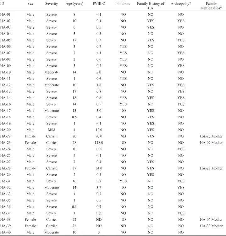
Approval by the ethics committees of all institutions was obtained. A total of 33 HA male patients (27 severe, 5 moderate, and 1 mild HA) and 6 females (patient’s
moth-ers) were included (Table 1). After signing the informed consent, patients had blood samples collected in EDTA tubes for DNA isolation. Additional information regarding disease clinical course, as well as treatment related infor-mation was collected.
DNA extraction
DNA was obtained from 200mL of whole blood using the DNA Blood Mini kit Qiagen, following manufacturer’s recommendation (Qiagen, Hilden Germany). DNA purity
Table 1- Clinical and demographic data of hemophilia A patients from Colombia.
ID Sex Severity Age (years) FVIII:C Inhibitors Family History of HA
Arthropathy* Family relationships^
HA-01 Male Severe 8 < 1 NO NO NO
HA-02 Male Severe 10 0.4 NO YES YES
HA-03 Male Severe 6 0.5 NO YES NO
HA-04 Male Severe 5 0.3 NO NO NO
HA-05 Male Severe 17 0.3 NO YES YES
HA-06 Male Severe 3 0.7 YES NO NO
HA-07 Male Severe 7 < 1 YES NO YES
HA-08 Male Severe 2 0.6 YES NO NO
HA-09 Male Severe 5 0.7 YES NO YES
HA-10 Male Moderate 14 2.0 NO NO NO
HA-11 Male Severe 1 0.6 YES NO NO
HA-12 Male Moderate 10 1.8 NO YES YES
HA-13 Male Severe 17 0.8 NO NO YES
HA-14 Male Severe 18 0.8 YES YES YES
HA-16 Male Severe 14 0.5 YES NO YES
HA-17 Male Moderate 13 3.0 NO YES NO
HA-18 Male Severe 0.5 0.4 NO YES NO
HA-19 Male Severe 1 < 1 NO YES NO
HA-20 Male Mild 4 12.0 NO YES NO
HA-22 Female Carrier 20 70.0 ND YES NO HA-20 Mother
HA-23 Female Carrier 28 118.0 ND NO NO HA-07 Mother
HA-24 Male Severe 10 0.5 NO NO YES
HA-25 Male Severe 5 < 1 NO NO NO
HA-27 Male Severe 7 0.4 NO YES NO
HA-28 Female Carrier 37 34.0 NO YES NO HA-27 Mother
HA-29 Male Severe 2 0.4 NO YES NO
HA-31 Male Severe 16 0.7 YES NO YES
HA-32 Male Moderate 14 3.7 NO NO YES
HA-33 Male Severe 1 0.7 NO NO NO
HA-35 Male Severe 1 0.5 NO NO NO
HA-36 Male Severe 0.5 0.4 NO NO NO
HA-37 Male Severe 1 0.2 NO NO YES
HA-38 Female Carrier 22 ND ND NO NO HA-06 Mother
HA-39 Female Carrier 23 ND ND NO NO HA-33 Mother
and concentration was determined in a Nanodrop 2000 spectrophotometer (Nanodrop, Wilmington, DE).
Inverse shifting PCR (intron 1 and intron 22 inversion analysis)
Intron 1 and 22 inversions were determined by in-verse shifting PCR as described previously (Rossettiet al., 2008). Briefly, 2mg of genomic DNA was digested with 20 U ofBclI restriction enzyme (Thermo Fisher Scientific, Massachusetts) for 4 h in a 50mL volume. Digested DNA was purified using Centricon 100 concentrators (Amicon, MA). Self-ligation was carried out in a 400mL reaction mix containing 3 U of T4 DNA ligase (Thermo Fisher Scien-tific, MA) at 15 °C for 12–14 h. Self-ligated circles were purified using Centricon 100 concentrators (Amicon) and adjusted to a final volume of 100mL. For intron 1 inversion analysis, 3mL was used for PCR amplification, while 6mL was used for intron 22 inversions. PCR amplifications were carried out in a 25mL reaction mix with 0.6mM of each primer: 200mM dNTP, 1.5 mM MgCl2, and 0.5 U GoTaq
Flexi DNA polymerase (Promega Corporation, WI) in a C100 BioRad thermal cycler with a protocol of 94 °C for 2 min followed by 30 cycles of 94 °C for 30 s, 56 °C for 1 min and 72 °C for 1.5 min, terminating with a final extension step of 5 min at 72 °C.
Amplified products were resolved in 2% Nusieve 3:1 agarose (Lonza, MD) in 1X SO buffer and visualized using SyberSafe stain (Invitrogen, CA).
High resolution melting analysis
High resolution melting (HRM) analysis was carried out using Precision Melt Supermix (BioRad, CA) and ana-lyzed in a 96 CFX real time system (BioRad), using primers covering all 26 exons of theF8gene, as described previ-ously (Linet al., 2008). Briefly, 2mL of genomic DNA (12.5 ng/mL) were used for each reaction containing 5mL of 2x Precision Melt Supermix, 2 mL primer mixture (10 pmol/mL each), and 1mL distilled water. An initial denatur-ing step at 95 °C for 2 min was followed by 45 cycles of 95 °C for 10 s, 53 °C for 30 s and plate reading 72 °C for 30 s. followed by HRM with 1 cycle of 95 °C for 30 s, 1 cycle at 60 °C for 1 min, 1 cycle at 65-95 °C (10 s/step, slope 0.2 °C/s) and plate reading. HRM analysis was done with the
Precision Melt analysis software (melt curve sensitivity set at 50, Tm difference threshold of 0.2 °C)
Microarray analysis
A total of 250 ng of genomic DNA was used for CytoScan 750 microarray (Affymetrix, CA) analysis. Briefly, genomic DNA was digested withNsp1 at 37 °C for 2 h. The digested DNA was purified and ligated to primer-adapters at 16 °C for 3 h, followed by PCR amplification. Amplified DNA was purified and digested with DNAseIat 37 °C for 35 min. Digested DNA was biotin-labeled for 4 h at 37 °C, hybridized to CytoScan 750K microarrays at 50 °C for 18 h and washed to remove unbound sample. Sam-ples were read with AGCC console and ChAS 3.1 (Chro-mosome Analysis Suite) from Affymetrix.
DNA sequencing
Samples with different HRM curves were sequenced using the 3.1 BigDye terminators cycle sequencing kit (Ap-plied Biosystems, CA), following manufacturer’s recom-mendations and analyzed in a 3500 ABI genetic analyzer (Applied Biosystems). DNA sequences were compared to the F8 gene reference sequence NM_000132.3 using SeqScape V5.4. (Applied Biosystems). Briefly, samples were amplified in a C1000 BioRad thermal cycler in 50mL volume reactions containing 10mLTaqpolymerase buffer, 5mL primer mix (10 pmol/mL each), 4mL dNTP (10 nM each), 2 mM MgCl2, and 1.25mLGoTaq DNA Polymerase (Promega, WI) and 5mL of genomic DNA (12.5 ng/mL). After an initial denaturation step at 95 °C for 2 min, 35 cy-cles of 95 °C for 10 s, 53 °C for 30 s and 72 °C for 30 s were run, followed by a final extension step of 5 min at 72 °C, PCR-amplified products were purified using PureLink quick PCR purification kit (Invitrogen) following manufac-turer’s recommendations. Purified fragments were quanti-fied in a Nanodrop 2000 system and 10 ng of PCR puriquanti-fied fragments were used for BigDye terminator sequencing. Mutation nomenclature was according to the Human Ge-nome Variation Society (HGVS) (den Dunnenet al., 2016)
Software prediction analysis
To determine the pathogenicity of new identified mis-sense variants, different functional prediction softwares ID Sex Severity Age (years) FVIII:C Inhibitors Family History of
HA
Arthropathy* Family relationships^
HA-41 Male Severe 14 0.7 NO YES YES
HA-42 Male Severe 19 < 1 YES NO YES
HA-43 Male Severe 11 0.7 NO NO YES
HA-45 Female Carrier 37 ND ND NO NO HA-16 Mother
ND, no data, Actual inhibitors or history of inhibitors; * Actual arthropathy or history of arthropathy; ^ Familial relationships between individuals in-cluded in this study.
were used, such as POLYPHEN-2, PROVEAN, Mutation Taster, and PhD-SNP (Capriottiet al., 2006; Adzhubeiet al., 2010; Choiet al., 2012; Schwarzet al., 2014).
Statistical analysis
A 2x2 cross-tab was used to analyze the associations between Inv22/inhibitor development and Inv22/ arthro-pathy by Fisher’s Exact test.
Results
Intron 22 and intron 1 inversions
Intron 22 inversion was detected in 14 out of 33 (42.4%) unrelated cases. Twelve samples were type I Inv22 and 2 samples type II Inv22. Three out of 33 samples (9.1%) were positive for Inv1 (Supplementary Figure S1). Two patients’ mothers were positive for Inv22 and an addi-tional mother was positive for Inv1.
HRM analysis
Samples negative for Inv22 and Inv1 were analyzed by HRM analysis (16 out of 33 patients (48.4%), and 3 mothers). Three samples (HA-07, HA-11, and HA-13) showed lack of amplification for one or moreF8 exons. One of the samples (HA-07), failed to amplify for exons 1 through 14. To verify this result, the same sample was am-plified from separate DNA isolates, showing the same re-sult. The patient’s mother (HA-23) showed amplification for all exons. Sample HA-13 failed to amplify exon 13 on separate occasions. Sample HA-11 failed to amplify exon 26. In all cases, poor DNA quality was excluded as the rea-son for failing PCR amplification since other exons were amplified in parallel simultaneously. In addition, other
samples used as controls for amplifications yielded the ex-pected PCR fragment. These results indicated that 3 out of 33 samples had large deletions (9.1%). Of the remaining samples, on average, HRM analysis showed different HRM curves in 3 to 4 fragments. Therefore, DNA sequencing was carried out on those fragments in order to detect the pathogenic variant present in each sample (see below).
Microarray analysis
Microarray analysis was carried out in those three samples that showed deletions by HRM analysis, as well as in the HA-07 mother. Sample HA-07 showed a large dele-tion involving exons 1 through 14 of 223,573 bp (arr[hg19] Xq28 (154153703-154377276)x0) that compromised 5 genes: F8, FUNDC2, MTCP1NB, MTCP1, and BRCC3. His mother had the same deletion as a heterozygous (arr[hg19] Xq28 (154153703-154377276)x1) (Supplemen-tary Figure S2). Patient HA-13 had a 17,979 bp deletion that involves part of intron 12, exon13 and intron 13 (arr[hg19] Xq28 (154163600-154181579)x0). Patient HA-11 microarray did not show any deletion in the micro-array analysis, even when the filter was lowered to a 1 kb size (Supplementary Figure S2).
DNA sequencing
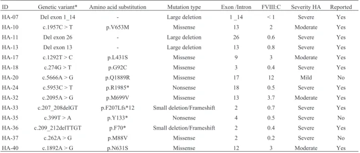
Initially, pathogenic variants were identified in nine of the 13 unrelated patients analyzed by HRM (Table 2, Supplementary Figure S3). Missense variants were present in seven samples, a nonsense variant in one sample, and a small frameshift deletion in one sample (Table 2). With this approach we did not identify the pathogenic variant in four patients. We sequenced the entire coding region with the exception of exon 14 in these four patients. Two additional
Table 2- Microarray and sequencing genetic variants identified in hemophilia A patients from Colombia.
ID Genetic variant* Amino acid substitution Mutation type Exon /Intron FVIII:C Severity HA Reported
HA-07 Del exon 1_14 - Large deletion 1 _14 < 1 Severe Yes
HA-10 c.1957C > T p.V653M Missense 13 2 Moderate Yes
HA-11 Del exon 26 - Large deletion 26 0.6 Severe Yes
HA-13 Del exon 13 - Large deletion 13 0.8 Severe Yes
HA-17 c.1292T > C p.L431S Missense 9 3 Moderate Yes
HA-18 c.274G > T p.G92C Missense 3 0.4 Severe Yes
HA-20 c.5666A > G p.Q1889R Missense 17 12 Mild No
HA-24 c.5953C > T p.R1985* Nonsense 18 0.5 Severe Yes
HA-32 c.2095A > G p.M699V Missense 13 3.7 Moderate Yes
HA-33 c.207_208delGT p.F207Lfs*12 Small deletion/Frameshift 2 0.7 Severe Yes
HA-35 c.399T > A p.Y133* Nonsense 4 0.5 Severe No
HA-36 c.209_212delTTGT p.F70* Small deletion/Frameshift 2 0.4 Severe Yes
HA-37 c.262A > G p.M88V Missense 2 0.2 Severe No
HA-40 c.1892A > G p.N631S Missense 12 3 Moderate Yes
pathogenic variants were identified: c.399T > A; p.Y133* in HA-35 and c.209-212delTTGT; p.F70* in HA-36, in-creasing the number of identified variants to 11 out of 13 unrelated patients. Most of these variants have been re-ported previously in hemophilia A databases: CHAMP CDC (https://www.cdc.gov/ncbddd/hemophilia/champs), Hemobase (http://www.hemobase.com), Factor VIII Vari-ant Database (http://www.factorviii-db.org), and the litera-ture, with the exception of three variants, two in severe HA c.262A > G, p.M88V, c.399T > A; p.Y133*, and another in mild HA c.5666A > G; p.Q1889R (Table 3, Supplementary Figure S3).
Genotype/phenotype correlations
Twenty male samples showed Inv22, Inv1, or large deletions. Nineteen of them showed a severe HA pheno-type. One patient (HA-12) had Inv22 with 1.8% FVIII:C. However, he presented a severe phenotype with history of arthropathy at the age of 8 and was receiving prophylaxis at the time (Table 1). The remaining were female carriers (two with Inv22, one Inv1, and one with large deletion). One fe-male showed FVIII activity (FVIII:C 35%), and the re-maining had normal FVIII activity.
Eleven samples had either point mutations or small deletions. Of these, seven patients had severe HA pheno-type. One of them, HA-33, had a frameshift deletion of two base pairs c.207-208delGT;p.F207Lfs12*, also found in his heterozygous mother (HA-39); HA-24 had a nonsense variant c.5953C > T; p.R1985*, and HA-18 had a missense variant c.274G > T, p.G92C (Table 2). The remaining sam-ples with missense variants had either moderate or mild phenotypes. Thus, a severe phenotype was mainly associ-ated with Inv22, Inv1, large deletions, frameshift, and non-sense variants, as described previously. Misnon-sense variants were mainly associated to moderate and mild phenotypes.
HA family history was present in 12/33 patients (36.3%). Arthropathy was found in 16/33 patients (48.4%). History of FVIII inhibitors was present in 11/33 patients (33.3%), similar to other populations (Gouwet al., 2011); there was no association of a particular genetic variant with FVIII inhibitors. Inversion 22 was present in 7 out of the 11 patients with inhibitors positive history (63.3%) compared to 9/26 (34.5%) inhibitors negative history (Fisher’s exact testp=0.151). A similar finding has been reported earlier (Oldenburget al., 2002; Goodeve and Peake, 2003;
Man-tilla-Capacho et al., 2007; Ghosh and Shetty, 2009; Tantawyet al., 2011; Kreuz and Ettingshausen, 2014).
Discussion
We used inverse shifting-PCR for Inv22 and Inv1 (Rossettiet al., 2005, 2008, 2011; Radicet al., 2009) fol-lowed by HRM analysis to identifyF8exon fragments that could harbor pathogenic variants, and DNA Sanger se-quencing. With this approach, we were able to detect patho-genic variants in 29 out of 33 affected male individuals. Two additional pathogenic variants were identified by se-quencing most of the entire coding sequence in two addi-tional patients (31/33, 94%).
As expected, 92.5% severe HA patients had Inv22, Inv1, frameshift, nonsense, and large deletions (25/27). The remaining two severe HA carried missense variants; one patient with a c.274G > T; p.G92C previously reported in the literature (Tuddenhamet al., 1994), and another with a c.262A > G;p.M88V not reported before. This variant was classified as a pathogenic variant in three out of four of the prediction analysis softwares used (Capriottiet al., 2006; Adzhubeiet al., 2010; Choiet al., 2012; Schwarzet al., 2014) (Tables 2 and 3, Supplementary Figure S3).
Microarray analysis was carried out in all the patients showing lack of amplification for one or more exons by HRM (Supplementary Figure S2). We chose to analyze large deletions by microarray analysis instead of MLPA since microarray analysis can further delineate deletion points, as well as other genetic regions involved. In addi-tion, MLPA analysis forF8would have shown what we had already detected in the HRM amplification, since MLPA probes are directed to exon sequences of the gene.
The HA-07 patient and his mother showed a large de-letion involving exon 1 through exon 14, with a total of 223,573 bp missing (Supplementary Figure S2). Similar deletions have been reported previously involving exons 1 through 14 (Tuddenhamet al., 1994; Kemball-Cook and Tuddenham, 1997; Giannelliet al., 1998; Kemball-Cooket al., 1998; Krebs et al., 2003; Lindvall and Swedenborg, 2006; Margaglione et al., 2008; Rallapalli et al., 2013; Rydzet al., 2013). In this case, the deletions involved four additional genes FUNDC2, MTCP1NB, MTCP1, and
BRCC3, a finding that has not been reported earlier since no microarray analysis had been performed.
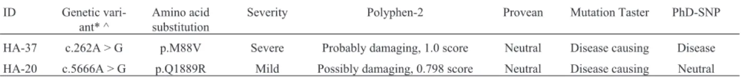
Table 3- Prediction software analysis of new genetic variants identified in Hemophilia A patients from Colombia
ID Genetic vari-ant* ^
Amino acid substitution
Severity Polyphen-2 Provean Mutation Taster PhD-SNP
HA-37 c.262A > G p.M88V Severe Probably damaging, 1.0 score Neutral Disease causing Disease
HA-20 c.5666A > G p.Q1889R Mild Possibly damaging, 0.798 score Neutral Disease causing Neutral
Patient HA-13 had a 17,979 bp deletion that involved part of intron 12, exon 13, and intron 13. Patient HA-11 failed to amplify exon 26 in several occasions by HRM. However, CytoScan 750 microarray analysis failed to de-tect a deletion, even when a 1 kb filter was used. Detailed analysis by CytoScan 750 microarray in this sample showed absence for probes (SNP and CNV markers) cover-ing this exon, thus explaincover-ing the lack of detection for this deletion in the microarray (Supplementary Figure S2).
Five patients had a moderate HA phenotype. One of them had Inv22 (FVIII:C 1.8%) and the remaining four sin-gle nucleotide missense variants, all of which had been published before: c.1292T > C;p.L431S (HA-17), c.2095A > G;p.M699V (HA-37), c.1892A > G;p.N631S (HA-40), and c.157C > T;p.V653M (HA-10) (Table 2, Supplemen-tary Figure S3).
A missense variant present in one HA mild patient and his heterozygous mother, c.5666A > G; p.Q1889R (12.4% and 70% FVIII:C activity, respectively) had not been published before. Two out of four prediction tools showed this variation as pathogenic (Capriottiet al., 2006; Adzhubeiet al., 2010; Choiet al., 2012; Schwarz et al., 2014) (Tables 2 and 3, Supplementary Figure S3).
The frequencies of pathogenic variants found in our study are similar to those described for other populations, including some Latin American studies. In Argentina, Inv22 frequencies have been reported between 39 and 41% (De Brasiet al., 2000, 2003; Rossettiet al., 2004), very similar to Venezuela in severe HA patients (Albanezet al., 2011), while in Mexico, the frequency reported was 45% (Mantilla-Capachoet al., 2007). In Costa Rica, Inv22 was found in 21/34 severe patients (61.7%) while no Inv1 was detected (Salazar-Sanchezet al., 2010). A slightly higher frequency for Inv1 was found in our study (9.1% of severe HA patients), compared to Argentinean patients.
High resolution melting analysis under standard melt-ing parameters allowed us to identify genetic variants by Sanger sequencing in nine of the remaining 13 patients (69%). High resolution melting analysis gave positive re-sults on average in three out of 52 fragments per sample. In four patients (4/33, 12.1%) we were not able to detect any variant with the approach used in the present study. These results could be due to either false negative HRM reactions, or genetic variants that are located outside of the regions tested here. HRM analysis is based on melting curves that are similar but distinguishable from each other, first, by dif-ferences in amplicon melting temperature (Tm), and sec-ond, by melting curves showing differences in homozygous and heterozygous samples (Ririeet al., 1997). Since only one amplicon is obtained in male HA patients, only differ-ences in melting temperature curves are evaluated in X-linked diseases. Thus, differences in homozygous and het-erozygous melt temperature curves are not accounted for.
In this regard, it is interesting that the genetic variant present in the HA-33 patient (a 2 bp deletion) was
sus-pected once the sample from the patient’s mother gave an abnormal melt pattern in exon 2 by HRM analysis that was not detected initially in the patient’s HRM analysis. Thus, it is possible that false negative HRM reactions are account-able for these results. We identified two additional patho-genic variants in two additional patients when we sequen-ced the entireF8coding regions with the exception of exon 14. The remaining two pathogenic variants could lie within exon 14 or within introns, outside of the regions tested here. Previous studies have shown that 2% of the genetic variants in HA are due to mutations found in non-coding se-quences (Halldenet al., 2012; Pezeshkpooret al., 2013). In addition, a third homologous recombination region has been described in HA (Pezeshkpooret al., 2012) that was not tested in our study. Thus, further studies are required to identify the pathogenic variants in those two samples.
Conclusion
For the first time in Colombia we have used a system-atic cost-effective molecular approach to detect HA patho-genic variants. This approach was used due to the high cost for methods such as next generation sequencing (NGS) in our health system. With this approach we were able to de-tect pathogenic variants in 94% of patients (31/33). Also, we identified three new genetic variants not reported previ-ously in the CHAMP CDC, Hemobase, Factor VIII Variant Database, and the literature. Further analysis is underway to identify the genetic variant responsible for the HA severe phenotype in the remaining two patients.
Acknowledgments
We would like to thank all the patients and their fami-lies for participating in this study. In addition, we would like to thank the Hemophilia Treatment Center from Clíni-ca Infantil Colsubsido and the Hemophilia Treatment Cen-ter from Fundación Hospital de la Misericordia (HOMI) for providing patients for the present study, as well as to the Centro de Investigaciones Clínicas de Colsubsidio. Also, we would like to thank the nurses Claudia Suarez Molano, Yadira Valderrama Vargas, and Angie Ubaque for their help and Servicios Médicos Yunis Turbay y Cia., for their support in the microarray analysis. All DNA sequencing was carried out at the Servicio de Secuenciación, Instituto de Genética Universidad Nacional de Colombia (SSIGMOL). This work financially was supported by a grant from División de Investigaciones Bogotá (DIB), Her-mes project #27597 Universidad Nacional de Colombia to JJY. LKY received support from “Convocatoria Nacional de Jóvenes investigadores e Innovadores 2015-Colciencias”.
References
haemo-philia A patients with inhibitors in Italy. Haemohaemo-philia 20:e243-250.
Adzhubei IA, Schmidt S, Peshkin L, Ramensky VE, Gerasimova A, Bork P, Kondrashov AS and Sunyaev SR (2010) A method and server for predicting damaging missense muta-tions. Nat Methods 7:248-249.
Ahmed RP, Ivaskevicius V, Kannan M, Seifried E, Oldenburg J and Saxena R (2005) Identification of 32 novel mutations in the factor VIII gene in Indian patients with hemophilia A. Haematologica 90:283-284.
Albanez S, Ruiz-Saez A, Boadas A, Bosch N and Porco A (2011) Identification of factor VIII gene mutations in patients with severe haemophilia A in Venezuela: Identification of seven novel mutations. Haemophilia 17:e913-918.
Antonarakis SE (1995) Molecular genetics of coagulation factor VIII gene and hemophilia A. Thromb Haemost 74:322-328. Antonarakis SE, Rossiter JP, Young M, Horst J, Moerloose P,
Sommer SS, Ketterling RP, Kazazian Jr HH, Négrier C, Vinciguerra Cet al.(1995) Factor VIII gene inversions in severe hemophilia A: results of an international consortium study. Blood 86:2206-2212.
Astermark J, Oldenburg J, Escobar M, White 2nd GC and Berntorp E (2005) The Malmö International Brother Study (MIBS). Genetic defects and inhibitor development in sib-lings with severe hemophilia A. Haematologica 90:924-931. Astermark J, Donfield SM, Gomperts ED, Schwarz J, Menius ED, Pavlova A, Oldenburg J, Kessing B, DiMichele DM, Sha-piro ADet al.(2013) The polygenic nature of inhibitors in hemophilia A: Results from the Hemophilia Inhibitor Genet-ics Study (HIGS) Combined Cohort. Blood 121:1446-1454. Brackmann HH, Oldenburg J and Schwaab R (1996) Immune
tol-erance for the treatment of factor VIII inhibitors—twenty years’ ‘Bonn protocol’. Vox Sang 70 Suppl 1:30-35. Brackmann HH, Schwaab R, Effenberger W, Hess L, Hanfland P
and Oldenburg J (2000) Antibodies to factor VIII in hemo-philia A patients. Vox Sang 78 Suppl 2:187-190.
Capriotti E, Calabrese R and Casadio R (2006) Predicting the in-surgence of human genetic diseases associated to single point protein mutations with support vector machines and evolutionary information. Bioinformatics 22:2729-2734. Choi Y, Sims GE, Murphy S, Miller JR and Chan AP (2012)
Pre-dicting the functional effect of amino acid substitutions and indels. PLoS One 7:e46688.
De Brasi C, Candela M, Cermelj M, Slavutsky I, Larripa I, Bianco RP and De Tezanos Pinto M (2000) Intron 22 factor VIII gene inversions in Argentine families with severe haemo-philia A. Haemohaemo-philia 6:21-22.
De Brasi CD, Rossetti LC and Larripa IB (2003) Rapid genotyp-ing ofXbaI andMspI DNA polymorphisms of the human factor VIII gene: Estimation of their combined heterozy-gosity in the Argentinean population. Haematologica 88:232-234.
den Dunnen JT, Dalgleish R, Maglott DR, Hart RK, Greenblatt MS, McGowan-Jordan J, Roux AF, Smith T, Antonarakis SE and Taschner PE (2016) HGVS recommendations for the description of sequence variants: 2016 update. Hum Mutat 37:564-569.
Ghosh K and Shetty S (2009) Immune response to FVIII in hemo-philia A: An overview of risk factors. Clin Rev Allergy Immunol 37:58-66.
Giannelli F, Green PM, Sommer SS, Poon M, Ludwig M, Schwaab R, Reitsma PH, Goossens M, Yoshioka A, Figuei-redo MSet al.(1998) Haemophilia B: Database of point mu-tations and short additions and deletions—eighth edition. Nucleic Acids Res 26:265-268.
Goodeve AC and Peake IR (2003) The molecular basis of hemo-philia A: Genotype-phenotype relationships and inhibitor development. Semin Thromb Hemost 29:23-30.
Gouw SC, Van Der Bom JG, Van Den Berg HM, Zewald RA, Van Amstel JKP and Mauser-Bunschoten EP (2011) Influence of the type of F8 gene mutation on inhibitor development in a single centre cohort of severe haemophilia A patients. Hae-mophilia 17:275-281.
Hallden C, Knobe KE, Sjorin E, Nilsson D and Ljung R (2012) In-vestigation of disease-associated factors in haemophilia A patients without detectable mutations. Haemophilia 18:e132-137.
Higuchi M, Kazazian Junior HH, Kasch L, Warren TC, McGin-niss MJ, Phillips 3rd JA, Kasper C, Janco R and Antonarakis SE (1991a) Molecular characterization of severe hemophilia A suggests that about half the mutations are not within the coding regions and splice junctions of the factor VIII gene. Proc Natl Acad Sci USA 88:7405-7409.
Higuchi M, Antonarakis SE, Kasch L, Oldenburg J, Economou-Petersen E, Olek K, Arai M, Inaba H, and Kazazian Junior HH (1991b) Molecular characterization of mild-to-mode-rate hemophilia A: Detection of the mutation in 25 of 29 pa-tients by denaturing gradient gel electrophoresis. Proc Natl Acad Sci USA 88:8307-8311.
Hoyer LW (1994) Hemophilia A. N Engl J Med 330:38-47. Kemball-Cook G and Tuddenham EG (1997) The Factor VIII
Mutation Database on the World Wide Web: The haemo-philia A mutation, search, test and resource site. HAM-STeRS update (version 3.0). Nucleic Acids Res 25:128-132. Kemball-Cook G, Tuddenham EG and Wacey AI (1998) The
fac-tor VIII structure and mutation resource site: HAMSTeRS version 4. Nucleic Acids Res 26:216-219.
Krebs H, Domsch C, Adelhard K, Brackmann HH, Graw J, Oldenburg J, Schwaab R and Schramm W (2003) The na-tional GTH haemophilia registry as database within the scope of the German human genome project. Hamosta-seologie 23:18-23.
Kreuz W and Ettingshausen CE (2014) Inhibitors in patients with haemophilia A. Thromb Res 134 Suppl 1:S22-26.
Lakich D, Kazazian Juniorr HH, Antonarakis SE and Gitschier J (1993) Inversions disrupting the factor VIII gene are a com-mon cause of severe haemophilia A. Nat Genet 5:236-241. Lin SY, Su YN, Hung Tsay W, Chiou SS, Chang CT, Ho HN and
Lee CN (2008) Mutation spectrum of 122 hemophilia A families from Taiwanese population by LD-PCR, DHPLC, multiplex PCR and evaluating the clinical application of HRM. BMC Med Genet 9:53.
Lindvall K and Swedenborg N (2006) UMAS Hemophilia Data-base. Stud Health Technol Inform 122:853.
Margaglione M, Castaman G, Morfini M, Rocino A, Santagostino E, Tagariello G, Tagliaferri AR, Zanon E, Bicocchi MP, Castaldo Get al.(2008) The Italian AICE-Genetics hemo-philia A database: Results and correlation with clinical phe-notype. Haematologica 93:722-728.
Oldenburg J, El-Maarri O and Schwaab R (2002) Inhibitor devel-opment in correlation to factor VIII genotypes. Haemophilia 8 Suppl 2:23-29.
Pezeshkpoor B, Rost S, Oldenburg J and El-Maarri O (2012) Iden-tification of a third rearrangement at Xq28 that causes severe hemophilia A as a result of homologous recombination be-tween inverted repeats. J Thromb Haemost 10:1600-1608. Pezeshkpoor B, Zimmer N, Marquardt N, Nanda I, Haaf T, Budde
U, Oldenburg J and El-Maarri O (2013) Deep intronic ‘mu-tations’ cause hemophilia A: Application of next generation sequencing in patients without detectable mutation in F8 cDNA. J Thromb Haemost 11:1679-1687.
Radic CP, Rossetti LC, Zuccoli JR, Abelleyro MM, Larripa IB and De Brasi CD (2009) Inverse shifting PCR based prenatal diagnosis of hemophilia-causative inversions involving int22h and int1h hotspots from chorionic villus samples. Prenat Diagn 29: 1183-1185.
Rallapalli PM, Kemball-Cook G, Tuddenham EG, Gomez K and Perkins SJ (2013) An interactive mutation database for hu-man coagulation factor IX provides novel insights into the phenotypes and genetics of hemophilia B. J Thromb Haemost 1:1329-1340.
Ririe KM, Rasmussen RP and Wittwer CT (1997) Product differ-entiation by analysis of DNA melting curves during the polymerase chain reaction. Anal Biochem 245:154-160. Rossetti LC, Candela M, Bianco RP, Pinto MT, Western A,
Goodeve A Larripa IB and De Brasi CD (2004) Analysis of factor VIII gene intron 1 inversion in Argentinian families with severe haemophilia A and a review of the literature. Blood Coagul Fibrinolysis 15:569-572.
Rossetti LC, Radic CP, Larripa IB and De Brasi CD (2005) Geno-typing the hemophilia inversion hotspot by use of inverse PCR. Clin Chem 51:1154-1158.
Rossetti LC, Radic CP, Larripa IB and De Brasi CD (2008) De-veloping a new generation of tests for genotyping hemo-philia-causative rearrangements involving int22h and int1h hotspots in the factor VIII gene. J Thromb Haemost. 6:830-836.
Rossetti LC, Radic CP, Abelleyro MM, Larripa IB and De Brasi CD (2011) Eighteen years of molecular genotyping the
he-mophilia inversion hotspot: from Southern blot to inverse shifting-PCR. Int J Mol Sci 12:7271-7285.
Rydz N, Leggo J, Tinlin S, James P and Lillicrap D (2013) The Canadian “National Program for hemophilia mutation test-ing” database: A ten-year review. Am J Hematol 88:1030-1034.
Salazar-Sanchez L, Jimenez-Cruz G, Mendez M, Chaverri P, Alvarado P, Schroder W, Wulff K, Sandoval M, Herrmann FH, Pavlova A et al.(2010) Molecular analysis of FVIII gene in severe HA patients of Costa Rica. Hamostaseologie 30 Suppl 1:S150-S152.
Sanger F, Nicklen S and Coulson AR (1977) DNA sequencing with chain-terminating inhibitors. Proc Natl Acad Sci USA 74:5463-5467.
Savage N (2014) Born in the Blood. Nature 515:S158–S159. Schwarz JM, Cooper DN, Schuelke M and Seelow D (2014)
MutationTaster2: Mutation prediction for the deep-sequencing age. Nat Methods 11:361-362.
Tantawy AA, Mackensen SV, El-Laboudy MA, Labib JH, Moftah F, El-Telbany MA and Mansour WA (2011) Health-related quality of life in Egyptian children and adolescents with he-mophilia A. Pediatr Hematol Oncol 28:222-229.
Tuddenham EG, Schwaab R, Seehafer J, Millar DS, Gitschier J, Higuchi M, Bidichandani S, Connor JM, Hoyer LW, Yoshioka Aet al.(1994) Haemophilia A: Database of nucle-otide substitutions, deletions, insertions and rearrangements of the factor VIII gene, second edition. Nucleic Acids Res 22:3511-3533.
Woods-Samuels P, Kazazian Junior HH and Antonarakis SE (1991) Nonhomologous recombination in the human ge-nome: deletions in the human factor VIII gene. Genomics 10:94-101.
Supplementary material
The following online material is available for this article: Figure S1-Inverse shifting-PCR for Inv22 and Inv1. Figure S2-CytoScan 750 (Affymetrix) microarray analy-sis.
Figure S3- Hemophilia A BigDye terminators direct se-quencing of Colombian HA patients.
Associate Editor: Maria Luiza Petzl-Erler