Comparison between three techniques for videosinuscopy in cattle
Comparação entre três técnicas para videosinuscopia em bovinos
Fernando Zanlorenzi BassoI Eduarda Maciel BusatoI Jéssica Rodrigues da SilvaI Rogério Luizari GuedesII Ivan Roque de Barros FilhoIII Peterson Triches DornbuschIII ISSN 1678-4596
ABSTRACT
Cattle have extensive paranasal sinuses that are susceptible to disease, most commonly sinusitis. The sinuscopy can be used to evaluate these structures, although there are no descriptions of this region for endoscopic anatomy, especially regarding the trocar position and the most appropriate type of endoscope. This study aimed to standardize the surgical approaches to sinuscopy in cattle by comparing the use of three endoscopes. Four accesses by trephination (one hole for each of the maxillary and frontal sinuses) were made in eight heads of slaughtered cattle. Each hole was inspected with three endoscopes: a 10mm flexible colonoscope with up to 180º of angulation, a 10mm 0° laparoscope and a 4mm 30º arthroscope. It was observed that all regions of the maxillary sinus were better visualized with the 4mm endoscope, and the structures of this sinus were less well visualized with the 10mm laparoscope. The frontal sinus was difficult to evaluate due to the tortuosity of its bony projections, and the cranial portion was not observed by the proposed accesses. The caudal regions of the frontal sinus such as the nuchal diverticulum and the back of the orbit had the greatest number of structures visualized by the 4mm endoscope, followed by the colonoscope. The comparative analysis showed that the 4mm endoscope was most efficient and could be adapted to sinuscopy in cattle.
Key words: endoscopy, videosurgery, nasal sinus, sinusitis, bovine.
RESUMO
Os bovinos apresentam seios paranasais extensos e passíveis de afecções, como a sinusite. A sinuscopia, técnica já utilizada em outras espécies, avalia os seios paranasais de modo pouco invasivo e não é descrita em bovinos. O presente estudo objetivou padronizar os acessos cirúrgicos para sinuscopia em bovinos, testando três técnicas de videoendoscopia.
Foram selecionadas oito cabeças de bovinos provenientes de abatedouro comercial, sendo realizada a trepanação dos seios maxilares e frontais de ambos os lados (um orifício por seio). Cada seio foi inspecionado com três óticas: um colonoscópio flexível com 10mm de diâmetro e até 180º de angulação, um laparoscópio rígido de 10mm e 0º e um artroscópio rígido de 4mm e 30º. Na região caudal do seio maxilar, os alvéolos e abertura maxilopalatina foram visualizadas com todas as óticas. A região caudodorsomedial e rostral do seio maxilar foram observadas com a ótica flexível e a rígida de 4mm, sendo que apenas esta adentrou no seio palatino. O seio frontal é de difícil visualização, devido à tortuosidade de suas projeções ósseas e sua porção cranial não foi observada pelo acesso proposto. A região caudal do seio frontal, o divertículo nucal e a área caudal à órbita tiveram o maior número de estruturas visualizadas com a ótica rígida de 4mm, seguida da flexível. A análise comparativa demonstra que a técnica utilizando a ótica rígida de 4mm permite a visualização de um maior número de estruturas com maior detalhamento e é a que mais se adapta à sinuscopia em bovinos.
Palavras-chave: endoscopia, videocirurgia, seios nasais, sinusite, bovino.
INTRODUCTION
The sinuses in cattle have peculiar characteristics, are underdeveloped in calves and acquire their full size after several years (DYCE et al., 2010). The frontal sinus presents rostral and caudal compartments that extend to the cornual processes. The maxillary sinuses are unique and
IPrograma de Residência Multiprofissional em Saúde, Universidade Federal do Paraná (UFPR), Curitiba, PR, Brasil.
IIPrograma de Pós-graduação em Ciências Veterinárias, Universidade Federal do Paraná (UFPR), 80035-050, Curitiba, PR, Brasil. E-mail: [email protected]. Corresponding author.
IIIDepartamento de Medicina Veterinária, Universidade Federal do Paraná (UFPR), Curitiba, PR, Brasil.
large, and enable communication with the palatine sinuses. They must be accessed via the hard palate, making the surgical approach quite difficult (SISSON & GROSSMAN, 1998).
Among the pathologies of the sinuses,
an inflammatory process called sinusitis stands
out. In cattle, the leading cause of frontal sinusitis
is associated with dehorning, as about 2% of
surgically dehorned animals develop this disease
(FIORAVANTI et al., 1999; SILVA et al., 2008). It can also be associated with respiratory infections, trepanations or fractures with frontal sinus
exposure, cysts or nasal cancer (SMITH, 2006).
Surgery by unqualified surgeons, the presence of foreign bodies and improper postoperative therapy
are also important etiologic factors in this species
(FIORAVANTI et al., 1996).
The diagnosis of sinusitis in cattle is
based on history and clinical examination findings
(DIRKSEN et al., 1993). In several species, in addition to a general clinical examination, some diagnostic
methods can be used such as regional radiographs,
sinucentesis, surgical exploration (sinusotomy),
tomography and sinuscopy; the latter is performed with rigid or flexible endoscopes (ALLISON, 1999; EMSHOFF et al., 1999; SMITH, 2006).
Sinuscopy has been performed in humans (BERTRAND & ROBILLARD, 1985; PETRUSON,
2004), horses (PERKINS et al., 2009a) and dogs
(JOHNSON, 2006), due to its practicality and lower postoperative morbidity compared to conventional
exploration techniques (SILVA et al., 2009). In
horses, sinuscopy is widely used to properly inspect the sinuses as well to collect samples and perform biopsies on those sites. In this procedure, the animal can be kept sedated in the quadrupedal position; sinus access occurs through trepanation, which allows for
the introduction of endoscopes (PERKINS et al.,
2009a; O’LEARY & DIXON, 2011). Until now, there have been no studies regarding sinuscopy in cattle, in terms of systematically describing the endoscopic
anatomy of the region, the access portals and the most appropriate type of lens.
This study aimed to compare the effectiveness of three different endoscopes in sinuscopic evaluation of the maxillary, palatine and frontal sinuses of cattle in a postmortem study. The experiment also aimed to standardize the minimally invasive surgical access for sinuscopy in
this species and to improve anatomical knowledge with an emphasis on the endoscopic anatomy of
those regions.
MATERIALS AND METHODS
Eight cattle heads were used, obtained from commercial slaughterhouses in Curitiba and nearby cities. The heads were received skinless, dehorned and partially stripped. The access to the sinuses was carried out by a trepanning technique, first with a drill, making a small skull opening, then amplified by rotational moves with a 20mm circular trephine. The chosen sites for trepanation were based on the species anatomy and facilitated by bone visualization, aiming for a bilateral evaluation of the frontal, maxillary and palatine sinuses. The access holes for the maxillary
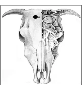
sinuses were located 3.7±0.9cm rostral to the eyeball and 2.1±0.3cm dorsal to the facial crest (Figure 1A). The access holes for the frontal sinuses were located 4.9±1.6cm rostral to the nuchal ridge and 2.8±0.5cm lateral to the midline (Figure 2A).
The equipment used for cavity inspection
included a flexible colonoscope with a diameter of 10mm and angles up to 180° (Karl Storz, Germany), a 10mm and 0° laparoscope (Karl Storz, Germany), and
a 4mm and 30° arthroscope (Karl Storz, Germany);
all were coupled to a laparoscopic unit composed by
a LED monitor, a microcam and a xenon light source (Telepack®, Karl Storz, Germany). The sinuses were
inspected with the three endoscopes, trying to identify
Figure 1 - Illustration of the sinuses in a bovine head in a left lateral view. A: access hole to the maxillary sinus; B: caudal and caudo-dorsomedial areas from maxillary sinus; C: maxilo-palatine opening; D: dental alveoli. Adapted from BUDRAS & HABEL,
the highest number of structures possible, according
to the literature and the local anatomy of this species,
being classified as 1: visible or 2: not visible. All inspections were documented individually for further assessment, recording and the identified structures were tabulated. Three independant evaluators were selected, one with experience in videoendoscopic/
videolaparoscopic procedures in another species, and
the other two with knowledge of cattle anatomy. The efficiency of these endoscopes was verified through their viewing capability and identification of structures by the surgeon evaluator. The group findings were statistically compared by the non-parametric Kruskal-Wallis test, followed by Dunn’s multiple comparison, using Graphpad Prism software, V5.
RESULTS AND DISCUSSION
The literature concerning cattle sinuscopy
is rare, making it difficult to compare the literature with the data obtained in this study. In horses, sinuscopy is
a tool used for the diagnosis, treatment and evaluation
of sinusitis (PERKINS et al., 2009b; DIXON et al., 2012.). Besides horses, there are reports of sinuscopy in dogs, but it is difficult to draw interrelationships between studies of these species with cattle because
they have anatomically different paranasal sinuses
(PETRUSON, 2004; JOHNSON, 2006).
During this study, a 20mm diameter circular
trephine was used, but smaller diameters such as 14 or 15mm may be used for the same purpose (PERKINS et al., 2009b). MACHADO & SILVA (2013) carried out an 8mm trepanation to compare rigid and a flexible
sinuscopy in horses, using a 4mm 30° rigid endoscope
and a flexible endoscope 4.8mm in diameter. Due the
10mm endoscope used in the present study, it was not
possible to work with smaller trephines.
The trephination areas and sinuses were selected based on anatomy, but they may be modified
according to the purposes of the exam (SMITH,
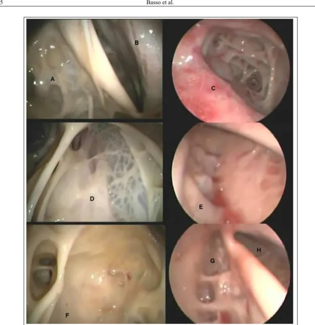
2006). The main identified areas are displayed in figure 3. Through the frontal sinus access, a caudal observation was made of this region, the nuchal diverticulum, the caudal region of the eyeball, but the exploration was complicated by the presence of large numbers of intrasinusal lamellae (Figure 3C).
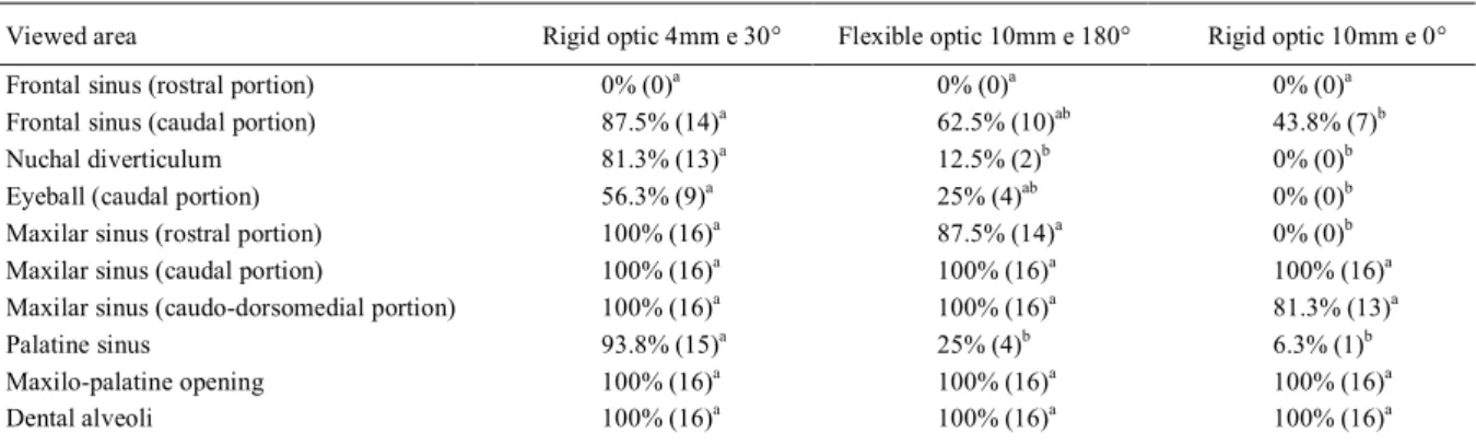
Data in percentages referring to viewing
capacity from different areas and techniques are
compiled on table 1. The visualization of the caudal
frontal sinus area (Figure 2C) varied according to the equipment used; the 4mm 30° arthroscope was most efficient (viewing rate of 87.5%). It was not possible to see the desired structure in only one of the eight heads, bilaterally, due the greater presence of bone irregularities therein. The colonoscope with
a diameter of 10mm and angles up to 180° ranked
second, with 62.5% successful visualizations, while the 10mm 0° endoscope had the lowest viewing rate among all tested endoscopes, as it was effective in less than half of the heads (43.75%). Observation of the nuchal diverticulum was possible only with the arthroscope and colonoscope, in 81.25% and 12.50% of accesses, respectively (Figure 2B). During the experiment, the caudal area to the eyeball was visualized by some accesses with the arthroscope (56.25%) and colonoscope (25%). The laparoscope proved to be ineffective for this purpose.
The rostral region of the frontal sinus
presents a tortuous anatomy, marked by intrasinusal lamellae, resulting in irregular areas (BUDRAS & HABEL, 2003; DYCE et al., 2010), which prevent
the insertion of endoscopes through the proposed
access. The cornual processes were not visible because the heads were obtained from previously
dehorned animals.
Inspection of the maxillary sinuses was easier and didactic when compared with the frontal sinuses, because the maxillae have a small number of tortuous bones and a more regular anatomy. This finding is in counterpoint to the purpose of the examination, since the major diseases of the bovine
paranasal sinuses are associated with dehorning,
and therefore good visualization of the frontal sinus
would be interesting (SILVA et al., 2008).
The caudal area of the maxillary sinuses
achieved excellent viewing with all endoscopes used (Figure 1B; Figure 3F). The caudodorsomedial portion of the same area (Figure 1B; Figure 3D) had slightly limited inspection when the laparoscope was used, because it was ineffective in three of the 16 views. The dental alveoli (Figure 1D; Figure 3A) and the maxillopalatine opening (Figure 1C; Figure 3C) were readily observed with all three endoscopes (viewing rate of 100%); however, the palatine sinus
(Figure 3E; Figure 3H) could not be accessed in all
heads. The most effective endoscopic access to the
palatine sinus was achieved with the arthroscope, which attained a 93.75% viewing rate, with only one not evaluated due a narrower maxillopalatine
opening than the others. The other endoscopes
showed poor efficiency to this area, with a viewing rate of 25% with the colonoscope and a 6.25% with
the laparoscope. The rostral region of the maxillary
sinus (Figure 3G) was inspected with 100% efficiency when using the arthroscope and 87.5% efficiency with the colonoscope; however, this viewing area was not accessible with the laparoscope.
Figure 3 - Explored anatomical areas identified during the video sinuscopy in cattle. A: dental alveolus; B: maxilo-palatine opening; C: tortuosity from frontal sinus; D: maxillary sinus, caudo-dorsomedial portion; E: palatine sinus; F: maxillary sinus, caudal portion; G: maxillary sinus, rostral portion; H:
CONCLUSION
A comparative analysis of the effectiveness
of different endoscopes shows that a rigid endoscope with 4mm and 30° is the most adaptable for cattle sinuscopy, because it has a smaller diameter and a higher angulation view, which are required to access structures with a narrow opening, such as the palatine sinus. Although, more accesses sites need to be tested, trying to optimize the viewing of the other endoscopes
used in this study. The maxillary and palatine sinus anatomies are more regular than the frontal sinuses,
which facilitates sinuscopic inspection of the first
ones. The proposed accesses to maxillary and palatine
sinuses are adequate, while the techniques for frontal sinus have limitations in the rostral sinus views.
BIOETHICS AND BIOSSECURITY COMMITTEE APPROVAL
This study was submitted to the Ethics Committee for animal use of the Agricultural sciences sector (Universidade Federal do Paraná (UFPR), Brazil), following the ethical principles of the Brazilian College of Animal Experimentation (COBEA), judged and approved under the process number 101/2010.
CONFLICT OF INTERESTS
There is no conflict of interests.
REFERENCES
BUDRAS, D.K.; HABEL, E.R. Bovine Anatomy. Germany: Schlütersche, 2003. p.34-36.
DIRKSEN, G. Sistema digestivo. In: ROSENBERGER. Exame clínico dos bovinos. Rio de Janeiro RJ: Guanabara Koogan, 1993. p.166-228.
DIXON, P.M. et al. Equine paranasal sinus disease: a long-term
study of 200 cases (1997–2009): treatments and long-term results of treatments. Equine Veterinary Journal, v.44, n.3,
p.272-5 276, 2012. Available from: <http://onlinelibrary.wiley.com/ doi/10.1111/j.2042-6 3306.2011.00427.x/abstract>. Accessed:
Mar. 25, 2014. doi: 10.1111/j.2042-73306.2011.00427.x.
DYCE, K.M. et al. Cabeça e pescoço ventral do ruminante. In:
_____. Tratado de anatomia veterinária. 4.ed. Rio de Janeiro: Elsevier, 2010. p.644-663.
EMSHOFF, R. et al. Idiopathic maxillary pain: prevalence of
maxillary sinus hyperreactivity in relation to allergy, chronic
mucosal inflammation, and eosinophilia. Oral Surgery, Oral Medicine, Oral Pathology, Oral Radiology and Endodontology,
v.87, n.6, p.685-690, 1999. Available from: <http://dx.doi. org/10.1016/S1079-2104(99)70161-7>. Accessed: Mar. 25, 2014.
doi: 10.1016/S1079-2104(99)70161-7.
FIORAVANTI, M.C.S. et al. Treatment of Subcutaneous Abscesses with Methacresolsulphonic Acid Associated with Nitrofurazone and Parenteral Application of Enrofloxacin. Anais da Escola de Agronomia e Veterinária - Universidade Federal de Goiás, v.26, n.2, p.1-8, 1996.
FIORAVANTI, M.C.S. et al. Use of metal clamps for skin suture after
Cattle dehorning. Ciência Rural, v.29, n.3, p.507-510, 1999. Available
from: <http://www.scielo.br/scielo.php?script=sci_arttext&pid=S0103-84781999000300021&lng=pt&nrm=iso&tlng=pt>. Accessed: Mar. 25,
2014. doi: 10.1590/S0103-84781999000300021.
JOHNSON, L.R. et al. Results of rhinoscopy alone or in conjunction with sinuscopy in dogs with aspergillosis: 46 cases (2001-2004).
Journal of the American Veterinary Medical Association,
v.228, n.5, p.738-742, 2006. Available from: <http://avmajournals. avma.org/doi/abs/10.2460/javma.228.5.738>. Accessed: Mar. 25,
2014. doi: 10.2460/javma.228.5.738.
Table 1 - View capacity (percentage and total number of animals) of the anatomical regions from paranasal sinuses in the evaluated cattle heads during the video-endoscopy techniques (n=16).
Viewed area Rigid optic 4mm e 30° Flexible optic 10mm e 180° Rigid optic 10mm e 0°
Frontal sinus (rostral portion) 0% (0)a 0% (0)a 0% (0)a
Frontal sinus (caudal portion) 87.5% (14)a 62.5% (10)ab 43.8% (7)b
Nuchal diverticulum 81.3% (13)a 12.5% (2)b 0% (0)b
Eyeball (caudal portion) 56.3% (9)a 25% (4)ab 0% (0)b
Maxilar sinus (rostral portion) 100% (16)a 87.5% (14)a 0% (0)b Maxilar sinus (caudal portion) 100% (16)a 100% (16)a 100% (16)a Maxilar sinus (caudo-dorsomedial portion) 100% (16)a 100% (16)a 81.3% (13)a
Palatine sinus 93.8% (15)a 25% (4)b 6.3% (1)b
Maxilo-palatine opening 100% (16)a 100% (16)a 100% (16)a
Dental alveoli 100% (16)a 100% (16)a 100% (16)a
MACHADO, T.S.L.; SILVA, L.C.L.C. Rigid and flexible endoscope
in sinoscopy and triangulation technique in equine paranasal sinus. Ciência Rural, v.43, n.12, p.2254-2260, 2013. Available from:
<http://www.scielo.br/scielo.php?script=sci_arttext&pid=S0103-84782013001200022&lng=pt&nrm=iso&tlng=en>. Accessed: Mar.
25, 2014. doi: 10.1590/S0103-7 84782013001200022.
O´LEARY, J.M.; DIXON, P.M. A review of equine paranasal
sinusites. A etiopathogenesis, clinical signs and ancilliary diagnostic techniques. Equine Veterinary Education, v.23, n.3,
p.148-159, 2011. Available from: <http://onlinelibrary.wiley.com/ doi/10.1111/j.20423292.201110.00176.x/abstract>. Accessed: Jun.
15, 2014. doi: 10.1111/j.2042-3292.2010.00176.x.
PERKINS, J.D. et al. Comparison of sinoscopic techniques for examining the rostral maxillary and ventral conchal sinuses of horses. Veterinary Surgery, v.38, p.607-612, 2009a. Available from: <http://
onlinelibrary.wiley.com/doi/10.1111/j.1532-950X.2009.00555.x/abst ract;jsessionid=379F60710F8DC5BEA35E910997B08097.f03t02>. Accessed: Mar. 25, 2014. doi: 16 10.1111/j.1532-950X.2009.00555.x.
PERKINS, J.D. et al. Sinoscopic treatment of rostral maxillary and ventral conchal sinusitis in 60 horses. Veterinary Surgery, v.38, p.
613-619, 2009b. Available from: <http://onlinelibrary.wiley.com/ doi/10.1111/j.1532-950X.2009.00556.x/abstract;jsessionid=5966 EC4FB8464AA7D961EB29B35177B1.f03t02>. Accessed: Mar. 25, 2014. doi: 10.1111/j.1532-950X.2009.00556.x.
PETRUSON, B. Sinuscopy in patients with titanium implants
in the nose and sinuses. Scandinavian Journal of Plastisc and
Reconstructive Surgery and Hand Surgery, v.38, n.2, p.86-93,
2004. Available from: <http://informahealthcare.com/doi/abs/1 0.1080/0284431031002324909>. Accessed: Mar. 25, 2014. doi:
10.1080/02844310310023909.
SISSON, S.; GROSSMAN, J.D. Anatomia de los animales domesticos. 5.ed. Barcelona: Salvat, 1998. 2v.
SMITH, B.P. Doenças do Sistema Respiratório. In: _____.
Medicina interna de grandes animais. Barueri: Manole, 2006. p.479-592.
SILVA, L.A.F. et al. Estudo retrospectivo sobre fatores de risco
e avaliação de quatro protocolos terapêuticos para sinusite em
um rebanho de 2491 bovinos (1998-2008). In: CONGRESSO BRASILEIRO DE MEDICINA VETERINÁRIA, 2008, Gramado, Rio Grande do Sul. Anais... Gramado: CONBRAVET, 2008.
Available from: <http://www.sovergs.com.br/conbravet2008/ anais/cd/resumos/R1028-10 3.pdf>. Accessed: Jun. 15, 2014.
SILVA, L.C.L.C. et al. Bilateral sinus cysts in a filly treated by
endoscopic sinus surgery (Case report). Canadian Veterinary Journal, v.50, p.417-420, 2009.