Universidade de Lisboa
Faculdade de Farmácia
Biochemistry of Staphylococcus aureus
adhesins:
Identification of a new vitronectin binding protein
on Staphylococcus aureus
Joana Sofia Igreja Amaral
Mestrado Integrado em Ciências Farmacêuticas
2019
Universidade de Lisboa
Faculdade de Farmácia
Biochemistry of Staphylococcus aureus
adhesins:
Identification of a new vitronectin binding
protein on Staphylococcus aureus
Joana Sofia Igreja Amaral
Monografia de Mestrado Integrado em Ciências Farmacêuticas apresentada à
Universidade de Lisboa através da Faculdade de Farmácia
Orientador: Doutor Giampiero Pietrocola
Co-orientador: Doutora Madalena Maria Vilela Pimentel, professora
associada
This work was done in partnership with the university of Pavia
in an Erasmus + program.
Abbreviations:
IsdB: iron-responsive surface determinant B
SDS-PAGE: sodium dodecyl sulphate poly acrylamide gel electrophoresis RT-qPCR: quantitative reverse transcription polymerase chain reaction ELISA: enzyme-linked immunosorbent assay
Vn: vitronectin
NEAT: conserved near iron transport
IsdA: iron-responsive surface determinant A IsdC: iron-responsive surface determinant C IsdD: iron-responsive surface determinant D
IsdE: iron-responsive surface determinant E IsdF: iron-responsive surface determinant F IsdH: iron-responsive surface determinant H RPMI: roswell park memorial institute BHI: brain heart infusion
CNS: coagulase negative species PCR: Polymerase Chain Reaction TSST-1: toxic shock syndrome toxin-1 SE: Staphylococcal enterotoxins
SSS: Staphylococcal scalded-skin syndrome CWA: cell wall anchored
LPXTG: leucine-proline-AA-threonine-glycine
MSCRAMM: microbial surface component recognizing adhesive matrix molecules SpsI: signal peptidase I
Fn: fibronectin Fbg: fibrinogen
FnBPA: fibronectin-binding protein A FnBPB: fibronectin-binding protein B ClfA: clumping factor A
ClfB: clumping factor B
CNA: collagen-binding proteins SdrS: serine-aspartate repeat protein S SdrD: serine-aspartate repeat protein D SdrR: serine-aspartate repeat protein R
Sbi: second binding protein G SasG: Surface protein G
Pls: plasmin sensitive surface protein MRSA: methicillin-resistant S. aureus VRSA: vancomycin-resistance S. aureus Hb: hemoglobin
Fur: ferric uptake repressor S protein: serum spreading factor
MAP kinase: kinase mitogen-activated protein BSA: Bovine serum albumin
SDS: Sodium dodecyl sulphate
Tris: trishydroxymethylaminomethane TCA: trichloroacetic acid
TEMED: Tetramethyl ethylenediamine
Resumo:
O número de casos de infeções por Staphylococcus aureus tende a crescer a cada ano. Porém, o inverso acontece quando se observa os níveis de eficácia dos antibióticos. Embora tenha havido muito progresso científico acerca dos fatores de virulência responsáveis pela proteção desta bactéria das defesas do ser humano, ainda existem algumas conexões desconhecidas entre
Staphylococcus aureus e o hospedeiro. Este estudo teve como objetivo investigar a capacidade
de IsdB, uma proteína presente na parede celular de Staphylococcus aureus, de se ligar à vitronectina, uma proteína humana presente no sangue e na matriz extracelular.
Como não há muita informação disponível sobre IsdB na literatura, este trabalho teve como um dos seus objetivos tentar perceber quais as condições ideias para a expressão desta proteína, uma proteína que pertence à família NEAT. Para o efeito células de S. aureus foram crescidas em diferentes meios de cultura e em diferentes estadios de crescimento e analisada a produção de IsdB por SDS-PAGE e western blot. A pesquisa mostrou que a expressão de IsdB é mais alta
em meios pobres em ferro e muito mais evidente na fase estacionária do crescimento bacteriano do que na fase exponencial. Este resultado foi confirmado por avaliação da transcrição do gene que codifica para IsdB, utilizando a técnica RT-qPCR. Foi demonstrado que a produção de IsdB começa somente após a fase exponencial, sugerindo um papel importante na virulência do
Staphylococcus aureus, pelo que deve ser estudado como um potencial alvo para possíveis
medicamentos preventivos.
Neste trabalho, foi ainda demonstrada, através de técnicas de ELISA, a existência de ligação entre IsdB e a vitronectina. Os dados obtidos revelaram uma constante de dissociação de 9,6nM e um nível de saturação dos recetores IsdB ao serem adicionados 250ng de Vn por poço. As experiências realizadas na presença de diferentes concentrações de NaCl, sugerem que esta ligação é dependente de NaCl, sendo que a ligação IsdB-Vn é inversamente proporcional à quantidade presente do sal. Com base nestes resultados, sugere-se que mais estudos sejam realizados para entender o mecanismo desta relação entre NaCl, IsdB e vitronectina.
Palavras-chave: Staphylococcus aureus, família NEAT, IsdB, fase estacionária, fase
exponencial, Vitronectina, NaCl.
Abstract:
The number of cases of S. aureus infections tend to grow every year while the effectiveness of antibiotics continues to decline. Although a lot of scientific progress has been made in terms of understanding the virulence factors involved in hiding this bacterium from host defences there are still some unknow connections between Staphylococcus aureus and the human body. This study aims to investigate a new possible bonding between IsdB from Staphylococcus aureus and vitronectin, a human protein present in the blood and extracellular matrix.
There isn´t much information available about IsdB in literature so we set out the goal to better understand this member of the NEAT family. The expression of IsdB protein in different mediums and in different stages of growth were analysed by SDS-PAGE followed by western blot techniques. The results have shown that IsdB expression is higher in pour-iron mediums and much more relevant in the stationary phase rather than the exponential one. These results were confirmed by transcription analysis of the IsdB coding gene by RT-qPCR. It was observed that IsdB is only produced after the exponential phase. Based on these results, IsdB seems to
have an important role in the pathogenesis of Staphylococcus aureus and should be further studied as a target for potential preventive medicines.
In this work, it is demonstrated through ELISA assays, that IsdB binds to vitronectin. The obtained results revealed a dissociation constant of 9.6nM and the saturation level of IsdB that was reached at a concentration of 250ng Vn. Experiments performed in the presence of different concentrations of sodium chloride show that the interaction between is the dependent on this salt, being inversely proportional to the present amount of sodium chloride. Based on the obtained data, it is suggested that more studies should be done to understand the mechanism of this relationship between NaCl, IsdB and vitronectin.
Keywords:
Staphylococcus aureus, NEAT family, IsdB, stationary phase, exponential phase,Vitronectin, NaCl.
General index
1. Introduction ... 11 1.1 Staphylococci: general characteristics
... 11
1.2 Staphylococcus aureus ... 11 1.2.1 - Structure and morphology ...
11
1.2.2 - Pathogenesis ... 12
1.2.3 - Virulence factors ... 13
1.2.4 - Cell Wall anchored proteins ... 14
1.2.4.1 - MSCRAMM family ... 16 1.2.4.2 - The Three-helical bundle motif family (Protein A) ... 18 1.2.4.3 - G5-E repeat family ...
19
1.2.4.4 - NEAT family ... 1 1.2.4.4.1 - IsdB ... 3 1.3 Vitronectin ... 3
2. Aim of the work ... 6
3. Materials and Methods ... 7
3.1 Bacterial strains and culture conditions ... 7
3.2 Proteins... 8
3.3 Antibodies ... 8
3.4 Media and buffers ... 8
3.5 SDS-PAGE (Sodium Dodecyl Sulphate Poly Acrylamide Gel Electrophoresis) and Sample preparation ... 9
3.7 Quantitative Reverse Transcriptase Polymerase Chain Reaction (RT-qPCR) ...11
3.8 Enzyme Linked Immuno Sorbent Assay (ELISA) ...12
4. Results and Discussion ...13
4.1 Western blot analysis of IsdB expression ...13
4.2 Analysis of IsdB-mRNA expression by RT-qPCR ...15
4.3 Saturation kinetic curve of Vitronectin ...16
4.4 Inhibitory effect of sodium chloride on IsdB-Vn interaction ...17
5. Conclusion ...38
Figure index
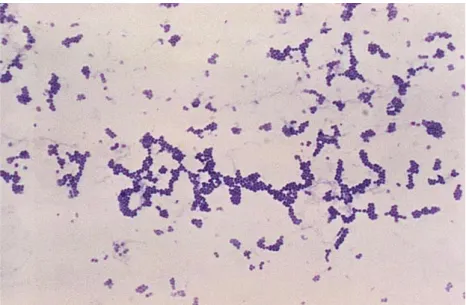
Figure 1: Microscopic observation of Staphylococcus aureus ……….10
Figure 2: Structure of Staphylococcus aureus. Source: Staphylococcus aureus infections….11 Figure 3: Sorting and surface display of cell wall-anchored proteins. ………17
Figure 4: The structure of the MSCRAMM family. ………. 18
Figure 5: The primary and tertiary structure of Protein A..……….………….19
Figure 6: The structure of the G5-E repeat family. ……….………20
Figure 7: Structure of IsdA, IsdB, IsdC and IsdH. ………...………22
Figure 8: Structure of IsdB. ……….…22
Figure 9: Structure of vitronectin.………... ………24
Figure 10.1: IsdB expression in both exponential and stationary phases using RPMI and BHI as growth mediums. ……….……….34
Figure 10.2: Expression of IsdB in RPMI and BHI. ………35
Figure 11: Expression of IsdB-mRNA in RPMI and BHI, in the two distinct phases of Staphylococcus aureus growth ……….36
Figure 12: Saturation curve of vitronectin ………...37 Figure 13: Effect of sodium chloride (NaCl) in the ligation between IsdB and vitronectin ….39
Table 1: TSM reagents and concentration in buffer ………23
Table 2: Electrophoresis buffer reagents ……… 24
Table 3: ELISA buffer reagents……… 24
Table 4: Temperatures values used in the RT-qPCR reaction………27
1. Introduction
1.1 Staphylococci: general characteristics
Staphylococci is a gender of bacteria that can live inside the men as a component of its normal flora or as an opportunistic microorganism (1). We can divide the different species of staphylococci in two main groups: coagulase positive (ex: Staphylococcus aureus) and coagulase negative (ex: Staphylococcus epidermidis) (2).
All species are known to colonize the human body, some like Staphylococcus aureus are located at the nose, pharynx, perineum, axillae and skin (3), others like Staphylococcus capitis are present in the head and regularly on adolescence (4) and finally, the skin is also a common place for species like Staphylococcus epidermidis to inhabit (5).
Scientific discoveries demonstrate that coagulase positive species (CPS), like Staphylococcus
aureus and Staphylococcus pseudintermedius are more inclined to become opportunistic
antigens and propagate illness than coagulase negative species (6). A good example of that is when we look at the more severe causes of infections by the staphylococci gender and we see S.
aureus as the primary suspect. The solemn conditions such as sepsis, pneumonia and
endocarditis are between the few that this bacterium can initiate (2). Besides that, coagulase negative species (CNS) are usually present in the normal microflora of healthy humans (7).
1.2 Staphylococcus aureus
1.2.1 - Structure and morphology
Staphylococcus aureus is a 1µm gram-positive cocci from the phylum Firmicutes that belongs
to the class Bacilli, order Bacillales and the Staphylococcaceae family (8).
When observed on a microscopic, S. aureus has a staphylococci morphology as it can be seen in Figure 1. In a macroscopic view of the colonies, they have gold coloration that is very characteristic. To have a better assurance of the identity of S. aureus, there are several tests that can be done. For instance the mannitol fermentation or the coagulase tests (which will be positive) and even some molecular tests like DNA amplification of specific regions through the polymerase chain reaction ( PCR) (9).
Figure 1: Microscopic observation of Staphylococcus aureus. The results of a Gram stain applied to
Staphylococcus aureus are shown, obtained from a microscopic view. The coloration of the bacteria
allows the identification of the Gram group of Staphylococcus aureus (Gram positive) and its morphology (staphylococci: cocci in irregular clusters). Adapted from (10).
S. aureus has a 2800bp circular genome which can encode a huge collection of virulence factors,
both cell-surface associated and secreted. These genes encode proteins responsible for: adherence to host cells, the destruction of the tissue and dissemination, iron uptake, lyse of cells, tampering with innate and adaptive immune responses and dodging antibody and complement-mediated immune responses by binding with proteins in bodily fluids.
1.2.2 - Pathogenesis
Staphylococcus aureus is known for possessing a frightful pathogenic ability to cause multiple
community and nosocomial infections (11). These gram-positive cocci are among the most significant causes of healthcare acquired infections and community-associated skin infections being considered by some authors, the most important ones (2).
Usually, S. aureus causes disease when there is an unbalance between man's defences and microbial multiplication. When the host defences are down, these bacteria produce many pathogenic factors, that help it evade the organism defences, endorse colonization, cause tissue damage and propagate for other places in the human body (12). There are two ways to cause an illness which are by invasion resulting in inflammation, or by producing enzymes or exotoxins. The toxic shock syndrome and staphylococcal scarlet fever are examples of diseases caused by toxic shock syndrome toxin-1 (TSST-1). The Staphylococcal scalded-skin syndrome (SSSS)is
triggered by exfoliative toxins, and, an additional example is that of the Staphylococcus enterotoxins (SEs), which are associated with foodborne diseases (13) (14).
Staphylococcus aureus is likewise, the etiologic agent number one of superficial lacerations,
such as skin inflammation and diabetic ulcer feet infections (15) and more severe and systemic infections like pneumonia, bacteraemia, endocarditis and osteomyelitis (13).
In the end, the consequence of the clash between the organism and this pathogen vary between life-threatening situations, an allergic reaction or a symptom-free colonization.
1.2.3 - Virulence factors
The remarkable range of virulence factors that Staphylococcus aureus has, makes the bacterium vastly adaptable, versatile and highly proficient of avoiding many host immune responses (12). These factors, demonstrated in Figure 2, include capsular polysaccharides, adhesins, toxins, immune dodging proteins and the ability to form biofilm (16,17). The focus of this work is on factors partaking in the first phases of infection, more concretely a adhesin named IsdB, instead of a produced virulence factors like TSST.-1.
Figure 2: Some examples of virulence factors of Staphylococcus aureus. CWA proteins such as protein A, collagen binding protein and fibronectin binding protein are represented as factors of virulence produced in the exponential-growth phase (A). In addition, TSST-1 and enterotoxin B are illustrated as secreted proteins and expressed in the stationary phase (A). The Figure also demonstrates the structure of S. aureus wall (B) and a representation of the domains of the clumping factor (C). Adapted from (18).
1.2.4 - Cell Wall anchored proteins
Staphylococcus aureus holds, in its cell wall, proteins that are linked covalently with
peptidoglycan, known as cell wall anchored (CWA) proteins. These proteins are known to be virulence factors, because they are vital for the development of pathogenesis(19,20). Different strains of Staphylococcus are known to express a different number of CWA proteins. S. aureus can have up to 24 different types, while coagulase-negative species like S. epidermidis and S.
lugdunensis are known to have fewer (21–23).
CWA proteins can be separated into four types of classes based on their structure and function: The Microbial Surface Component Recognizing Adhesive Matrix Molecules family (MSCRAMM), the three-helical bundle motif family, G5-E repeat family and the conserved near iron transport (NEAT) family.
Function
A typical characteristic of these proteins is that they can be multifunctional. These functions can include colonization of host tissues, because they are crucial factors in adhesion and invasion of cells and tissues, evasion of host defences and the development of biofilm (19,20,24,25). Because of these multifunctional proteins, Staphylococcus aureus can act as an opportunistic pathogen that invades the host and effectively provokes an infection. Consequently, pursuing therapeutics like vaccines that target these proteins could prevent infections by this bacteria (26). Structure
There are some elements in the structure of cell-wall anchored proteins that are common to all of them. Analysing their amino acid sequence, is noted that all of them have a N-terminal exposed on the cell surface that interacts with the host and a C-terminal that interacts with the peptidoglycan coat (24,27). In the NH2-terminal there is a secretory signal sequence (Sec signal)
responsible for directing, the produced protein to the cellular machinery located at the membrane for secretion. During the secretion process, this signal is cleaved, having served its propose (19,20). Regarding the C-terminus, this is featured by a cell wall sorting sequence consisting of a LPXTG (leucine-proline-AA-threonine-Glycine) typical motif, a hydrophobic domain and residues with a positive charge. All of these elements constitute a sorting signal, that helps the protein to bond itself to the peptidoglycan layer (19,20,25).
As it was said before, the Sec signal at the N-terminal leads the immature CWA protein to the cell Sec apparatus, where is cleaved by SpsI (signal peptidase I). Then, the presence or absence of the signal peptide motif YSIRK-G/S will determinate weather the protein is guided to their secretion and anchorage near the place where the synthesis of peptidoglycan is happening, or, if the proteins will go to the poles of the cell (cases where the motif is absent) (19,28,29). Sorting
The deposition of CWA proteins in the cell is a very complex procedure because it depends on numerous factors like cell division, morphogenesis and gene expression. This last one, specifically, is also dependent on other variants like the bacteria phase growth (exponential or stationary) or if the bacteria is grown in the presence or absence of iron (25,30–33).
In the C-terminus, the different elements (LPXTG motif, hydrophobic segment, and the positive charge residues) of the sorting signal are responsible for different tasks (Figure 3). The hydrophobic transmembrane segment holds the CWA protein inside the membrane (19), the positive charge residues blocks the secretion of the chain into the extracellular medium (34) and, in the end the LPXTG motif serves as the substrate for sortase A. This transpeptidase (sortase A) cleaves the ligation between the threonine and glycine present in the LPXTG and secures the C-terminal of the peptide employing the thiol present in the cysteine that composes its active site (35). This acyl intermediate is attacked by the amino group of pentaglycine of lipid II (36), and then this newly created complex is integrated into the cell wall by the means of the penicillin-binding proteins (25).
Figure 3: Sorting and surface display of cell wall-anchored proteins. The Figure demonstrates the sorting steps that CWA proteins suffer in order to attached themselves to the cell wall. The hydrophobic domain (blue cylinder) and the positive charge (signal +) hold the protein in place whereas the LPXTG motif, which is represented by the letters LPXTG, suffers degradation by sortase. Then, lipid II bounds itself to the acyl-enzyme intermediate, forming a lipid II-linked intermediate, which will then originate a CWA surface protein. Adapted from (19).
In conclusion, the three components of the sorting signal must be present for the anchoring of the proteins to the cell wall to be successful: the absence of the charged positive residues means that the protein gets released into the culture medium as an alternative to becoming attached, and the reducing hydrophobic domain results in less protein anchored (19).
1.2.4.1 - MSCRAMM family
The MSCRAMM proteins are known to bound to elements of the extracellular matrix or blood plasma, like collagen, fibrinogen (Fbg) and fibronectin (Fn) (34,35). Initially, the acronym MSCRAMM was used for any surface protein that intervened in the ligation between S. aureus and the elements mention above (35). Nevertheless, it was soon discovered that there are other families of surface proteins and that several MSCRAMMs have more distinct functions than adhesion to host cells (17,18). In 2015, Foster et al. (19) advised the use of the acronym only to
proteins that own two domains IgG-like folded and that are functionally and structurally alike. These Ig-G domains are located at the N terminal represented as the A region in Figure
4. This N terminus is divided into three subdomains: N1, N2 and N3. The Ig-G domains are only present in the subdomains N2 and N3.
Figure 4: The structure of the MSCRAMM family. ClfA, ClfB, FnBPA, FnBPB and CNA have similar amino acid sequences such as a signal sequence (S) and a N-terminal ligand binding (A) composed by three domains (N1, N2, N3). In addition to that, the C-terminus of the proteins are formed with a cellwall-binding region (W), a membrane-spanning segment (M) and the C-terminal tail (C). For each protein, the positions of LPXTG motif are indicated. Adapted from (37).
Besides structure, it is important to mention that there are two known ways for these proteins to bind to the host cells: The Duck, Lock and Latch mechanism (18) and collagen hug.
Based on Foster et al. (17), we can consider being part of this family of CWA proteins, the fibronectin-binding proteins (FnBPA and FnBPB), the clumping factor (ClfA and ClfB), the collagen-binding protein (CNA), the bone sialoprotein-binding protein (Bbp) and the serineaspartate repeat protein (SdrS, SdrD, SdrR).
Duck, Lock and Latch mechanism
Almost all MSCRAMMS are known to form connections to elements of the extracellular matrix or blood plasma, using this Duck, Lock and Latch method, that is based on the N2 and N3
domains. These domains include two β-sheets organized into an IgG fold, and between them exists a hydrophobic channel that can hold ligands. A good example is the case of ClfA, FnBPA and FnBPB, that use this hydrophobic pocket to get attached to the C-terminal of the γ-chain of fibrinogen. In a second step, there is a conformational change of N3, that makes this domain able to surround the peptide to lock it in place, and then creating a new β-sheet of N2 described as the latch (17,36). The ClfB also uses this method to get attached to the α-chain of fibrinogen, cytokeratin 10 and loricrin (17).
Collagen Hug mechanism
The collagen-binding protein (CNA) uses N1 and N2 domains instead of N2 and N3. Between N1 and N2 is essential the presence of an extensive linker region, that allows the adjustment needed for binding to collagen, which has a very dense structure (triple-helical array of three separate polypeptide chains). In addition to that essential factor, there is also the stabilization given by the linker and its interaction with the β-sheet in N1 (37).
1.2.4.2 - The Three-helical bundle motif family (Protein A)
This family is known for the formation of three or more helical bundle motifs. As it can be seen in Figure 5, this family is characterized by a signal sequence (S), five IgG-binding domains (E,A,B,C,D) and a proline-rich C-terminal, represented by Xr (38). The three-helical bundle motif family is represented by one member which is protein A.
Figure 5: The primary and tertiary structure of Protein A. Protein A contains the signal sequence (S) and the sorting signal (SS) that is characteristic to all CWA proteins. The five N-terminal triple-helical bundle domains (EABCD) that bind to IgG and other ligands are followed by the repeat-containing Xr region and the non-repetitive Xc region. Adapted from (19).
Protein A (SpA) is produced by gene spa and has a mass of ± 40kDa (molecular weight=42 000 g/mol) (39,40). It is organized into four or five immunoglobulin binding domains with 56-61 residues each, a variable repeat region Xr, and a conserved one named Xc, responsible for holding the attachment sequence (41). One domain of SpA is arranged in a three-helical bundle of antiparallel alpha-helices (42,43).
This CWA protein can be produced by almost all of Staphylococcus aureus strains and is known for binding to Fc and F(ab)2 regions of immunoglobulins (Igs). By doing so, it can prevent the
action of vaccines, because it can connect with B-cells and trigger the apoptosis of this cells (40,44,45). Protein A can also monopolize the antibody response, restricting the host response to other virulence factors that was necessary for its protection against Staphylococcus aureus (45,46).
1.2.4.3 - G5-E repeat family
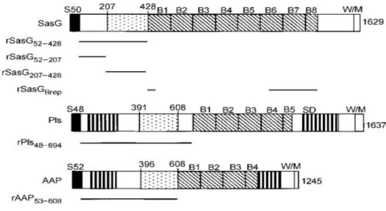
The name G5-E comes from the present of “G5” domains in the B multidomain region. These G5 are approximately 78 residues arranged in a tandem array, followed by 50 residues spacer named “E” (47–49). The letter G comes from the five preserved glycine residues that appear on the fold (47). In relation to the C-terminal this is poorly formed by faulty B repeats and cell wall attachment structures, whereas the N-terminal (region A) consist of two subdomains, one with 157 residues and a second one with 212 residues (50,51). The G5-E repeat family has two members: S. aureus surface protein G (SasG) and plasmin sensitive (Pls). Both of them have a domain A, having a role in attachment to the epithelial cells and a B repeat domain involved in the intercellular ligations using a mechanism of homodimerization dependent on zinc (49). Figure 6 shows the structure of these two proteins together with the accumulation associated protein (Aap) that has a very similar structure and is present in Staphylococcus epidermidis (51,52).
Figure 6: The structure of the G5-E repeat family. Primary structures of SasG and Pls of S. aureus and the AAP of S. epidermidis are shown. The proteins have similar segments such as the signal sequences (S), the B repeats (B), the serine–aspartate (SD) repeats and the wall/membrane spanning regions
(W/M). The dotted box between the S and the B regions represents the conserved domain in the
Nterminal region (also referred to as A domain). The SD dipeptide short repeats of the Pls and AAP are identified as a vertical bar. Adapted from (50).
Knowledge on Pls is lower than the SasG. Pls is a 230kDa protein, very susceptible to plasmin and that is usually found in methicillin-resistant Staphylococcus aureus (MRSA) (53,54). SasG has a very similar structure when compared with the others CWA anchored proteins. The signal sequence and the repetitive domain B are all present in its composition (51). This protein uses the domain A and B to link to the nasal epithelium and to other bacteria, creating a biofilm, respectively (52). Another major factor of this protein is its ability to form β-sheet structures which allows observation in an electron microscopy (48,52). One crucial aspect is that the minimum requirement for biofilm formation is the presence of five B domains. In the first step, the A domain is cut by an unknow proteinase (51), and then the exposed B structures of adjacent cells interact with each other, in a way that is Zn 2+ dependent, resulting in cell aggregation, combined with biofilm creation (47,51,56). Some SasG proteins with more than four B repeats have a very special feature of blocking other CWA proteins, such as ClfB with fibrinogen (49,52,54).
1.2.4.4 - NEAT family
The presence of iron is essential for a pathogen, because it has a very important role in its growth and cell activity. Indeed, one of the host innate responses is restricting the availability of iron (57–59).
Inside the human host, iron is distributed in heme groups which, are part of hemoglobin and myoglobin and also of proteins like transferrin and lactoferrin (57). About 75% of the human iron is placed in hemoglobin (Hb) so, pathogens like, Staphylococcus aureus, have acquired ways to break up and isolate the group heme from hemoglobin (58,60). One of these ways is producing toxic hemolysins which lyse erythrocytes and release Hb (61,62).
Another important factor that is challenging is the size of S. aureus cell wall (20-80nm) (58). So, in order to gain access to heme, Staphylococcus aureus have developed the iron-responsive surface determinant (Isd) system to transport it through the peptidoglycan and into the cytoplasm (57,58,62).
The production of these proteins is regulated by the global ferric uptake repressor, Fur which is known to repress the translation of the Isd proteins when the concentration of iron in the environment is high (31,63,64).
Horsburgh et al., in 2000 (63), examined the S. aureus genome and found genes with a high similarity to iron-regulated proteins from other bacteria. Before these genes there were operons that other scientists like, Xiong et al., (65) and Heinrichs et al., (17) had previously identified as ferrochrome-uptake operon (fhu) and sirA (the gene that produces the SirA an Iron-regulated ABC transporter siderophore-binding protein), respectively. While these two authors proved the homology of these two sequences for the Fur box, Horsburgh et al. (63) was able to continue their studies and proved that both were repressed by Fur when Staphylococcus aureus was grown in iron rich media. Considering these findings, this author concluded that the only factor that regulates S. aureus iron uptake was Fur (17,63,65,66).
Nevertheless, Fur is not exclusive of Staphylococcus aureus, other bacteria like Helicobacter
pylori and Yersinia pestis have also their iron uptake regulated by this repressor when grown in
iron-rich mediums (67,68).
The isd locus, which is controlled by Fur, encodes nine proteins: IsdA and IsdC (heme), IsdB (hemoglobin) and IsdH (hemoglobin-haptoglobin) binding proteins; IsdD-IsdE-IsdF which act as a membrane transport and IsdG and IsdI which are monooxygenases (57).
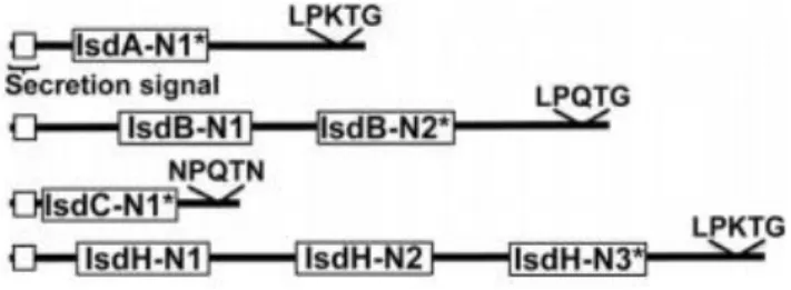
As we can see in Figure 7, all the Isd binding proteins (Isd A, IsdB, IsdC and IsdH) have one, two or three NEAT (conserved near iron transport) domains (62,66).
Figure 7: Structure of IsdA, IsdB, IsdC and IsdH. The white box at the N-terminus indicates the secretion signal of each protein. NEAT domains are represented in each of these protein as IsdX-Ny, where X is the name of the protein (A, B, C or H) and y the order of the NEAT domain started from the N-terminus region. The * indicates the heme binding NEAT domain of each protein. Adapted from (62). The NEAT domain has ± 120 amino acid residues and is notorious for its eight-stranded βsandwich fold. In this particular conformation, it possess a hydrophobic pocket with a tyrosine that allows the ligation to heme (62). Generally, methemoglobin (metHb) bounds to IsdB-NEAT2 or IsdH-NEAT3 (69,70). In a second step, heme is moved across the
peptidoglycan wall by IsdA-NEAT and IsdC-NEAT to the membrane transporter IsdEF. Then, after internalization, iron is released from heme by the action of IsdG or IsdI (hemedegrading enzymes) (62,71).
1.2.4.4.1 - IsdB
IsdB (±72kDa) has five very important segments (Figure 8): NEAT1 (IsdBN1), with 145-270 residues; NEAT2 (IsdBN2), composed by 338-458 residues; a linker region that separates these two domains and the usual N (40-144 residues) and C (459-613 residues) terminal region (72):
Figure 8: Structure of IsdB. The black bar represents the signal sequence, whereas the white bar is the LPQTG sortase signal. IsdB-N1 and IsdB-N2 are shown in two different shades of grey. Adapted from (73).
The difference between IsdBN1 and IsdBN2 is that the first one is known for binding hemoglobin, but it cannot connect with heme (or Haem) whereas the second one binds with both. So it can be concluded that IsdBN2 is the only domain responsible for the extraction of heme from Hb and also that it is the one responsible for the transportation of heme to IsdA-N1 and IsdC-N1 (73,74). Because IsdB is known to allocate heme to IsdA at a rate 8 times bigger than to IsdC, it can be presumed that the pathway of heme is IsdB-N2-IsdA-N1-IsdC-N1 (31,73,75).
Some studies have proven that by deleting the isdB gene, but not the isdH, there is a decrease in the hemoglobin-staphylococcus aureus binding, and as a consequence, the iron-uptake by the bacteria is hindered (76). In this work, the authors show a decrease in the virulence of
Staphylococcus aureus after a mutation in the isdB gene (72,76). In short, we can assume that
IsdB is the main Hb-binding protein for iron uptake in Staphylococcus aureus, and thus is very important to further study this protein (67).
1.3 Vitronectin
Vitronectin is a 75kDa protein also named as S protein (serum spreading factor) or epibolin present in plasma at a concentration of 200±400 mg/ml. This glycoprotein is present in blood and in the extracellular matrix, and is known for binding to collagen, plasminogen, glycosaminoglycans, urokinase-receptor, plasminogen activation inhibitor-1, complement, heparin and thrombi-antithrombin III complexes (77).
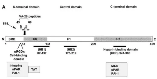
In terms of structure (Figure 9), vitronectin has an amino terminal composed by 44 amino acids responsible for binding to the plasminogen activator inhibitor-1 (77,78). Next to this domain, there is an RGD sequence that englobes three amino acids: arginine, glycine and aspartic acid. This structure has the function of supervising the connection of vitronectin to the extracellular matrix, through the specialized integrin receptor (77,79). Nearby the RGD domain, this glycoprotein has a short stretch of amino acids, 53 to 64, that holds two sulphated tyrosine residues (56 and 59 – Figure 9 represented as Y56S/Y59S) and has two very important functions. One of these purposes is forming the ligation between vitronectin and thrombinantithrombin III complex, and the other is defusing the charge present at the carboxyl terminal by the cationic domain, 348-379. This neutralization between the cationic 348 to 379 amino acids and the anionic 53 to 64 domain is implicated in the stabilization of the molecule (77).
Figure 9: Structure of vitronectin. Schematic structure of vitronectin demonstrating the positions of the cell-binding motif RGD, the sulphated tyrosine residues (Y56S/Y59S) and the high affinity heparinbinding domain (HBD) are shown. The grey region (CR) connects the N-terminal and the central domains and includes a stretch of acidic residues (53–64) within which, consequently, contain the Y56S and Y59S. The C-terminal domain residues 348–376 contain the main heparin-binding domain (HBD) and are composed of several highly charged residues. Adapted from (80).
Most of vitronectin comprises a region (132 to 459) that contains six hemopexin repeats and a disulphide bridge (274 to 453). The binding to plasminogen occurs at the carboxyl terminal, between amino acids 332 to 348 (81). After this domain, there is a group of basic amino acids
(348-376) that contribute for heparin, glycosaminoglycan and plasminogen activator inhibitor1 binding (82).
Inside the blood vessels, we can find vitronectin in two types: as a single chain or into two chains held together by the disulphide bridge mentioned above. However, this same protein is observed in plasma as a very stabilized folded monomer, because of the interaction concerning the cationic and anionic amino acids asserted before (77).
Vitronectin has several important roles in cell adhesion and proteolysis. It is known that this protein can interact with integrins (ex: avb3 or avb5) and induce a diversity of signalling pathways responsible for gene expression, intracellular iron transport, lipid metabolism and cytoskeletal reformation. One example, of a pathway that these integrins receptors can induce is the MAP kinase (mitogen-activated protein). In addition to that, vitronectin is also acknowledged for its inhibition of the cytolytic process, that comes from blocking the action of the complement system and the perforin. Furthermore, one of its vital roles in the human organism is being part of both the development and the destruction of coagulum. This ambiguous function comes from the fact that vitronectin can either prevent the action of antithrombin III and, thus, preserving thrombin, or, it can destroy the blood clots by activating the tissue-type plasminogen activator, which in turn converts plasminogen to plasmin, that consecutively destroys fibrin clots. Another way, which vitronectin can control plasminogen is when it binds to plasminogen activator inhibitor-1 and stops the production of plasmin (77). Furthermore, this glycoprotein can connect to the urokinase receptors to the extracellular matrix, allowing them to activate the urokinase mediated proteolysis, therefore, permitting damage to occur to the basement membrane and, as such, enabling the metastasis of tumours (83).
To conclude, some gram-positive pathogens such as Streptococcus pneumoniae and
Streptococcus pyogenes use vitronectin in order to bound with human cells and therefore, infect
the host (84).
2. Aim of the work
USA statistics analyses reveal that between the years of 2001 and 2009, more than 12 000 and less than 23 000 dollars have been spent, per year, in hospitalizations caused by Staphylococcus
aureus (85,86). This coupled with the fact that some types of this bacteria have acquired
resistance to traditional antibiotics such as, methicillin and vancomycin (named MRSA and VRSA) expose a great need to try to understand the ways S. aureus interacts with human organism. Therefore, the study of virulence factors becomes extremely important to make advancements in the creations of vaccines and others prophylactic medicines that focus on preventing future infections by Staphylococcus aureus (87).
Although a lot of research has been done on some cell wall anchored proteins referred before like fibronectin, protein A, clumping factor and fibrinogen, there are not a lot of studies about the neat domain. As a result, the aim of this work was to show, that besides the normal function of binding to hemoglobin, IsdB is a protein that also binds to vitronectin inside the human host (62,73).
There were four main goals. The first one was to confirm that this protein is manly expressed in iron-poor mediums. In addition to that, some experiments were done, in order to see in which phase of the bacterial growth, IsdB presented a higher expression (stationary or exponential). The third goal of this thesis was to confirm the existence of an interaction between IsdB and vitronectin, and, subsequently study the hypothesis of this interaction being dependent on the presence of NaCl.
3. Materials and Methods
3.1 Bacterial strains and culture conditions
The strain of Staphylococcus aureus used was the SH1000 Δspa, provided by Prof. Timothy J. Foster and Prof. Joan A. Geoghegan of the Microbiology Department, Moyne Institute of Preventive Medicine, Trinity College, Dublin, Ireland. This 8325-4 derived strain has a functional rsbU, is sigB positive (88) and a spa mutant (89).
One Erlenmeyer flask with the bacterium strain were grown at 37ºC with constant shaking (150 rpm) using a brain heart infusion (BHI) medium. The process was done overnight to reach the stationary phase in an incubator shaker (Scientific Co Classic C24KC). In the next day,
staphylococcal cells (from stationary phase) were obtained from the culture by a 10 minutes centrifugation step (4 000 rpm), and then they were washed with PBS (Phosphate Buffered Saline) and resuspended at the appropriate density (OD600nm =0.7; UV-Vis Spectrophotometer
used: JASCO V-630). In order to obtain the exponential phase, the other half of the bacterial culture was diluted (1:40) in a fresh batch of BHI medium and incubated at 37ºC with shaking until the OD600nm=0.7 was achieved.
3.2 Proteins
a) Vitronectin
Human vitronectin (Vn) was purified from human plasma using the method of HeparinSepharose affinity chromatography by Steven K. Akiyama and prepared at a concentration of
1.8mg/ml (90).
b) BSA
The Bovine Serum Albumin (BSA) was obtained from Sigma (St Louis, MO) and prepared at a concentration of 2%.
3.3 Antibodies
The polyclonal antibodies used in this study were all available at Professor Pietrocola’s laboratory:
1) Anti-IsdB rabbit: Was raised in a rabbit using immunisation procedures and purified by affinity chromatography using protein G-Sepharose columns.
2) Sheep anti-human vitronectin antibody: Affinity biologicals INC. (Ancaster, Canada) 3) Anti-rabbit and anti-sheep Horse Radish Peroxidase (HRP)-conjugated secondary
antibodies: Dako Cytomation (Glostrup, Denmark).
3.4 Media and buffers
After adding and mixing the following reagents, the pH was adjusted to 7.3: Table
1: TSM reagents and concentration in buffer
Reagents Concentration in the buffer
Sucrose 0.5M
Tris-HCL [pH 7.5] 1mM
MgCl2 10 mM
Tris-Glycine-SDS electrophoresis buffer
This buffer was prepared at a 10X concentration:
Table 2: Electrophoresis buffer reagents
Reagents Concentration in buffer
Tris 30g/l
Glycine 140.4g/l
Sodium-dodecyl-sulfate 10g/l
The pH was adjusted to 8.3 and the buffer was filtrated using a pleated filter.
Enzyme Linked Immuno Sorbent Assay (ELISA) buffer Table 3: ELISA buffer reagents
Reagents Concentration in buffer
NaCl 5M 0.3M
Tween 20 0.04M
3.5 SDS-PAGE (Sodium Dodecyl Sulphate Poly Acrylamide Gel
Electrophoresis) and Sample preparation
Overnight cultures of S. aureus SH1000 Δspa strains either in RPMI or BHI medium (VWR International Srl) were diluted (1:40) in fresh medium and grown at 37°C to obtain the exponential phase (OD600 about 0.7). Then, cells of both phases (stationary and exponential)
were harvested by centrifugation, the supernatant was recovered, and the pellets were resuspended in phosphate buffer saline (PBS) to obtain an OD600=1. In a second step, the
samples were harvested by centrifugation and washed with TSM. The pellets were then resuspended in TSM containing 0.1mg/ml of Lysostaphin and incubated at 37°C for 20 minutes. After incubation, they were harvested by centrifugation and proteins in supernatant were precipitated by incubation with 7.5% trichloroacetic acid (TCA) (v/v) on ice for 20 minutes. In parallel, the culture supernatant recovered from each sample was also subjected to protein precipitation with 7.5% TCA (v/v) on ice for 20 min. The precipitates were collected by centrifugation, washed twice with ice-cold acetone and then let air-dry overnight. The next day, the pellets were resuspended in loading buffer, subjected to 10% SDS-PAGE in reducing conditions and proteins were electro-transferred to a nitrocellulose membrane.
Because we want to reveal IsdB with a predicted molecular mass of 72kDa , a 12.5% running gel (12.5% acrylamide; 25% 0.5M Tris-HCL pH 6.8; 0.08% ammonium persulfate; 0.08% TEMED (tetramethyl ethylenediamine) and a 4.8% staking gel (4.8% acrylamide; 16% 0.5M Tris-HCL pH 6.8; 0.13% ammonium persulfate; 0.13% TEMED) were prepared. The gel was left to polymerized for about 20-30 minutes, and then placed in a vertical migration
electrophoresis tank (material used: Mini protean IITM, from Bio-Rad and wide Mini-Sub Cell GT electrophoresis cell with PowerPac Basic power supply) with the running buffer (20mM Tris-HCL pH 8.3; 0.2M glycine; 0.1% SDS).
Before loading the samples on the gel, an equal volume of SDS-PAGE loading buffer (0.1% (v/v) bromophenol blue; 20% (v/v) glycerol, 2% SDS in 50 mM Tris-HCL pH 6.8) was added and samples were boiled for 5 minutes. The molecular weight standard Precision Plus Protein Standard, from BioRad, Hercules, CA was also loaded in the gel.
Initially, the gel ran at 80V voltage in order to give time for the proteins to reach the separating gel, subsequently there was an increased in the voltage to 150V. The gel was then transferred on a Nitrocellulose blotting membrane (0.45μm NC) (GE Healthcare) for western blotting.
In order to transfer the gel to a nitrocellulose blotting membrane, the membrane had to be wet with water and left for 15 minutes in a blot buffer (0,4M glycine, 40mM Tris-HCL, 20% methanol, pH 8.3). Then, it was assembled in a sandwich, where the gel and the membrane were surrounded by two sheets of Whatman® blotting paper and sponge on each side. When inserted in the vertical blot tank, the membrane was positioned at the positive pole while the gel was situated at the negative charge. Proteins were transferred to the membrane at 100 V for 1 hour.
To block the membrane, it was submerged in 5% (w/v) skim milk (Sigma) in PBST (0.1%Tween 20 in PBS) at 4ºC, overnight.
The membrane was incubated with the first antibody (polyclonal rabbit against IsdB (1:5000 in 1% skim milk) for 1h, at 22ºC. After a few washes with PBST, the membrane was incubated with a secondary antibody (HRP-conjugated α-rabbit (1:10000 in milk 1%)) for 1h.
In the end the ECL Advance Western blotting detection kit (GE Healthcare) and an ImageQant TM LAS 4000 mini-biomolecular imager (GE Healthcare) were used for development and detection of the bands obtained.
3.7 Quantitative Reverse Transcriptase Polymerase Chain Reaction
(RT-qPCR)
Cells from S. aureus SH1000 Δspa strains in both BHI and RPMI medium were grown until a OD600=0.7. After growth, it was added an RNAprotect Bacteria Reagent (Qiagen) in a
proportion of 1 (volume of cells) to 2 (volume of buffer), followed by an incubation for 5 mins at 25ºC and a centrifugation (10 min at 5000 x g). The RNA extraction was done by means of the Quick-RNA Fungal/Bacterial Miniprep Kit (cat. no. R2014, Zymo Research) protocol. In order to eliminate any DNA contamination, the samples went through a DNase treatment using the TURBO DNA-free kit (cat.no.AM1907, Invitrogen) after the extraction.
In a third step, the RNA samples were subjected to an RT-qPCR reaction using the iTaq Universal SYR Green One-step Kit (cat no.172-5150, Bio-Rad). The following table shows the temperatures used in each cycle of the procedure.
Table 4: RT-qPCR reaction conditions
Reverse Amplification Melt-Curve Analysis
Transcription Reaction Polymerase Activation and DNA denaturation Denaturation at 95°C Annealing/ Extension + Plate Read at 60°C Cycles 10 min at 50°C 1 min at 95°C 10 s 10-30 s 35-40 65-95°C 0.5°C increment 2-5 s/step
3.8 Enzyme Linked Immuno Sorbent Assay (ELISA)
With the purpose of characterizing the interaction between IsdB and human Vitronectin, two ELISA assays were made.
a) Saturation kinetics of the binding between IsdB and human Vitronectin
IsdB (200μg/well) was immobilized on wells of a microplate (11 wells) and incubated overnight at 4ºC. The wells were then washed three times with 0.1% (v/v) Tween 20 in PBS (PBST) and incubated with bovine serum albumin (BSA, 2% v/v) in PBS (200µl/well) for 1 h at room temperature in order to block unspecific binding sites. Increasing concentrations (from 0 to 10mg/ml) of human Vitronectin were then added in 100μL PBS and incubated 1 hour at room temperature. After washing three times with PBST, a polyclonal sheep a-Vn antibody (10mg/ml in BSA 1%) was added to the microtiter plate and incubated for 1 hour at room temperature. Following incubation, the plate was again washed three times with PBST and incubated with HRP-conjugated α-sheep antibody (1:1000 in BSA 1%) for 45 minutes at room temperature. Vitronectin interaction with IsdB was detected by incubation of the wells with orthophenylenediamine dihydrochloride (OPD Dako Cytomation). The absorbance at 490nm was read with a microplate reader (Microplate Reader, model 680, BioRad, Richmond, CA).
a) Inhibitory effect of sodium chloride on IsdB-Vn interaction
Microtiter wells were coated with 2μg/ml of IsdB in PBS and incubated at 4°C overnight. The wells were then incubated with BSA 2% (v/v) in PBS (200μl/well) for 1 hour at room temperature. Increasing concentrations (from 0M to 1M) of NaCl together with a fixed concentration of the Vn (250ng/well in PBS) were then added to the wells and incubated for 1
hour at room temperature. After washing, a polyclonal sheep a-Vn antibody (5mg/ml in BSA 1%) was added and incubated for 1 hour at room temperature. Bound protein was revealed by using an HRP-conjugated α-sheep secondary antibody (1:1000 in BSA 1%) for 45 minutes at room temperature. The absorbance at 490nm was read with a microplate reader.
4. Results and Discussion
4.1 Western blot analysis of IsdB expression
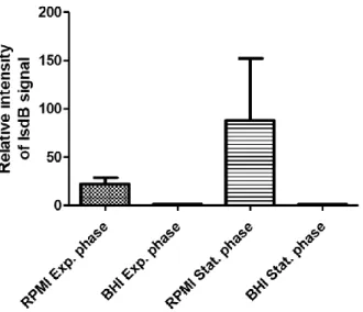
In order to analyse the effect of iron in the expression of IsdB, S. aureus SH1000 Δspa was grown in an iron-rich medium (BHI) and iron-poor medium (RPMI). The production of the protein was analysed by Western blot using an anti-IsdB antibody, after an SDS-PAGE to separate the protein by size. As shown in Figure 10.1 a protein with ±70kDa, the predicted molecular weight of IsdB, could be detected with the anti-IsdB antibody.
The results show that the expression of IsdB is not equal in all tested conditions. The relative intensity of the IsdB signal in the different mediums and in the distinct phases of Staphylococcus
aureus growth is represented in Figure 10.2. As can be observed, a higher expression of IsdB
occurs in cells grown in RPMI poor) (panels A and C), while in the BHI medium (iron-rich) the expression of IsdB is almost non-existent. (panels B and D).
Figure 10.1: IsdB expression in exponential and stationary phase cells grown in RPMI or BHI. IsdB proteins were separated in a 12,5% SDS-PAGE, transferred to a nitrocellulose membrane and revelled using a polyclonal rabbit against IsdB as a first antibody and an HRP-conjugated α-rabbit as the second one. A) RPMI exponential phase B) BHI exponential phase C) RPMI stationary phase D) BHI stationary phase.
Figure 10.2: Relative intensity of IsdB expression in RPMI and BHI mediums. The data was revealed by an ImageQant TM LAS 4000 mini-biomolecular imager from GE Healthcare. The results are an average of two independent experiments.
These results confirm that Staphylococcus aureus only produces IsdB in poor-iron mediums, which is in agreement with the results obtained by Hempel et al. (57) and Horsburgh et al. (63). In both experiments (N=2) the expression of IsdB in the RPMI medium and in the stationary phase was higher than the one obtained in the exponential phase for the same medium. However, the ratio between stationary phase and exponential is 5.3 in one experiment (Figure 10.1) and 1.5 in the second experiment, resulting in a very high standard deviation between these two values (Figure 10.2).
The possible reason for a lower signal (RPMI stationary phase) in the second experiment might be due to a low incubation time with the antibodies, non-proper transference to the nitrocellulose membrane (ex: the transfer time was cut short or the sandwich was assembled incorrectly), or over-dilution of the antibodies. More repetitions of this experiment should be performed in order to draw more accurate conclusions about the expression of IsdB in RPMI in stationary phase.
4.2 Analysis of IsdB-mRNA expression by RT-qPCR
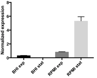
The previous results suggest that IsdB has a higher expression in the stationary phase. To clarify this a quantification of the correspondent mRNA was performed using the same cells growth conditions. In order to measure the levels of mRNA in the different situations (BHI-exponential phase, BHI-stationary phase, RPMI-exponential phase, BHI-stationary phase) an RT-qPCR experiment was performed. The data was normalized using a reference gene, rpoC (encodes for DNA-directed RNA polymerase subunit) that presents a constant expression in the conditions referred.
As observed in Figure 11 transcription of the IsdB gene is also increased in cells grown in RPMI compared to cells grown in BHI media. The graphic shows that in iron-poor conditions (RPMI) the expression of mRNA-IsdB in stationary phase is 15 times higher than the exponential. However, in the cells grown in iron-rich conditions (BHI) the opposite happens, there is an expression of mRNA-IsdB higher in the exponential phase. Some mistakes might have happened during the RT-qPCR of the BHI culture that could explain these results (eg: DNA impurities). In the end, these results of the BHI media aren´t relevant because IsdB is a protein manly expressed in RPMI conditions.
Figure 11: Normalized expression of IsdB-mRNA in RPMI and BHI, in the two phases of Staphylococcus
aureus growth. The data was revealed by Roche LightCycler LC480 and normalized with the rpoc gene, that presents invariable expression in the conditions used (BHI exponential phase; BHI stationary phase; RPMI exponential phase and RPMI stationary phase. The results are an average of three independent experiments. 0 2 4 6 8
So, taking these findings into account, the data referred above contributes to a clear understanding that the transcription of isdb in RPMI medium is induced at the stationary phase. Quantification of mRNA was performed with the use of RT-qPCR method due to its higher sensitivity when compared to other methods such as the Northern Blot or RPA (ribonuclease protection assay).
4.3 Saturation kinetic curve of Vitronectin
The main goal of this thesis was to identify a new interaction between IsdB of Staphylococcus
aureus and human vitronectin. For this an ELISA experiment was performed as described in
methods. IsdB protein (200μg/well) was immobilized in the wells of a microplate and after a series of washing steps, increasing concentrations of vitronectin ranging from 143nM to 0.14nM w added to different wells, in order to obtain a saturation curve. The results obtained (Figure 12), show the correlation between the absorbance obtained at 490nm of the connection IsdB-Vn and the different concentrations of Vn added in each well. Using the software GraphPad prism it was obtained a Kd value of 9.604±0.2668nM and a saturation concentration of Vn of 250ng/well.
Vn added (nM)
Figure 12: Saturation curve of vitronectin. IsdB proteins were immobilized on an ELISA plate at a concentration of 200µg/well. To detect the signal, an OPD (o-phenylenediamine dihydrochloride) was added, and then the absorbance was measured at 490 nm with the Microplate Reader (model 680) from BioRad. The Kd value obtained was 9.604±0.2668nM and the saturation concentration of Vn was 250ng/well (calculated using the software GraphPad prism). The results are an average of two independent experiments.
Fuquay et al. (92) (1986) was the first investigator to perform studies about the interaction between Staphylococcus aureus and protein S, also known as vitronectin (89). In 1993, Liang
et al. (93) found that 0.32μg of Vn bound to 109 Staphylococcus aureus at the saturation stage,
and that the dissociation constant of Vn to this pathogen was 7.4×10⁻¹⁰M (0.7nM±0.08nM). No studies about the connection of IsdB of S. aureus to vitronectin have been performed so far. However, because it was obtained a value of 9.6nM as the dissociation constant of this connection, it can be assumed that there are more CWA proteins responsible for the interaction between S. aureus and Vn, that has a Kd 13 times lower than the Kd obtained. To concluded, the connection S. aureus and Vn is 13 times stronger than the one between IsdB and Vn. In this experiment, the software that we used (GraphPad prism) was also able to calculate the saturation level of Vn (250ng/well). This result is extremely important, because it provides information about how much of vitronectin do we have to put in the wells to have a saturation of the receptors of IsdB (200μg/well).
Even though the results confirm the attachment in vitro, additional studies in vivo should be performed to support this evidence.
4.4 Inhibitory effect of sodium chloride on IsdB-Vn interaction
A lot of studies have shown the tolerance of Staphylococcus aureus to high concentrations of NaCl. Most of these studies are performed in iron-rich mediums (ex: BHI) and all of them show that increasing concentrations of NaCl (until 10%) reduce the growth of Staphylococcus aureus in an inversely proportional manner (94,95). With the purpose of discovering if the interaction of IsdB with vitronectin in iron-pour mediums (RPMI) was also compromised by the concentration of sodium chloride and in the same manner that occurs in BHI mediums, we performed an ELISA (Enzyme Linked Immuno Sorbent Assay). For this, IsdB was immobilized on the wells (8 wells) of a microplate and then we added a fix concentration of Vitronectin (250ng/well) with increasing quantities of NaCl. The [Vn] used (250ng/well) was chosen based on previous work that reported this as the amount necessary to cause saturation of the IsdB receptors (93). In the studies mentioned above, is shown that at a concentration of 10% (±1.7M) or higher, NaCl has a highly detrimental effect on the growth of Staphylococcus aureus (95).
Based on these studies, we decided to use an interval of NaCl concentrations between 1 and 0,0156M. In Figure 13, is represented the data analysis of the values obtained from this ELISA test. The percentages were calculated by dividing the absorbance obtained in each well with the absorbance achieved without NaCl (only the interaction of IsdB-Vn). As expected, the percentage of vitronectin binding to IsdB is inversely proportional to the concentration of salt in the wells.
NaCl concentration (M)
Figure 13: Effect of sodium chloride (NaCl) in IsdB and vitronectin binding. 200μg/well of IsdB were immobilized in the wells of a microplate at 4ºC overnight. The absorbance of Vn was read at 490 nm, with a positive control without NaCl. The percentages were calculated by dividing the signal obtained in each well per the signal given by the positive control and multiply by 100. The results are an average of three independent experiments.
Nevertheless, the two lower points of the curve present a high standard deviation, which means that some technical errors may have occurred. Some factors that could also explain an inconsistent results of ELISA repetitions are, inadequate washing steps or unpredictable fluctuations of room temperature.
5. Conclusion
The diversity of cell wall anchored proteins presents in Staphylococcus aureus, which are important virulence factors, allows this microorganism to survive the most hostile environments and sidestepping the defences of the human body. This thesis aimed to identify a new interaction between the protein of Staphylococcus aureus and vitronectin.
Based on the results of IsdB expression in different mediums, it can be concluded that this bacterium induces its expression in iron-pour conditions. In addition, our results indicate that IsdB is only transcribed in an after-growth stage. Coupled with the fact that the majority of antibiotics focus only on growing cells, these findings suggest that IsdB has a rule on bacterial survival in nutrient-poor environments (96). In other words, IsdB can be a very useful target to combat this gap in the antibacterial therapy, since there aren’t any antibiotics targeting the stationary phase.
By analysing the interaction between IsdB and vitronectin, it can be concluded that this ligation has a Kd of 9.6nM. Besides that, other conclusions can be taken about the same experiment. More concretely, the saturation level of IsdB with 200µg/well when adding 250ng of vitronectin. These data provide new insights that may be very useful for further research on this matter. On the final experiment this value of Vn was used in order to make sure that all IsdB was connected to this human protein. In addition, we proved that sodium chloride, a known bacterial killer has a negative impact on the ligation between Vn and IsdB. To better understand the implications of these results, future studies could address the mechanism by which NaCl obstructs the bonding between IsdB and Vn.
In conclusion, with an increasing rate of hospitalization costs due to Staphylococcus aureus infections, it becomes relevant to investigate IsdB interactions with human proteins, such as vitronectin.
References
1. Lane MJ, Roy AF, Kearney MT, Pucheu-haston CM. Characterization, distribution, antimicrobial resistance and resistance risk factors in staphylococci isolated from cats from 2001 to 2014. Vet Med Sci. 2018;4(18):315–25.
2. Otto M. Staphylococcus colonization of the skin and antimicrobial peptides. Expert Rev Dermatol. 2011;5(10):183–95.
3. Wertheim HFL, Verveer J, Boelens AM, Belkum A Van, Verbrugh HA, Vos MC. Effect of Mupirocin Treatment on Nasal, Pharyngeal and Perineal Carriage of Staphylococcus aureus in Healthy Adults. Expert Rev Dermatol. 2005;49(4):1465–7. 4. Schleifer KH. Isolation and Characterization of Staphylococci from Human Skin II .
Descriptions of Four New Species : Staphylococcus warneri , Isolation and Characterization of Staphylococci from Human Skin. Int J Syst Evol Microbiol.
2015;25(1):62–79.
5. Schleifer KH. Fusobacterium simiae , a New Species from Monkey Dental. Vet Med Sci. 1982;32(2):191–164.
6. Devriese LA, Vancanneyt M, Baele M, Vaneechoutte M, Graef E De, Snauwaert C, et al. Staphylococcus pseudintermedius sp, a coagulase-positive species from animals. Int J Syst Evol Microbiol. 2019;55(4):1569–73.
7. Goetz C, Tremblay YDN, Lamarche D, Blondeau A, Gaudreau AM, Labrie J, et al. Coagulase-negative staphylococci species affect biofilm formation of other coagulasenegative and coagulase-positive staphylococci. J Dairy Sci. 2017;100(8):6454–64.
8. Dayan GH, Mohamed N, Scully IL, Cooper D, Begier E, Eiden J, et al. Expert Review of Vaccines Staphylococcus aureus : the current state of disease , pathophysiology and strategies for prevention for prevention. Expert Rev Vaccines. 2016;15(11):1373–92. 9. Tristan A, Bes M, Etienne J, Vandenesch F, Lina G, Ying L. Use of multiplex PCR to
identify Staphylococcus aureus adhesins involved in human hematogenous infections. J Clin Microbiol. 2003;41(9):4465–7.
10. Helmenstine AM. Gram Stain Procedure in Microbiology [Internet]. Thoughco. 2019 [cited 2019 Jul 6]. Available from: https://www.thoughtco.com/gram-stain-procedure4147683
11. Oliveira D, Borges A, Simões M. Staphylococcus aureus toxins and their molecular activity in infectious diseases. Toxins (Basel). 2018;10(6):1–19.
12. Dayan GH, Mohamed N, Scully IL, Cooper D, Begier E, Eiden J, et al. Staphylococcus aureus: the current state of disease, pathophysiology and strategies for prevention. Expert Rev Vaccines. 2016;15(11):1373–92.
13. Seyyed Mousavi MN, Mehramuz B, Sadeghi J, Alizadeh N, Oskouee MA, Kafil HS. The pathogenesis of Staphylococcus aureus in autoimmune diseases. Microb Pathog. 2017;111(1):503–7.
14. Bukowski M, Wladyka B, Dubin G. Exfoliative toxins of Staphylococcus aureus. Toxins (Basel). 2010;2(5):1148–65.
15. Dunyach-Remy C, Essebe CN, Sotto A, Lavigne JP. Staphylococcus aureus toxins and diabetic foot ulcers: Role in pathogenesis and interest in diagnosis. Toxins (Basel). 2016;8(7):1–20.
16. Bunney, P. E., Zink, A. N., Holm, A. A., Billington, C. J., Kotz CM. Staphylococcus aureus aggregation and coagulation mechanisms, and their function in host-pathogen interactions. Physiol Behav. 2017;96(1):139–48.
17. Heinrichs JH, Gatlin LE, Kunsch C, Choi GH, Hanson MS. Identification and characterization of SirA, an iron-regulated protein from Staphylococcus aureus. J Bacteriol. 1999;181(5):1436–43.
18. Lowy FD. Staphylococcus aureus infections. N Engl J Med. 1998;339(8):520–32. 19. Geoghegan, J. A. and Foster JT. Cell Wall-Anchored Surface Proteins of Staphylococcus
aureus: Many Proteins, Multiple Functions. Curr Top Microbiol Immunol. 2017;409(1):95–120.
20. Bunney, P. E., Zink, A. N., Holm, A. A., Billington, C. J., Kotz CM. Adhesion, invasion and evasion: the many functions of the surface proteins of Staphylococcus aureus. Physiol Behav. 2017;176(1):139–48.
21. McCarthy AJ, Lindsay JA. Genetic variation in staphylococcus aureus surface and immune evasion genes is lineage associated: Implications for vaccine design and hostpathogen interactions. BMC Microbiol. 2010;10(1):10–173.
22. Bowden MG, Chen W, Singvall J, Xu Y, Peacock SJ, Valtulina V, et al. Identification and preliminary characterization of cell-wall-anchored proteins of Staphylococcus epidermidis. Microbiology. 2005;151(5):1453–64.
23. Heilbronner S, Holden MTG, van Tonder A, Geoghegan JA, Foster TJ, Parkhill J, et al. Genome sequence of Staphylococcus lugdunensis N920143 allows identification of putative colonization and virulence factors. FEMS Microbiol Lett. 2011;322(1):60–7. 24. Schneewind O, Fowler A, Faull KF. Structure of the Cell Wall Anchor of Surface
Proteins in Staphylococcus aureus. Am Assoc Adv Sci. 2016;268(4):103–6.
25. Bierne SD and H. Spatial Organization of Cell Wall-Anchored Proteins at the Surface of Gram-Positive Bacteria. Curr Top Microbiol Immunol. 2014;404(4):177–201.
26. Glowicz. Sortases, surface proteins and their roles in Staphylococcus aureus disease and vaccine development. Physiol Behav. 2017;176(5):139–48.
27. Foster TJ, McDevitt D. Surface-associated proteins of Staphylococcus aureus: Their possible roles in virulence. Fems Microbiol Lett. 1994;118(3):199–205.
28. Cregg KM, Wilding EI, Black MT. Molecular cloning and expression of the spsB gene encoding an essential type I signal peptidase from Staphylococcus aureus. J Bacteriol. 1996;178(7):5712–8.
29. DeDent A, Bae T, Missiakas DM, Schneewind O. Signal peptides direct surface proteins to two distinct envelope locations of Staphylococcus aureus. Eur Mol Biol Organ J. 2008;27(20):2656–68.
30. Bischoff M, Dunman P, Kormanec J, Macapagal D, Murphy E, Mounts W, et al. Microarray-Based Analysis of the Staphylococcus aureus B Regulon. J Bacteriol. 2004;186(7):4085–99.
31. Hazmanian SK, Skaar EP, Gaspar AH, Humayun M, Gornicki P, Jelenska J, et al. Passage of heme-iron across the envelope of Staphylococcus aureus. Science (80- ). 2003;299(2):906–9.
32. McAleese FM, Walsh EJ, Sieprawska M, Potempa J, Foster TJ. Loss of Clumping Factor B Fibrinogen Binding Activity by Staphylococcus aureus Involves Cessation of Transcription, Shedding and Cleavage by Metalloprotease. J Biol Chem.