Ciências da Saúde
Effects of dyes on Candida spp. viability
Ana Rita Gomes de Elvas
Dissertação para obtenção de Grau Mestre em
Medicina
(ciclo de estudos integrado)
Orientador: Professor Doutor José Martinez de Oliveira
Co-orientador: Mestre Ana Palmeira de Oliveira
iii
Dedicatory
To my mother, Olga, and my father, Artur, because without them nothing would be possible. To my family, for all the support.
v
Acknowledgments
A special thanks to:
- Professor Doutor Martinez de Oliveira for clinical vision and shared scientific knowledge. - Mestre Ana Palmeira de Oliveira for the long hours of work in lab, for the determinant presence on moments of biggest disorientation and for share with me the enthusiasm about investigation.
- My parents for the constant support and for helping me be that what I am today. - Renato, for all the waiting hours and for all the kindness to listen my concerns. - My cousins and uncles for always inspiring me more.
- Grandmothers for being an example of life.
- Martinha, Marina, Patrícia e Ana Filipa for their support and share experiences.
- Teresa, Cátia e Andreia for reminding me how difficult it was to get here, but also how rewarding.
vii
Resumo Alargado
As infecções por Candida têm aumentado significativamente nos últimos anos, em larga medida como consequência do aumento dos casos de imunossupressão. C. albicans e C.
glabrata são as espécies mais frequentemente isoladas. Estima-se que aproximadamente 75%
de todas as mulheres terão pelo menos um episódio de candidose vulvovaginal durante toda a sua vida e que 5-10% destas irá experimentar episódios subsequentes.
Embora a C. albicans seja responsável por 80-90% de todas as infecções, as vaginites causadas por Candida não-albicans têm aumentado durante os últimos anos, estimando-se que apenas cerca de 50% dos casos de candidose vulvovaginal causada por Candida
não-albicans respondem à terapia oral ou local convencional com azóis. Nos últimos anos têm
surgido estudos que relatam a crescente resistência de espécies não-albicans aos antifúngicos clássicos. Este problema emergente salienta a necessidade do estudo de alternativas eficazes para tratar a candidose vulvovaginal. Neste contexto, os tratamentos tradicionais têm sido revistos e novas modalidades terapêuticas têm sido procuradas e propostas.
Vários corantes são reconhecidos pelas suas propriedades anti-microbianas. Contudo o seu perfil de actividade e de segurança não está, na maioria dos casos, definido. Da confirmação da sua actividade antimicrobiana e da segurança da sua aplicação in vivo, depende a confirmação do seu interesse no tratamento de infecções mucocutâneas, assim como no tingimento de roupa de uso hospitalar ou uso íntimo, com vista à prevenção da re-infecção e no revestimento de materiais médicos, ou outros produtos que se pretendam estéreis.
O violeta de genciana tem sido tradicionalmente usado no tratamento da candidose mucocutânea. Contudo, a informação existente sobre o seu espectro antimicrobiano não é suficiente para que se possa definir o seu valor clínico. O azul-de-metileno apresenta ampla aplicação na clínica. Porém, a sua actividade anti-séptica, apesar de referida na literatura, não é ainda completamente compreendida.
O objectivo deste estudo é avaliar a actividade in vitro do violeta de genciana e do azul-de-metileno em diferentes espécies de Candida, comparar a sua actividade com a de antifúngicos clássicos e contribuir para o conhecimento do mecanismo de acção destes produtos.
Foram estudadas dezanove estirpes de Candida: C. tropicalis (n=4), C. albicans (n=5),
C. parapsilosis (n=4), C. glabrata (n=4) e C. krusei (n=2). As estirpes clínicas foram isoladas
de casos graves de candidose vulvovaginal. O efeito antifúngico foi avaliado pela determinação da concentração mínima inibitória (CMI) com base no micrométodo descrito na referência CLSI M27-A3, após 48 horas de incubação a 37 ºC. O crescimento da levedura foi
viii
visualmente comparado para cada concentração com a amostra controlo. O efeito fungicida foi verificado com base na determinação da concentração mínima letal (CML), adoptando-se o protocolo proposto por Canton (2003). Foram incluídos controlos de crescimento em etanol. Todas as determinações foram realizadas em duplicado e apenas resultados concordantes de três experiências independentes foram considerados. O protocolo do CLSI M27-A3 também foi utilizado para a avaliação do efeito da anfotericina B e fluconazol sobre as estirpes de
Candida testadas. Por fim, o mecanismo de acção do corante sobre as leveduras foi avaliado
por citometria de fluxo com base na metodologia descrita por Pina Vaz (2010).
No caso do violeta de genciana, as CMI e CML foram aproximadamente as mesmas para todas as estirpes estudadas, excepto para a C. glabrata. Os valores de CMI variaram entre 0,03 µg/mL e 0,24 µg/mL e de CML variam entre 0,03 µg/mL e 0,98 µg/mL. C. albicans e C. tropicalis foram as espécies mais sensíveis. O efeito antifúngico não foi influenciado pelo etanol a 20% e a 96%. O violeta de genciana mostrou uma potente actividade fungicida contra todas as estirpes de Candida.
Para o azul-de-metileno, as CMI variaram entre 0,039 mg/mL e 0,078 mg/mL e as CML entre 0,039 mg/mL e 0,3125 mg/mL para as estirpes sensíveis. Uma estirpe de C. parapsilosis foi apenas inibida pelo corante. As C. glabrata e C. krusei são resistentes às máximas concentrações utilizadas (5 mg/mL).
Os resultados da citometria de fluxo revelaram baixa marcação celular por iodeto de propídio, mostrando que o mecanismo principal de morte celular após exposição ao violeta de genciana não é a lesão primária da membrana. As leveduras expostas ao azul-de-metileno sofreram marcação pelo iodeto de propídio, mostrando que este corante provoca lesão primária da membrana.
O violeta de genciana apresenta actividade fungicida mesmo para as estirpes resistentes ao fluconazol. O azul-de-metileno apresenta um perfil de acção semelhante ao do
fluconazol.
A candidose vulvovaginal recorrente que não é controlada com protocolos terapêuticos clássicos precisa de abordagens diferentes. O violeta de genciana e o azul-de-metileno parecem ser drogas com potencial para serem usadas nos casos de candidose vulvovaginal recorrentes.
Palavras-chave
Violeta de genciana; Azul-de-metileno; Actividade anti-Candida; Candidose vulvovaginal recorrente; Tratamento alternativo.
ix
Abstract
Several dyes are widely recognized for their anti-microbial properties, although there spectrum of activity and security profiles are not always defined.
Gentian violet (GeV) has been traditionally used to treat mucocutaneous candidosis. However, information concerning its antimicrobial spectrum required to support its clinical value is scarce.
Methylene blue (MB) is widely used in clinic. Nevertheless, its antiseptic activity, although reported in the literature, is also not yet fully understood.
The aim of this study is to evaluate the in vitro activity of GeV and MB against different Candida species. Nineteen strains of Candida will be studied. Clinical strains were isolated from clinical resistant cases of Candida infections. The anti-Candida activity of GeV and MB was evaluated according to CLSI protocol M27-A3. The mechanism of action it was evaluated by flow cytometry.
About GeV, the minimal inhibitory concentrations (MIC) and minimal lethal concentrations (MLC) were approximately the same for all strains studied, except for C.
glabrata. C. albicans and C. tropicalis were the most sensitive species. The antifungal effect
was not influenced by alcohol at 20% and 96%. GeV showed a potent fungicidal activity against all strains of Candida. GeV doesn’t cause primary lesion of cytoplasmic membrane.
Not all tested strains were susceptible to MB. The C. glabrata and C. krusei were resistant to the highest concentration used (5 mg/mL). In contrast to GeV, MB cause membrane lesion.
Recurrent vulvovaginal candidosis, especially if resistant to classical therapeutic protocols ask for new approaches. GeV and MB continue to stand up as potential drugs to be used topically isolated or in addition to oral antifungal drugs in clinical resistant vulvovaginal candidosis.
Keywords
Gentian violet; Methylene Blue; Anti-Candida activity; Vulvovaginal candidosis applicant; Alternative treatment.
xi
Publications resulting from this work
- Oral presentation entitled “In vitro anti-Candida activity of gentian violet”, on V Annual CICS Symposium (abstract and certificate: Annex 1);
- Oral presentation entitled “In vitro anti-Candida activity of gentian violet”, on European College for the study of vulval disease Congress (abstract and certificate: Annex 2);
- Poster entitled “In vitro anti-Candida activity of gentian violet”, on European College for the study of vulval disease Congress (abstract and certificate: Annex 2).
xiii
Index
Dedicatory... iii
Acknowledgments ... v
Resumo Alargado ... vii
Abstract... ix
Publications resulting from this work ... xi
Index ... xiii
Figure List ... xv
Table List ... xvii
List of Acronyms ... xix
Chapter 1: Introduction ... 1
1.1 Candidosis ... 1
1.2 Dyes ... 1
1.2.1 Gentian Violet ... 2
1.2.2 Methylene Blue ... 2
Chapter 2: Material and Methods ... 4
2.1 Yeasts isolates ... 4 2.2 Drugs ... 4 2.3 Antifungal activity ... 4 2.4 Mechanism of action ... 5 Chapter 3: Results ... 6 3.1 Yeasts isolates ... 6
3.2 Determination of the anti-Candida activity ... 6
3.2.1 Susceptibility to Gentian Violet ... 6
3.2.2 Susceptibility to Methylene Blue ... 7
3.2.3 Susceptibility to Fluconazole ... 8
xiv
3.3 Mechanism of action ... 10 Chapter 4: Discussion ... 13 4.1 Future Perspectives ... 16 Chapter 5: Conclusion ... 17 References ... 18 Annexes ... 22xv
Figure List
Figure 1: Histogram representing PI stained cells. AF: autofluorescence; Live: viable – non
treated cells (viability control). Dead: death- cells treated with 70% ethanol (death control). MLC: cells treated with MLC concentration of GeV during 60 minutes. ... 10
Figure 2: PI stained cells comparison between: Live – non treated cells (viability control);
Dead – cells treated with 70% ethanol (death control); 1/2MLC – cells treated during 1h with half MLC; MLC – cells treated during 1h with MLC; 2MLC – cells treated during 1h with double MLC of GeV. ... 10
Figure 3: Histogram representing PI stained cells. AF: autofluorescence; Live: viable – non
treated cells (viability control). Dead: death- cells treated with 70% ethanol (death control). MLC: cells treated with MLC of MB during 60 minutes. ... 11
Figure 4: Kinetic study showing the percentage (%) of PI stained cells after treatment with
xvii
Table List
Table 1: Tested species and source. ... 6 Table 2: MIC and MLC of a hydro-alcoholic solution of GeV. ... 7 Table 3: MIC and MLC of an aqueous solution of MB. ... 7 Table 4: Susceptibility to fluconazole classification of yeasts according to M27-A3 protocol on:
Susceptible (S), Resistant (R), Depending-dose Susceptibility (S-DD) and Intermediate Susceptibility (I). ... 8
Table 5: Susceptibility to amphotericin B. ... 9 Table 6: Death percentage with different MB concentrations. ... 12
xix
List of Acronyms
AF Autofluorescence
AIDS Acquired immune deficiency syndrome
ATCC American Type Culture Collection
CFU Colony Forming Units
CMI Concentração Mínima Inibitória
CML Concentração Mínima Letal
CSLI Clinical and Laboratory Standards Institute
DMSO Dimethylsulfoxide
DNA Deoxyribonucleic acid
GeV Gentian Violet
GRP Gabinete de Relações Públicas
HIV Human immunodeficiency virus
MB Methylene Blue
MIC Minimal Inhibitory Concentration
MLC Minimal Lethal Concentration
NADPH Nicotinamide adenine dinucleotide phosphate
PI Propidium Iodide
RPMI Royal Park Memorial Institute
RVVC Recurrent Vulvovaginal Candidosis
UBI Universidade da Beira Interior
1
Chapter 1: Introduction
1.1 Candidosis
Candida infections, both mucocutaneous and systemic, have increased significantly in
recent years in consequence of the increasing numbers of AIDS, chemotherapy and immunosuppressive drugs, extensive burns and the use of broad spectrum antibiotics (1, 2).
C. albicans and C. glabrata are the most frequent isolated species (1, 3-5).
Vulvovaginal candidosis (VVC) is a mucocutaneous infection caused by Candida spp., involving the vulva and vagina. This disease is frequently referred as vulvovaginal candidiasis, thrush or moniliasis and is a common worldwide problem, particularly in fertile women, representing about 20-30% of vaginal infections (6, 7). It has been estimated that approximately 75% of all women will have at least one episode of VVC throughout their lifetime and 5-10% of them will experience more than one episode (4).
Although, C. albicans is responsible for 80-90% of all infections, vaginitis caused by non-albicans Candida have increase in frequency during the last years being clinically indistinguishable from those caused by C. albicans (3, 6). This change in the pattern of
Candida infection is thought to be related to single-dose treatments, low-dosage azole
maintenance regimens and use of over-the-counter antimycotics (8). It has been estimated that only approximately 50% of all cases of VVC caused by non-albicans Candida spp. respond to conventional oral or local therapy with azoles (6). In a recent study a progressive decrease in sensitivity to fluconazole by C. albicans isolates in women with recurrent vulvovaginal candidosis (RVVC) has been reported, apparently as the result of long-term fluconazole therapy (9). Other studies reporting the resistance of non-albicans species to classical antifungals have been published (10-15). In fact, RVVC, defined as four or more episodes of VVC occurring in a 12-months period and frequently caused by non-albicans strains (especially
C. glabrata), become more prevalent in the past few years (4, 16).
This emergent problem stresses the study of efficient alternatives to treat RVVC. In this context, conventional and traditional treatments have been reviewed (6, 17, 18) and new therapeutic modalities have been studied (19-23).
1.2 Dyes
A dye is a substance that can be applied in solution or dispersion to a substrate, giving them a colourful appearance. Generally, substrates are textile fibbers, but paper, hair, plastics, cosmetics, food, among many others, may also be stained (24, 25). Dyes are
2
characterized by their ability to absorb visible light and are used by humans since prehistoric times (26).
Natural dyes (animal, mineral or derived from plants) are known for a long time by their colouring properties and medicinal effect, namely its antimicrobial activity (27). These characteristics are shared with some synthetic dyes (28).
Their antimicrobial effect has been traditionally called for the treatment of mucocutaneous infections, although the security of its use has not been completely studied. The potential interest on the use of these products as pharmaceutical drugs for the management of mucocutaneous recurrent infections, or clothes coating, particularly intimate clothes, and hospital textiles, in order to prevent microorganisms’ transmission, stresses the need for clarification of their effective antimicrobial activity against specific microorganisms and its mechanism of action.
In this work two dyes were studied, gentian violet (GeV) and methylene blue (MB). Both compounds are traditionally recognized for their antimicrobial effects. However, in vitro activity of both has been poorly investigated.
1.2.1 Gentian Violet
Gentian violet (GeV) is a triphenylmethane dye mixture used to paint hair, paper or textiles. It is derived from coal tar and has been widely used as an antiseptic. Its antimicrobial activity has been traditionally recognized and is recommended for candidosis treatment (4, 6, 29, 30). Despite being widely used, its in vitro antifungal activity has been poorly investigated. Recent studies reported the activity of GeV against Candida clinical isolates from the oral cavity of HIV-infected patients and its effect on virulent properties of
C. albicans, namely inhibiting germ tube formation and enzymatic expression of yeasts (31,
32).
1.2.2 Methylene Blue
Methylene blue (MB) is an odourless composite of green salts, aromatic, heterocyclic, soluble in water, producing a blue solution. It has broad applications, such as bacteriological dye and vital indicator to identify cellular viability. It is also used to treat metehemoglobinemia and on the detection of precancerous lesions and cancer, as an in vivo epithelial marker (33). Its anti-Candida effect has been studied in association with lightherapy (34-37). Apparently, MB is a photosensitizer that absorbs energy from light generating an activated form of oxygen, that it is believed to be the main cytotoxic agent (38).
3
Although the well known antifungal properties of phenothiazine dyes, MB antifungal effect out of this association context is not full characterised (revised by Ohlow et al. (39)). Just as GeV, it is described as an antiseptic product (40).
The aim of this study was to evaluate the anti-Candida activity of GeV and MB against strains from collection and isolates from patients with clinical resistant Candida infections. The activity of fluconazole and amphotericin B, two classical antifungals, against selected yeasts strains was also determined and compared with GeV and MB susceptibility profile. In addition, studies were performed to clarify the action mechanism of both dyes.
4
Chapter 2: Material and Methods
2.1 Yeasts isolates
18 clinical strains of Candida were studied: 4 C. tropicalis; 4 C. albicans; 4 C.
parapsilosis; 4 C. glabrata; 2 C. krusei. In addition strain 10231 from American Type Culture
Collection (ATCC) was also used. Clinical strains were obtained from severe Candida infections, some with recurrent Candida infections mucocutaneous disease and showing variable resistance to fluconazole. Such isolates had been characterized to species level using API 32C (BioMérieux, Vercieux, France). The strains were kept frozen in Brain–Heart Broth (Difco Laboratories, Detroit, MI, USA) with 40% glycerol at -70 ºC until testing. For each experiment, the yeasts were subcultured twice from frozen stocks on Sabouraud agar (Difco) and incubated at 37ºC for 24h.
2.2 Drugs
A 1% (m/v) hydro-alcoholic GeV solution (Amresco, USA) was prepared in 20% ethanol. We also prepared a similar solution in 96% ethanol, according to the Portuguese Pharmacopeia recommendations (41). Both solutions were then passed through a 0.22 µm sterilizing filter.
A 50 mg/mL solution of MB was prepared on sterile water and subsequently diluted in RPMI 1640 (Sigma, Portugal), the culture medium, in order to obtain the work solution with 10 mg/mL. The solution was passed through a 0,22 µm sterilizing filter.
Serial dilutions were then performed on RPMI 1640 according to CLSI reference M27-A3 protocol (42).
Amphotericin B (Bristol-Myers Squibb, New York) and fluconazole (Pfizer, Groton, CT) were used as classical antifungal drugs for comparison.
2.3 Antifungal activity
Minimal inhibitory concentration (MIC) of GeV against Candida spp. was determined by the CLSI reference M27-A3 micromethod, after 48 hours of incubation at 37ºC (42). Yeast growth was visually compared for each concentration with the control sample. Only the 100% growth inhibition, visually evaluated, was taken as MIC.
The minimal lethal concentration (MLC) was determined based on the modified protocol proposed by Canton et al (43).
5
Growth controls in ethanol were included. All determinations were performed in duplicate and only concordant results from three independent experiments were considered.
CLSI protocol M27-A3 was also used for the evaluation of amphotericin B and fluconazole effect on Candida strains tested.
2.4 Mechanism of action
The effect of dyes on yeasts was enlightened by flow cytometry, as previously
reported by Pina Vaz et al. (44). Briefly, after incubating 106 cells/mL for 1 hour at 37ºC with
dye at half MLC, MLC and double MLC, the cells were stained with propidium iodide (PI, Sigma), 1 µg/mL, protected from light, at room temperature, during 15 minutes. PI is a fluorescent probe usually used to stain non-viable cells, because dead or dying cells with primary lesion of cytoplasmic membrane can incorporate PI (45). Following the staining step, the cells were analyzed on a FACS Calibur Cytometer (BD Biosciences, Sydney) at FL3 (620 nm-red).
Autofluorescence was detected by using treated and PI stained cells; non-treated and stained cells served as viability control and yeasts cells non-treated with 70% ethanol for 10 min were used as a death control.
For kinetic studies, fungal cells were incubated with the dye during 5, 10, 15, 30 e 45 minutes at MLC and then stained with PI using the protocol described above.
After treatment with each dye for 60 minutes, cells were platted on agar medium for evaluation of the number of Colony Forming Units (CFU).
6
Chapter 3: Results
3.1 Yeasts isolates
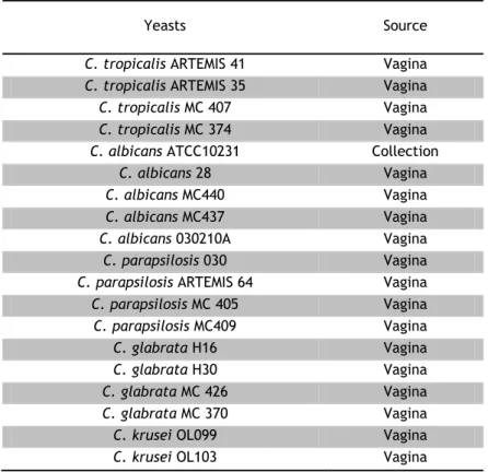
Table 1 shows the strains of Candida selected for the study and their source.
Table 1: Tested species and source.
Yeasts Source
C. tropicalis ARTEMIS 41 Vagina
C. tropicalis ARTEMIS 35 Vagina
C. tropicalis MC 407 Vagina
C. tropicalis MC 374 Vagina
C. albicans ATCC10231 Collection
C. albicans 28 Vagina
C. albicans MC440 Vagina
C. albicans MC437 Vagina
C. albicans 030210A Vagina
C. parapsilosis 030 Vagina
C. parapsilosis ARTEMIS 64 Vagina
C. parapsilosis MC 405 Vagina C. parapsilosis MC409 Vagina C. glabrata H16 Vagina C. glabrata H30 Vagina C. glabrata MC 426 Vagina C. glabrata MC 370 Vagina
C. krusei OL099 Vagina
C. krusei OL103 Vagina
3.2 Determination of the anti-Candida activity
3.2.1 Susceptibility to Gentian Violet
The results show that GeV is active against all Candida species tested (Table 2). MIC values ranged from 0.03 µg/mL and 0.24 µg/mL. C. albicans and C. tropicalis yeasts proved to be more susceptible to GeV (MIC between 0.03 µg/mL and 0.12 µg/mL). C. parapsilosis, C.
glabrata and C. krusei were less sensitive (MIC between 0.06 µg/mL and 0,12 µg/mL), but still
highly susceptible with MIC values significantly lower than the concentration of 1% solution of GeV clinically used.
Table 2 also shows that MLC values range from 0.03 µg/mL and 0.98 µg/mL. MIC and MLC are approximately the same for all yeasts except for C. glabrata, which presents a MLC between two to four times higher than MIC (0.12 µg/mL - 0.98 µg/mL).
7
Table 2: MIC and MLC of a hydro-alcoholic solution of GeV.Yeasts Susceptibility to GeV
MIC 48h (µg/mL) MLC 48h (µg/mL) C. tropicalis ARTEMIS 41 0,03-0,06 0,03-0,06 C. tropicalis ARTEMIS 35 0,03-0,06 0,06 C. tropicalis MC 407 0,03-0,12 0,06-0,12 C. tropicalis MC 374 0,03-0,06 0,06 C. albicans ATCC10231 0,06 0,06-0,12 C. albicans 28 0,03 0,06 C. albicans MC440 0,06 0,06 C. albicans MC437 0,06 0,06 C. albicans 030210A 0,03-0,06 0,03-0,06 C. parapsilosis 030 0,12-0,24 0,12-0,24 C. parapsilosis ARTEMIS 64 0,06 0,06-0,12 C. parapsilosis MC 405 0,12 0,12-0,24 C. parapsilosis MC409 0,12 0,12 C. glabrata H16 0,06 0,12 C. glabrata H30 0,12 0,49 C. glabrata MC 426 0,12 0,49-0,98 C. glabrata MC 370 0,12 0,24 C. krusei OL099 0,12 0,12 C. krusei OL103 0,12 0,12
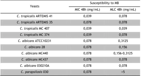
3.2.2 Susceptibility to Methylene Blue
The results concerning the susceptibility to MB (Table 3) show that not all strains are susceptible to this dye. C. glabrata and C. krusei are resistant to the highest concentration used. It is important to indicate that, for a concentration of 5 mg/mL the dye colour makes it impossible the visual evaluation of yeast growth. In contrast to GeV the MIC and the MLC are not coincident for all tested strains.
Table 3: MIC and MLC of an aqueous solution of MB.
Yeasts Susceptibility to MB MIC 48h (mg/mL) MLC 48h (mg/mL) C. tropicalis ARTEMIS 41 0,039 0,078 C. tropicalis ARTEMIS 35 0,078 0,078 C. tropicalis MC 407 0,039 0,039 C. tropicalis MC 374 0,039 0,078 C. albicans ATCC10231 0,078 0,3125 C. albicans 28 0,078 0,156 C. albicans MC440 0,078 0,156-0,3125 C. albicans MC437 0,078 0,078 C. albicans 030210A 0,078 0,078 C. parapsilosis 030 0,078 >5
8
C. parapsilosis ARTEMIS 64 0,039 0,078 C. parapsilosis MC 405 0,039 0,078 C. parapsilosis MC409 0,039 0,078 C. glabrata H16 ≥5 >5 C. glabrata H30 ≥5 >5 C. glabrata MC 426 ≥5 >5 C. glabrata MC 370 ≥5 >5 C. krusei OL099 ≥5 >5 C. krusei OL103 ≥5 >53.2.3 Susceptibility to Fluconazole
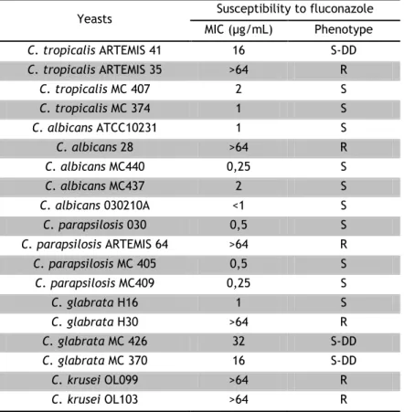
Table 4 shows the susceptibility to fluconazole, a classical antifungal used to treat candidosis. Some yeasts (C. tropicalis ARTEMIS 35, C. albicans 28, C. glabrata H30, C. krusei OL099, C. krusei OL103, C. parapsilosis ARTEMIS 64) are resistant to fluconazole (MIC> 64 µg/mL), and one strain of C. tropicalis and two of C. glabrata were susceptible dose dependent.
Table 4: Susceptibility to fluconazole classification of yeasts according to M27-A3 protocol on:
Susceptible (S), Resistant (R), Depending-dose Susceptibility (S-DD) and Intermediate Susceptibility (I). Yeasts Susceptibility to fluconazole
MIC (µg/mL) Phenotype C. tropicalis ARTEMIS 41 16 S-DD C. tropicalis ARTEMIS 35 >64 R C. tropicalis MC 407 2 S C. tropicalis MC 374 1 S C. albicans ATCC10231 1 S C. albicans 28 >64 R C. albicans MC440 0,25 S C. albicans MC437 2 S C. albicans 030210A <1 S C. parapsilosis 030 0,5 S C. parapsilosis ARTEMIS 64 >64 R C. parapsilosis MC 405 0,5 S C. parapsilosis MC409 0,25 S C. glabrata H16 1 S C. glabrata H30 >64 R C. glabrata MC 426 32 S-DD C. glabrata MC 370 16 S-DD C. krusei OL099 >64 R C. krusei OL103 >64 R
9
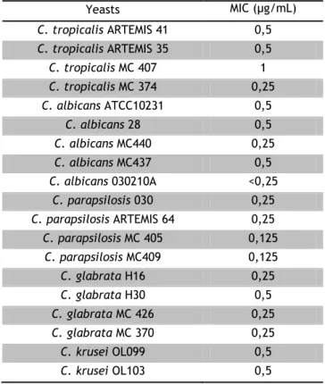
3.2.4 Susceptibility to Amphotericin B
In Table 5 we show the MIC values of amphotericin B. All strains presented a similar susceptibility profile, with MIC range between 0,125 µg/mL and 1 µg/mL.
Table 5: Susceptibility to amphotericin B.
Yeasts MIC (µg/mL) C. tropicalis ARTEMIS 41 0,5 C. tropicalis ARTEMIS 35 0,5 C. tropicalis MC 407 1 C. tropicalis MC 374 0,25 C. albicans ATCC10231 0,5 C. albicans 28 0,5 C. albicans MC440 0,25 C. albicans MC437 0,5 C. albicans 030210A <0,25 C. parapsilosis 030 0,25 C. parapsilosis ARTEMIS 64 0,25 C. parapsilosis MC 405 0,125 C. parapsilosis MC409 0,125 C. glabrata H16 0,25 C. glabrata H30 0,5 C. glabrata MC 426 0,25 C. glabrata MC 370 0,25 C. krusei OL099 0,5 C. krusei OL103 0,5
10
3.3 Mechanism of action
Flow cytometry results showed that GeV fungicidal effect is not related with a primary lesion of the cytoplasmic membrane, since PI was not able to enter the cell (Figure1).
Figure 1: Histogram representing PI stained cells. AF: autofluorescence; Live: viable – non treated cells
(viability control). Dead: death- cells treated with 70% ethanol (death control). MLC: cells treated with MLC concentration of GeV during 60 minutes.
This result was not influenced by the GeV concentration, since similar percentage of stained cells was obtained for MLC and double MLC determinations (Figure 2). Cell death was confirmed by CFU assessment.
Figure 2: PI stained cells comparison between: Live – non treated cells (viability control); Dead – cells
treated with 70% ethanol (death control); 1/2MLC – cells treated during 1h with half MLC; MLC – cells treated during 1h with MLC; 2MLC – cells treated during 1h with double MLC of GeV.
1/2MLC MLC 2MLC
11
Cytometric study of MB reveals a fungicidal effect due to primary lesion of cytoplasmic membrane, as represented on Figure 3.
Figure 3: Histogram representing PI stained cells. AF: autofluorescence; Live: viable – none treated
cells (viability control). Dead: death- cells treated with 70% ethanol (death control). MLC: cells treated with MLC of MB during 60 minutes.
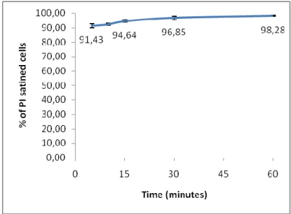
The MB fungicidal effect is quick and effective as it is shown by the cytometric results: after 5 minutes of cells treatment with MB, the percentage of death cells was high (91,43%) and after 60 minutes it was about 98,28%. The kinetic study it is represented in Figure 4.
Figure 4: Kinetic study showing the percentage (%) of PI stained cells after treatment with MLC during
5, 10, 15, 30, 45 and 60 minutes, that is, dead cells. MLC
12
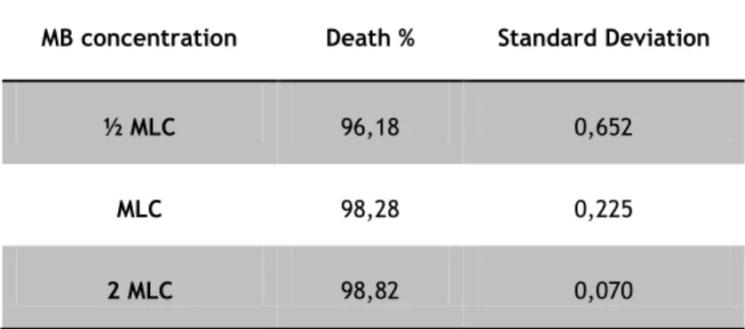
The differences between dye concentrations are represented on the follow table.
Table 6: Death percentage with different MB concentrations.
MB concentration Death % Standard Deviation
½ MLC 96,18 0,652
MLC 98,28 0,225
13
Chapter 4: Discussion
The methodology used is in accordance with international accepted guidelines and allows for a comparison with other studies.
The results obtained with GeV are consistent with previously reported studies, although differences in methodology have been noted. Traboulsi et al. (2008) tested GeV in oral cavity isolates from individuals HIV seropositive. As the dye is considered water insoluble, they used dimethylsulfoxide (DMSO) as solvent (31). In this case, we chose to test the GeV solution as it is used on traditional therapies, say a hydro-alcoholic solution. However, MIC values obtained by them ranged from 0.03 µg/mL and 0.25 µg/mL, approximately the same values reported here (31).
An important fact that should be mentioned is that the GeV showed a fungicidal effect for the inhibitory concentration, that is, the MIC values and MLC were similar for most species tested. The inhibitory effect of the hydro-alcoholic solution of GeV against Candida
spp. was not influenced by the presence of ethanol, since both solutions tested showed the
same results and solvent controls had no effect on growth inhibition (data not shown).
The results emphasize the use of traditional GeV for the treatment of VVC, especially in recurrent cases resistant to classical treatment. Being effective, cheaper than other antifungal agents and easy to apply it is understandable that GeV is still being used.
Despite the fact that GeV is being widely used in clinical settings, its mode of action is not fully understood. Cytometric results showed that yeasts treated with GeV were not stained with PI, showing that GeV doesn’t cause primary lesion of cytoplasmic membrane. However, it was not possible to determine the exactly mechanism of action of GeV because the required resources like metabolic markers were not available for the present study. Regardless this limitation, our results are in accordance with previous studies as discussed below.
Initial studies to elucidate the mechanism of action of GeV reported its reaction with the DNA of living cells by inhibiting its synthesis and causing damage to bacterial DNA (46, 47). The literature also mention a possible effect by producing free radicals through GeV microsomal reduction (48). More recent studies have shown that brief exposure of C. albicans to low concentrations of GeV (0.5 µg/mL and 2 µg/mL) led to a reduction of three virulence factors (phospholipase activity, proteinase activity and germination rate) (32). In fact, these possible mechanisms of action led to another contentious issue: the safety of GeV. The demonstration, in vitro, of the interaction of GeV with DNA (46) and carcinogenicity in mice points to a possible toxic effect (49). However, the carcinogenicity effect is dose-dependent
14
and GeV concentrations used in these studies were remarkably higher than those reported here, approximately one hundred times higher than MIC determined in our work. Tolba et al. revealed that GeV reduces or stops the absorption of nitrate nitrogen, inhibiting the synthesis of proteins and peptides, and at concentrations above 10 ppm (≈ 10 µg/mL) reduces the output of carbon dioxide (50). Despite these effects at the cellular level of GeV seem to indicate a level of toxicity, GeV has been used for a long time, even in children without complications (51). Moreover, its clinical use in treating acute episodes of VVC or recurrent cases unresponsive to conventional antifungal agents is limited to one or two applications (30). Besides, our results show that GeV is not able to disrupt the cytoplasmic membrane and is not probable to passively enter to the cell nucleus.
The clinical benefit of GeV use, especially in difficult cases to handle, seems to overcome some possible side effects (52). In fact, a recent study reported the safety and efficacy of using GeV in oral thrush, on a total of 17 patients, nine achieved clinical success, but purplish, cracked lips and dry mouth were reported as adverse effects (53). Oral irritation was also reported in another study (54). However, these effects can be overcome, as with the development of a pharmaceutical formulation suitable for reducing the effect of color and mucosal irritation is possible.
The results show that MB is a dye with antifungal activity. All C. glabrata and C.
krusei tested strains were resistant at the maximum concentrations tested (MIC ≥ 5 mg/mL
and MLC > 5 mg/mL). Due to the dye colour, we were not able to visually evaluate the yeasts growth for concentrations equal or higher than 5 mg/mL.
Although not extensively studied, the MB antimicrobial activity is being reported in the literature. Regarding its anti-Candida effect, Ofoegbu (2007) reported a success treatment of a sepsis by C. albicans in a premature newborn with MB continuous infusion during 8 weeks combined with systemic antifungal (flucytocine and liposomal amphotericin) (55). On the other hand, another study refereed the absence of MB fungicidal activity. These results could be explained by the low concentration tested and the insufficient exposure time (56).
The account of the routine use of MB with few toxic reactions described reveals the safety of the product and reinforces the possibility of its use as alternative therapy and/or complementary treatment of candidosis. This dye is frequently used for the evaluation of the digestive anastomosis integrity. It is also claimed to be neuroprotective and it is a promise drug in Alzheimer’s disease treatment. Some reports also refer the use of MB to treat several conditions, namely ifosfamide-induced encephalopathy, and to early detection of cancer and precancerous lesions (33, 57-59).
15
Regarding its mechanism of action, MB seems to have anti-oxidant action properties, inhibiting the activity of NADPH oxidase and myeloperoxidase (60). Our results show that MB is a potent fungicidal compound that causes a primary lesion of the cytoplasmic membrane. PI was able to stain yeast cells treated with MB after 5 minutes of treatment. This shows that MB has a potent fungicidal effect right from the 5 minutes exposure. When cells were treated with half MLC after one hour about 96% were dead and about 98% were dead with MLC and twice MLC.
The mechanisms of action of classical antifungals are well characterised. Fluconazole acts by inhibiting ergosterol synthesis and so changing membrane permeability. On other hand, amphotericin B bounds to ergosterol, creating channels or pores, and then increasing cell permeability. By this, it is easily to understand that previously and/or simultaneously treatment with fluconazole reduces amphotericin B activity.
Tested strains represent the most common species isolated from clinic cases. The susceptibility results to classical antifungals are in accordance with previous studies and clinical observations (61, 62). All Candida spp. showed a low MIC value for amphotericin B, while for fluconazole, C. glabrata and C. krusei were the species with higher MIC value and six strains were resistant to it.
The study of new drugs target it has been focused on proposing new ways to inhibit or kill yeasts or to find a pathway to avoid pathogenic mechanisms, such as avoiding the biofilm formation.
In vitro GeV was highly fungicidal to all tested strains, even to those resistant to fluconazole. This is an important finding because it shows that this dye can be an important drug in treating infections due to emerging resistant strains.
MB shows an identical profile than fluconazole. It was not possible to determine MIC or MLC to C. glabrata and C. krusei showing that probably these are resistant species to MB.
So, this study reinforces the important role of these new molecules, especially GeV that presents a fungicidal effect at very low concentrations and also because of the extent clinical experience with these dyes.
16
4.1 Future Perspectives
It is imperative to continue studies on this area mostly because of the lack of drugs to control fungal infections and because of a crescent number of resistant cases.
It is important to study further on the mechanism of action of these dyes, to evaluate its activity in vivo and to develop a drug delivery system that both avoid the inconvenient of colour staining and allows for an easy application.
Ex vivo studies on epithelial cellular cells from vulva and vagina are also relevant to
17
Chapter 5: Conclusion
In conclusion, the GeV is fungicidal for all strains tested at a concentration significantly lower than that used in clinical practice. These results support the use of GeV as a complementary treatment for the management of clinical cases of mucocutaneous Candida infections, especially of those resistant to conventional treatment regimens.
This study shows that the effect of GeV is species dependent and not related to the antifungal susceptibility to classical therapy, that is, fluconazole and amphotericin B, making the GeV a potential alternative in the treatment of VVC resistant to classical antifungals.
It was observed that GeV does not cause primary lesion of the cytoplasmic membrane, but more studies are necessary to clarify its mode of action.
MB also showed an antifungal activity. It is fungicidal to the majority of tested strains. However, it was not possible to determine MIC values to C. glabrata and C. krusei showing that probably these are resistant species. This pattern is identical to that observed with fluconazole.
Cytometric results show that MB has potent and quick fungicidal effect on C. albicans ATCC10231. The cells were stained with PI which means that MB causes primary lesion of cytoplasmic membrane.
So, the tested dyes represent a potential group of drugs to be used on mucocutaneous candidosis.
18
References
1. Costa-de-Oliveira S, Pina-Vaz C, Mendonca D, Goncalves Rodrigues A. A first
Portuguese epidemiological survey of fungaemia in a university hospital. Eur J Clin Microbiol Infect Dis. 2008 May;27(5):365-74.
2. Lopez-Martinez R. Candidosis, a new challenge. Clin Dermatol. 2010 Mar
4;28(2):178-84.
3. Fernandes RV, Ana; Cerqueira, Fátima. Candida species distribution in clinical
samples. Revista da Faculdade de Ciências da Saúde Porto : Edições Universidade Fernando Pessoa. 2009:264-71.
4. Mardh PA, Rodrigues AG, Genc M, Novikova N, Martinez-de-Oliveira J, Guaschino S.
Facts and myths on recurrent vulvovaginal candidosis--a review on epidemiology, clinical manifestations, diagnosis, pathogenesis and therapy. Int J STD AIDS. 2002 Aug;13(8):522-39.
5. Matthews R, Burnie J. The epidemiology and pathogenesis of candidiasis: applications
in prevention and treatment. Bull Inst Pasteur. 1998;96:249-56.
6. das Neves J, Pinto E, Teixeira B, Dias G, Rocha P, Cunha T, et al. Local treatment of
vulvovaginal candidosis : general and practical considerations. Drugs. 2008;68(13):1787-802.
7. Sobel JD, Faro S, Force RW, Foxman B, Ledger WJ, Nyirjesy PR, et al. Vulvovaginal
candidiasis: epidemiologic, diagnostic, and therapeutic considerations. Am J Obstet Gynecol. 1998 Feb;178(2):203-11.
8. Sobel JD. Vulvovaginal candidosis. Lancet. 2007 Jun 9;369(9577):1961-71.
9. Shahid Z, Sobel JD. Reduced fluconazole susceptibility of Candida albicans isolates in
women with recurrent vulvovaginal candidiasis: effects of long-term fluconazole therapy. Diagn Microbiol Infect Dis. 2009 Jul;64(3):354-6.
10. Sobel JD. Limitations of antifungal agents in the treatment of Candida vaginitis:
future challenges. Drug Resist Updat. 1999 Jun;2(3):148-52.
11. Spinillo A, Capuzzo E, Gulminetti R, Marone P, Colonna L, Piazzi G. Prevalence of and
risk factors for fungal vaginitis caused by non-albicans species. Am J Obstet Gynecol. 1997 Jan;176(1 Pt 1):138-41.
12. Fidel PL, Vazquez JA, Sobel JD. Candida glabrata: An important fungal pathogen for
the 21st century. Clinical Microbiology Newsletter. 2001;23(22).
13. Rogers TR. Antifungal drug resistance: does it matter? Int J Infect Dis. 2002 Mar;6
Suppl 1:S47-53.
14. Rogers TR. Antifungal drug resistance: limited data, dramatic impact? Int J Antimicrob
Agents. 2006 Jun;27 Suppl 1:7-11.
15. Donders G, Bellen G, Byttebier G, Verguts L, Hinoul P, Walckiers R, et al.
Individualized decreasing-dose maintenance fluconazole regimen for recurrent vulvovaginal candidiasis (ReCiDiF trial). Am J Obstet Gynecol. 2008 Dec;199(6):613 e1-9.
16. Workowski KA, Berman SM. Sexually transmitted diseases treatment guidelines, 2006.
19
17. Palmeira-de-Oliveira A, Salgueiro L, Palmeira-de-Oliveira R, Martinez-de-Oliveira J,
Pina-Vaz C, Queiroz JA, et al. Anti-Candida activity of essential oils. Mini Rev Med Chem. 2009 Oct;9(11):1292-305.
18. Lima IdO, Oliveira idAG, Lima EdO, Farias NMP, Souza ELd. Atividade antifúngica de
óleos essenciais sobre espécies de Candida. Revista Brasileira de Farmacognosia. 2006;16(2):197-201.
19. Pina-Vaz C, Rodrigues AG, Sansonetty F, Martinez-De-Oliveira J, Fonseca AF, Mardh
PA. Antifungal activity of local anesthetics against Candida species. Infect Dis Obstet Gynecol. 2000;8(3-4):124-37.
20. Palmeira-de-Oliveira A, Ribeiro MP, Palmeira-de-Oliveira R, Gaspar C,
Costa-de-Oliveira S, Correia IJ, et al. Anticandida Activity of a Chitosan Hydrogel: Mechanism of Action and Cytotoxicity Profile. Gynecologic and Obstetric Investigation. 2010;70(4):322-27.
21. Pinto E, Pina-Vaz C, Salgueiro L, Goncalves MJ, Costa-de-Oliveira S, Cavaleiro C, et al.
Antifungal activity of the essential oil of Thymus pulegioides on Candida, Aspergillus and dermatophyte species. J Med Microbiol. 2006 Oct;55(Pt 10):1367-73.
22. das Neves J, Pinto E, Amaral M, Bahia M. Antifungal activity of a gel containing
Thymus vulgaris essential oil against Candida species commonly involved in vulvovaginal candidosis. Pharm Biol. 2009;47(2):151-3.
23. Pina-Vaz C, Sansonetty F, Rodrigues AG, Martinez-De-Oliveira J, Fonseca AF, Mardh
PA. Antifungal activity of ibuprofen alone and in combination with fluconazole against Candida species. J Med Microbiol. 2000 Sep;49(9):831-40.
24. Allen RLM. Colour Chemistry. London: Nelson; 1971.
25. Guaratini CCI, Zanoni MVB. Corantes Têxteis. Química Nova. 2000;23(1):71-8.
26. Zollinger H. Color Chemistry. Second, revised ed.: VCH, Weinheim; 1991.
27. Chengaiah B, Rao KM, Kumar KM, Alagusundaram M, Chetty CM. Medicinal Importance
of Natural Dyes - A review. International Journal of PharmaTech Research. 2010;2(1):144-54.
28. Oros G, Cserhati T, Forgacs E. Antifungal activity of some trityl-based synthetic dyes.
Environ Toxicol Chem. 2002 Jun;21(6):1206-12.
29. Urdaneta E, Benaim Pinto V, Gavaller B. [Candidiasis.]. Mycopathologia. 1961 Aug
30;15:317-42.
30. White DJ, Johnson EM, Warnock DW. Management of persistent vulvo vaginal
candidosis due to azole-resistant Candida glabrata. Genitourin Med. 1993 Apr;69(2):112-4.
31. Traboulsi RS, Mukherjee PK, Ghannoum MA. In vitro activity of inexpensive topical
alternatives against Candida spp. isolated from the oral cavity of HIV-infected patients. Int J Antimicrob Agents. 2008 Mar;31(3):272-6.
32. Ying S, Qing S, Chunyang L. The effect of gentian violet on virulent properties of
Candida albicans. Mycopathologia. 2010 Apr;169(4):279-85.
33. Chen Y-W, Lin J-S, Fong JH-J, Wang I-K, Chou S-J, Wu C-H, et al. Use of methylene
blue as a diagnostic aid in early detection of oral cancer and precancerous lesions. British Journal of Oral and Maxillofacial Surgery. 2007;45:590-1.
20
34. Munin E, Giroldo LM, Alves LP, Costa MS. Study of germ tube formation by Candida
albicans after photodynamic antimicrobial chemotherapy (PACT). J Photochem Photobiol B. 2007 Jul 27;88(1):16-20.
35. Trindade GS, Farias SL, Rumjanek VM, Capella MA. Methylene blue reverts multidrug
resistance: sensitivity of multidrug resistant cells to this dye and its photodynamic action. Cancer Lett. 2000 Apr 14;151(2):161-7.
36. Teichert MC, Jones JW, Usacheva MN, Biel MA. Treatment of oral candidiasis with
methylene blue-mediated photodynamic therapy in an immunodeficient murine model. Oral Surg Oral Med Oral Pathol Oral Radiol Endod. 2002 Feb;93(2):155-60.
37. de Souza SC, Junqueira JC, Balducci I, Koga-Ito CY, Munin E, Jorge AO.
Photosensitization of different Candida species by low power laser light. J Photochem Photobiol B. 2006 Apr 3;83(1):34-8.
38. Tardivo JP, Giglio AD, Oliveira CSd, Gabrielli DS, Junqueira HC, Tada DB, et al.
Methylene blue in photodynamic therapy: From basic mechanisms to clinical applications. Photodiagnosis and Photodynamic Therapy. 2005;2:175-91.
39. Ohlow MJ, Moosmann B. Phenothiazine: the seven lives of pharmacology's first lead
structure. Drug Discov Today. 2011 Feb;16(3-4):119-31.
40. Steczko J, Ash SR, Brewer L, Guillem A. In vitro and in vivo evaluation of efficacy of
citrate/methylene blue/parabens/IPA solution as a skin disinfectant. J Infect. 2010 Jan;60(1):36-43.
41. CETEMED-ANF. Formulário Galénico Português. Lisboa: Centro Tecnológico do
Medicamento and Associação Nacional de Farmácias; 2007.
42. Reference method for broth dilution antifungal susceptibility testing of yeast;
approved standard-Third Edition. (2008).
43. Canton E, Peman J, Viudes A, Quindos G, Gobernado M, Espinel-Ingroff A. Minimum
fungicidal concentrations of amphotericin B for bloodstream Candida species. Diagn Microbiol Infect Dis. 2003 Mar;45(3):203-6.
44. Pina-Vaz C, Rodrigues AG. Evaluation of antifungal susceptibility using flow
cytometry. Methods Mol Biol. 2010;638:281-9.
45. Shapiro H. Pratical Flow Cytometry. New York: Alan R Liss. 1988.
46. Rosenkranz HS, Carr HS. Possible hazard in use of gentian violet. Br Med J. 1971 Sep
18;3(5776):702-3.
47. Wolfe AD. Influence of cationic triphenylmethane dyes upon DNA polymerization and
product hydrolysis by Escherichia coli polymerase I. Biochemistry. 1977 Jan 11;16(1):30-3.
48. Harrelson WG, Jr., Mason RP. Microsomal reduction of gentian violet. Evidence for
cytochrome P-450-catalyzed free radical formation. Mol Pharmacol. 1982 Sep;22(2):239-42.
49. Littlefield NA, Blackwell BN, Hewitt CC, Gaylor DW. Chronic toxicity and
carcinogenicity studies of gentian violet in mice. Fundam Appl Toxicol. 1985 Oct;5(5):902-12.
50. Tolba MK, Saleh AM. Studies on the Mechanism of Fungicidal Action of Crystal Violet
21
51. Shrand H. Thrush in the newborn. Br Med J. 1961 Dec 9;2(5266):1530-3.
52. Donders GG, Bellen G, Mendling W. Management of recurrent vulvo-vaginal candidosis
as a chronic illness. Gynecol Obstet Invest. 2010;70(4):306-21.
53. Wright SC, Maree JE, Sibanyoni M. Treatment of oral thrush in HIV/AIDS patients with
lemon juice and lemon grass (Cymbopogon citratus) and gentian violet. Phytomedicine. 2009 Mar;16(2-3):118-24.
54. Horsfield P, Logan FA, Newey JA. Letter: Oral irritation with gentian violet. Br Med J.
1976 Aug 28;2(6034):529.
55. Ofoegbu BN, Agarwal RP, Lewis MA. Methylene blue irrigation-treatment of renal
fungal balls causing acute renal failure in a preterm infant. Acta Paediatr. 2007 Jun;96(6):939-40.
56. Souza RC, Junqueira JC, Rossoni RD, Pereira CA, Munin E, Jorge AO. Comparison of
the photodynamic fungicidal efficacy of methylene blue, toluidine blue, malachite green and low-power laser irradiation alone against Candida albicans. Lasers Med Sci. 2010 May;25(3):385-9.
57. Smith S, McGeehin W, Kozol RA, Giles D. The efficacy of intraoperative methylene
blue enemas to assess the integrity of a colonic anastomosis. BMC Surg. 2007;7:15.
58. Rojas JC, John JM, Lee J, Gonzalez-Lima F. Methylene blue provides behavioral and
metabolic neuroprotection against optic neuropathy. Neurotox Res. 2009 Apr;15(3):260-73.
59. Oz M, Lorke DE, Petroianu GA. Methylene blue and Alzheimer's disease. Biochem
Pharmacol. 2009 Oct 15;78(8):927-32.
60. Heydrick SJ, Reed KL, Cohen PA, Aarons CB, Gower AC, Becker JM, et al.
Intraperitoneal administration of methylene blue attenuates oxidative stress, increases peritoneal fibrinolysis, and inhibits intraabdominal adhesion formation. J Surg Res. 2007 Dec;143(2):311-9.
61. Pina-Vaz C, Sansonetty F, Rodrigues AG, Costa-Oliveira S, Tavares C,
Martinez-de-Oliveira J. Cytometric approach for a rapid evaluation of susceptibility of Candida strains to antifungals. Clin Microbiol Infect. 2001 Nov;7(11):609-18.
62. Pinto PM, Weikert-Oliveira Rde C, Lyon JP, Cury VF, Arantes RR, Koga-Ito CY, et al. In
vitro antifungal susceptibility of clinical isolates of Candida spp. obtained from patients with different predisposing factors to candidosis. Microbiol Res. 2008;163(5):579-85.
22
Annexes
Annex 1
IN VITRO ANTICANDIDA ACTIVITY OF GENTIAN VIOLET
Gomes-de-Elvas, A.R1; Palmeira-de-Oliveira, R.1; Gaspar, C.1; Gouveia, P1,2; Martinez-de-Oliveira, J.1,3; Palmeira-de-Oliveira, A.1
1 – Health Sciences Research Center (CICS), Faculty of Health Sciences, University of Beira Interior, Covilhã, Portugal
2 – Clinical Pathology Laboratory, Hospital Center Cova da Beira, Covilhã, Portugal
3 – Women and Child Health Department, Hospital Center Cova da Beira, Covilhã, Portugal
Objective:
Gentian violet has been used traditionally to treat vulvovaginal candidosis. There is, however, scarce information concerning its antimicrobial spectrum, required to define its clinical value. This study was designed to evaluate in vitro activity of gentian violet against Candida spp.
Study design:
Ten strains of Candida were studied: 4 C. albicans (1 American Type Culture Collection strain, 3 clinical strains); 2 C. krusei clinical strains; 4 C. glabrata clinical strains. Clinical strains were isolated from recurrent vulvovaginal candidosis cases.
A 1% (m/v) hydro-alcoholic gentian violet (Amresco, USA) was prepared dissolving 1g of violet gentian on 9g of 20% ethanol, and adding purified sterile water up to 100 mL final volume. This solution was then passed through a 0,22 µm sterilizing filter.
The anticandida activity of gentian violet was studied according to CLSI reference M27-A3 protocol. Minimal inhibitory concentrations (MIC) were determined after 24 and 48 hours of incubation at 37ºC. Yeast growth was visually compared for each concentration with the control sample.
The minimal lethal concentration (MLC) was determined based on the modified protocol proposed by Canton et al (2003).
Controls were performed with RPMI medium containing ethanol 20% in the same concentration as the samples. All determinations were performed in duplicate.
Results:
Gentian violet showed to have a potent in vitro activity against Candida strains tested. MIC values achieved for C. albicans were lower (0,06 µg/ml) than for C. krusei and C. glabrata (0.12 µg/ml). For C.
albicans and C. krusei MIC and MLC were the same. For C. glabrata MLC was higher than MIC (0.49-0.98
µg/ml).
This effect was not influenced by the 20% alcohol solvent as all tested strains and respective controls exhibited similar growth either when present or not.
Conclusion:
Gentian Violet has a fungicidal effect against C. albicans and C. krusei. Its activity against C. glabrata revealed a different pattern of susceptibility.
Gentian violet is considered a potential drug to be used in clinical resistant vulvovaginal candidosis. Keywords: vulvovaginal candidosis; gentian violet; recurrence
24
Annex 2
IN VITRO ANTI-CANDIDA ACTIVITY OF GENTIAN VIOLET
Gomes-de-Elvas, A.R1; Palmeira-de-Oliveira, R.1; Gaspar, C.1; Gouveia, P1,2; Martinez-de-Oliveira, J.1,3; Palmeira-de-Oliveira, A.1
1 – Health Sciences Research Center (CICS), Faculty of Health Sciences, University of Beira Interior, Covilhã, Portugal 2 – Clinical Pathology Laboratory, Hospital Center Cova da Beira, Covilhã, Portugal
3 – Women and Child Health Department, Hospital Center Cova da Beira, Covilhã, Portugal