Multiplex real-time PCR and blood culture for identification of
bloodstream pathogens in patients with suspected sepsis
H. Westh1*, G. Lisby1*, F. Breysse2, B. Bo¨ddinghaus3, M. Chomarat2, V. Gant4, A. Goglio5, A. Raglio5, H. Schuster4, F. Stuber6, H. Wissing7and A. Hoeft6
1) Department of Clinical Microbiology 445, Hvidovre Hospital, Hvidovre, Denmark,2) Laboratoire de Microbiologie, Centre Hospitalier Lyon-Sud, Pierre-Be´nite, France,3) Institut fu¨r Med. Mikrobiologie, Johann Wolfgang Goethe-Universita¨t, Frankfurt/Main, Germany,4) Centre for Infectious Diseases and International Health, University College Windeyer Building, Windeyer Institute of Medical Sciences, London, UK,5) U.O Microbiologia e Virologia, Ospedali Riuniti Bergamo, Largo G. Barozzi, Bergamo, Italy ,6) Klinik und Poliklinik fu¨r Ana¨sthesiologie und Operative Intensivmedizin, Universita¨tsklinik Bonn, Bonn, Germany and7)Klinik f. Ana¨sthesiologie, Intensivmedizin und Schmerztherapie, Johann Wolfgang Goethe-Universita¨t, Frankfurt/Main, Germany
Abstract
Severe sepsis is increasingly a cause of death. Rapid and correct initial antimicrobial treatment reduces mortality. The aetiological agent(s) cannot always be found in blood cultures (BCs). A novel multiplex PCR test (SeptiFast(alpha version)) that allows identification of 20 bacterial and fungal species directly from blood was used, comparatively with BC, in a multicentre trial of patients with suspected bacterial or fungal sepsis. Five hundred and fifty-eight paired samples from 359 patients were evaluated. The rate of positivity was 17% for BC and 26% for SeptiFast. Ninety-six microorganisms were isolated with BC, and 186 microorganisms were identified with SeptiFast; 231 microrganisms were found by combining the two tests. Of the 96 isolates identified with BC, 22 isolates were considered to be contaminants. Of the remaining 74 non-contaminant BC isolates available for comparison with SeptiFast, 50 were identified as a species identical to the species identified with SeptiFastin the paired sample. Of the remaining 24 BC isolates for which the species, identified in the BC, could not be detected in the paired SeptiFast sample, 18 BC isolates were identified as a species included in the SeptiFast master list, and six BC isolates were identified as a species not included in the SeptiFastmaster list. With SeptiFast, 186 microorganisms were identified, 12 of which were considered to be contaminants. Of the 174 clinically relevant microorganisms identified with SeptiFast, 50 (29%) were detected by BC. More than half of the remaining microorganisms identified with SeptiFast (but not isolated after BC) were also found in routine cultures of other relevant samples taken from the patients. Future clinical studies should assess whether the use of SeptiFastis of significant advantage in the detection of bloodstream pathogens.
Keywords:Blood culture, LightCycler, multiplex, PCR, sepsis, SeptiFast
Original Submission:26 February 2007;Revised Submission:18 September 2008;Accepted:18 September 2008 Editor: D. Mack
Clin Microbiol Infect2009;15:544–551
Corresponding author and reprint requests:H. Westh, Department of Clinical Microbiology 445, Hvidovre Hospital, Kettega˚rd Alle 30, DK-2650 Hvidovre, Denmark
E-mail: henrik.westh@hvh.regionh.dk
*These authors contributed equally to this work.
LightCycler, SeptiFast, MGRADE, MagNA Lyser and AmpErase are trademarks of Roche Molecular Diagnostics.
Introduction
Sepsis represents a rising healthcare burden. The incidence of sepsis is increasing, as is the number of sepsis-related deaths [1]. There were 659 935 cases of sepsis reported in the USA in 2000, with a bias towards men (relative risk 1.3)
and with an average mortality rate of 18%. Ten bacterial species (fungi were not investigated) found in the SENTRY Antimicrobial Surveillance Program (1997–2002) accounted for 89–92% of all isolates. The ranking of the presence of these species was very similar across North America, Latin America, and Europe [2]. In the USA, a nationwide hospital study of 24 179 nosocomial bloodstream infections showed that the nine most frequent bacterial pathogens were all included in the ten most frequent pathogens found in the SENTRY surveillance, with Candida being the fourth most common pathogen isolated [3].
crucial for successful treatment of septic patients [4–7]. Many years of research in BC technology has led to improved culture media and automated BC systems with increased sensitivity, inactivation of antimicrobial agents, rapid detection of microbial growth, and improved detec-tion of fungi and fastidious microorganisms [8,9]. Despite such advances, the rates of positive BC from patients with different categories of sepsis vary greatly, depending on the degree of sepsis [10–12].
PCR assays developed for specific detection of pathogens in the blood were described as early as 1993 [13–16].
Further development led to broad-spectrum PCR
assays, allowing more universal detection of microorganisms [17–20]. Such broad-spectrum PCR methodology has been hampered by problems of contamination. Contaminating microbial DNA can be introduced either during the sampling process or by handling in the microbiology laboratory [18,21–23]. In particular, PCR assay kit components, such as reagents for DNA extraction [24] and polymerases, are usu-ally contaminated [25,26]. In addition, reservations have been voiced concerning the ability of PCR to achieve the required sensitivity, because of small sample volumes and the per-ceived necessity for an initial (and time-consuming) enrich-ment step involving microbial growth [27]. The diversity of the pathogens concerned necessitates the incorporation of multiple probes for multiple targets, once again prompting questions about the ease of use of the test and the time required to obtain results.
SeptiFasthas recently been used in the molecular diagnosis of sepsis in neutropenic patients [28] and in emergency room, intensive-care unit and general medicine patients with suspected bloodstream infection [29]. We describe a large multicentre evaluation of SeptiFast, which was designed to be sensitive and rapid and to allow the identification of 20 spe-cies of bacteria and fungi that are responsible for up to 95% of all positive BCs.
Materials and Methods
Material
This multicentre study was initiated and performed in six centres; in each centre, 31–129 episodes were included between June and October 2004. An episode was defined as a BC and a simultaneously obtained blood sample for the SeptiFast test. All patients included were clinically suspected to have bacterial or fungal sepsis. Signs of the systemic inflammatory response syndrome (temperature, heart rate, respiratory rate, and white blood cell count) were recorded for all patients and registered in a case report form. Data
registered in the case report form also included antimicrobial therapy and the suspected focus of infection. A patient could be included with more than one episode (one to three epi-sodes per patient). The results of the SeptiFasttest were not used to guide clinical treatment. The relevant institutional or regional review boards or ethics committees approved the research protocol, and participants gave written informed consent, except in one centre, where this was not required by the local ethics committee.
Methods
Blood culture, blood for the SeptiFast test and supplementary microbiological samples. Skin disinfection was performed twice, with ethanol (70%) or propanol (70%), and blood for the BC was drawn by a phlebotomist wearing sterile gloves. A single venipuncture was used to draw samples for 2·2 bottles of BacT/Alert (Biomerieux S.A., Marcy-l Etoile, France) (30– 40 mL of blood) or three bottles of BACTEC (BD Diagnos-tics, Sparks, MD, USA) (25–30 mL of blood). Immediately after blood was drawn for BC (8–10 mL per BC bottle), 5 mL of whole blood was collected in sterile VACUETTE EDTA K2E tubes (Greiner Bio-One, Frickenhausen, Ger-many) for the alpha version of the SeptiFasttest (see below). Each BC was performed in a pair of aerobic/anaerobic bot-tles. Blood for one or two additional BC sets was collected from each patient within a 24-h period and included in epi-sode evaluation. The BCs were analysed using the semi-auto-mated blood culture systems BACTEC or BacT/ALERT, according to laboratory-defined standard operating proce-dures, and time to culture positivity was registered. System-atic collection of samples from other body sites was not part of the protocol. Microbiological results from supplementary samples were obtained only when clinical indications were present. Identification of microorganisms from a suspected infectious focus within 48 h of the episode was used to resolve discrepancies in the results.
The SeptiFast kit. The internal transcribed sequences located between the bacterial 16S and 23S ribosomal RNA genes and the fungal 18S and 5.6S ribosomal RNA genes were selected as the targets for amplification and microorganism identification (Roche Diagnostics GmbH, Penzberg, Germany) [30,31]. Information concerning the sequences of primers and probes is proprietary. A SeptiFasttest was taken to be positive when an internal hybridization probe emitted a fluorescent signal above a defined threshold level. The spe-cies identification of a positive SeptiFasttest was based upon a subsequent melting curve analysis.
the microorganism (Table 1). All reagents, instruments and disposables were obtained from Roche Molecular Diagnostics (Roche Diagnostics GmbH, Penzberg, Germany).
Sample preparation for the SeptiFast test and the PCR procedureThe preparation of DNA and testing were performed as recommended by the manufacturer, using the alpha version of the SeptiFast lys kit, the SeptiFast prep kit, and the LightCycler SeptiFast kit, which were similar but not identical to the commercially available products. The princi-pal difference between the alpha version of the SeptiFast kit and the commercial SeptiFast kit is the automated identifica-tion of species and controls by the SeptiFast identification software, where low concentrations of streptococci and coagulase-negative staphylococci (CoNS) are not displayed as positive results [30]. These ‘low copy number positives’ can be found manually by examining the amplification and melting curve data obtained from the ‘Gram-positive’ capillary.
All MGRADE reagents and disposables used in the
Septi-Fast test were produced using stringent DNA-depleting procedures as stated by the manufacturer. The mechanical lysis of the bacteria was performed using the SeptiFastlys kit and the MagNA Lyser instrument. After the MagNA Lyser procedure was performed, the internal control (IC) of the LightCycler SeptiFast kit was added to each sample and to the negative control (NC). Manual DNA extraction was
performed according to the manufacturer’s instructions, using the SeptiFast prep kit. Blood (in two aliquots of 1.5 mL) was lysed in the MagNA Lyser, using glass beads. Subsequently, total DNA was extracted from 2 mL and eluted in a final volume of 300lL. Fifty microlitres was used for each LightCycler capillary. The amount of DNA available for amplification in each SeptiFast capillary originated from 1/3 mL of blood, as compared with the 30–40 mL of blood obtained for a BC set.
Potential amplicon contaminations were eliminated using AmpErase (Roche Diagnostics). The eluates were then sub-jected to multiplex real-time PCR analysis, using the LightCy-cler 2.0 instrument (Roche). LightCycler data were only considered valid if the corresponding assay controls (reagent control and the IC of the NC) were in the assignedTmrange
and the NC was negative. A complete SeptiFast workflow included samples from seven patients and was analysed in 6 h.
Data analysis and interpretation A non-contaminant, positive BC result was assumed to represent a true infection accord-ing to previously published data [32–34]. Microorganisms contained in the SeptiFast master list (Table 1) were identi-fied by characteristic peaks recognized with LightCycler soft-ware and by manual analysis of Tmvalues. The SeptiFast test
was recorded as negative when the IC was positive and no other signals were detected. SeptiFast samples with a nega-tive IC (as a sign of potential inhibition) were included in the study as negative results.
Whether microorganisms identified with the SeptiFasttest represented a true infection was evaluated retrospectively by considering the identity of the microorganism and the focus of infection as diagnosed by the clinician, and by comparing the BC results with findings from other clinical specimens.
Evaluation of BC and SeptiFast test contaminants Typical BC contaminants (CoNS, Streptococcus spp., Propionibacterium
spp., and Bacillus spp.) were identified by the local investiga-tors. Generally, isolates were considered to be contaminants if only one positive BC result was available within 48 h. If two BC results were obtained with different samples from the same patient within 48 h, including one positive and one negative result, the positive BC was considered to be contaminated. However, if both results within this time per-iod were positive, they were considered to indicate infection. In cases where three samples were drawn from the same patient within the same 48-h period, the patient was consid-ered to have an infection if two of the three samples or all three samples yielded the same microorganism. The BC was considered to be contaminated if only one of three samples from the 48-h period was culture-positive [32–36].
TABLE 1.Multiplex PCR test SeptiFast master list; the
bacteria and fungi listed can be detected by a
three-capillary multiplex real-time LightCycler 2.0 system (limits
of detection of microorganisms are described in the
footnotesa)
Gram-negative Gram-positive Fungi
Escherichia colib Staphylococcus aureusc Candida albicansc
Klebsiella
(pneumoniaec/oxytocac) Coagulase-negativestaphylococcid Candida tropicalis c
Serratia marcescensb Streptococcus pneumoniaec Candida parapsilosisc
Enterobacter
(cloacaec/aerogenesc) Streptococcus
spp.e
Candida glabrata
Proteus mirabilisb Enterococcus faeciumc Candida kruseic
Pseudomonas aeruginosab Enterococcus faecalisc Aspergillus fumigatusb
Acinetobacter baumanniic – – Stenotrophomonas maltophiliac – –
aLimit of detection of microorganisms as described in the package insert of the
commercial assay: all microorganisms in the SeptiFastmaster list found at con-centrations of 100 CFU/mL.
bMicroorganisms found in 20/20; analysis at 3 CFU/mL. cMicroorganisms found in 20/20; analysis at 30 CFU/mL.
dThe coagulase-negative staphylococci that can be identified with the
commer-cial assay are described in the package insert asS. epidermidis,S. hemolyticus, S. hominis,S. pasteuri,S. warneri,S. cohnii,S. lugdenensis,S. capitis,S. caprae,S. sap-hrophyticus, andS. xylosis.
eTheStreptococcusspecies that can be identified with the commercial assay are
Evaluation of a SeptiFast test result as contaminant was performed for CoNS and Streptococcus spp. on the basis of the following criteria: (i) BC bottles were negative; (ii) time to positive BC with contaminant was more than 24 h; (iii) microorganisms were not found in other culture specimens; and (iv) the crossing point (CP) was higher than 35 cycles.
This CP cut-off value was calculated from an in-house (Roche) experiment, in which a SeptiFasttest was performed with samples from healthy participants and in which low copy numbers and high CP values were found for occasional CoNS and less frequent streptococci (data not shown). No other bacteria, or fungi, were found in these healthy participants.
Statistical methods The McNemar test was used for testing the differences between paired proportions. Comparisons of episodes and isolates/microorganisms were made using chi-square tests, with Yates’ correction when the number of samples was <20.
Overall agreement between blood culture and SeptiFastAnalyses of overall agreement between the findings from the SeptiFast test and BC were performed as follows: first, as an episode-to-episode comparison (Table 2)—positive episode agreement between the two tests could be achieved in spite of the identification of different clinical isolates/microorganisms with the two tests; and second, as an isolate-to-microorganism comparison (Table 3)—this could be considered to be a more direct comparison of the two tests than the episode-to-episode comparison. In the assessment of agreement, contaminant episodes or contaminant isolates/microorganisms were included, even though the two test systems could be contaminated at different phases and therefore could not be expected to find the same contaminant [37].
Results
Patients
Three hundred and fifty-nine patients were included in the study. From these patients, 558 episodes fulfilled the inclu-sion criteria of the study, with BC and SeptiFast samples obtained simultaneously. Of these, 382 episodes were nega-tive in both BC and SeptiFast, and 176 episodes were posi-tive in at least one test system (Table 2). The 176 posiposi-tive episodes resulted in a total of 231 isolates/microrganisms found by either of the two methodologies (Table 4). Seventy episodes (12.5%) in which the IC was negative in the
Septi-Fasttest were included in the study as SeptiFastnegatives.
BC and SeptiFastepisodes
For BC, the positive episode rate was 17% (96/558). Of the 96 positive episodes, 74 episodes contained clinical isolates and 22 episodes contained contaminant BC isolates only. The positive episode rate of SeptiFastwas 26% (144/558). Of the 144 SeptiFast-positive episodes, 138 contained clinical microorganisms (six contaminants were found with clinical microorganisms), and six episodes contained contaminants only. The BC contamination rate was 3.9% (22/558), and the contamination rate for SeptiFastwas 2.2% (12/558). Excluding contaminants, the positive rate of SeptiFast was twice as high as that of BC (25%, 138/558 vs. 13%, 74/558; Table 2).
BC and SeptiFastisolates/microorganisms
In this study, a single microorganism was detected in 74 non-contaminant positive BCs. Polymicrobial infection, how-ever, was detected by SeptiFastat an average of 1.3 microor-ganisms per sample (112 episodes with one microorganism, 18 episodes with two microorganisms, six episodes with three microorganisms, and two episodes with four microor-ganisms). Fifty of the 74 positive BC isolates (68%) were
TABLE 2.Episode agreement between blood culture and
SeptiFasttest
Blood culture
Positive Negative Contaminant Total
SeptiFast
Positive 58 77 3 138
Negative 16 382 16 414
Contaminant 0 3 3 6
Total 74 462 22 558
Significantly more episodes were positive by SeptiFast(p <0.0001). Overall percentage agreement, (58 + 382 + 3)/558: 79% (95% CI 76–83%). Agreement of SeptiFast with positive blood culture, 58/74: 78% (95% CI 67–87%).
Agreement of SeptiFast with negative blood culture, 382/462: 83% (95% CI 79–86%).
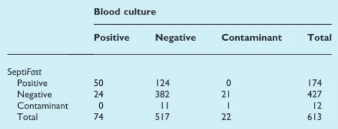
TABLE 3.Isolate/microorganism agreement between blood
culture and SeptiFasttest
Blood culture
Positive Negative Contaminant Total
SeptiFast
Positive 50 124 0 174
Negative 24 382 21 427
Contaminant 0 11 1 12
Total 74 517 22 613
Significantly more microorganisms were identified by SeptiFast, p <0.0001. Overall percentage agreement, (50 + 382 + 1)/613: 71% (95% CI 67–74%). Agreement of SeptiFast with positive blood culture, 50/74: 68% (95% CI 56–78%).
detected with both systems. Of the 24 BC-positive but
Septi-Fast-negative isolates, six isolates were not included and 18 isolates (ten different species) were included in the list of microorganisms that can be detected by SeptiFast (Table 1). The 174 SeptiFast clinical microorganisms (Table 3) were detected in 138 episodes. Of the 174 microorganisms, 50 (29%) microorganisms were also detected by BC. Of the 124 microorganisms detected only by SeptiFast, 67 (54%) could be confirmed as clinical pathogens by culture of the same microorganism/species from a relevant anatomical site within the same clinical time frame. The remaining 57 microorgan-isms found using SeptiFast only could not be confirmed, as the microorganism did not grow in culture from a clinically relevant site or because samples from such a site were not obtained. In total, 117/174 (67%) microorganisms found using SeptiFast could be confirmed by culture. The isolates/micro-organisms found using BC and/or SeptiFast are shown in Table 4.
Episodes where Staphylococcus aureus was found by BC and using SeptiFast had a mean CP of 26.8 (standard devia-tion 3.8), whereas episodes that were SeptiFast-positive and
BC-negative had a mean CP of 29.7 (standard deviation 3.8). This difference in CP suggests that the amount of S. aureus
DNA present in SeptiFastsamples where the paired BC sam-ple is negative is significantly lower than the amount of
S. aureusDNA present in SeptiFastsamples where the paired BC sample is positive.
Low-level contamination in SeptiFast
Low-level contamination (included as a negative result) in the SeptiFast PCR (a CP higher than 35 cycles) was seen in 57 episodes due to CoNS and in two cases due to Streptococcus
spp. (11%, in 558 episodes).
Agreement between BC and SeptiFastresults
The overall episode-to-episode agreement (positives with positives plus negatives with negatives) between SeptiFast
and BC was 79% (Table 2). For positive SeptiFastresults, the agreement with BC was 78%, and for negative SeptiFast
results, the agreement with BC was 83% (Table 2).
The overall microorganism-to-isolate agreement between SeptiFast and BC was 71% (Table 3). For positive SeptiFast
results, the agreement with BC was 68%, and for negative SeptiFast results, the agreement with BC was 74% (Table 3).
In the absence of a laboratory reference standard for the diagnosis of sepsis, we compared the two tests in three dif-ferent ways: (i) the BC result is 100% accurate; (ii) the
Septi-Fast result is 100% accurate; and (iii) the combined findings by BC or SeptiFast, excluding contaminant isolates/microor-ganisms, are 100% accurate. An analysis of the positive find-ings consequently leads to different sensitivity rates for BC and SeptiFast. On the basis of these definitions, the sensitivity of BC (ability to find a positive result) would be 29% if
Septi-Fast were used as the standard. On the other hand, the sen-sitivity of SeptiFast would be 68% if BC were the reference standard. If all non-contaminant findings by BC or SeptiFast
were regarded as true positives, the sensitivity of BC would be 37%, and that of SeptiFastwould be 88%.
Influence of antimicrobial therapy on BC and SeptiFasttest
results
SeptiFast detected 124 microorganisms in patients for whom the paired BC was negative. In 64 (52%, 95% CI 42–61%) of these episodes, at the time of sampling the patient had received antimicrobial therapy considered likely to be effec-tive against the microorganism detected using SeptiFast. BC detected 74 isolates. In 27 (37%, 95% CI 27–49%) of these BC episodes, at the time of sampling the patient had received antimicrobial therapy effective against the microorganism.
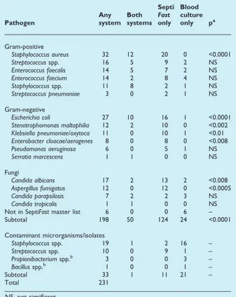
TABLE 4.Number of microorganisms/isolates detected
with SeptiFastor blood culture
Pathogen
Any system
Both systems
Septi
Fast
only Blood culture only pa
Gram-positive
Staphylococcus aureus 32 12 20 0 <0.0001 Streptococcusspp. 16 5 9 2 NS Enterococcus faecalis 14 5 7 2 NS Enterococcus faecium 14 2 8 4 NS Staphylococcusspp. 11 8 2 1 NS Streptococcus pneumoniae 3 0 2 1 NS
Gram-negative
Escherichia coli 27 10 16 1 <0.0001 Stenotrophomonas maltophilia 12 2 10 0 <0.002 Klebsiella pneumoniae/oxytoca 11 0 10 1 <0.01 Enterobacter cloacae/aerogenes 8 0 8 0 <0.008 Pseudomonas aeruginosa 6 0 5 1 NS Serratia marcescens 1 1 0 0 NS
Fungi
Candida albicans 17 2 13 2 <0.008 Aspergillus fumigatus 12 0 12 0 <0.0005 Candida parapsilosis 7 2 2 3 NS Candida tropicalis 1 1 0 0 NS Not in SeptiFastmaster list 6 0 0 6 – Subtotal 198 50 124 24 <0.0001
Contaminant microrganisms/isolates
Staphylococcusspp. 19 1 2 16 – Streptococcusspp. 10 0 9 1 – Propionibacteriumspp.b 3 0 0 3 –
Bacillusspp.b 1 0 0 1 –
Subtotal 33 1 11 21 –
Total 231
NS, not significant.
aThe McNemar test was used for testing the difference between paired
propor-tions (SeptiFastonly vs. blood culture only).
Time to positive BC or SeptiFasttest
The time to a positive result was documented in 36 of 50 episodes during which the same microorganism was detected using BC and SeptiFast. The median time to the first positive BC signal (equalling the time to Gram stain—not the time to the final species identification) was 2 days (range: 1–10 days). If SeptiFasthad been performed on a once-daily basis (not as batched runs, as in this study), the average time from obtain-ing the sample to the SeptiFast result would be a median of 18 h (range: 6–30 h).
Discussion
The purpose (and design) of this study was to compare SeptiFast test results with BC results—not to assess the potential clinical value of the SeptiFast test when used in addition to BC. This latter issue can only be examined by controlled clinical trials evaluating the impact of SeptiFasttest results on patient care and outcome variables.
We observed more episodes of circulating bacterial and/ or fungal DNA detected using SeptiFast than episodes in which microorganisms were detected using BC, as observed in other studies [28,29]. In particular, we found more epi-sodes withS. aureus,Escherichia coli,Candida albicans, Aspergil-lus fumigatus, Stenotrophomonas maltophilia, Klebsiella pneumoniae, Klebsiella oxytoca, Enterobacter cloacae, and Ente-robacter aerogenes. Although the numbers are small, it appears that, for Candida spp. andA. fumigatus, the SeptiFast
methodology was more sensitive than conventional BC, sug-gesting that some fungal infections in intensive-care unit patients might currently be undiagnosed. The detection of
A. fumigatusDNA was unexpected, as this microorganism is difficult to detect with current conventional technology [38,39]. In only two of five patients, A. fumigatus infection was confirmed by other diagnostic methods, autopsy and bronchoalvelolar lavage, as the cause of endocarditis and pneumonia, respectively. Diagnostic tests for Aspergilluswere not performed for the other three patients. Recently, two neutropenic patients with haematological malignancies have also been found to be A. fumigatus-positive using SeptiFast
and bronchoalvelolar lavage [28]. Specifically designed studies will be needed to investigate the clinical significance of posi-tive SeptiFastresults forA. fumigatusin suspectedA. fumigatus
infections. Patients who were positive for S. aureus by both BC and SeptiFast had lower CPs (more target) than did patients for whom BC was negative and SeptiFastpositive for
S. aureus. This may explain, in part, why BC was negative in these cases. The interpretation of Streptococcus spp. and CoNS as non-contaminant microorganisms was based on
simultaneous findings of these microorganisms using SeptiFast
and culture. This could be imprecise, as species determina-tion cannot be performed for Streptococcus spp. and CoNS detected with the SeptiFasttest. The contamination rate was slightly lower for SeptiFast(2.2%) than it was for BC (3.9%). This was primarily due to the CP cut-off value of 35 cycles defined in the software for CoNS and streptococci.
The most important advantage of BC over SeptiFastis that susceptibility testing of an isolate can be performed, allowing the implementation of specifically targeted antimicrobial or antifungal therapy. BC also has an advantage over SeptiFast
with respect to microorganisms not included in the SeptiFast
master list. In some cases, microorganisms were identified by BC that theoretically should have been found using SeptiFast. This probably happened in cases of low-level bacteraemia, where there was no target for SeptiFastin the sample tested. Unfortunately, IC DNA in the SeptiFast assay was not ampli-fied in 12.5% of episodes; therefore, the SeptiFast test gave no information. This was due to either inhibition of the PCR reaction or inappropriate sample preparation, both of which must be addressed in future improvements of the assay [40].
Early appropriate antimicrobial treatment of sepsis has been demonstrated in several studies to improve survival [4–7,41]. The diagnosis of bacteraemia can be complicated in patients receiving antimicrobial treatment, and all current BC systems have been modified in an attempt to reduce the effect of antimicrobials in the BC bottle [42]. The advantage of a DNA-based detection system (as compared with BC) is that the microorganism causing sepsis does not have to be viable at the time of sampling. Although our data in this respect are limited, owing to the design of the study,
Septi-Fast may be particularly advantageous for patients receiving antibiotics.
One question that must be answered by future studies concerns the clinical relevance of microorganisms detected only by SeptiFast. It is not clear whether DNAaemia as revealed by the SeptiFast test reflects true infection. Analysis of BCs and other routine clinical microbiology samples revealed that 67% of the microorganisms detected using SeptiFastcould be confirmed by culture. This is in agreement with the 69% confirmation rate found in a previous study [29]. The present study was not designed to evaluate the clinical significance of microbial DNAaemia, which is certainly not the same as, and therefore not directly comparable with, bacteraemia. We believe that further studies are needed to address this issue.
We observed an ‘overall agreement’ between SeptiFast
replaced by much more sensitive nucleic acid amplification methods [43]. Finally, if any non-contaminant microorganisms found using BC or SeptiFasttesting were viewed as true pos-itives in septic patients, 37% of the microorganisms were found using BC as opposed to 88% using SeptiFasttesting.
The SeptiFasttechnology could represent an advantageous addition to BC technology, and seems to hold promise for enhanced detection of bacteria and fungi in patients with sus-pected sepsis. This new test will not replace BC, which will still be required as a prerequisite for identification of micro-organisms, and in particular for susceptibility testing.
Transparency Declaration
All authors’ laboratories have received research funding from Roche Molecular Diagnostics to perform the trial. Roche Molecular Diagnostics provided financial support for manu-script preparation. Roche Molecular Diagnostics provided assistance in study design, data acquisition, and editing of the manuscript. H. Westh, G. Lisby, A. Hoeft and F. Stuber have been on the speakers’ bureau for Roche Molecular Diagnos-tics.
References
1. Martin GS, Mannino DM, Eaton S, Moss M. The epidemiology of sep-sis in the United States from 1979 through 2000.N Engl J Med2003; 348: 1546–1554.
2. Biedenbach DJ, Moet GJ, Jones RN. Occurrence and antimicrobial resistance pattern comparisons among bloodstream infection isolates from the SENTRY Antimicrobial Surveillance Program (1997–2002). Diagn Microbiol Infect Dis2004; 50: 59–69.
3. Wisplinghoff H, Bischoff T, Tallent SM, Seifert H, Wenzel RP, Edmond MB. Nosocomial bloodstream infections in US hospitals: analysis of 24,179 cases from a prospective nationwide surveillance study.Clin Infect Dis2004; 39: 309–317.
4. Garnacho-Montero J, Garcia-Garmendia JL, Barrero-Almodovar A, Jimenez-Jimenez FJ, Perez-Paredes C, Ortiz-Leyba C. Impact of ade-quate empirical antibiotic therapy on the outcome of patients admit-ted to the intensive care unit with sepsis.Crit Care Med2003; 31: 2742–2751.
5. Zaragoza R, Artero A, Camarena JJ, Sancho S, Gonzalez R, Nogueira JM. The influence of inadequate empirical antimicrobial treatment on patients with bloodstream infections in an intensive care unit. Clin Microbiol Infect2003; 9: 412–418.
6. Leibovici L, Shraga I, Drucker M, Konigsberger H, Samra Z, Pitlik SD. The benefit of appropriate empirical antibiotic treatment in patients with bloodstream infection.J Intern Med1998; 244: 379–386. 7. Valles J, Rello J, Ochagavia A, Garnacho J, Alcala MA.
Community-acquired bloodstream infection in critically ill adult patients: impact of shock and inappropriate antibiotic therapy on survival. Chest 2003; 123: 1615–1624.
8. Magadia RR, Weinstein MP. Laboratory diagnosis of bacteremia and fungemia.Infect Dis Clin North Am2001; 15: 1009–1024.
9. Cockerill FR 3rd, Wilson JW, Vetter EAet al.Optimal testing param-eters for blood cultures.Clin Infect Dis2004; 38: 1724–1730. 10. Members of the American College of Chest Physicians. Definitions
for sepsis and organ failure and guidelines for the use of innovative therapies in sepsis.Crit Care Med1992; 20: 864–874.
11. Rangel-Frausto MS, Pittet D, Costigan M, Hwang T, Davis CS, Wen-zel RP. The natural history of the systemic inflammatory response syndrome (SIRS). A prospective study.JAMA1995; 273: 117–123. 12. Sands KE, Bates DW, Lanken PNet al.Epidemiology of sepsis
syn-drome in 8 academic medical centers. Academic Medical Center Consortium Sepsis Project Working Group. JAMA 1997; 278: 234– 240.
13. Iralu JV, Sritharan VK, Pieciak WS, Wirth DF, Maguire JH, Barker RH Jr. Diagnosis ofMycobacterium aviumbacteremia by polymerase chain reaction.J Clin Microbiol1993; 31: 1811–1814.
14. Kami M, Fukui T, Ogawa Set al.Use of real-time PCR on blood sam-ples for diagnosis of invasive aspergillosis. Clin Infect Dis 2001; 33: 1504–1512.
15. Maaroufi Y, Heymans C, De Bruyne JM et al. Rapid detection of Candida albicans in clinical blood samples by using a TaqMan-based PCR assay.J Clin Microbiol2003; 41: 3293–3298.
16. Song JH, Cho H, Park MY, Na DS, Moon HB, Pai CH. Detection of Salmonella typhi in the blood of patients with typhoid fever by poly-merase chain reaction.J Clin Microbiol1993; 31: 1439–1443. 17. Rothman RE, Majmudar MD, Kelen GDet al.Detection of
bactere-mia in emergency department patients at risk for infective endocardi-tis using universal 16S rRNA primers in a decontaminated polymerase chain reaction assay.J Infect Dis2002; 186: 1677–1681. 18. Klaschik S, Lehmann LE, Raadts A, Hoeft A, Stuber F. Comparison of
different decontamination methods for reagents to detect low con-centrations of bacterial 16S DNA by real-time-PCR. Mol Biotechnol 2002; 22: 231–242.
19. Corless CE, Guiver M, Borrow R, Edwards-Jones V, Fox AJ, Kacz-marski EB. Simultaneous detection ofNeisseria meningitidis, Haemophi-lus influenzae, and Streptococcus pneumoniae in suspected cases of meningitis and septicemia using real-time PCR.J Clin Microbiol2001; 39: 1553–1558.
20. Klaschik S, Lehmann LE, Raadts Aet al.Detection and differentiation of in vitro-spiked bacteria by real-time PCR and melting-curve analy-sis.J Clin Microbiol2004; 42: 512–517.
21. Garcia-Erce JA, Grasa JM, Solano VMet al.Bacterial contamination of blood components due to Burkholderia cepacia contamination from clorhexidine bottles.Vox Sang2002; 83: 70–71.
22. Oie S, Kamiya A. Microbial contamination of antiseptics and disinfec-tants.Am J Infect Control1996; 24: 389–395.
23. Dave J, Springbett R, Padmore H, Turner P, Smith G.Pseudomonas cepaciapseudobacteraemia.J Hosp Infect1993; 23: 72–73.
24. Peters RP, Mohammadi T, Vandenbroucke-Grauls CM, Danner SA, van Agtmael MA, Savelkoul PH. Detection of bacterial DNA in blood samples from febrile patients: underestimated infection or emerging contamination?FEMS Immunol Med Microbiol2004; 42: 249–253. 25. Corless CE, Guiver M, Borrow R, Edwards-Jones V, Kaczmarski EB,
Fox AJ. Contamination and sensitivity issues with a real-time univer-sal 16S rRNA PCR.J Clin Microbiol2000; 38: 1747–1752.
26. Mohammadi T, Reesink HW, Vandenbroucke-Grauls CM, Savelkoul PH. Optimization of realtime PCR assay for rapid and sensitive detec-tion of eubacterial 16S ribosomal DNA in platelet concentrates.J Clin Microbiol2003; 41: 4796–4798.
27. Peters RP, van Agtmael MA, Danner SA, Savelkoul PH, Van-denbroucke-Grauls CM. New developments in the diagnosis of bloodstream infections.Lancet Infect Dis2004; 4: 751–760.
29. Louie RF, Tang Z, Albertson TE, Cohen S, Tran NK, Kost GJ. Multi-plex polymerase chain reaction detection enhancement of bacteremia and fungemia.Crit Care Med2008; 36: 1487–1492.
30. Roche Molecular Systems. LightCyclerSeptiFast Test Mgrade. For use with the LightCycler 2.0 Instrument (serial number 1415001 and higher). 04623886001 ed. 2005.
31. Iwen PC, Hinrichs SH, Rupp ME. Utilization of the internal tran-scribed spacer regions as molecular targets to detect and identify human fungal pathogens.Med Mycol2002; 40: 87–109.
32. Weinstein MP. Blood culture contamination: persisting problems and partial progress.J Clin Microbiol2003; 41: 2275–2278.
33. Weinstein MP, Towns ML, Quartey SM et al. The clinical signifi-cance of positive blood cultures in the 1990s: a prospective com-prehensive evaluation of the microbiology, epidemiology, and outcome of bacteremia and fungemia in adults. Clin Infect Dis1997; 24: 584–602.
34. Reimer LG, Wilson ML, Weinstein MP. Update on detection of bac-teremia and fungemia.Clin Microbiol Rev1997; 10: 444–465. 35. Richter SS, Beekmann SE, Croco JLet al.Minimizing the workup of
blood culture contaminants: implementation and evaluation of a labo-ratory-based algorithm.J Clin Microbiol2002; 40: 2437–2444. 36. Hall KK, Lyman JA. Updated review of blood culture contamination.
Clin Microbiol Rev2006; 19: 788–802.
37. Fischer JE, Bachmann LM, Jaeschke R. A readers’ guide to the inter-pretation of diagnostic test properties: clinical example of sepsis. Intensive Care Med2003; 29: 1043–1051.
38. Hope WW, Walsh TJ, Denning DW. Laboratory diagnosis of invasive aspergillosis.Lancet Infect Dis2005; 5: 609–622.
39. Klont RR, Meis JF, Verweij PE. Critical assessment of issues in the diagnosis of invasive aspergillosis. Clin Microbiol Infect2001; 7 (suppl 2): 32–37.
40. Burkardt HJ. Standardization and quality control of PCR analyses.Clin Chem Lab Med2000; 38: 87–91.
41. Fraser A, Paul M, Almanasreh Net al.Benefit of appropriate empiri-cal antibiotic treatment: thirty-day mortality and duration of hospital stay.Am J Med2006; 119: 970–976.
42. Vetter E, Torgerson C, Feuker Aet al.Comparison of the BACTEC MYCO/F lytic bottle to the isolator tube, BACTEC plus aerobic F/bottle, and BACTEC anaerobic lytic/10 bottle and comparison of the BACTEC plus aerobic F/bottle to the isolator tube for recovery of bacteria, mycobacteria, and fungi from blood.J Clin Microbiol2001; 39: 4380–4386.