Kamila Kappke Dias
Bachelor degree in Biotechnology
Safety Assessment of Polymeric Nanoparticle
Carriers for Drug Delivery in Human
Osteoblasts
Dissertation to obtain the Master Degree in
Biochemistry for Health
Supervisor: Professor Maria João Silva, PhD, Instituto Nacional de Saúde
Doutor Ricardo Jorge, Lisboa
Co-Supervisor: Professor Ana Bettencourt, PhD, Faculdade de Farmácia,
Universidade de Lisboa
III
Kamila Kappke Dias
Bachelor degree in Biotechnology
Safety Assessment of Polymeric Nanoparticle
Carriers for Drug Delivery in Human
Osteoblasts
Dissertation to obtain the Master Degree in
Biochemistry for Health
Supervisor: Professor Maria João Silva, PhD, Instituto Nacional de Saúde
Doutor Ricardo Jorge, Lisboa
Co-Supervisor: Professor Ana Bettencourt, PhD, Faculdade de Farmácia,
Universidade de Lisboa
Jury:
President: Professor Maria Teresa Nunes Mangas Catarino, PhD, FCT-UNL
Argue: Elisabete de Jesus Oliveira Marques, PhD, FCT-UNL
Supervisor: Professor Maria João Silva, PhD, Instituto Nacional de Saúde
Doutor Ricardo Jorge, Lisboa
V
Copyright
“Safety Assessment of Polymeric Nanoparticle Carriers for Drug Delivery in Human Osteoblasts”
Copyright © Kamila Kappke Dias, Faculdade de Ciências e Tecnologias, Universidade Nova de
Lisboa
The Faculty of Sciences and Technology and the NOVA University of Lisbon have the right,
forever and without geographical limits, to file and publish this dissertation through printed copies
reproduced in paper or by digital means, or by any other mean known or that is invented, and to
disclose it through scientific repositories and to allow its copyright and distribution for
non-commercial educational or research purposes, provided that the author and editor are credited.
A Faculdade de Ciências e Tecnologias e a Universidade Nova de Lisboa têm o direito, perpétuo
e sem limites geográficos, de arquivar e publicar esta dissertação através de exemplares
impressos reproduzidos em papel ou de forma digital, ou por qualquer outro meio conhecido ou
que venha a ser inventado, e de a divulgar através de repositórios científicos e de admitir a sua
cópia e distribuição com objetivos educacionais ou de investigação, não comerciais, desde que
VII
“I would rather have questions that can’t be answered that answers which can’t be questioned”
IX
Acknowledgments
First of all, I would like to thank to my mentors Doctor Maria João Silva from Departamento
de Genética Humana – Instituto Nacional de Saúde Dr. Ricardo Jorge and Doctor Ana
Bettencourt from Faculdade de Farmácia, Universidade de Lisboa, that allowed me to work their
laboratories, for all the guidance, the support, the knowledge and for always being available to
discuss my crazy ideas. Without them, none of this would be possible.
To my non-official mentors, Doctor Henriqueta Louro and Doctor Lídia Gonçalves, for all
the teaching, the limitless patience and for making feel welcome all the time.
To both institutions, Insituto Nacional de Saúde Dr. Ricardo Jorge and Faculdade de
Farmácia da Universidade de Lisboa who gave me the opportunity to perform this study, for the
accommodation and technical support along this project.
I also want to show my gratitude to all my lab colleagues for all the help and the fun
moments, specially Célia Ventura, for all the laughs, the friendship and the support.
To all friends from Coimbra and Lisboa for their fellowship, and for making this journey
way better than it was.
Lastly, to my family, specially to my mom, for her support and affection, and to Tiago, for
always being right by side and for never stop encouraging me.
XI
Abstract
Nanoparticles (NPs) applied to pharmaceutics constitute an innovative approach to
improve drug release profiles on targeted sites. The assessment of their biocompatibility and
safety for human health plays also a major role in the development process. The objective of this
work was to characterize the cellular interactions and potential toxicity of polymeric nanoparticles,
in human osteoblasts.
Poly(methyl methacrylate) (PMMA) and Eudragit® RL 100 (Eud) were used to produce
PMMA and PMMA-Eud (50:50) NPs (average size range of 500 nm) by single-emulsion with
solvent evaporation methodology. Their physicochemical properties (size distribution, surface
charge, morphology and aggregation/agglomeration states) were analysed. Their safety
evaluation was conducted in “normal” and differentiated MG63 cells. Cell uptake, cyto- and
genotoxicity were characterized using several endpoints: cell viability (MTT assay), oxidative
stress production (H2DCFDA assay), DNA and chromosome damage (Comet and Micronucleus
assays).
The results confirmed the successful cellular uptake of PMMA and PMMA-Eud. Both NPs
were neither cytotoxic nor able to produce oxidative stress in differentiated cells, although a
moderated toxicity was detected in undifferentiated cells. As to the genotoxic potential, both NPs
induced primary DNA damage (comet assay) in osteoblasts, especially in short-term exposure.
Noteworthy, none of the NPs caused chromosome alterations, indicating that the DNA lesions
were not converted into permanent genetic damage. However, an increased cell proliferative
capacity was noted for PMMA that needs confirmation.
In conclusion, PMMA and PMMA-Eud are promising nanocarriers in drug delivery
systems. Their in vitro safety assessment in osteoblasts indicated that both NPs are
biocompatible but display a weak genotoxicity that needs further investigation, e.g., using other
endpoints or in vivo models. The utilization of cells under different specialization status improved
data reliability. Moreover, understanding how physicochemical features relate to toxicity will
support the design of safer formulations for biomedical purposes as envisaged by the
safer-by-design concept.
XIII
Resumo
A aplicação de nanopartículas (NPs) à área farmacêutica constitui uma resposta
inovadora para melhorar os perfis de libertação de fármacos em órgãos-alvo. No entanto, a
avaliação da sua biocompatibilidade e segurança para a saúde humana constituem uma fase
limitante. O objetivo deste trabalho foi caracterizar o potencial tóxico de NPs poliméricas e
possíveis interações com osteoblastos de origem humana.
Foi utilizado poli(metil metacrilato) (PMMA) e Eudragit® RL 100 (Eud) para a produção
das NPs (tamanho médio: 500nm), através do método de emulsão simples com evaporação do
solvente. Seguiu-se uma caracterização das suas propriedades físico-química incluindo a
distribuição de tamanhos, carga superficial, morfologia e formação de agregados/aglomerados.
A avaliação de segurança foi realizada em células “normais” e diferenciadas de osteoblastos
(MG-63). Foi analisada a capacidade de internalização destas NPs assim com a sua cito- e
genotoxicidade através de vários parâmetros: viabilidade celular (ensaio do MTT), stress
oxidativo (ensaio do H2DCFDA) e danos ao nível do DNA e da estrutura cromossómica (ensaios
do Cometa e dos Micronúcleos).
Os resultados confirmaram uma internalização bem-sucedida tanto para o PMMA como
para o PMMA-Eud. Ambas as NPs não demonstraram citotoxicidade nem capacidade de induzir
stress oxidativo em células diferenciadas, apesar de uma toxicidade moderada ter sido detetada
em células indiferenciadas. Quanto ao potencial genotóxico, ambas as NPs induziram danos
primários ao nível do DNA, especialmente em exposições mais curtas. Nenhuma NP causou
alterações cromossômicas, indicando que as lesões induzidas ao DNA não foram convertidas
em danos genéticos permanentes. No entanto, foi observado um aumento da capacidade
proliferativa das células quando expostas a NPs de PMMA que ainda necessitam de confirmação.
Em conclusão, o PMMA e o PMMA-Eud apresentam propriedades distintas para uma
aplicação na veiculação de fármacos. A sua avaliação de segurança in vitro em osteoblastos
indicou que ambas as NPs são biocompatíveis, mas apresentam uma genotoxicidade moderada
que necessita de ser explorada, através de outros parâmetros ou modelos in vivo. A utilização
de células sob diferentes estados de especialização aumentou a sensibilidade dos ensaios. Além
disso, compreender a relação entre as características físico-químicas e uma potencial toxicidade
irá promover a produção de formulações mais seguras para fins biomédicos como previsto pelo
conceito “safe-by-design”.
XV
Table of Contents
1.
Introduction ... 1
1.1
Nanoparticles and Nanotechnologies ... 1
1.2
Bone grafts and Osteomyelitis ... 2
1.2.1
Bone structure and formation ... 2
1.2.2
Bone grafts ... 2
1.2.3
Bone infections: Osteomyelitis ... 3
1.3
Novel nanoparticles-based therapeutics for Osteomyelitis ... 5
1.3.1
Polymeric nanocarriers for drug delivery ... 5
1.3.2
Nano-sized Poly(methyl methacrylate) and Eudragit formulations ... 5
1.3.2.1 PMMA and PMMA-Eud Production ... 7
1.4
Characterization of physicochemical properties ... 8
1.5
Cellular interactions and potential toxicity of nanoparticles ... 10
1.5.1
Nano-bio interactions ... 10
1.5.2
Nanotoxicology ... 11
1.5.3
In vitro experimental models ... 12
1.5.3.1 Osteoblasts ... 13
1.5.4
Cellular uptake ... 14
1.5.5
Cytotoxicity assessment ... 14
1.5.6
Oxidative stress ... 15
1.5.7
Genotoxicity assessment ... 16
1.5.7.1 Characterization of the DNA damage: Comet assay ... 16
1.5.7.2 Characterization of chromosome damage: Cytokinesis-Block
Micronucleus Assay ... 17
2.
Objective ... 21
3.
Methodology ... 23
3.1
Nanoparticles Production ... 23
3.1.1. Single Emulsion Solvent Evaporation (SESE) method ... 23
3.2
Physicochemical Characterization... 24
3.2.1
Particle Size Analysis ... 24
3.2.2
Surface charge ... 25
3.2.3
Scanning Electron Microscopy (SEM) Analysis... 26
3.3
Cellular Assays ... 27
3.3.1
Cell Maintenance ... 27
3.3.2
Differentiation assays ... 27
3.3.2.1 Alkaline phosphatase activity ... 28
3.3.2.2 Calcium deposition ... 28
3.3.3
NPs Solution ... 28
3.3.4
Uptake Assay ... 29
3.3.4.1 Fluorescence Microscopy ... 30
3.3.5
Cytotoxic Assays ... 31
3.3.5.1 Viability Assay ... 31
3.3.6
Reactive Oxygen Species (ROS) production ... 32
3.3.7
Genotoxicity Assays ... 33
XVI
3.3.7.2 Cytokinesis-Block Micronucleus Assay (CBMN) ... 34
4.
Results ... 37
4.1
Nanoparticles Production ... 37
4.2
Psychochemical Characterization ... 37
4.2.1
Particle Size Analysis ... 37
4.2.2
Surface Charge ... 40
4.2.3
Morphology ... 40
4.3
Cellular Assays ... 42
4.3.1
Cell culture ... 42
4.3.2
Differentiation Assays ... 44
4.3.2.1 AP Activity ... 44
4.3.2.2 Calcium deposition ... 45
4.3.4
Uptake Assay ... 45
4.3.4.1 Fluorescence Microscopy ... 46
4.3.5
Cytotoxicity and oxidative stress induction ... 47
4.3.6
Genotoxicity Assessment ... 50
4.3.6.1 Characterization of DNA damage by the Comet Assay ... 50
4.3.6.2 Characterization of chromosome alterations by the CBMN Assay ... 51
5.
Discussion ... 55
5.1
Nanoparticles Production ... 55
5.2
Physicochemical characterization ... 57
5.3
Cell Assays ... 59
5.3.1
Cell differentiation ... 59
5.3.2
Cellular uptake ... 60
5.3.3
Cyto- and Genotoxicity of PMMA and PMMA-Eud NPs ... 61
5.3.3.1 Cytotoxicity Assessment ... 61
5.3.3.2 Genotoxicity Assessment ... 63
6.
Conclusions and Future Perspectives ... 67
7.
References ... 69
8.
Annexes ... 76
Annex 1: Normalization assays with BSA protein ... 76
Annex 2: Results of cytotoxicity assays ... 78
Annex 3: Results of genotoxicity assays ... 79
XVII
Table of Figures
Figure 1: Biofilm formation process on a medical device ... 4
Figure 2: PMMA structure ... 6
Figure 3: Chemical structure of Eudragit® RL 100. ... 7
Figure 4: Physicochemical interactions between nanoparticles and biological
compartments. ... 9
Figure 5: Safe-by-design concept. ... 11
Figure 6: The mechanisms of nanotoxicity under cellular structures. ... 12
Figure 7: Osteoblast formation and differentiation. ... 13
Figure 8: Formation of a fluorescent compound by the specific probe (H2DCFDA)
indicating oxidative stress levels ... 15
Figure 9: The various possible fates of cultured cytokinesis-blocked cells following
exposure to cytotoxic/genotoxic agents... 18
Figure 10: NPs production process ... 24
Figure 11: Standard Electrophoretic cell, containing two gold electrodes (with opposite
charges).. ... 25
Figure 12: Sample holder containing double sided carbon adhesive ... 27
Figure 13: Structure of MTT and the coloured formazan product. ... 31
Figure 14: Sample distribution on slides for comet assay in FPG and Non FPG
treatments. ... 34
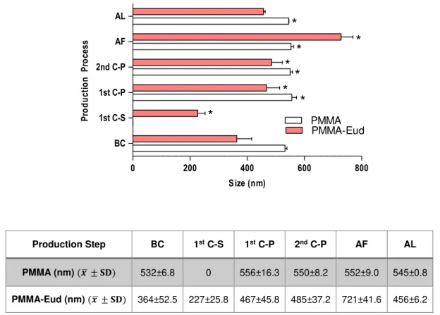
Figure 15: Production phases and its influence in size range distribution ... 38
Figure 16: Incubation process and the effects on size distribution. ... 39
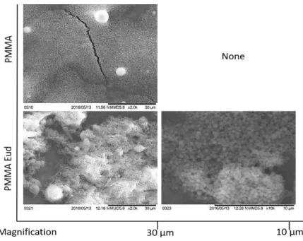
Figure 17: NPs dispersed in dH2O. ... 41
Figure 18: Size measurement of PMMA-Eud dispersed in dH2O. ... 41
Figure 19: NPs incubated for 3 hours with undifferentiated MG-63. ... 42
Figure 20: Normal MG-63 in subconfluence ... 43
Figure 21: Cell culture of normal and differentiated MG-63 during five days ... 43
Figure 22: Absorbance values in AP assay ... 44
Figure 23: Uptake assay considering two tested concentrations of PMMA and
PMMA-Eud. ... 46
Figure 24: Results of the viability assessment of PMMA and PMMA-Eud in
undifferentiated MG-63 cells ... 48
Figure 25: Results of the viability assessment of PMMA and PMMA-Eud in
differentiated MG-63 cells ... 49
Figure 26: ROS production by H2DCFDA assay in differentiated cells.. ... 49
Figure 27: Results of the Comet Assay for undifferentiated MG-63 cells after 3 hours
exposure to PMMA and PMMA-Eud.. ... 50
Figure 28: Results of the Comet Assay for undifferentiated MG-63 cells after 24 hours
exposure to PMMA and PMMA-Eud. ... 51
Figure 29: Micronucleated binucleated cells (MNBN) frequency per 1000 BNC cells.. 52
Figure 30: CBPI estimated for undifferentiated (A) and differentiated (B) MG-63cells
after treatment with PMMA and PMMA-Eud for 64h ... 52
Figure 31: RI estimated for undifferentiated (A) and differentiated (B) MG-63 cells. .... 52
XIX
List of Tables
Table 1: Conditions applied for each formulation ... 23
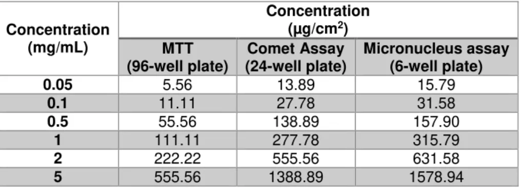
Table 2: NPs concentration used on cellular assays in different well-plates ... 29
Table 3: Yield of production of PMMA and PMMA-Eud after lyophilisation ... 37
Table 4: Surface charge of NPs dispersed in different medium composition ... 40
Table 5: Absorbance values of differentiated and normal MG-63 and HEK293T cell line
... 45
XXI
List of Acronyms and Abbreviations
Abbreviation Meaning
ATCC American Type Culture Collection
AP Alkaline Phosphatase
BCA Bicinchoninic Acid
BNC Binucleated Cell(s)
BSA Bovine Serum Albumin
CBMN Cytokinesis-Block Micronucleus
CBPI Cytokinesis-Block Proliferation Index
Cyt-B Cytochalasin-B
DCM Dichloromethane
DMSO Dimethyl Sulfoxide
EDTA Ethylenediamine Tetraacetic Acid
Eud Eudragit® RL 100
EMS Ethyl Methanesulfonate
FDA Food and Drug Administration
FPG Formamido-Pyrimidine-DNA-Glycosilase
ICH International Council for Harmonisation
ISO International Organization for Standardization
LDV Laser Doppler Velocimetry
LD Laser Diffraction
H2DCFDA 2-7’ dichlorodihydrofluorescein diacetate
MMA Methylmethacrylate
MMC Mitomycin C
MNi Micronucleus
MNBNC Micronucleated Binucleated cell(s)
MRSA Methicillin Resistant Staphylococcus aureus
MTT [3-(4,5-dimethylthiazol-2-yl)-2,5-diphenyltetrazolium bromide]
NB Nuclear Bud
NM Nanomaterial
NP Nanoparticle
NPB Nucleoplasmatic Bridges
OD Optical Density
OECD Organisation for Economic Co-operation and Development
PBS Phosphate Buffered Saline
PMMA Poly(methyl methacrylate)
PVA Poly(vinyl alcohol)
XXII
RI Replicative Index
RPMI Roswell Park Memorial Institute
SB Strand Breaks
SD Standard Deviation
QSAR Quantitative Structure Activity Relationship
RANK Receptor Activator for Nuclear Factor K
RANKL Receptor Activator for Nuclear Factor K Ligand
RFU Relative Fluorescence Units
ROS Reactive Oxygen Species
RT Room Temperature
SEM Scanning Electron Microscopy
SESE Single Emulsion Solvent Evaporation
SDS Sodium Dodecyl Sulfate
TB Trypan Blue
1
1. Introduction
1.1 Nanoparticles and Nanotechnologies
The rapid expansion of nanotechnology has been widely spread in our society and
becoming increasingly important in multiple areas of interest not only in the medicine and
pharmacology, but also in food industry (packing and additives), cosmetics, eletronical devices,
paints, clothing, etc (Arora et al. 2012). Different consumer products with nanoparticles (NPs) in
their composition are available and daily consumed. When Richard Feynman faced the nanoscale
(10-9 m), he also discovered new properties and functions from common chemical elements not
described for classical laws of physics (Sanchez & Sobolev 2010). NPs present size-dependent
properties. Their surface-volume ratio comprises a higher percentage of surface atoms leading to
reactive materials that tend to interact with other molecules (Klabunde 2009). Besides the high
reactivity, NPs also present particular physicochemical features, including electrical conductivity,
optical and magnetic properties that render them very attractive for multiple industrial and
biomedical applications.
One of the controversies around nanotechnology is the definition of nanomaterial (NM).
The European Commission published a specific recommendation (2011/696/EU) proposing a
definition for nanomaterial where 50% or more particles (in an unbound, aggregate or an
agglomerate state) belongs to a distribution range between 1-100nm (Rauscher et al. 2015).
However, the nanoscale is also the scale at which properties of materials are different than they
are at the macro or microscale, although this characteristic is not comprised in the definition. For
this reason, in nanomedicine, it also include particles up to 1000 nm, regarding potential materials
with new medical applications (Wagner et al. 2006).
Among the above mentioned as distinct and valuable properties of nanomaterials,
nanomedicine has emerged with the intention of adopting these materials for medical diagnosis
or therapeutics to improve health strategies. In fact, the ability of NPs to cross biological barriers
can provide applications in drug delivery, imaging and diagnostic, therapies and novel of drug
discovery (Wagner et al. 2006). Among all this diverse world that is encompassed by the
nanomedicine, an approach for solving osteomyelitic infections and further problems associated
2
1.2 Bone grafts and Osteomyelitis: A challenge to find new therapeutic approaches
1.2.1 Bone structure and formation
Bone is a complex and mineralized form of connective tissue that provides mechanical
support to the whole body (Murugan & Ramakrishna 2005). With the ability of self-regeneration
and self-remodelling during lifetime, bones are a well-protected organ remotely predisposed to
infections and fractures (Grabowski 2015). Osteogenesis can be accomplished by
intramembranous ossification or by endochondral ossification. The first one is assigned to the
development of craniofacial bones and the second is responsible for the other structures present
in the body (Gilbert 2000).
The endochondral ossification is conducted by multiple and synchronized actions
performed by different cell types (Gilbert 2000). Bone formation begins with mesenchymal cells
condensation and posterior differentiation into chondrocytes that are cells able to form
cartilaginous tissue. Chondrocytes proliferate until they become hypertrophic and apoptosis
events are induced (Clarke 2008). This gap allows vascularization and the influx of osteoblasts.
These cells mediate bone matrix formation and mineralization, leading to a complete replacement
of cartilage by bone. Bone resorption is carried out by osteoclasts, another important constituent
of bone (Grabowski 2015). Bone is described as a dynamic tissue because is constantly being
resorbed by osteoclasts and replaced by osteoblasts, thus assuring bone remodelling (Ducy et
al. 2000).
1.2.2 Bone grafts
In spite of bone being a well-protected organ of our body, it is still prone to degeneration,
pathology and trauma that may lead to the destruction of bone integrity. This tissue has the ability
of self-regeneration, but this feature also tends to be reduced by age and cumulative injuries.
Thus, external intervention is often required. Murugan et. al (2005) reports 550.000 cases of
surgical interventions related to bone grafting per year in U.S.A. and it tends to increase. Bone
grafting can be defined as a replacement of the damaged area and restoring of bone volume and
structure (Bagherifard 2017). There are different ways to perform bone grafts: it can be
autologous, allograft or synthetic. Autologous grafts are associated with the use of bone from the
same individual. Usually, it is harvested from non-essential zones such the iliac crest or Gerdy’s
tubercle (tibia). Allografts are similar to autografts, but the bone material is removed from another
patient. It can be harvested or donated by bone/tissue banks (Finkemeier 2002). Synthetic grafts
are related to the development of manufactured materials that can mimic bone architecture and
3
Autografts and allografts are regularly surgeon’s first choice. It happens because these types of inserts are easily re-vascularized and well-accepted by the body (Finkemeier 2002). The
grafts are also osteoinductive and osteogenic, which is essential to regenerate the lost bone and
recover its normal activity. However, these procedures present further problems leading to donor
site morbidity, excessive inflammation, pathogen transfer (allografts), among others (Bagherifard
2017). A statistical study has shown rates between 9 and 21% of major and minor complications
associated with autologous bone grafts (Finkemeier 2002). Besides that, surgical procedures
represent a huge obstacle as well.
The search of synthetic materials that enable bone regeneration has dramatically
increased over the last years. This search has included not only the creation or adaption of
bio-inert materials, but also the introduction of bioactive compounds to integrate and regenerate the
lost tissue. Despite being a broad field of interest, the most used biomaterials are bioactive
ceramics or glasses, biological and synthetic polymers. Hydroxyapatite-based material, hydrogels
(e.g. polyethylene glycol), bioactive glasses with Ca2+, polylactide, polyglycolide, poly(methyl
methacrylate) and polyesters are just a few examples of a very extended list of the available
materials that can be used in orthopaedic implants (Stevens 2008; Yu et al. 2015)
However, surgical interventions and medical devices implantation, promote an imbalance
of the immune system. This can lead to pathogenic organisms’ migration to the body leading to
infection development, secondary to surgeries and orthopaedic implants.
1.2.3 Bone infections: Osteomyelitis
Among pathogenic microorganisms, Staphylococcus aureus and Staphylococcus
epidermidis are by far the most commonly involved in joint infections. Usually, bone and joint
infections associated to these bacteria are defined as osteomyelitis. This condition generally
results in bone destruction and necrosis, and the spread of inflammation to other regions (Birt et
al. 2017). When bacteria competently enter in the host tissue and are able to reproduce, they
induce an acute inflammatory reaction. S. aureus expresses on their surface adhesins (e.g.
laminin and fibronectin) that promote attachment to the host (Foster 1996). The specific receptor
that promotes adherence to collagen is particularly associated with strains that cause
osteomyelitis and septic arthritis.
S. aureus and S. epidermidis also have a predisposition to form biofilms on medical
devices. Biofilms represent a complex group of microbial cells that adhere and colonize the
surfaces representing a serious problem not only for the patient but also a public health issue
(Donlan 2001). A better understanding of biofilm formation mechanism can provide new
perspectives of successful treatments. As shown in figure 1, this process comprises different
stages: the first one starts with the adhesion of free bacteria on the medical device. They start to
4
formation and posterior stabilization. A cumulative cell grow, leads to the production of several
layers on the prosthetic surface. Polysaccharides are produced as well, forming a barrier to
protect microbes and enabling a matured biofilm. A significant decline on nutrients is experienced,
so bacterial cells disperse from the mature film and enter into the bloodstream spreading the
infection to other tissues (Veerachamy et al. 2014).
Figure 1: Biofilm formation process on a medical device. Adapted from Veerachamy et al. 2014
In a dense cell assembly, it is also expressed an altered phenotype, gene expression and
protein production. This strongly reduces the chances of eradicating the infection. For this reason,
an early and accurate diagnosis can help to decrease the spread of the disease. The diagnostic
procedures used in osteomyelitis often requires bone biopsy. After that, cell culture of infected
bone, peripheral blood cell counts, erythrocytes sedimentation rates and serum protein C-reactive
are analysed and are usually increased if the patient experienced osteomyelitis (Lew & Waldvogel
2004). However, different studies revealed variations on these markers, appearing increased or
decreased due to other infections, as the initial or advanced stages of the infection also affect the
results (Davis 2005). Imaging methods also play a major role in diagnosing skeletal infections.
Radiography, for example, can assess soft tissues, narrow joint spaces and bone destruction. But
between 10-21 days of infection, bone destruction is still not clear. Other different imaging
techniques with high power resolution are able to perform an accurate diagnosis. A computed
tomography or a magnetic resonance imaging using radiopharmaceuticals may present very
detailed results (Lew & Waldvogel 2004). The major drawback associated with these processes
are the high costs, making them not generally available for the entire population.
Treatments associated with osteomyelitis are essentially based on antibiotics use. A
combined antimicrobial and surgical procedure are usually considered when osteomyelitis reach
an advanced stage (chronic osteomyelitis) (Davis 2005). Diagnosis already represents an
obstacle and choosing adequate antibiotics may represent another problem. The approach tends
to use a two-drug combination trying to cover the most recurrent microorganisms in bone
infections. Long and invasive administrations represent high costs and further complications
5
of Methicillin-resistant S. aureus (MRSA) and Vancomycin-resistant S. aureus (VRSA) strains has
been reducing the prospect of infection eradication (Lew & Waldvogel 2004). Other antibiotics,
such daptomycin, have shown interest results regarding fewer side effects for the patient and
active penetration in biofilms, another significant barrier to antimicrobial agents (Mascio et al.
2007).
With the present alternatives, osteomyelitis still represents a great financial burden and
reduces life quality for patients. New diagnose, and treatments are essential to overcome these
problems and to assure improvement in life expectancy.
1.3 Novel nanoparticles-based therapeutics for Osteomyelitis
Novel materials and formulation at the nanoscale are being developed to act like drug
delivery-systems revealing great benefits. NPs are able to promote an effective delivery of high
doses of a drug at target sites during larger periods of time and with reduced systemic
toxicity(Bettencourt & Almeida 2014). In other words, it will regulate the biodistribution and
enhance the therapeutic index of drugs. It may represent an appropriated option to eradicate the
biofilm formation.
1.3.1 Polymeric nanocarriers for drug delivery
Polymeric NPs represent a milestone on the drug delivery field. This is due to their
increased colloidal stability, good chemical resistance, and the easy surface functionalization.
This nano-scaled drug delivery system can provide a controlled release and an efficient targeting
process (Goldberg et al. 2011).
1.3.2 Nano-sized Poly(methyl methacrylate) and Eudragit formulations
Poly(methyl 2-methylprop-2-enoate) (IUPAC) or, more commonly, Poly(methyl
methacrylate) (PMMA) is a synthetic and amorphous homopolymer of methylmethacrylate (MMA)
monomer (Bettencourt & Almeida 2012). As presented in figure 2, PMMA is a building block of
6
Figure 2: PMMA structure. In red are presented oxygen atoms and in blue are hydrogen atoms. Adapted from PubChem Compound Database; accessed on Feb. 3, 2017.
PMMA can also be described as a thermoplastic with glass transition temperature of
105ºC (Bettencourt & Almeida 2012). It is soluble in most organic solvents, but poorly soluble in
water (50.5 mg/mL, at 25ºC). However as it comes in contact with water, the contact angle tends
to decrease and the NPs become slightly hydrophilic (PubChem Compound Database; accessed
on Feb. 3, 2017). Their polymerized form is found in many products, in multiple areas. Due to its
optical properties, PMMA is often used as implantable intraocular and contact lenses, and even
as a glass substitute (Santos et al. 2011). It also has applicability in dental and mandibular
implants, but it is in orthopaedic surgery the most important appliance of PMMA. This polymer is
often used as a bone cement for total hip replacement or for other joints such knee, shoulders,
and elbow, for almost 40 decades. Therefore, PMMA is defined as a bioinert and biocompatible
polymer with remarkable toxicological safety record, being a Food and Drug Administration (FDA)
approved material (Bettencourt & Almeida 2014). For musculoskeletal infections, the use of
PMMA as a nanocarrier for the local delivery system may be a more efficient alternative to the
conventional antibiotics administration. For this purpose the drug is entrapped or dispersed into
a cavity and surrounded by the polymer membrane (Kong 2015).
The major drawbacks associated with this polymer are the fact of being a
non-biodegradable and hydrophobic material. The first one may require surgery to remove the material
which is painful to patient and high-cost associated. The hydrophobicity affects the drug realising
profiles for undefined periods favouring the growth of resistant strains of bacteria (Gomes et al.
2013). Strategies to improve drug release profiles and to avoid drug retention in the reservoir
include the synthesis of PMMA composites with hydrophilic polymers.
Eudragit® RL 100 (Eud) is a synthetic polymer, industrially produced and commercialized
by Evonik Industries, Germany. Eud is a copolymer of ethyl acrylate, methyl methacrylate and
7
Figure 3: Chemical structure of Eudragit® RL 100. Evonik brochure, accessed on May 12, 2016.
It has in its composition quaternary ammonium groups (positively charged particle)
conferring permeability to the polymer (Evonik, accessed on May 12, 2016). Eud is mostly used
in pharmaceutical formulations to obtain a controlled and desirable drug delivery performance. It
is insoluble at physiologic pH and able to limited swelling, representing a suitable material for drug
dispersions (Das et al. 2010). Some studies with PMMA-Eud formulations in antibiotic-loaded
microparticles have been already performed showing improved results in permeability,
encapsulation, and release profiles when compared with PMMA formulations (Ferreira et al.
2015). Nanoparticles with both formulations were produced with the purpose of exploring new
medical features at nanoscale conditions.
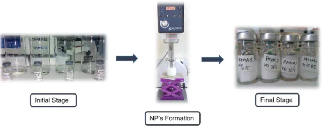
1.3.2.1 PMMA and PMMA-Eud Production
Several techniques have been developed for the last decades to produce micro- and
nanoparticles. Particles can be prepared from a preformed polymer or by direct polymerization
from a monomer solution (Bettencourt & Almeida 2012). The first methodology uses a preformed
polymer and it is frequently applied for polymers that cannot be formed by radical polymerization
[e.g.,: poly(lactic acid), poly(glycolicacid), etc.]. It comprises salting-out, solvent evaporation,
supercritical fluid technology and other processes depending on the main purposes of the study.
Direct polymerization, on the other hand, requires a chemical (e.g. ammonium persulphate) or
physical initiation (e.g. gamma radiation) and encloses several techniques being emulsion-based
procedures the most described in the literature (Ferreira 2015).
PMMA nanospheres have already been characterized and produced by different
techniques, but the emulsification-solvent evaporation with a single oil-in-water emulsion (o/w) is
still one of the most popular methods. This technology implies an emulsification of PMMA polymer
(hydrophobic) in an organic phase using dichloromethane (DCM) as a solvent. Non-ionic
surfactants such as poly(vinyl alcohol) (PVA) are also added, providing hydrophilicity to the
8
step is important to prevent aggregation, a major problem in micro and nanoparticle production.
PVA was demonstrated to be an effective surfactant in PMMA formulations. After the
emulsification process, the solvent is evaporated by stirring at RT, resulting in precipitation and
consequent formation of polymer particles.
In spite of being a fast and easy technique to execute, single-emulsion with solvent
evaporation (SESE) has some concerns related to the use of organic solvents and surfactants
that may bring some toxicological issues or even deposit in the formulations. Following the
International Conference on Harmonization (ICH) guidelines that classify DCM as a Class 2
solvent (“Solvents to be limited”), it restrains the maximal residual concentration to 600 ppm (ICH 2011). Taking this into account, it was demonstrated by Florindo et al. (2010), that is possible to
remove great amounts of this solvent after evaporation and keep far behind the established
values. All results were confirmed by nuclear magnetic resonance spectroscopy (Bettencourt &
Almeida 2012) (Florindo et al. 2010). Furthermore, the washing steps taken by protocol also help
to remove some chemical depositions in nanoparticles.
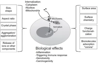
1.4 Characterization of physicochemical properties
The most relevant physicochemical properties that have been shown to affect NP
behaviour are presented in figure 4 as their possible impact on cells. Size constitutes the most
obvious characteristic to consider. It regulates NP internalization and the ability to trigger or shield
our immune system. At the nano level, reductions in size turn into a high surface-to-volume ratio,
leading to extremely reactive NPs. Thus, particle size could be directly proportional to its toxicity.
For example, smaller particles (<100 nm) tend to cause a higher inflammatory reaction due to
their biopersistence in the body (Sutariya & Pathak 2015).
Shape/morphology and surface charge seems to affect NP uptake as well. NPs assume
various shapes including fibres, tubes, spheres, etc. This characteristic is deeply connected to
membrane wrapping process. Sphere NPs have already shown easier endocytosis when
compared to rod or fibre NP (Gatoo et al. 2014). Associated with shape is also toxic. Studies with
single- and multi-walled carbon nanotubes present great differences regarding cytotoxic events
(Oberdörster 2010). On the other hand, cationic NPs display improved internalization efficacy
when compared with anionic or neutral NP, but also induce a higher toxicity when interacting with
cellular components (Louro, et al. 2015). Besides, when NPs enter in a biological fluid, a protein
coating is formed around the surface. This phenomenon is called “protein corona” and may
enhance or reduce the cell uptake (Hocherl et al. 2012). NPs association or dissociation of
proteins and current exchange with free proteins present on the boundaries will change some of
their physicochemical properties and will mediate the biological response among surfaces and
receptors or in the endocytic pathways (Nel et al. 2009). These interactions promote shifts in
9
Figure 4: Physicochemical interactions between nanoparticles and biological compartments. Adpated from Louro et al. 2015.
Another important property for analysis is the dynamic behavior of NPs, i.e., aggregates
and agglomerates formation. Agglomerates are formed when NPs are dispersed and held
together by weak physical interactions leading to the formation of precipitates. This is an easily
reversible process. Aggregates instead, are formed by strongly bounded NPs forming a cluster
and, for that reason, the process is irreversible (Sokolov et al. 2015). These phenomena are
determined by size, surface charge, composition of NPs and the chosen dispersant medium.
Accumulation of aggregates for extended periods of time may lead to toxicity and reduced uptake
mechanisms for larger particles (Gatoo et al. 2014). Regarding these properties, the size,
superficial charge, morphology and aggregation/agglomeration state were evaluated during this
work.
Particles size were accessed and measured by laser diffraction, during the NPs
production process, in order to correlate how alterations in temperature, medium composition and
centrifugal forces can induce size variations. Surface charge was estimated by zeta potential (ζ)
after the production process in different dispersant mediums to evaluate how medium
compositions interact with the NPs surfaces. Finally, the morphology and consequent
agglomerate/aggregate formation was evaluated by Scanning Electron Microscopy (SEM)
considering the NPs in their “native state” dispersed in dH2O and after an incubation with human
10
1.5 Cellular interactions and potential toxicity of nanoparticles
1.5.1 Nano-bio interactions
The nano-sized materials and biological structures are within the same size ranges, which
facilitate the nano-bio interactions. Indeed, these interactions consist of physicochemical
interactions, kinetics and thermodynamic exchanges between the nanomaterial surface and
biological entities such as membranes, proteins, organelles and DNA. The differences existent
among physical states of a NP and the biological substrates lead also to solid-liquid interactions.
When the solid NP is exposed to liquid environments, cells experiment several effects on this
interface. The first interactions happen at cellular membrane level where specific (receptor-ligand)
and non-specific binding occur. From these interactions, structural, functional and conformational
changes may be triggered and developed in biomolecules. Membrane structures are capable of
wrap and uptake a NP e.g., by endocytosis, leading to new cellular interactions (inter and
intracellular effects) (Nel et al. 2009).
In this perspective, severe effects can be devised from the existing nano-bio interactions
at cellular and molecular levels. The importance of understanding and categorizing these
outcomes is fundamental to assure the safety of nanomaterials and nanodevices. Recent efforts
around the world have been made to recognize the benefits of nanotechnology while minimizing
the potential risks. In this context, the “safe-by-design” concept has gained substantial importance over the last years. Reducing population exposure and assuring safe manufacturing
processes and reliable products are the gold standard of this approach (Louro et al. 2015). In
other words, a material/product should be engineered in its less hazardous nanoform (e.g.size,
shape) and in an cost-effective way. Prediction tools such Quantitative Structure Activity
Relationships (QSARs), read across and high-throughput screening (in vitro/in vivo) should be
adopted in the early stage to select better formulations (Dekkers et al. 2016). The next step to
consider is the exposure risks for the consumers and the environment, and reduce them as
possible. Industrial safety procedures include not only secure infrastructures but also safe
conditions for workers to handling nanomaterials, storage and transport them. Building a product
based on these pillars (safe design, safe use of products and safe industrial procedures) will
potentiate a safety course not only in our health but also in the ecosystems (figure 5)
11
Figure 5: Safe-by-design concept is held by 3 important pillars: Safe design, Safe use of the products and Safe industrial procedures to obtain the final product.
The challenge remains on the fact that nanomaterials with similar chemical composition
have quite different behaviors when compared to macroscale materials. In fact, a variation on a
specific size, shape, or superficial charge seems to influence its toxicity (Dekkers et al. 2016).
Thus, the chemical and physical properties of each nanomaterial should be deeply characterized
to better evaluated its potential adverse effects to human health and to the environment.
1.5.2 Nanotoxicology
Furthermore, cell interactions must also be considered within the safety assessment of a
nanomaterial. Nanotoxicology is an area of toxicology that addresses the acute and chronic
adverse effects of nanomaterials taking into account their psychochemical properties.
Nanotoxicology diverges for the conventional toxicology area in that due to their small size, NPs
behave differently, resulting in a distinct toxic profile (Bhattacharjee & Brayden 2015).
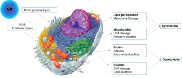
The mechanisms inherent to NPs toxicity comprise different endpoints and can mediate
cytotoxic or genotoxic responses by biological entities. As presented in figure 6, NPs can directly
or indirectly induce cellular dysfunctions with impacts on essential cell components: membrane,
mitochondria and nuclear compartment. The outcomes from this unbalance, will produce DNA
lesions including strand breaks, oxidized and alkylated bases, bulky adducts and intra/inter-strand
cross-links (Pillco & Peña 2014). Moreover, genetic instability will modulate inflammatory
responses (i.e. macrophages and neutrophils) leading to genotoxicity and cell death (Jiang & Gao
2017). Eventually, chronic inflammation can occur in case of biopersistency and accumulation of
NPs in the body. Repair pathways and antioxidant mechanisms work as a first mechanism of
cellular defence and DNA repair systems for more depleting effects that cannot be easily repaired. Safe
industrial production
Safe use of products Safe
12
Figure 6: The mechanisms of nanotoxicity under cellular structures. Adapted from (Jiang & Gao 2017), picture available on http://classes.midlandstech.edu/carterp/Courses/bio210/chap03/lecture1.htm.
There are relevant bioassays to assess the toxicity of chemicals and also international
recommendations about the most adequate battery of assays that should be used to ensure that
a given substance is safe for human health. Considering that NPs are synthesized for biomedical
applications, the focus will be on the one hand to understand their biocompatibility and, on the
other hand, to assess their potential toxicity. For this purpose, there are still few standard
operating procedures to allow the standardization of bioassays towards a complete evaluation of
the harmful effects generated by NPs.
1.5.3 In vitro experimental models
Cell culture processes are an essential tool for diverse areas and applications. Cancer cells
are commonly used since they can be established in simple culture media and proliferate
indefinitely, contrarily to non-transformed cells. The most standard systems used for bioassays
are adherent two-dimensional (2D) cell monolayer (Edmondson et al. 2014). These cultures
represent the gold standard for research although they still provide limited information about the
whole-organisms responses (Ravi et al., 2015). In this work, MG-63 a human
osteosarcoma-derived cell line from American Type Culture Collection (ATCC ® CRL- 1427 ™) was chosen to
assess NPs toxicity. This cell line was derived from an explant culture of a osteosarcoma tissue
from a 14-year-old caucasian male. It presents a fibroblast morphology and is adherent under
culture conditions. This osteoblastic cell line can be induced to differentiation in culture, thereby
allowing to comparatively analyse the toxic effects of nanoparticles in different stages of cell
13
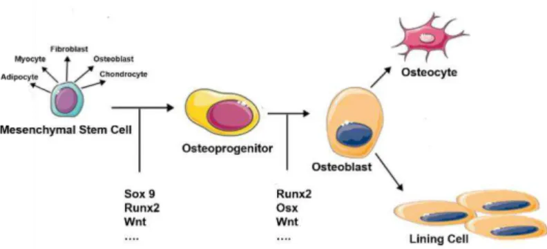
1.5.3.1 Osteoblasts
Osteoblasts arise from a mesenchymal stem cell as other different cell types such
adipocytes, myocytes, chondrocytes, and fibroblasts (figure 7). Differentiation of osteoblasts is
coordinated by several genes, but Runx2, Sox 9 and Osterix are the most important ones. After
differentiation, osteoblasts can follow two different paths: they can differentiate into osteocytes
embedded in a bone matrix, or in lining cells disposed in the bone surfaces (Grabowski 2015).
Figure 7: Osteoblast formation and differentiation. Adapted from Arboleya & Castañeda 2013; Grabowski 2015.
Osteoblasts are involved in bone matrix formation and in the regulation of the osteoclasts
activity. Osteoblasts have an important role in the organic phase of bone matrix composition
(Murugan & Ramakrishna 2005). They secrete collagen (mostly Type I) and non-collagenous
proteins. In general, it serves to regulate bone mineral deposition and bone cell activity. The main
non-collagenous protein present in bone is alkaline phosphatase. It is a glycosylated protein
linked to osteoblast surface, but it can also be found free in mineralised matrix (Clarke 2008,
Boskey 2013). This glycoprotein is imperative in bone mineralization, although it is still a poorly
understood process (Grabowski 2015). Furthermore, additional glycoproteins are also
synthesized by osteoblasts enabling great amounts of calcium and other minerals deposition.
Respecting to osteoblast-osteoclast interaction, it involves important signalling bone
mechanisms and the immune system. Osteoblasts trigger osteoclast differentiation via
RANK-RANKL (Receptor Activator for Nuclear Factor K Ligand) (Jayakumar & Silvio 2014). Osteoclasts
are derived from the macrophage lineage, supporting one of the main functions of this cell line
which is bone destruction. Keeping the number and the activity of osteoclasts well controlled is a
healthy issue. For instance, if many osteoclasts are active, they will destroy too much bone and
osteoporosis will arise, and so, other disorders (Gilbert 2000).
As already referred, osteoblasts are an intrinsic part of bone growth, development, and
maintenance. For that reason, the supervision of biochemical and morphological changes of
14
1.5.4 Cellular uptake
When a NP is being studied to be used as a nanocarrier, high uptake efficiency must be
demonstrated. Different characteristics of NPs can influence the capability of being uptaken by
the cell (Singh & Ramarao 2013). Among them, size, chemical composition and the surface
charge seem to be those that most influence the NPs internalization. Different mechanisms can
be responsible for the cellular uptake of NPs: It can occur by diffusion, by specific transport
channels (for smaller particles), or by endocytic pathways. The latter one involve invaginations of
the cell membrane (e.g. clathrin or caveolae-mediated systems) or even extensions of cell
membrane, including macropinocytosis and phagocytosis (Hocherl et al. 2012). Several studies
explore the uptake mechanism without distinguish between the level of internalized and the
adsorbed results from NPs. The fluorescence resulting from the interaction between the proteins
adsorbed to NPs surface and in the cell membrane provide a global but not a correct signal
associated to NPs internalization. This may lead to an unappropriated determination of NPs
concentration that actually are internalized. The methodology employed in this work intends to
differentiate the intracellular fluorescence by internalized particles from the background
fluorescence that comes from the adsorption of nanoparticles on the cell surface. External
fluorescence was removed using the vital dye Trypan Blue (TB) that is incapable of penetrating
in intact cell membranes and can efficiently quench that background (Vranic et al. 2013).
1.5.5 Cytotoxicity assessment
From a pharmacological point of view, cytotoxicity assays are used as a first screening to
test different experimental conditions, observe cellular response and suggest the most
appropriated concentration-range to further explore. There are many assays available to
characterize the cytotoxic potential of a compound. These assays are usually indicators of cellular
damage. Better results are obtained when different endpoints are tested and discussed and thus,
complementary assays can be used, depending of the study objective. However, concerns related
to NPs adsorption to dyes (e.g. Carbon nanotubes) frequently used to measure cell viability
through colorimetric assays may produce false positives and this aspect should be taken into
account when choosing a methodology (Bhattacharjee & Brayden 2015). In order to figure out
possible cytotoxic outcomes from a PMMA and PMMA-Eud exposure, cell viability and production
15
1.5.6 Oxidative stress
Reactive Oxygen Species (ROS) are chemical species formed upon incomplete reduction
of oxygen. It includes O2-, H2O2 and HO·. These are extremely reactive molecules and interact
with different biomolecules such as proteins, phospholipidic bilayer and even with DNA molecules
(D’Autréaux & Toledano 2007). For this reason, ROS can induce several adverse effects on cell metabolism, but also can serve as an important intracellular messenger/signalling molecule. In
spite of harmful side effects, these molecules are also constantly produced as by-products of
aerobic respiration with no damage to cells. To aerobic organisms, it is imperial to assure total
detoxification of ROS by cell defence mechanisms in order to maintain a balance between
production and removal of oxygen species. When it is not possible to preserve this homoeostasis,
oxidative stress is triggered (Held 2012).
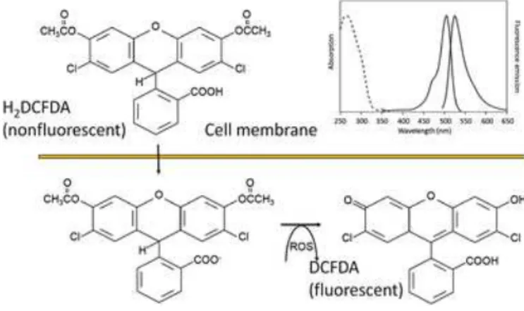
Oxidative stress can be quantified in cells in culture based on specific probes, e.g., 2-7’
dichlorodihydrofluorescein diacetate (H2DCFDA). This molecule can pass through cell
membranes (figure 8) being hydrolyzed by intracellular esterases. The result is a charged
compound that stays trapped inside the cell. If intracellular ROS exists, this enzyme will oxidize
H2DCF to DCF, converting a non-fluorescent compound to a highly fluorescent dye (Chin et al.
2011).
Figure 8: Formation of a fluorescent compound by the specific probe (H2DCFDA) indicating oxidative stress levels.
Adapted from Chin et al. 2011
It is a fast and easy technique that provides information on stress levels from cell
populations. This probe is not specific for an individual reactive species, but instead, it seems to
16
1.5.7 Genotoxicity assessment
Genotoxicity assays are presented as a landmark on safety assessment not only for NPs
but also for the characterization of other potentially harmful compounds. A NP or a genotoxic
agent can interact with DNA or other cellular targets, compromising the integrity of the genetic
material. NPs can promote genetic damage either by primary or secondary interactions with DNA
molecules. Primary mechanisms can occur by direct or indirect NP-DNA association. Direct
primary DNA damage requires NPs internalization into the nuclear compartment provoking
physical injury into DNA structure. These will lead to DNA lesions (strand breaks and intercalating
NPs with DNA base pairs) or even mutagenic events if it compromises the DNA repair systems.
Indirect interactions presuppose that the damage is induced by other molecules (i.e. proteins,
repair enzymes, unbalanced ROS production, etc.) that will affect cell replication and division
cycles or even the by-products of inflammation that is often triggered by the NPs into the cellular
environment. The secondary interactions can be described as the ones that happen between
different and organized cells, mostly like it goes on in the in vivo situation, during a body stimulus
(Evans et al. 2016). It combines different cellular responses and mechanisms that correspond to
a more realistic genotoxic evaluation of an NP. This approach can only be explored using in vivo
models, even though, a plenty of in vitro tests must be performed in a first-line of action to
characterize and assess NPs toxicity and to support a safe-by-design practice. Considering the
primary interplay of NP-DNA, only a few studies were reported suggesting that NPs can enter in
the nucleus and promote direct physical injury to DNA structure. Lovrié et al. (2010) showed that
quantum dots within the size range of 2-3nm are able to induce genotoxicity by direct interaction
with the main nucleus. Since PMMA and PMMA-Eud are NPs of 500 ± 50 nm size, that pathway
will not be considered as a probable mechanism of genotoxic induction. Instead, an indirect
genetic damage can be expected to occur.
Alterations at DNA level are intimately connected to several human genetic diseases
including cancer. DNA is constantly exposed to mutagenic compounds that can cause serious
damage to the human genome. Therefore, the identification of compounds that may have a
mutagenic or carcinogenic activity is essential not only for drug development but for controlling
human and environmental exposure. Different genotoxicity assays have been developed to detect
DNA damage. Procedures that require the use of small cell samples and are able to evaluate
DNA damage based on single cell analysis are always valuable.
1.5.7.1 Characterization of the DNA damage: Comet assay
The Single-cell electrophoresis or Comet Assay is a fast and consistent technique to
assess DNA damage and repair in individual cells (Glei et al. 2016). For many reasons, the comet
assay is an essential tool in toxicological research. It allows to understand background levels of
17
their repair capacity (Collins 2004). The comet assay in vivo, has already been reported in OECD
Guideline 489 as a standard test to execute the “Testing of Chemicals” (OECD 2014). It has multiple applications in the detection of genotoxic potential, monitoring tests
(ecological/environmental or even human biomonitoring) and in clarifying fundamental
mechanisms of DNA damage and repair (Collins 2004). The standard comet assay and a
modification using a DNA repair enzyme were used in this work.
The method relies on the migration of lysed cells embedded in agarose on a microscope
slide, where an electric current is applied. Agarose assures that DNA is immobilized for the
electrophoresis run (Vandghanooni & Eskandani 2011). Alkaline single-cell electrophoresis is
performed at a high pH (≈13). This alkaline environment in electrophoresis allows the unwinding of the supercoiled DNA structure. Loops that contain breaks are then extended by electrophoresis
process, forming a “comet tail” (Louro, et al. 2015). The alkaline medium also makes comet tails more pronounced and easier to detect (Collins 2004). Using this methodology, it is possible to
quantify the level of DNA Strand-Breaks (SB), but it is even possible to increase the assay
sensitivity and selectivity by applying lesion-specific enzymes, particularly, glycosylases. These
enzymes are able to convert oxidised bases into DNA breaks, increasing the comet tail (Collins
et al. 2008). Formamidopyrimidine DNA glycosylase (FPG) is an enzyme capable of detecting
adenine and guanine oxidation and convert it into a break (Collins 2004). During this work, the
comet assay was executed with and without this modification in order to determine also the level
of oxidative lesions comparatively to untreated cultures. Another important aspect to take into
account is the comet scoring. Only cells with a clear head and tail should be scored. The
percentage of DNA in tail corresponds to the intensity of the comet tail, and it is directly related
to the DNA breakage frequency (Glei et al. 2016). The DNA lesions quantified by the comet assay
correspond to primary and reversible lesions that can be repaired or, on contrary, lead cell to
death if it is highly damaged.
1.5.7.2 Characterization of chromosome damage: Cytokinesis-Block Micronucleus Assay
Severe DNA damage may be not reversible by cellular mechanisms of repair thereby
resulting in permanent damage to the cell. On the other hand, the cell cycle can be blocked in an
attempt to allow DNA repair and, if this is not possible, the programmed cell death (apoptosis)
can be triggered. Another possibility is the progress of cell division where the DNA damage is
transmitted to daughter cells either as gene mutations or as chromosome aberrations, inducing
deleterious defects and leading to cell transformation (e.g. carcinogenesis). The in vitro
18
validation and described in OECD Guideline 487 as standard procedure for detect genotoxic
events at the chromosome level.
The Micronucleus assay is one of the most important in vitro procedures to assess genetic
damage and characterize cytotoxicity and genotoxicity of a specific compound/chemical (Fenech
2000). When cells experience high levels of toxicity, micronucleus (MNi) can reflect a pronounced
effect of chromosome damage (loss or breakage). They originate from chromosome fragments
(acentric fragments) and/or whole chromosomes that are incapable of migrating to the poles of
the cell at anaphase stage (Fenech et al. 2003). A nuclear membrane is formed around the
genetic material and this corpuscle remains in the cytoplasm, morphologically similar to main
nuclei but smaller, which gave origin to the term “micronucleus” (Fenech 2000). MNi could be
only expressed in “active cells” that are able to complete cell cycle, so it is important to restrict
and distinguish cell population between dividing and non-diving cells. The CBMN assay is a slight
modification form of the traditional methodology since it allows this selection by addition of
cytochalasin-B (Cyt-B). Cyt-B is an inhibitor of actin polymerisation, essential to individualize the
two daughter cells in cytokinesis phase and thus its effect results in a binucleated cell following
the mitosis (Fenech 2007).
CBMN is also used to measure other important endpoints related to chromosomal
abnormalities such as: nucleoplasmatic bridges (NPBs); nuclear buds (NBs); cell viability
(necrotic and apoptotic aspect) (figure 9) and cytostatic effects.
19
The NPBs are formed from dicentric chromosomes that are pulled to opposite poles of the
cell in anaphase, the NBs and MNi have homologous structure, but NBs are still linked to the main
nucleus and is an important biomarker for gene amplification. Cytostasis represents the ratio
between mononucleated, binucleated and multinucleated cells in a population. All of these
parameters improve the detection of possible toxicological effects induced by a chemical
21
2. Objective
This projected was aimed at assessing the safety of polymeric nanoparticles developed
as nanocarriers for drug delivery, in a human osteoblast cell line. To achieve this purpose, the
following specific objectives were defined:
1) Production of plain PMMA and PMMA-Eud (50:50) with an average distribution size
range of 500 nm.
2) Physicochemical characterization of both sets of nanoparticles considering the
different conditions experienced not only in particles production and storage but also
in cellular studies.
3) Biosafety evaluation through cytotoxicity and genotoxicity characterization in a