Ana Rita Marques Lemos
Licenciada em Biologia
Molecular targeting of proteins by homocysteine:
implications in familial and clinical
hypercholesterolemia
Dissertação para obtenção do Grau de Mestre em
Genética Molecular e Biomedicina
Orientador: Sofia de Azeredo Pereira, Professora Doutora
NOVA Medical School, UNL
Co-orientador: Mafalda Bourbon, Professora Doutora
Instituto Nacional de Saúde Doutor Ricardo Jorge
Júri:
Presidente: Prof. Doutora Margarida Casal Ribeiro Castro Caldas Braga
Arguente: Prof. Doutor Rui Manuel Amaro Pinto
Vogal: Prof. Doutora Sofia de Azeredo Gaspar Pereira
iii Molecular targeting of proteins by homocysteine: implications in familial and clinical
hypercholesterolemia
Copyright Ana Rita Marques Lemos, FCT/UNL, UNL
A Faculdade de Ciências e Tecnologia e a Universidade Nova de Lisboa têm o direito, perpétuo e
sem limites geográficos, de arquivar e publicar esta dissertação através de exemplares impressos
reproduzidos em papel ou de forma digital, ou por qualquer outro meio conhecido ou que venha a ser
inventado, e de a divulgar através de repositórios científicos e de admitir a sua cópia e distribuição
com objectivos educacionais ou de investigação, não comerciais, desde que seja dado crédito ao
iv
v
The results discussed in this thesis originated:
Publications in international scientific journals:
Marinho A, Dias C, Pinheiro P, Lemos AR, Antunes A, Marques MM, Monteiro E, Miranda J,
Pereira S. 2015. Nevirapine modulation of paraoxonase-1 in the liver: an in vitro three-model
approach. European Journal of Pharmaceutical Sciences. DOI: 10.1016/j.ejps.2015.11.019
Dias CG, Lemos AR, Maia S, Almeida MG, Morello J, Coelho NR, Caixas U, Monteiro EC, Soto K,
Pereira SA. 2015. Monitoring the lactonase activity of paraoxonase-1 in HIV-patients: a plausible
target of active infection. Viral Immunology (submitted).
Oral communications in national meetings:
Lemos AR, Pereira SA, Bourbon M. 2015. Paraoxonase-1 activity in Familial
Hypercholesterolemia. Jornadas Intercalares das Dissertações Anuais dos Mestrados. Faculdade
de Ciências e Tecnologia – Universidade Nova de Lisboa, Lisboa, Portugal.
Marinho AT, Dias CG, Lemos AR, Caixas U, Soto K, Branco T, Antunes AMM, Marques MM,
Monteiro EC, Pereira SA. 2015. Apolipoprotein A1 sex-differences and nevirapine-induced liver
injury. XLV Reunião Anual da Sociedade Portuguesa de Farmacologia/ XXXIII Reunião de
Farmacologia Clínica/ XIV Reunião de Toxicologia, Lisboa, Portugal.
Poster communications:
Soto K, Dias CG, Campos P, Lemos AR, Marinho AT, Papoila AL, Pereira SA. 2015.
Paraoxonase-1 in early kidney dysfunction in HIV+ patients under protease inhibitors. ISN World
Congress of Nephrology, Cape Town, South Africa.
Lemos AR, Dias CG, Coelho NR, Casimiro NF, Bourbon M, Pereira SA. 2015. Molecular targeting
of proteins by homocysteine: implications in familial and clinical hypercholesterolemia. iMed 7.0
Conference, Lisbon, Portugal.
Proceedings:
Dias CG, Casimiro N, Coelho N, Lemos AR, Papoila AL, Maia S, Soto K, Pereira SA. 2015. Serum
non-protein bound homocysteine levels are related with chronic kidney disease progression in
vi
vii
A
GRADECIMENTOS
Vários agradecimentos são devidos a diversas pessoas por me terem ajudado a chegar até este
momento tão importante.
À Professora Doutora Sofia Pereira, pela forma como me recebeu no laboratório de Farmacologia,
pelos ensinamentos, motivação e disponibilidade constantes.
À Professora Doutora Mafalda Bourbon por me ter aberto as portas do Instituto Ricardo Jorge e,
através do projecto eCOR, me ter dado a conhecer o “grandioso” mundo das doenças
cardiovasculares.
À Joana Canilho, técnica do INSA, por toda a disponibilidade e simpatia a solucionar as minhas
dúvidas e a fornecer as infindáveis caixas de alíquotas.
Obrigada às meninas do lab: Aline, Nádia, Clara, Inês (e Mª João, Joanas, Bernardete, Cristiana)
que me receberam da melhor forma e me ajudaram nos momentos de dúvida. Aline por ter sido a
minha primeira professora de “PONs” e de HPLC. A tua genuína simpatia ajudou muito em momentos
mais stressantes. “Clarofsky”, a minha “co-co-orientadora”, ajudante e amiga. Aquelas sessões de
karaoke minion são inesquecíveis! Obrigada por tudo, mesmo.
Obrigada à Catarina por ser a “formiguinha” trabalhadora que veio revolucionar o laboratório para
melhor e a quem recorri em situações de pânico relacionados com o HPLC!
Nuno (Mr. Bifanas!), o “homem-da-última-ronda-para-verificar-que-os-aparelhos-estão-todos
desligados” e com quem partilhei tantas vezes a honra de sermos os últimos a sair do laboratório.
Nada como um hplc a largar “bolhas” pra nos fazer ficar mais um bocadinho a olhar pro infinito...
Melissa (Felícia para mim!) e Lena, há “São Joões” que não se esquecem! ;) Obrigada pela vossa
amizade e pelos crepes de chocolate a meio da tarde!
O maior agradecimento vai para as pessoas mais importantes.
À minha mãe e heroína, a quem tudo devo. Incluindo a réstia de sanidade mental nestes últimos
meses. Ao meu pai, que me mostrou recentemente que nunca é tarde para arriscar e reconstruir a
vida.
Às minhas avós Dulce e Blandina e aos meus avôs, Rui e Fernando, todos eles os meus ídolos
por diferentes razões. “Becas” e “Nandinho”, não sei se me vêem, mas espero que tenham partido
orgulhosos da vossa neta.
Ao Meeestre, confidente e grande amigo, não há palavras que descrevam a importância que tens
na minha vida. Muita da responsabilidade de estar onde estou é tua, por toda a inacreditável
confiança que sempre tiveste em mim e por tudo o que me transmitiste ao longo dos anos. Muito te
devo e estarei sempre grata por isso.
À minha madrinha de praxe e amiga, desde os tempos em que eu era uma caloirinha com 18
anos, no Porto, até agora 6 anos depois, obrigada pelas mensagens de preocupação, e pela
amizade.
Last, but not least, às minhas meninas e companheiras desta aventura de mestrado, Sara, Joana,
ix
R
ESUMO
As doenças cardiovasculares (DCVs) são uma das principais causas de morte mundialmente e um
dos factores que está na sua origem é a hipercolesterolémia.
A hipercolesterolémia pode ter uma base genética (hipercolesterolémia familiar, HF) e não
genética (hipercolesterolémia clínica, HC), sendo a primeira muito mais severa, originando
aterosclerose prematura.
Se por um lado a função patofisiológica da homocisteína (Hcy) na DCV é ainda controversa, por
outro, a S e N-homocisteinilação de proteínas oferece um novo paradigma a ser considerado na
patogénese vascular da hipercolesterolémia.
Neste sentido, o presente estudo ambiciona revelar novos conceitos sobre a ligação de Hcy a
proteínas, na HC e HF.
Foram incluídos no estudo 187 indivíduos: 65 normolipidémicos e 122 hipercolesterolémicos.
As fracções de Hcy total (tHcy) e livre (fHcy) foram quantificadas em amostras de soro após
validação de um método de HPLC-FD, para avaliar a S-homocisteinilação. A actividade de lactonase
(LACase) da enzima paraoxonase-1 (PON1) foi quantificada por um ensaio colorimétrico para avaliar
a N-homocisteinilação.
Os níveis de tHcy não diferem entre grupos. Não obstante, a fracção fHcy diminui nos grupos
hipercolesterolémicos, com maior evidência para a população HF. Consequentemente, parece haver
um aumento de S-homocisteinilação, independentemente da terapia de redução lipídica (TRL).
Também a actividade LACase é mais baixa neste grupo, mesmo com TRL, por isso, o risco de N
-homocisteinilação parece ser maior.
Além disso, a diminuição dos rácios LACase/ApoA1 e LACase/HDL na população HF mostra que
a lipoproteína de alta densidade (HDL) está disfuncional nesta população apesar da concentração ser
normal.
Os resultados suportam a hipótese de que a função patofisiológica da Hcy na hipercolesterolémia
pode residir na sua capacidade de fazer modificações pós-traducionais em proteínas. Este fenómeno
é particularmente evidente na condição de HF. No futuro será interessante identificar quais as
proteínas-alvo envolvidas na progressão da patologia vascular.
PALAVRAS-CHAVE: Hipercolesterolémia; Doença cardiovascular; Homocisteinilação de proteínas;
x
xi
A
BSTRACT
Cardiovascular diseases (CVDs) are one of the leading causes of death and disability worldwide
and one of its underlying causes is hypercholesterolemia. Hypercholesterolemia can have genetic
(familial hypercholesterolemia, FH) and non-genetic causes (clinical hypercholesterolemia, CH), the
first much more severe, with occurrence of premature atherosclerosis.
While the pathophysiological role of homocysteine (Hcy) on CVD is still controversial, molecular
targeting of protein by S and N-homocysteinylation offers a new paradigm to be considered in the
vascular pathogenesis of hypercholesterolemia.
On this regard, the present study aims to give new insights on protein targeting by Hcy in both CH
and FH conditions.
A total of 187 subjects were included: 65 normolipidemic and 122 hypercholesterolemic.
Total (tHcy) and free (fHcy) fractions were quantified in serum samples after validation of an
HPLC-FD method, to assess S-homocysteinylation. Also, the lactonase (LACase) activity of paraoxonase-1
(PON1) was quantified by a colorimetric assay, as a surrogate of N-homocysteinylation.
tHcy does not differ among groups. Nevertheless, fHcy declines in the hypercholesterolemic
groups, with more evidence to the FH population. Consequently, there seems to be an increase of S
-homocysteinylation, regardless of lipid lowering therapy (LLT). Also, despite of LLT use, LACase
activity is lower in FH, thus the risk for protein N-homocysteinylation seems to be higher.
Moreover, the decrease in LACase/ApoA1 and LACase/HDL ratios in FH, shows that HDL is
dysfunctional in this population, despite its normal concentration values.
Data supports that the pathophysiological role of Hcy on hypercholesterolemia may reside in its
ability to post-translationally modify proteins. This role is particularly evident in FH condition.
In the future, it will be interesting to identify which target proteins are modified and thus involved in
vascular pathology progression.
KEYWORDS: Hypercholesterolemia; Cardiovascular disease; Protein homocysteinylation;
xiii
T
ABLE OF CONTENTS
AGRADECIMENTOS…...VII
RESUMO...IX
ABSTRACT...XI
TABLE OF CONTENTS...XIII
INDEX OF FIGURES...XV
INDEX OF TABLES ...XVII
ABBREVIATIONS...XIX
1.INTRODUCTION...1
1.1 Cardiovascular disease’s burden in the XXI century………3
1.2 Hypercholesterolemia pathophysiology………...3
1.3 Protein homocysteinylation and paraoxonase-1 detoxification………...5
1.4 Objectives……….10
2.MATERIALS AND METHODS………11
2.1 Study population and patient’s inclusion………..13
2.2 Homocysteine quantification: total, free and S-homocysteinylated protein fraction..…………...14
2.3 PON1 lactonase activity assessment………...17
2.4 Statistical analysis………...18
3.RESULTS……….19
3.1Development and validation of a homocysteine quantification method………..21
3.2 Anthropometric and clinical data of the included patients……….24
3.3 Serum homocysteine levels………...26
3.4 PON1 lactonase activity……….27
4.DISCUSSION………29
5.REFERENCES………..37
xv
I
NDEX OF FIGURES
Figure 1.1 Percentage of deaths caused by cardiovascular diseases in Portugal (adapted from
WHO, 2014)………..3
Figure 1.2 Pathophysiology of familial hypercholesterolemia (adapted from Nordestgaard et al.,
2013)………...5
Figure 1.3 Protein targeting by homocysteine and vascular protection conferred by lactonase
activity (adapted from Dias et al., 2015)……….………..9
Figure 2.1 Quantification of total and free homocysteine fraction in serum……….16
Figure 2.2 Quantification of lactonase activity in serum (N-Homocysteinylation surrogate) (adapted
from Dias et al., 2015)………...18
Figure 3.1 Homocysteine calibration curve………...21
Figure 3.2 Representative chromatogram of a standard sample with 50 µM of homocysteine.…..22
Figure 3.3 Representative chromatogram of a patient sample………...23
Figure 3.4 Comparison between serum total homocysteine concentration measured at INSA
laboratory and the concentration measured at the Translational Pharmacology
laboratory……….23
Figure 3.5 Percentage of patients with non-quantifiable levels of free homocysteine (<0.625
µM)………26
Figure 4.1 Hypothesis of paraoxonase-1 as a target for homocysteine and a plausible explanation
xvii
I
NDEX OF TABLES
Table 1.1 Cardiovascular consequences of S and N-homocysteinylated proteins………...7
Table 2.1 Lipid reference values based on ESC guidelines for the management of dyslipidemias………..13
Table 3.1 Accuracy and inter-assay precision for the LLOQ and QC samples of homocysteine quantification method……….22
Table 3.2 Anthropometric and clinical data………24
Table 3.3 Lipid lowering therapy use by hypercholesterolemic population………...24
Table 3.4 Lipid and lipoproteins profile: exploring the effect of disease and treatment...25
Table 3.5 Total homocysteine levels in the studied groups………26
xix
A
BBREVIATIONS
5-HETEL 5-hydroxy-6E,8Z,11Z,14Z-eicosatetraenoic acid lactone
5,6-DHETL 5,6-dihydroxytrienoic acid-1,5 lactone
δ-iodolactone 5-hydroxy-6-iodo-8,11,14-eicosatrienoic acid δ-lactone
AA Araquidonic acid
AIDS Acquired immunodeficiency syndrome
ApoA1 Apolipoprotein A1
ApoB Apolipoprotein B
AREase Arylesterase activity
BSA Bovine serum albumin
CBS Cystathionine ß-synthase
CH Clinical hypercholesterolemia
CHD Coronary heart disease
CV Coefficient of variation
CVD Cardiovascular disease
Cys Cysteine
Cys-Gly Cysteinyl-glycine
DHC Dihydrocoumarin
ESC/EAS European Society of Cardiology / European Atherosclerosis Society
EDTA Ethylenediaminetetra-acetic acid
FH Familial hypercholesterolemia
fHcy Free homocysteine
GSH Glutathione
Hcy Homocysteine
HcyTL Homocysteine-thiolactone
HDL High-density lipoprotein
HeFH Heterozygous familial hypercholesterolemia
HHcy Hyperhomocysteinemia
HLOQ Higher limit of quantification
HMG-CoA 3-hydroxy-3-methylglutaryl-coenzyme A
HPLC-FD High-performance liquid chromatography with fluorescence detection
INSA National Institute of Health Dr. Ricardo Jorge
LACase Lactonase activity
LDL Low-density lipoprotein
LLOQ Lower limit of quantification
LLT Lipid lowering therapy
Lys Lysine
xx
MS Methionine synthase
NAC N-acetilcysteine
N-Hcy N-homocysteinylation/N-homocysteinylated
o-HPPA 3-(o-hydroxyphenyl) proprionic acid
ox-LDL Oxidized LDL
PBS Phosphate-buffered-saline
POase Paraoxonase activity
PON1 Paraoxonase-1
PS Physiological serum
PUFAs Polyunsaturated fatty acids
QC Quality control
ROS Reactive oxygen species
RT Room temperature
SBD-F 7-Fluorobenzofurazan-4-sulfonic acid ammonium salt
S-Hcy S-homocysteinylation/ S-homocysteinylated
TC Total cholesterol
TCA Trichloroacetic acid
TCEP Tris(2-carboxyethyl)phosphine hydrochloride
tHcy Total homocysteine
t-PA Tissue plasminogen activator
1
2
3
1.1 Cardiovascular disease’s burden in the XXI century
Cardiovascular diseases (CVDs) are a group of disorders of the heart and blood vessels that
include coronary heart disease (CHD), cerebrovascular and congenital heart disease, among others
(WHO, 2015).
According to the World Health Organization (WHO), approximately 17 million people die of CVD
every year, particularly heart attacks and strokes. This number is increasing each year and it is
expected to reach 23.3 million people until 2030 (WHO, 2015). CVDs occur almost equally in men and
women. Although heart attacks and strokes are major killers in all parts of the world, 80% of premature
deaths from these causes could be avoided by controlling the main risk factors: tobacco, unhealthy
diet and physical inactivity (WHO, 2015). CVD is the leading cause of death in developing and
developed countries, Portugal included (Figure 1.1) (WHO, 2015). It causes more than half of all
deaths across the European Region and it represents 46 times the number of deaths and 11 times the
disease burden caused by acquired immunodeficiency syndrome (AIDS), tuberculosis and malaria
combined, in Europe (WHO, 2015).
Figure 1.1 Percentage of deaths caused by cardiovascular diseases in Portugal (adapted from
WHO, 2014). NCDs: Non-communicable diseases
1.2 Hypercholesterolemia pathophysiology
Hypercholesterolemia is characterized by high serum levels of cholesterol, and its deposition in the
artery wall leads to atherosclerosis, the underlying cause of CVD (Çı̇ftçı̇ et al., 2015).
Hypercholesterolemia can have genetic (familial hypercholesterolemia, FH) and non-genetic causes
(clinical hypercholesterolemia, CH), being the first much more severe, with occurrence of premature
atherosclerosis (Marais, 2004).
FH is a monogenetic condition transmitted through an autosomal dominant pattern with an
estimated prevalence of 1 in 500 in the heterozygous (HeFH) form in most populations (Hutter et al.,
2004) and is characterized by permanent high levels of circulating low-density lipoprotein (LDL)
Cardiovascular diseases 32%
Cancer 28% Chronic respiratory diseases 6% Diabetes 5% Other NCDs 15% Communicable, maternal, perinatal and nutritional conditions 10% Injuries 4%
Proportional mortality (% of total deaths, all ages, both sexes)
4
particles (Medeiros et al., 2014). The homozygous form of the disease is very rare (1 in 1 million) and
very severe with onset in the first two years of life (Marais, 2004).
There are currently three systems at use for a clinical diagnostic of FH after which people are
selected for genetic testing. The diagnostic criteria used by the National Institute of Health Doutor
Ricardo Jorge (INSA) for diagnosing a person with HeFH was adapted from the Simon Broome Heart
Register Group in the UK (Bourbon and Rato, 2006; Scientific Steering Committee, 1999) and it
reflects on LDL and total cholesterol (TC) levels above 190 mg/dL and 290 mg/dL, respectively, as
well as family history of hypercholesterolemia and premature CVD (Annex I). Usually, triglyceride
levels are normal. If not treated, 50% of male before 50 and 30% of female before 60 years old will
likely develop CHD (Kim and Han, 2013). Sometimes there are also physical symptoms that result
from cholesterol deposition within macrophages on extra vascular tissues (Bourbon and Rato, 2006;
Kim and Han, 2013) and facilitate medical diagnosis, for example, xanthomas in the Achilles tendon,
knees or finger tendons, which appear irregular and thickened (Bourbon and Rato, 2006; Marais,
2004), xanthelasmas which are cholesterol deposits on the eyelids and arcus cornealis that appears
from cholesterol infiltration around the cornea (Kim and Han, 2013).
In 1980, Goldstein and Brown – Nobel prize recipients for their research on cholesterol metabolism
– discovered the molecular basis of the disease which is the mutations occurring on LDL receptor
gene (LDLR) causing half or total defective LDL receptors (LDLR) at the surface of several cell types,
including hepatocytes (Avis et al., 2007; Bourbon and Rato, 2006). The studies that followed over the
years, contributed for the discovery of two other genes connected to the FH phenotype: apolipoprotein
B (APOB) and proprotein convertase subtilisin/kexin type 9 (PCSK9) (Bourbon and Rato, 2006).
LDLR mutation is found in most FH cases and more than 1000 different mutations have been
described (Kim and Han, 2013; Medeiros et al., 2010). The fact that these malfunctioning receptors
can’t make the uptake of LDL particles into the cells, particularly the liver, for processing and
degradation, leads to the accumulation on vessel walls initiating the atherogenic process and
premature CVD (Avis et al., 2007; Medeiros et al., 2014).
ApoB is an apolipoprotein associated with LDL and the only particle connecting LDL to its receptor
on the liver. When the mutation occurs on the APOB gene, the apolipoprotein formed cannot bind
LDL, hence, the clinical phenotype is similar to the ones in LDLR mutations (Austin et al., 2004;
Bourbonand Rato, 2006).
The last gene found to be associated with FH was PCSK9 in 2004 (Bourbon and Rato, 2006).
PCSK9 codes the information for a serine protease that helps regulate the amount of cholesterol in the
bloodstream by controlling the number of LDL receptors at the hepatocytes surface (Maxwell et al.,
2005). Apparently, the protein does its function by breaking down the receptors before they reach the
cell’s surface, interfering with their recycling (Horton et al., 2007). The mutation changes a single
amino acid and provokes a gain of function of the protein that exacerbates its activity, i.e., the
degradation rate of the receptors is higher (Cameron et al., 2006). Fewer receptors implicate higher
LDL levels on the bloodstream.
A schematic representation of FH onset and its physiological implications can be seen in Figure
5
Figure 1.2 Pathophysiology of familial hypercholesterolemia (adapted from Nordestgaard et al.,
2013). ApoB: Apolipoprotein B; LDL: Low-density lipoprotein; LDLR: Low-density lipoprotein receptor; PCSK9: Proprotein convertase subtilisin/kexin type 9.
Besides the defects on LDL receptors, patients with HeFH develop a damaged flow-mediated
dilatation of the brachial artery (de Jongh et al., 2002) and an increased intima media thickness of the
carotid artery (Wiegman et al., 2004) that result in functional and morphological changes on the blood
vessels since infancy (Avis et al., 2007). In 2005, Sankatsing and co-authors showed that these
abnormalities are representative markers of atherosclerotic vascular disease (Sankatsing et al., 2005),
so it is important that treatment starts at a young age.
1.3 Protein homocysteinylation and paraoxonase-1 detoxification
Homocysteine (Hcy) is part of a group of amino acids that possesses a thiol group (-SH), called
aminothiols, where cysteine (Cys), cysteinylglicine (Cys-Gly) glutathione (GSH) and N-acetilcysteine
(NAC) are also members.
Hcy can enter two metabolic pathways. It is either converted by transsulfuration to cystathionine
and then into Cys or it is remethylated to methionine (Met). The first process requires the enzyme
Cystathionine ß-synthase (CBS) and vitamin B6 as a cofactor; in the second, vitamin B12 and
5,10-methyl-tetrahydrofolate serve as cofactors for methionine synthase (MS) (Hankey and Eikelboom,
1999; Jakubowski, 2006).
The only source of Hcy in the human body is through protein ingestion (Yilmaz, 2012). Normal total
Hcy (tHcy) levels in the human body range from 5 to 15 µM and higher values are classified as
moderate (16-30 µM), intermediate (31-100 µM) and severe (>100 µM) hyperhomocysteinemia (HHcy)
(Hankey and Eikelboom, 1999). The term “total Hcy” refers to the pool of all Hcy fractions in the blood
(Yilmaz, 2012).
HHcy is an established risk factor for the onset and development of atherosclerosis in susceptible
6
atherosclerosis in patients affected by FH in both males and females (Pisciotta et al., 2005). Every
increase of 2.5 µM in plasma Hcy may be associated with an increase of stroke risk of about 20%
(Malinowska et al., 2012). Although severe HHcy is rare, moderate HHcy is a rather frequent factor in
the general population (Jakubowski, 2008), which increases the vascular risk and the formation of
homocysteine-thiolactone (HcyTL). For instance, tHcy above 20 µM is associated with a nine-fold
increase of the myocardial infarction and stroke risk (Malinowska et al., 2012).
Despite these observations, pathophysiological role of Hcy on CVD is still controversial. On one
hand, tHcy is accepted as an independent risk factor. On the other hand, studies aimed at lowering its
plasma concentrations showed no significant reduction on cardiovascular risk (Cheng, 2013). This
might indicate that by monitoring tHcy itself, valuable information might be lost. In fact, 80% of the total
amount of homocysteine in circulation is bound to proteins (Hankey and Eikelboom, 1999).
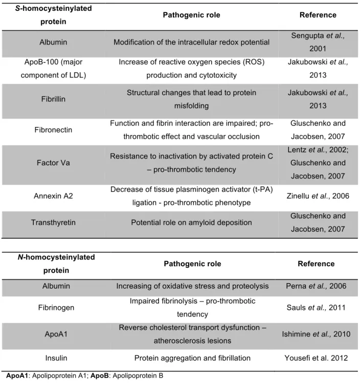
Molecular targeting of proteins by N-homocysteinylation (due to acylation of free amino groups
mediated by Hcy toxic metabolite, HcyTL) or by S-homocysteinylation (formation of disulphide bonds
with protein’s Cys residues mediated by Hcy itself) may have a role on the pathogenesis of
hypercholesterolemia (Malinowska et al., 2012).
Both N- and S-homocysteinylation have well-established pathophysiological consequences owed
to protein structure modification, as protein inactivation, cell damage, oxidative stress, activation of
autoimmune response and enhanced thrombosis (Zinellu et al., 2012) (Table 1.1).
There are two different types of disulphides in protein structures that have different functional roles:
one type of disulphides stabilizes proteins by cross-linking their polypeptide chains while others are
redox reactive and thus may react with free aminothiols, such as Hcy (Jakubowski, 2013). These
alterations can change the redox balance of proteins (Malinowska et al., 2012) Plasma S-thiolated
proteins have been detected in healthy humans, in patients with cardiovascular disease and in several
cell types after oxidant exposure (Zinellu et al., 2006).
Proteins are N-homocysteinylated at rates proportional to the amount of lysine (Lys) residues
present, and a physiologic concentration of HcyTL of 10 nM is sufficient to start the process
(Jakubowski, 2006; Zinellu et al., 2006). N-Hcy-proteins levels in human plasma are several times
higher than the plasma levels of HcyTL (0-35 nM), varying from 0.1 µM to 13 µM, meaning that most
HcyTL formed establishes immediate bonds with the amino groups of proteins (Jakubowski, 2006).
HcyTL formation occurs in all cells tested thus far, including human vascular endothelial cells,
when Methionyl-tRNA syntethase selects by error Hcy instead of Met in an editing reaction of protein
synthesis (Jakubowski, 2006; Yilmaz, 2012). This process is favoured when the two other pathways
are impaired by lack of vitamin B6, B12 and/or folate (B9) intake, by high-Met diet or by genetic
alterations of enzymes involved in Cys/Met metabolism – CBS, MS and/or methylenetetrahydrofolate
reductase (Jakubowski, 2006).
Generally, the percentage of tHcy that binds to proteins as N-Hcy in human plasma of healthy
individuals is 3–7% (Jakubowski, 2008). Increased total plasma Hcy concentrations lead to increased
production of HcyTL and N-homocysteinylation which in turn favours the number of -SH sites available
for S-homocysteinylation (Zinellu et al., 2006). The opposite also happens. Human serum albumin has
7
between Cys34 residue and a free Cys aminothiol was N-homocysteinylated faster than
mercaptoalbumin (the form with free reduced Cys34residue) (Jakubowski, 2006). Lys525 residue of
albumin is two times more reactive to N-homocysteinylation when the protein is in its disulphide form
than when Cys34 is reduced, leading to a final product much more susceptible to proteolysis
(Jakubowski, 2006).
Table 1.1 Cardiovascular consequences of S and N-homocysteinylated proteins.
S-homocysteinylated
protein Pathogenic role Reference
Albumin Modification of the intracellular redox potential Sengupta et al., 2001
ApoB-100 (major
component of LDL)
Increase of reactive oxygen species (ROS)
production and cytotoxicity
Jakubowski et al.,
2013
Fibrillin Structural changes that lead to protein misfolding
Jakubowski et al.,
2013
Fibronectin Function and fibrin interaction are impaired; pro-thrombotic effect and vascular occlusion
Gluschenko and
Jacobsen, 2007
Factor Va Resistance to inactivation by activated protein C – pro-thrombotic tendency
Lentz et al., 2002;
Gluschenko and
Jacobsen, 2007
Annexin A2 Decrease of tissue plasminogen activator (t-PA)
ligation - pro-thrombotic phenotype Zinellu et al., 2006
Transthyretin Potential role on amyloid deposition Gluschenko and Jacobsen, 2007
N-homocysteinylated
protein Pathogenic role Reference
Albumin Increasing of oxidative stress and proteolysis Perna et al., 2006
Fibrinogen Impaired fibrinolysis – pro-thrombotic
tendency Sauls et al., 2011
ApoA1 Reverse cholesterol transport dysfunction –
atherosclerosis lesions Ishimine et al., 2010
Insulin Protein aggregation and fibrillation Yousefi et al. 2012
ApoA1: Apolipoprotein A1; ApoB: Apolipoprotein B
High-density lipoprotein (HDL) and LDL have also been subject of homocysteinylation studies. LDL
interaction with HcyTL causes aggregation of the lipoprotein and higher uptake by cultured
macrophages (Zinellu et al., 2006). Ferretti and co-workers theorized that the oxidative stress caused
-8
homocysteinylated LDL by membrane receptors with intracellular release of Hcy after hydrolytic
degradation (Ferretti et al., 2004). The authors have also reported that LDL incubation with 100 µM
HcyTL for two hours causes N-homocysteinylation of ≈10% of apoB-100 Lys residues (Ferretti et al.,
2004).
As to HDL, it is the major carrier of hydroperoxides in plasma and that happens as consequence of
oxidation at inflammation sites and re-entry into circulation or by transfer of oxidized lipids from LDL to
HDL (Ferretti et al., 2010). An increase in lipid hydroperoxides and other oxidation markers
destabilizes the lipoprotein structure, facilitating HcyTL access to HDL amino groups (Ferretti et al.,
2010).
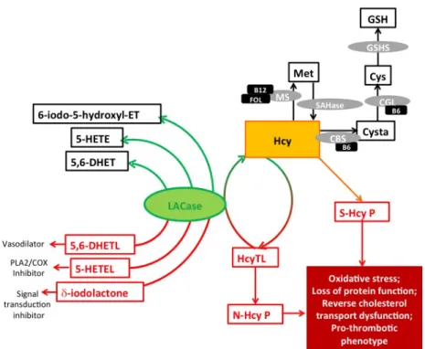
HcyTL is detoxified by the lactonase (LACase) activity of Paraoxonase-1 (PON1), an
HDL-associated enzyme, hence avoiding protein N-homocysteinylation and its nefarious effects on the
organism (Yilmaz, 2012) (Figure 1.3).
LACase is nowadays considered to be the native function of this enzyme in mammal’s organisms
(Jakubowski, 2000). It was first proposed by Jakubowski that PON1 evolved to metabolise HcyTL
(Jakubowski, 2000). Further on, other investigators found that it is also able to hydrolyse other classes
of endogenous lactones, specifically, aromatic and aliphatic lactones that arise from oxidation of
polyunsaturated fatty acids (PUFAs) and result in modulation of the local anti-inflammatory response
(Draganov et al., 2005; Khersonsky and Tawfik, 2005). 5-hydroxy-6E,8Z,11Z,14Z-eicosatetraenoic
acid lactone (5-HETEL) and 5,6-dihydroxytrienoic acid-1,5 lactone (5,6-DHETL) are lactones
originated through one of araquidonic acid (AA) oxidative pathways, the lipoxygenase pathway
(Draganov and Teiber, 2008). 5-HETEL inhibits platelet neutrophil phospholipase A2, an inflammatory
mediator that unchains the AA cascade, and cycloxygenase, the enzyme involved in the oxidative
pathway that originates thromboxane A2 and prostaglandins (Draganov and Teiber, 2008). 5,6-DHETL
is a potent vasodilator that is rapidly metabolised by PON1 to 5,6-dihydroxyeicosatrienoic acid
(5,6-DHET), though it is not known what the physiological effect of this metabolism in vivo might be
(Draganov and Teiber, 2008). 5-hydroxy-6-iodo-8,11,14-eicosatrienoic acid δ-lactone (δ-iodolactone)
inhibits signal transduction pathways that are activated by local growth factors (e.g. epidermal and
9
Figure 1.3 Protein targeting by homocysteine and vascular protection conferred by lactonase
activity (adapted from Dias et al., 2015). 5-HETEL: 5-hydroxy-eicosatetraenoic acid lactone; 5,6-DHETL: 5,6-dihydroxytrienoic acid-1,5 lactone; δ-iodolactone: 6-Iodo-5-hydroxy-8,11,14-eicosatrienoic trienoic acid δ-lactone; CBS: Cystathionine ß-synthase; CGL: cystathionine γ-lyase; Cys: Cysteine; Cysta: Cystathionine; ER: Endoplasmic reticulum; FOL: Folate; GSH: Glutathione; GSHS: Glutathione synthetase; Hcy: Homocysteine;
HcyTL: Homocysteine-thiolactone; LACase: Lactonase; Met: Methionine; MS: Methionine synthase; N-Hcy P: N -homocysteinylated protein; S-Hcy P: S-homocysteinylated protein; SAHase: S-adenosylhomocysteine hydrolase.
Human PON1 is a calcium-dependent glycoprotein, one calcium ion with a structural role and the
other with a catalytic function, mainly expressed in the liver (Perła-Kaján and Jakubowski, 2012). It
was firstly classified as an aryldialkylphosphatase (EC 3.1.8.1) and later, it was also designated by
Aldrige, in 1953, an A-esterase for its ability to hydrolyse organophosphates (Mackness and
Mackness, 2013). It retains the hydrophobic sequence on the N-terminal region, which allows the link
with HDL (Draganov et al., 2005; Farid and Horii, 2012). 95% of PON1 content exists on a subclass of
HDL, HDL3, which is responsible for most of HDL antioxidant capacity and contains also
apolipoprotein A1 (ApoA1) and clusterin (Farid and Horii, 2012; Gugliucci et al., 2012; Mackness and
Mackness, 2013). PON1 activity largely depends on its association with apoA1 and phospholipids in
HDL, although a minor, free form can be found (Gugliucci et al., 2012).
It is believed to have antiatherogenic and antioxidant properties and a broad substrate specificity
going from drug metabolism and nerve agents detoxification (sarin and soman) to the metabolism of
organophosphates (paraoxon, diazoxon), aromatic esters (phenyl acetate) and several classes of
lactones (HcyTL, PUFA lactones and statins with lactone structure) (Draganov et al., 2005;
10
1.4 Objectives
General aim
Molecular targeting of protein by Hcy offers a new paradigm to be considered in the pathogenesis
of hypercholesterolemia. This work aimed to give new insights on protein targeting by Hcy in
hypercholesterolemia condition with genetic (familial hypercholesterolemia, FH) and non-genetic
aetiology (clinical hypercholesterolemia, CH).
Specific aims
ü To optimize and validate a method for Hcy fractions quantification;
ü To compare tHcy and its fractions (fHcy and S-Hcy-P) in normolipidemic, CH and FH patients;
ü To explore S-protein bound fraction in normolipidemics, CH and FH patients;
ü To assess N-protein bound fraction through the use of LACase activity as a surrogate;
ü To ascertain the effect of lipid lowering drugs use in LACase and in tHcy and its fractions, in
11
12
13
2.1 Study population and patient’s inclusion
The Portuguese Familial Hypercholesterolemia Study has been implemented in 1999 at the INSA
following WHO recommendations for a universal FH screening, in 1998 (WHO, 1998). People with a
clinical diagnosis of FH have been recruited by clinicians all over the country to perform genetic tests
to identify a possible mutation (Medeiros et al., 2015). Parallel to this, there is a national study being
developed at INSA named “eCOR” that was designed to estimate the prevalence of major
cardiovascular risk factors in the general Portuguese population.
The eCOR project has so far, a total of 1685 individuals included and approximately 20% are under
lipid lowering therapy. Randomized samples have been collected from 5 Portuguese sub-regions
defined for statistic means (North, Centre, Lisboa, Alentejo and Algarve) and stratified by age and sex
in order to be representative of the Portuguese adult population, aged between 18-79 years old. Data
collection includes an individual survey, physical examination and clinical history. Serum samples
were collected after a 12-hour fasting, by venipuncture, in healthcare centres across the country and
sent to INSA.
The current work was conducted in accordance with the Declaration of Helsinki. The study protocol
and database have been approved by the National Institute of Health Ethics Committee and the
National Data Protection Commission, respectively. Written informed consent was obtained from all
participants before their inclusion in the study.
A subgroup has been selected and divided into three groups according to their TC levels to
quantify LACase activity and Hcy fractions: subjects from the eCOR with normal cholesterol levels;
patients previously diagnosed with CH and patients diagnosed with FH. Both hypercholesterolemic
groups were diagnosed at INSA, following Simon Broome criteria (Annex I). Lipid reference values
adopted in this study were followed according to the guidelines established by the European Society
of Cardiology for the general population (ESC/EAS, 2011) (Table 2.1).
Table 2.1 Lipid reference values based on ESC guidelines for the management of
dyslipidemias.
Parameter Recommended value (mg/dL)
Total cholesterol < 190
Low-density lipoprotein <115
High-density lipoprotein >40 for men; >50 for women
Apolipoprotein B < 120
Apolipoprotein A1 >120 for men; >140 for women
14
2.2 Homocysteine quantification: total, free and S-homocysteinylated protein fraction
I. RATIONAL
Like other aminothiols, Hcy exists mostly in the protein-bounded form (70-80%) due to the reactivity
of its sulfhydryl group (-SH) but it also circulates in the blood in its free oxidized form as homocysteine
dimers or mixed disulphides (20-30%) and, in less extent, in a free reduced form (1%) (Hankey and
Eikelboom, 1999).
Molecular targeting of proteins by Hcy may occur by N-homocysteinylation or by S
-homocysteinylation (Malinowska et al., 2012).
In most studies and even in clinical routine analysis, the contribution of Hcy to CVD relies on tHcy
quantification, which gives limited knowledge, hence probably leading to the loss of valuable
information. In this sense, it would be important to understand the contribution from each Hcy fraction
in CVD. To achieve this, a high sensibility quantification method is required. High-performance liquid
chromatography with fluorescence detection (HPLC-FD) allows the detection of free and total Hcy,
having the advantage of differentiating from other thiols present in the sample (Cys, Cys-Gly, GSH
and NAC). S-Hcy-protein fraction is extrapolated by subtracting the free to the tHcy concentration
(Przemysław et al., 2011). By adapting the conceptual idea for fHcy separation from Przemyslaw and
co-workers and the analytical method described by Nolin and colleagues (Nolin et al., 2007), a new
method that allowed the quantification of Hcy fractions was optimized.
Whenever available, the values of tHcy obtained were compared to those obtained in clinical
setting.
II. STANDARDS PREPARATION FOR THE CALIBRATION CURVE AND QUALITY CONTROL
Stock solution
A L-Homocysteine (Sigma-Aldrich) 10mM stock solution was prepared by dissolving the
appropriate amount of the solid analyte in 1mL of deionized and distilled water.
Standards preparation
Three calibration curves were prepared for validation purposes.
The standards for the calibration curve were prepared by successive dilutions of the stock solution
in phosphate-buffered-saline (PBS) in order to obtain ten different concentrations of L-Homocysteine
ranging from 0.625 to 50 µM.
The quality control (QC) samples were prepared from distinct stock solutions by successive
dilutions in PBS to obtain final concentrations of Hcy of 37.50, 18.75 and 4.375 µM (QC1, QC2, QC3
15
III. METHOD VALIDATION
The validation criteria were defined according to guidebooks regarding the validation of
bioanalytical methods (US-FDA, 2001; EMA, 2011).
Linearity of the method: Three calibration curves were prepared from different stock solutions and
using 10 standards within the concentration range: 0.625 µM (lower limit of quantification, LLOQ) to 50
µM (higher limit of quantification, HLOQ).
The calibration curves were plotted by linear regression of the chromatographic peak area
(mV*min) as function of analytes concentration (µM) to assess the linearity of the method. Also, the
slopes and Y-intercept (Y0) of the curves where compared in order to access reproducibility. The
average back-calculated concentrations were also assessed.
Lower limit of quantification: The LLOQ for Hcy was established at 0.625 µM. In order to validate
the LLOQ, five runs of this standard were analysed for accuracy and inter-assay precision studies.
Accuracy: Accuracy was defined as the closeness to the theoretical concentration of the QC and
LLOQ samples. Mean concentration of each QC sample analysed should be within 100±15% of the
theoretical concentration, except for LLOQ, where 100±20% is acceptable. This parameter was
calculated using the equation 1.
Inter-assay precision: Inter-assay precision was assessed by the coefficient of variation (CV)
obtained from the analysis of QC samples in five runs on different days. The CV of QC should not
exceed 15%, except for LLOQ for which a 20% variation is acceptable.
Stability: To understand the stability of the main solutions used in this protocol, standards with
Tris(2-carboxyethyl)phosphine hydrochloride (TCEP) and 7-fluorobenzofurazan-4-sulfonic acid
ammonium salt (SBD-F) were frozen at -80 ºC, overnight and analysed on the next day.
Stock-solutions were also submitted to three freeze-thaw cycles.
The stability of Hcy was also tested on two separate 50 µM standards after being submitted to two
different conditions: a) -20 ºC and b) 4 ºC, both overnight.
Selectivity and interference: Interference with other endogenous compounds (Cys, Cys-Gly, GSH
and NAC) was tested.
Carryover: Sample carryover was evaluated by inserting vials of methanol after every five samples.
Accuracy (%) = ___________________ Obtained concentration
16
IV. STANDARD OPERATING PROCEDURE
Sample preparation: The method herein described allows the quantification of tHcy and was
adapted from a method previously developed by Nolin and colleagues (Nolin et al., 2007). Briefly, the
analytes present in 50 µL of serum samples were first reduced with 5 µL of TCEP (100g/L,
Sigma-Aldrich) and incubated at room temperature (RT) for 30 minutes. After that, proteins were precipitated
with 45 µL of trichloroacetic acid solution (TCA, 100 g/L) (Roth), containing 1.0 mM of
ethylenediaminetetra-acetic acid (EDTA, Sigma-Aldrich). The resulting mix was centrifuged, the pellet
discarded and a volume of 25 µL of the supernatant was collected and added to a new tube containing
5 µL of 1.55 M sodium hydroxide (NaOH, VWR), 62.5 µL of 0.125 M sodium tetraborate buffer (pH 9.5,
Sigma-Aldrich) with 4.0 mM EDTA and 25 µL of SBD-F (1 g/L, Sigma-Aldrich) in borate buffer (0.125
M with 4.0 mM EDTA). Samples were vortexed and incubated in the dark, at 60 ºC during 1 hour, for
derivatization. A final volume of 30 µL of the derivatized solution was transferred to microvials and
placed in the refrigerated autosampler (8 ºC). A 10 µL aliquot was injected into the HPLC system for
analysis.
The main procedure steps order was changed for the fHcy fraction extraction, based on
Przemyslaw methodological concept (Przemysław et al., 2011): first precipitation and removal of
proteins, and then reduction and derivatization of the supernatant containing the free fraction (Figure
2.1).
Equipment and chromatographic conditions: HPLC analysis was performed on a Shimadzu system
(LC-10AD VP) using a reversed-phase C18 LiChroCART 250-4 column (LiChrospher 100 RP-18,
5µm, VWR) at 29 ºC. Detector was set at excitation and emission wavelengths of 385 and 515 nm,
respectively. Mobile phase consisted of acetate buffer 0.1 M (pH 4.5, VWR) and methanol (VWR)
[99:1 (v/v)]. The analytes were separated in an isocratic elution for 20 min at a flow rate of 0.8 mL/min.
Figure 2.1 Quantification of total and free homocysteine fraction in serum.
17
2.3. PON1 lactonase activity assessment
I. RATIONAL
LACase activity is still the least studied PON1 activity, possibly because it was the last PON1
activity to be discovered and because previous methods have disadvantages that hamper a practical
clinical application (Billecke et al., 2000; Gaidukov and Tawfik, 2005; Rock et al., 2008). On the other
hand, it is also the one that raises most interest because of increasing evidence that it is the
endogenous PON1 activity. Over the years, different authors came to the conclusion that PON’s family
has a high affinity for lactones so it is nowadays proposed that the native function of PON1 is the
metabolism of endogenous lactones, namely HcyTL and other arisen from oxidation of
polyunsaturated fatty acids (PUFAs) (Draganov et al., 2005; Jakubowski, 2000; Khersonsky and
Tawfik, 2005). This activity is responsible for HcyTL detoxification converting it back to Hcy, which can
then enter Met and Cys metabolism, hence avoiding HcyTL deleterious effects (Yilmaz, 2012).
Therefore, LACase activity might be a surrogate of protein N-homocysteinylation.
The LACase method is based on the stoichiometric production of 3-(o-hydroxyphenyl) propionic
acid (o-HPPA) resulting from the hydrolytic reaction of the substrate dihydrocoumarin (DHC) (Figure
2.3). This reaction is monitored spectrophotometrically at 405 nm, following the colour change of the
pH indicator phenol red (from red to yellow). This method was developed and validated at
Translational Pharmacology lab (Dias et al., submitted).
II. STANDARDS PREPARATION FOR THE CALIBRATION CURVE
The stock solutions were prepared by adding the appropriate amount of o-HPPA (Sigma-Aldrich) to
freshly prepared HEPES buffer (2.0 mM, pH 8.0) (Roth), containing calcium chloride (CaCl2, 1.0 mM)
(BDH Chemicals Ltd Pool England) and 0.005% albumin from bovine serum (BSA, Roth). Six standard
samples were prepared for the calibration curve by diluting the stock solutions in physiological serum
(PS). For correction of the non-enzymatic hydrolysis of o-HPPA, a blank sample was prepared using
PS and HEPES buffer.
The accuracy and precision of two QC concentrations (QC1 - 4.92 mM and QC2 - 6.55 mM of
o-HPPA), as well as the LLOQ (1.29 mM of o-HPPA) and the HLOQ (10.24 mM of o-HPPA) were
evaluated using a previously defined criterion (Dias et al., 2014).
III. STANDARDOPERATINGPROCEDURE
LACase activity was assayed by measuring the extent of the hydrolysis of DHC, using a
spectrophotometric method adapted to a 96-well microplate (Figure 2.3). Briefly, serum samples were
diluted in the proportion of 1:5 in PS and incubated during 10 minutes, at 37 °C, as well as the
previous prepared standards. Then, 190 µl of freshly prepared HEPES buffer (2.0 mM, at pH 8.0)
containing CaCl2 (1.0 mM), BSA (0.005%), phenol red (106 µM, Fluka) and DHC (1.0 mM,
18
405 nm on a microplate reader (Biotrack II plate reader, Amersham Biosciences). The activity was
directly obtained from the calibration curve and expressed as kU/L, which is the amount of enzyme
producing 1 mM of o-HPPA per minute. As DHC is toxic by inhalation or skin contact substrate (Ceron
et al., 2014), proper clothing was used.
Figure 2.2 Quantification of lactonase activity in serum (N-Homocysteinylation surrogate)
(adapted from Dias et al., 2015). DHC: dihydrocoumarin; LACase: Lactonase activity; o-HPPA: 3-(o-hydroxyphenyl) propionic acid; PS: physiological serum; RT: Room temperature.
2.4 Statistical analysis
Statistical analysis was performed using GraphPad Prism® version 6.0 (GraphPad Software Inc.,
CA). Data was expressed as median [interquartile range, IQR] or frequencies (%), whenever
applicable. Normality of groups was tested using D’Agostino & Pearson and Shapiro-Wilk test.
Chi-Square test was used to assess data independence. Correlations between parameters were explored
using Pearson or Spearman’s rank correlation test according to the normality of groups. Comparisons
between groups were performed using One-way ANOVA or Unpaired t test for normal distributions
and Kruskal-Wallis or Mann-Whitney U for groups that did not pass the normality test. Values of
p<0.05 were considered significant. Comparisons with more than two variables were performed using
Two-way ANOVA test. The F-test was used to explore differences between the slopes and the
elevations of the calibration curves in the validation of the Hcy method. Variability among data was
19
20
21
3.1 Development and validation of a homocysteine quantification method
I. LINEARITY OF THE METHOD
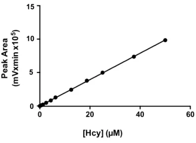
The linear regression model showed to be the most suitable for fitting a function to the points
obtained (p value of Run Tests>0.05 and deviation from linearity non significant). There were no
differences between the slopes and the elevations of the calibration curves (p value of F tests <0.001).
The correlation coefficient, r2, was higher than 0.999, which is a good indicator of adjustment of all
calibration curves. The 95% confidence interval for the Y-intercept when X=0 contained zero. The
average back-calculated concentrations obtained were close to the expected theoretical values at
each tested concentration and presented differences lower than 14%.
Figure 3.1 Homocysteine calibration curve. m= 19750±84; y(0)= -583±1817.
II. LOWER LIMIT OF QUANTIFICATION
The LLOQ for Hcy was 0.625 µM. The accuracy and inter-assay precision results are presented in
(Table 3.1).
III. ACCURACY
The accuracy values obtained for QC1, QC2 and QC3 ranged between 96% and 98% (Table 3.1).
IV. INTER-ASSAY PRECISION
The inter-assay precision for all samples evaluated was higher than 94% (Table 3.1). 10
5 15
22
Table 3.1 Accuracy and inter-assay precision frequencies for the LLOQ and QC samples of
homocysteine quantification method.
LLOQ: lower limit of quantification; QC1: quality control 1; QC2: quality control 2; QC3: quality control 3.
V. STABILITY
Stability performance showed no interference for using SBD-F and TCEP defrost one time
(Kruskal-Wallis test).
Three freeze–thaw cycles had no effect on the stability of tHcy present in the stock-solutions.
Hcy standards can be maintained overnight at 4 ºC and -20ºC (Kruskal-Wallis test).
VI. SELECTIVITY AND INTERFERENCE
Hcy retention time was not affected by the presence of other analytes in all standards tested
(Figure 3.2).
Figure 3.2 Representative chromatogram of a standard sample with 50 µM of homocysteine.
Legend: A – Cysteine peak (5.5 min); B – Homocysteine peak (7.8 min); C – Cys-Gly (8.7 min); D
– GSH (12.5 min); E – NAC peak (17 min).
Standard (µM) Accuracy (%) Inter-assay precision (%)
LLOQ (0.625) 93 94
QC3 (4.357) 98 98
QC2 (18.750) 98 97
QC1 (37.500) 96 97
0.0 2.5 5.0 7.5 10.0 12.5 15.0 17.5 min
23
A chromatogram from a patient sample is shown for comparison effects (Figure 3.3).
Figure 3.3 Representative chromatogram of a patient sample.
Legend: B – Homocysteine peak (7.9 min).
VII. CARRYOVER
There was no evidence for carryover phenomenon.
As a further validation step of the method, tHcy concentrations herein obtained were compared
with Hcy values provided by the INSA lab, whenever available (Figure 3.4).
Figure 3.4 Comparison between serum total homocysteine concentration measured at INSA
laboratory and the concentration measured at the Translational Pharmacology laboratory.
(Spearman r=0.892, p<0.001).
0 10 20 30 40 50
0 10 20 30 40 50
Serum tHcy-Translat. Pharm. lab (µM)
Se ru m t H c y -I N SA [ µ M] B
0.0 2.5 5.0 7.5 10.0 12.5 15.0 17.5 min
24
3.2 Anthropometric and clinical data of the included patients
A total of 187 individuals (53% men) were included in the study: The normolipidemic group (TC
<190 mg/dL) included 65 subjects; the clinical hypercholesterolemia group (CH) was composed by 76
patients and familial hypercholesterolemia group (FH) by 46 patients.
The characterization of the studied population is presented on Table 3.2. No sex (Chi-square test)
or age (Kruskal-Wallis test with Dunn’s multiple comparison test) differences were found between
groups.
Table 3.2 Anthropometric and clinical data.
a
Data is presented in number, % or Median [IQR]; bChi-square test. CH: Clinical hypercholesterolemic; FH: Familial hypercholesterolemic; LLT: under lipid lowering therapy; n: number of subjects.
The number of patients on lipid lowering therapy (LLT) varied significantly among groups, reaching
approximately half of the CH patients and almost 75% of the FH population (Chi-square test, p<0.001).
Table 3.3 describes the number of patients under the different LLT regimens.
Table 3.3 Lipid lowering therapy use by hypercholesterolemic population.
aData is presented in number. CH: Clinical hypercholesterolemic; FH: Familial hypercholesterolemic; n: number of
subjects.
Parameter (Unit)a Normolipidemia CH FH p
valueb
n 65 76 46
Men (%) 49 62 46 ns
Age (years) 44 [39-50] 44 [36-57] 43 [35-50] ns
LLT (%) 0 47 74 <0.001
Parametera CH FH Total
Statins 24 16 40
Fibrates 1 0 1
Statin + Nicotinic acid 1 0 1
Statin + Ezetimibe 4 17 21
Statin + Fibrates 2 0 2
Fibrates + Ezetimibe 1 0 1
Omega 3 1 0 1
Unknown 2 1 3
25
Statins is the most prescribed class of drugs in both populations, as single prescription and also in
combination with ezetimibe. Considering the fraction where they are used as single therapeutic, the
prevalence is of 67% in the CH group, and 47% in the FH. This fraction is less expressive in the FH
group because of a combined therapy of statins plus ezetimibe that reaches 50% of the patients.
Simvastatin and rosuvastatin seem to be the most administered statins in both groups and in both
types of prescriptions (single and combined), followed by atorvastatin, pravastatin and pitavastatin.
Considering that, the data herein obtained were analysed comparing patients on treatment vs.
patients not receiving any LLT medication (Table 3.4).
Table 3.4 Lipid and lipoproteins profile: exploring the effect of disease and treatment.
a
Data presented as number or Median [IQR]; *Kruskal-Wallis with Dunn’s multiple comparisons test vs normolipidemic; bTwo-way ANOVA with Bonferroni post test: +CH vs FH and # non-LLT vs LLT in CH.
ApoA1: Apolipoprotein A1; CH: Clinical hypercholesterolemia; FH: Familial hypercholesterolemia; HDL: High density lipoprotein; LDL: Low density lipoprotein; LLT: under lipid lowering therapy; Non-LLT: not under lipid lowering therapy; TC: Total cholesterol
CH patients have, as expected, higher levels of TC and LDL than normolipidemic patients (
Kruskal-Wallis test, p<0.001). CH non-LLT group has a significantly lower HDL concentration (Kruskal-Wallis
test, p<0.05) and CH LLT group has higher ApoA1 levels (Kruskal-Wallis test, p<0.01).
FH patients had also elevated TC and LDL comparatively to normolipidemics (Kruskal-Wallis,
p<0.001) and to CH disease group (Two-way ANOVA, p<0.05), while HDL and ApoA1 levels did not
differ.
LLT use affected particularly HDL and ApoA1, with higher levels in the treated groups (Two-way
ANOVA, p<0.01).
The percentage of patients with HDL<40 mg/dL was higher in CH non-LLT patients (68%), followed
by FH non-LLT (33%), 32% in normolipidemic, 25% in CH LLT and 18% on FH LLT.
Parameter (mg/dL)a
Normo
lipidemia CH FH
p
valueb
Non-LLT LLT Non-LLT LLT
n 65 40 36 12 34
TC 164 [153-177] 226[212-253]*** 241[210-275]*** 234[214-261]*** 256[224-299]*** +
LDL 92 [83-108] 154[139-173]*** 162[134-180]*** 166[149-182]*** 179[156-226]*** +
HDL 50 [37-61] 38 [33-48]* 56 [40-67] 44 [36-47] 50 [44-57] ##
26
3.3. Serum homocysteine levels
Table 3.5 Total homocysteine levels in the studied groups.
a
Data presented as Median [IQR]. CH: Clinical hypercholesterolemia; FH: Familial hypercholesterolemia; LLT:
under lipid lowering therapy; Non-LLT: not under lipid lowering therapy; NS: Non-significative; tHcy: total homocysteine
Serum tHcy levels did not differ among the studied groups. Furthermore, most of patients had tHcy
levels within the normal range (5-15 µM).
Five patients (2 Normolipidemic and 3 CH) presented moderate HHcy (16-30µM) and two (1 CH
and 1 FH) had intermediate HHcy (31-100 µM). On the selected population, no severe HHcy (>100
µM) was found.
None of the samples presented a tHcy concentration higher than the HLOQ (50 µM).
The levels of fHcy varied greatly among groups, and it was not quantifiable (<0.625 µM) in 9% of
normolipidemia condition and in about 90% of FH patients (Chi-square test, p<0.001) (Figure 3.5).
Figure 3.5 Percentage of patients with non-quantifiable levels of free homocysteine (<0.625
µM). Chi-square test, p<0.001. CH: Clinical Hypercholesterolemia; FH: Familial Hypercholesterolemia; LLT: Lipid lowering therapy; Non-LLT: Not under lipid lowering therapy
Moreover, while the effect of therapy in FH did not influence the percentage of quantifiable fHcy, in
CH patients the values dropped from 65% in non-LLT to 22% in LLT group.
In the normolipidemia group, the median [IQR] value of fHcy was 1.5 [1.1-2.1] µM, which
corresponds to 15% of tHcy and the median [IQR] value of S-Hcy-protein was 9 [7-11] µM, 85% of
tHcy.
Parameter (Unit)a Normolipidemia CH FH p value
Non-LLT LLT Non-LLT LLT
Serum tHcy (µM) 10 [8-13] 9 [7-12] 9 [8-12] 9 [6-10] 8 [6-10] ns
27
3.4 PON1 lactonase activity
Accuracy, intra and inter-assay precision data of the method were higher than 94, 93 and 92%,
respectively, for the QCs, LLOQ and HLOQ. Calibration curves were linear (r2= 0.999 ± 0.0002) and
there were no differences between their slopes and elevations (F-test).
LACase results are shown in Table 3.6.
Table 3.6 Lactonase activity in the study population.
a
Data presented as Median [IQR]; *Kruskal-Wallis with Dunn’s multiple comparisons test vs Normolipidemic;
b
Two-way ANOVA with Bonferroni post test: +CH vs FH and # non-LLT vs LLT in CH.
ApoA1: Apolipoproteina A1; CH: Clinical hypercholesterolemia; FH: Familial hypercholesterolemia; HDL: High density lipoprotein; LACase: Lactonase; LLT: under lipid lowering therapy; Non-LLT: not under lipid lowering therapy; NS: Non-significative; tHcy: total homocysteine
Regarding the LACase activity, the FH group had a lower activity than CH patients (Two-way
ANOVA, p<0.001) (Table 3.7).
CH non-LLT patients had higher LACase/HDL and LACase/ApoA1 ratios than normolipidemic
patients (Kruskal-Wallis, p<0.01) and presented differences in both ratios towards the treated CH
group (Two-way ANOVA, p<0.05).
Also, FH patients had lower ratios than CH, regardless of therapy (Two-way ANOVA, p<0.001).
Parameter (Unit)a
Normo
lipidemia CH FH
p valueb Non-LLT LLT Non-LLT LLT
LACase
(kU/L) 8 [5-10] 10 [8-12] 10 [8-12] 5 [4-7] 7 [3-9]
+++
LACase/
tHcy 0.9[0.5-1.2] 1.1 [0.8-1.3] 1.0 [0.7-1.4] 0.6 [0.4-1.2] 0.7 [0.5-1.2] ns LACase/
HDL 0.15[0.11-0.19] 0.22[0.16-0.33]
** 0.18[0.12-0.24] 0.11[0.09-0.17] 0.13[0.06-0.19]
+++ #
LACase/
ApoA1 0.05[0.04-0.07] 0.07[0.05-0.10]
*
0.06[0.04-0.08] 0.04[0.03-0.05] 0.04[0.02-0.06]
28
29
30


