1
Vol.59: e16160297, January-December 2016 http://dx.doi.org/10.1590/1678-4324-2016160297
ISSN 1678-4324 Online Edition
BRAZILIAN ARCHIVES OF BIOLOGY AND TECHNOLOGY
A N I N T E R N A T I O N A L J O U R N A L
Influence of mulberry leaf extract on serum adiponectin,
visfatin and lipid profile levels in type 2 diabetic rats
Sepideh Barzin Tond
1; Soudabeh Fallah
2; Zahra Salemi
1,3*; Morteza Seifi
41 Department of Biochemistry, Arak University of Medical Sciences, Arak, Iran; 2Department of Biochemistry, Iran
University of Medical Sciences, Tehran, Iran; 3Molecular and Medicine Research Center, Arak University of Medical Sciences, Arak, Iran; 4Department of Medical Genetics, Faculty of Medicine and Dentistry, University of Alberta, Edmonton, AB, Canada.
ABSTRACT
The effect of ethanolic mulberry leaf extract (MLE) and mulberry leaf powder (MLP) on glycemic control, serum adiponectin, visfatin and lipid profile in type2 diabetic rats have been investigated. 30 male wistar rats randomly divided into 5 groups. One group was randomly assigned as control (I) and diabetes was induced in others by administration of streptozotocin (STZ) (55 mg/kg body weight) 15 minutes after the administration of nicotinamide (110 mg/kg body weight) intraperitoneally. Finally, fasting blood glucose (FBG), lipid profile, adiponectin and visfatin were assessed after 6 weeks. Lipid profiles including serum FBG, total cholesterol, triglycerides, low-density lipoprotein cholesterol (LDL-c), very low-density lipoprotein cholesterol (VLDL) and visfatin significantly decreased and high-density lipoprotein cholesterol (HDL-c) and adiponectin increased in the two groups of treated diabetic rats in comparison to the diabetic control (p<0.05). For all the investigated factors, there was no significant difference between two treatment methods. However, MLP was more effective than MLE in improving visfatin. Results showed that MLE and MLP possess hypoglycemic and hypolipidemic activities and play an important role in regulating the secretion of adipokines such as adiponectin and visfatin.
Key words: Mulberry leaf extract, mulberry leaf powder, type2 diabetes, adiponectin, visfatin
*Authors for correspondence: [email protected]
Braz. Arch. Biol. Technol. v.59: e16160297, Jan/Dec 2016
INTRODUCTION
Type 2 diabetes mellitus (T2DM) is one of the most common health problems in most countries of the world. Global prevalence of diabetes is predicted to increase to 366 million by 2030, largely owing to an aging population, increased urbanization and more sedentary lifestyles (1). 90-95% of diabetic cases suffer from T2DM, which is a heterogeneous disorder characterized by impaired cellular responses to insulin known as insulin resistance and followed by progressive partial pancreatic beta cell dysfunction (2, 3). T2DM is mainly characterized by the development of increased morbidity and mortality for cardiovascular disease; it is also characterized by dramatic microangiopathic complications, such as retinopathy, nephropathy, and neuropathy (4).
Evidence for the importance of plant extracts in the management of type 2 diabetes is emerging. Medicinal plants are frequently considered to be less toxic and free from side effects than synthetic ones (5). World health organization (WHO) encourages, recommends and promotes traditional / herbal remedies in natural health care programs because these drugs are easily available at low cost, safe and people have faith in them (6).
Flavonoids are polyphenolic compounds with low molecular weight and play a major role in the cell synthesis (7). These compounds are abundant in fruits and vegetables and increasing evidence demonstrates a positive relationship between consumption of flavonoids-rich foods and disease prevention (8). As for as diabetes mellitus is concern, flavonoids play a vital role in all aspects and its mechanism is well known (9). Recent reports indicate that mulberry leaves and leaf-dried extracts are a rich source of polyphenol antioxidants, including phenolic acids and flavonoids, such as Caffeic acid, 5- Caffeoylquinic acid , Kaempferol 3-O-(6"-malonyl-glucoside), Quercetin 3-O-(6″-O-malonyl)-β-D-glucoside (10). Mulberry leaf, contains 18 sugar-mimic alkaloids and other active compounds, such as dietary fiber,
rutin, isoquercitin, and astragalin.
1-deoxynojiricmycin (DNJ) is present in high concentration, accounting for 50% of iminosugars (11). The leaves are a valuable, yet low-cost material that can be used in reducing the risk and treatment of T2DM, and cardiovascular system, nervous system, as well as in weight loss (12). Adipocytes produce and release a variety of proteins, collectively termed adipokines that exhibit
important metabolic and inflammatory properties (13). Adiponectin is a 244 –residue protein and unlike most other adipokines, circulating adiponectin levels tend to be low in obese patients and increase with weight loss and with the use of insulin-sensitizing drugs. It increases fatty acid oxidation and glucose uptake in the muscle and reduces the synthesis of glucose in the liver (14). Visfatin is mainly produced by visceral fat, but it can be found in liver, skeletal muscle, bone marrow and lymphocyte. It exerts insulin-mimetic effects in vivo and in vitro and is upregulated in obesity, metabolic syndrome, and diabetes. Visfatin and insulin at similar concentrations have a comparable ability to induce glucose uptake and to inhibit glucose release. Visfatin activates insulin receptor though visfatin binds to this receptor at a distinct site from that of insulin. It is also implicated in dyslipidemia, hypertension, and generally atherosclerotic-related diseases. Subjects with metabolic abnormalities exhibit increased visfatin concentrations (15). We have recently shown that
biochanin A possessed hypoglycemic and
hypolipidemic activities and increased visfatin secretion (16). Treatment of diabetes has been under scientific investigation, and several strategies are being developed to manage it. Dietary modulation is the first –treatment option. Polyphenol derived from plants, have been put on test. In the present study, we aimed to investigate the effect of MLE and MLP on adipokine secretion in rats, with particular attention on the adiponectin and visfatin, each with unique properties.
MATERIAL AND METHODS
Chemicals and Reagents
Streptozotocin and nicotinamide were purchased from Sigma-Aldrich. All other chemicals used in this study were of analytical grade obtained from E Merck. Serum concentration of fasting blood glucose (FBG), total cholesterol, triglycerides (TG), and high-density lipoprotein cholesterol (HDL-c) were measured enzymatically using commercial kits (Pars Azemoon, Tehran, Iran) and spectrophotometer (JENWAY 6505, Europe Union). The serum low-density lipoprotein cholesterol (LDL-c) and very low-density lipoprotein cholesterol (VLDL) were calculated by the Friedewald formula (17) as follows:
Seifi, M et al. 3
Insulin was determined by ELISA (Bioassay technology laboratory Shanghai, China). The concentration of serum adiponectin and visfatin
was determined by commercial enzyme
immunoassay kits (Bioassay technology laboratory Shanghai, China) and Elisa plate reader Bio Tek ELX800TM (VT, U.S.A).
Preparation of Mulberry Leaf Extracts (MLE) and Isolation
Leaves of Morus alba were collected from a farm in Tehran, Iran. It was washed thoroughly under running tap water, shade dried, and ground to a fine powder using an electric blender. 2200g of dried leaves powder were extracted three times with 96%ethanol by maceration at room temperature. The mixture was filtered, evaporated in vacuum evaporator to give 112g of extract. The obtained dry extract was suspended in water followed by
extraction with hexane, CHCl3 and ethyl acetate for three times consecutively (18).
Measurement of Total Flavonoids Content The total flavonoids content (TFC) in ethanolic extract of mulberry leaves and its fractions were determined using AlCl3 reagent. Briefly, 2.5 ml of each sample (and/or quercetin as the standard), previously dissolved in 90% ethanol, was mixed with 2.5 ml of a 2% AlCl3 solution in 90% ethanol. After 40 min, the absorbance of the yellow color
was measured at 415 nm. The TFC [as μg quercetin
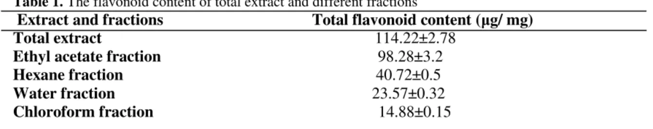
equivalents/mg of sample] for the sample was calculated on the basis of a linear calibration curve obtained using quercetin (y=0.0169x+0.3526, r2=0.995) (19). The highest amounts of flavonoids were found in total extract and ethyl acetate extract respectively (Table 1). The extract was kept in 4◦C until further used.
Table 1. The flavonoid content of total extract and different fractions
Extract and fractions Total flavonoid content (μg/ mg) Total extract 114.22±2.78
Ethyl acetate fraction 98.28±3.2 Hexane fraction 40.72±0.5 Water fraction 23.57±0.32 Chloroform fraction 14.88±0.15
Preparation of Mulberry Leaf Powder (MLP) Dried mulberry leaf powder was administered by inclusion in the diet. To prepare MLP, standard feed was mixed with powdered mulberry leaves at 25% level (20).
Animals
Male Wistar rats (200-250 g) were purchased from the central animal house, Tehran University of Medical Sciences, and maintained in an air conditioned room (23±2ºC) and humidity (55±5%) with a 12 h light/12 h dark cycle. All the rats were provided with commercially available rat normal pellet diet (which contained carbohydrate 60% (w/w), fat 12% (w/w), protein 17.5% (w/w) and fiber 8% (w/w)) and water ad libitum. The study protocols were approved by the institutional
animals’ ethics committee of Arak University of
Medical Sciences, Arak, Iran (With protection code of animal subject in medical research 93-176-32).
Induction and selection of type 2 diabetic rats Type2 diabetes was induced in male wistar rats by a single-day intraperitoneal (IP) injection of
55mg/kg b.w. STZ, 15 minutes after the IP administration of 110 mg/kg b.w. of nicotinamide.
STZ was dissolved in citrate buffer, pH 4.5 and nicotinamide was dissolved in normal saline, while the respective control rats were given vehicle citrate buffer and normal saline (21). Diabetes induction was confirmed through measurement of blood glucose level with glucometer and rats with blood glucose levels more than 126 mg/dl were considered diabetic and selected for further studies (22).
Experimental design
Braz. Arch. Biol. Technol. v.59: e16160297, Jan/Dec 2016 3. sham: diabetic (0.4 ml ethanol and 0.1 ml water);
Group 4. diabetic + MLE (600mg /kg MLE dissolved in 0.4 ml ethanol and 0.1 ml water) and Group 5. diabetic + MLP (standard feed mixed with powdered mulberry leaves at 25% level). Treatments were continued for 6 weeks (Table 2). The initial and final body weight and FBG of all rats were determined before and after treatment, in
fasting condition. Blood glucose level was determined using a glucometer (Accu-Chek Advantage II, Roche Diagnostics, Mannheim, Germany). The animals were anesthetized using Ketamine (75 mg/kg b.w) and Xylazine (10 mg/ Kg b.w) intraperitoneally. Blood sample was collected by cardiac puncture and serum was separated immediately.
Table 2. Ingredient contents of food with or without MLE and MLP
V
control, Diabetic control and Sham
groups
Carbohydrate 60% 60% 45% Fat 12% 12% 9% Protein 17.5% 17.5% 13.12% Fiber 8% 8% 6% Water 2.5% 2.5% 1.87% MLE - 600mg/kg b.w - MLP - - 25%
Statistical analysis
All the data were expressed as mean± standard deviation (S.D.) of three replicates for six rats in each group. Stata software, version 13 (Stata Corp, College Station, TX, USA) was used for all statistical analysis. Normality assumption was checked using Shapiro Wilk test. One-way ANOVA was applied for determining differences between mean of the variable sin the studied groups. Post hoc Test (Tukey) was used to compare the data. Values of p<0.05 were considered statistically significant.
RESULTS
Fasting Blood Glucose Levels
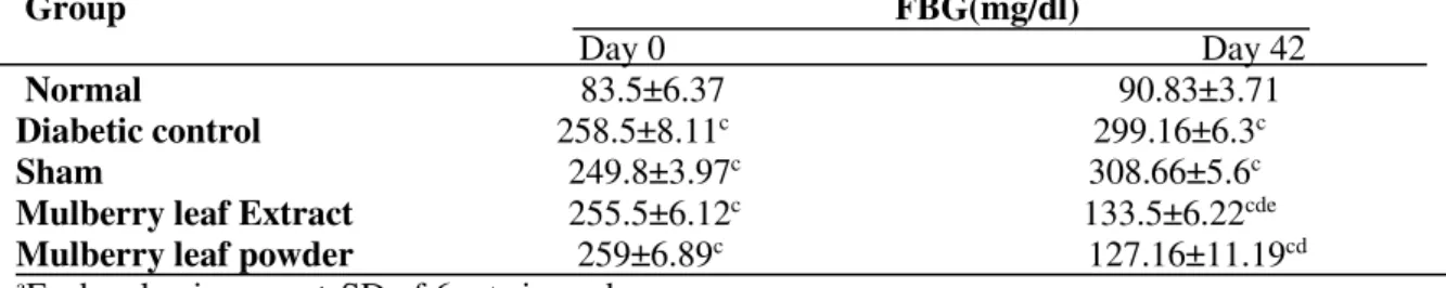
Table 3 presents the effect of MLE and MLP on changes of fasting blood glucose in normal and diabetic rats. Serum glucose level was measured in all rats on 0 day and 42thday of oral administration of MLE and MLP. Three days after STZ injection, fasting blood glucose significantly increased in diabetic rats when compared to control group (p=0.001) (0day). Oral administration of MLE and MLP for 42 days, showed highly significant effect and FBG decreased in treated rats compared to untreated diabetic rats (P=0.001). No significant difference was observed between two treatment methods.
Table 3. Effect Oral Administration of MLE and MLP on Fasting Blood Glucose Levels in Studied Rats a,b
Group FBG(mg/dl)
Day 0 Day 42 Normal 83.5±6.37 90.83±3.71 Diabetic control 258.5±8.11c 299.16±6.3c Sham 249.8±3.97c 308.66±5.6c Mulberry leaf Extract 255.5±6.12c 133.5±6.22cde Mulberry leaf powder 259±6.89c 127.16±11.19cd aEach value is mean ± SD of 6 rats in each group
bAbbreviations: FBG, fasting blood glucose. cP< 0.05 in comparison with normal rats. dP< 0.05 in comparison with diabetic rats. eP< 0.05 in comparison with sham rats.
Seifi, M et al. 5
Serum Insulin Levels
Serum insulin levels decreased significantly in diabetic rats and after treatment with MLP and MLE for 42 days, insulin secretion increased significantly in all treated diabetic rats. Oral administration of MLP was more effective in insulin secretion than MLE, but the difference was not significant (data not shown).
Serum Lipids Levels
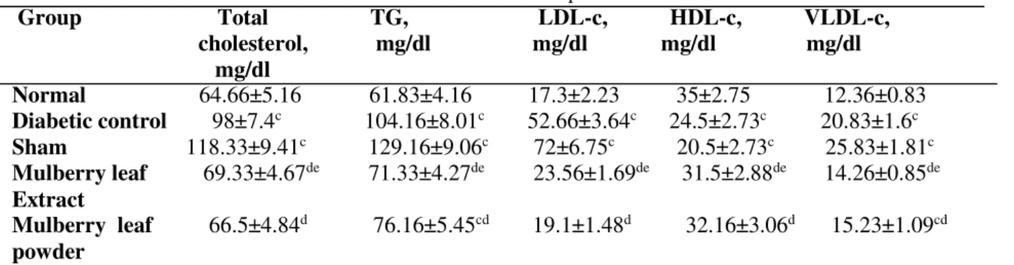
The effect of MLP and MLE administration on lipid profile was shown in table 4. The total cholesterol, triglyceride, VLDL and LDL concentrations in the serum were significantly higher in type-2 diabetic
rats than in the control rats (P=0.001). The administration of MLE and MLP suppressed the increase in the total cholesterol, triglyceride, VLDL and LDL levels in the serum of diabetic rats. MLE and MLP administration decreased total cholesterol and triglyceride levels significantly to almost the control levels. Serum HDL-c was significantly lowered by diabetes induction (P=0.001); however, it was higher in MLE and MLP supplemented groups compared to the untreated diabetic groups (P=0.001). There was no significant difference between the effect of MLE and MLP administration in improving lipid profile.
Table 4. Effect Oral Administration of MLE and MLP on Serum Lipids in Studied Rats a,b
Group Total TG, LDL-c, HDL-c, VLDL-c, cholesterol, mg/dl mg/dl mg/dl mg/dl mg/dl
Normal 64.66±5.16 61.83±4.16 17.3±2.23 35±2.75 12.36±0.83 Diabetic control 98±7.4c 104.16±8.01c 52.66±3.64c 24.5±2.73c 20.83±1.6c Sham 118.33±9.41c 129.16±9.06c 72±6.75c 20.5±2.73c 25.83±1.81c Mulberry leaf 69.33±4.67de 71.33±4.27de 23.56±1.69de 31.5±2.88de 14.26±0.85de Extract
Mulberry leaf 66.5±4.84d 76.16±5.45cd 19.1±1.48d 32.16±3.06d 15.23±1.09cd powder
aEach value is mean ± SD of 6 rats in each group
bAbbreviations: TG, triglycerides; LDL-c, low-density lipoprotein cholesterol; HDL-c, high-density lipoprotein
cholesterol and VLDL-c, very-low-density lipoprotein cholesterol.
cP< 0.05 in comparison with normal rats. dP< 0.05 in comparison with diabetic rats. eP< 0.05 in comparison with sham rats.
Adiponectin andVisfatin Levels
Table 5 shows the effect of MLP and MLE on changes in adiponectin and visfatin concentration in normal and diabetic rats. Serum adiponectin levels were found to be reduced in diabetic rats significantly (P=0.001). The mean serum level of adiponectin in groups receiving MLE and MLP treatment were significantly increased, when compared to untreated diabetic rats (P=0.001), but there was no significant difference between MLE
and MLP in increasing adiponectin levels in serum. The visfatin levels significantly increased in diabetic rats when compared to control group (P=0.001). Oral administration of MLP was significantly effective in 42 days and it returned to near normal in diabetic rats (P=0.001). MLE supplementation was also significantly effective in reducing visfatin levels (P= 0.001), but MLP was more effective than MLE (P= 0.002).
Table 5. Effect Oral Administration of MLE and MLP on Adiponectin and Visfatin Levels in Studied Rats a,b
Group Adiponectin (mg/l) Visfatin (ng/l) Normal 5.73±0.47 254.50±9.13 Diabetic control 3.75±0.25b 329.16±10.80b Sham 2.61±0.22b 333.83±8.88b Mulberry leaf Extract 4.95±0.39bcd 274.66±18.51cd Mulberry leaf powder 5.00±0.60bc 242.50±13.86ce
Braz. Arch. Biol. Technol. v.59: e16160297, Jan/Dec 2016
dP< 0.05 in comparison with sham rats.
eP< 0.05 in comparison with treatment with Mulberry leafExtract
DISCUSSION
To determine the effect of various therapeutic agents such as MLE and MLP, we used nicotinamide and streptozotocin (STZ) to induce diabetes mellitus in rats. Streptozotocin-nicotinamide (STZ-NA)-induced diabetic rats exhibited moderate hyperglycemia associated with the less of postprandial early phase insulin secretion, as well as 50% decrease in pancreatic insulin content (23). The results from this study suggest that MLE and MLP exhibit significant anti-diabetic properties by decreasing blood glucose levels, increasing the plasma insulin level and improving lipid profile in STZ-NA diabetic rats. Our results showed that, in all treated rats the increased levels of glucose were lowered significantly, but the level of FBG in treatment groups was still higher than the normal group. Additionally, in diabetic rats, degranulation or reduction of insulin secretion, arise from
destruction of β-islet cells of pancreas. By the
administration of MLE and MLP, the reduced glucose levels suggested that they likely increase the insulin secretion, which in turn, raise glucose uptake by tissues (24). Elevation of the serum insulin in treated rats could be due to the insulinotropic substances, which induce the intact
functional β-cells of the langerhans to produce
insulin, or protect the functional β-cells from
further deterioration, so that they remain active and produce insulin. It seems that the hypoglycemic effect of MLE and MLP are due to the increased level of serum insulin and the enhancement of peripheral metabolism of glucose (25).On the other hand, the hypoglycemic activity of mulberry leaves maybe attributed to the high fiber content, the presence of trigonelline bases and moran A and/or moranoline in mulberry leaves. Mulberry leaf extract also contain other compounds with significant hypoglycemic activity in diabetic rats (26). One of major constituent of Morus alba is deoxynojirimycin (DNJ) (27). DNJ, as a
competitive inhibitor of intestinal α-glucosidase,
affecting carbohydrate digestion and absorption,
resulting in suppressed postprandial
hyperglycemia. Intake of DNJ inhibited D-glucose uptake at the intestinal brush border membrane because of its similar size and, to some extent, structure to D-glucose (12).
Diabetes is commonly associated with
abnormalities in plasma lipid and lipoprotein levels. Abnormalities in lipid profile was associated with an increased risk of coronary heart disease, therefore ideal treatment for diabetes, in addition to glycemic control, should have a favorable effect on lipid profile (28). According to our results, diabetic condition in rats significantly increased serum total cholesterol (TC), LDL-c, TG and VLDL-C, while HDL cholesterol was significantly decreased. Administration of MLE or MLP improved these values to the normal ones. A significant reduction in serum triglyceride, total cholesterol, LDL-c and a significant increase in the HDL-c was observed. The triglyceride and cholesterol lowering effect of MLE and MLP is due to the stimulatory effect of
quercetin on β-oxidation of fatty acid (12). Several
studies have already investigated various alkaloids, flavonoids and phytochemicals in white mulberry leaves to improve dyslipidemia, especially hypercholesterolemia (29) and inhibit oxidation of LDL-cholesterol (30). The hypolipidemic effect of mulberry leaf extract in healthy non-diabetic human subjects was investigated and showed no significant difference in serum lipid profile, indicating the hypolipiemic effect of mulberry leaf tea may not be similar between normal and diabetic persons (31). Rechel Dorothy et al. tested the hypolipidemic effects of mulberry leaf extract and found total cholesterol and triglycerides were significantly decreased in diabetic mulberry tea high group (DBTH), compared to diabetic control group (DBC) and diabetic mulberry tea low group (DMTL) and suggested the possible hypolipidemic effects of mulberry leaf tea where DNJ might be involved. Since it has been shown to be effective in decreasing lipid accumulation not only via
increasing β-oxidation, but also by increasing
adiponectin levels and activating AMP-activated protein kinase (AMPK) in isolated rat liver (32). Insulin resistance maybe responsible for dyslipidemia, because insulin has an inhibitory action on HMG-coA reductase, a key enzyme in metabolism of cholesterol rich LDL-cholesterol particle (33).
White adipose tissue is an active endocrine organ that secretes many biologically active mediators
referred to as “adipokines” that have significant
impact on energy consumption, carbohydrate and
Seifi, M et al. 7
physiological functions such as immunity and inflammation (34). Adiponectin is defined as an anti-diabetic hormone secreted by adipose tissue. It was associated with many metabolic disorders, including obesity, insulin resistance, obesity related cardiovascular and fatty liver diseases (35). Our data showed that treatment with MLE and MLP, created a significant increase in serum level of adiponectin. Other studies also showed adiponectin has an adverse connection between FBG, TG, Total cholesterol and direct relation with HDL (36). Adiponectin concentration is known to be lower in insulin-resistant states such as obesity and T2DM (37). A significant number of the metabolic actions of adiponectin are dependent on the activation of AMP-dependent kinase (AMPK) (38). AMPK is a fuel-sensing enzyme and is activated by adiponectin, probably through a cAMP–dependent pathway (39). AMPK is activated when ATP is required, and one of its main stimulator is the AMP/ATP ratio, although it has been suggested that AMP kinase activation is the final common pathway of a number of insulin sensitizers including leptin and metformin (40). Metformin increases glucose uptake, reduces hepatic glucose production and increases fatty acid oxidation with the aim of increasing ATP production. The reduction of hepatic and skeletal muscle triglyceride increases insulin sensitivity (41). Adiponectin also enhances the transcription of other genes involved in fatty acid metabolism, most notably peroxisome proliferator-activated
receptor-α (PPARreceptor-α) (42), the target of the liberate group of lipid lowering agents. Activation of PPARα leads to
an increase in levels of molecules involved in free fatty acid transport, such as CD36, and energy dissipation, such as uncoupling protein2 (41) which also increases fatty acid oxidation. Adipocytes have an increasingly recognized role in the endocrine and paracrine control of metabolism and inflammation which appears to play a role in fatty acid and glucose metabolism through a change in insulin sensitivity and activation of fuel oxidation by
AMPK and PPARα. Adiponectin has a plethora of
anti-inflammatory and anti-atherogenic actions including inhibitors of tumor necrosis factor (TNF) signaling cascade, essentially acting as a TNF antagonist (43). Our results showed treatment of diabetic rats with MLE and MLP significantly leads to elevated adiponectin level. There was no difference between two treatment methods. Effect of Morus alba leaves on adiponectin secretion in murine 3T3L1 cells showed they significantly
stimulate lipid accumulation in cells and increase two key transcription factors involved in adipocyte differentiation, CCAAT/enhancer binding protein alpha (C/EBPα) and PPARγ, consequently. The
expression of both aP2 and adiponectin is increased, as is the secretion of adiponectin. The results suggest that Morus alba leaves treatment may also improve insulin sensitivity by upregulating the secretion of adiponectin (44). Our results showed visfatin significantly increased in diabetic group and oral administration of MLE and MLP reduced it significantly, effectiveness of MLP was more than MLE in reducing visfatin. Studies revealed visfatin level increased in T2DM (45). Haider et al. showed that the release of visfatin by adipocytes in response to hyperglycemia is dependent on the duration and extent of glucose (46). Some Other phenomena may also be involved in the increase of visfatin in type 2 diabetes. For instance, increase of visfatin levels may be a result of beta-cell deterioration, which is common in newly-diagnosed type 2 diabetic patients (47). It has insulin mimetic effect and the affinity of visfatin for insulin receptor was found to be similar to that of insulin. Fukuhara et al. identified the potential role of visfatin as insulin mimetic and found that visfatin stimulated insulin receptor in a different way compared with insulin (48). Derdemezies et al. showed that quercetin reduces visfatin secretion. Similarly Chung et al. demonstrated that quercetin improves anti-inflammatory pathway and insulin resistance (49). In another study, Omer et al. showed quercetin shows protective effects in experimental diabetes, possibly by decreasing oxidative stress and
preservation pancreatic β_cell integrity (50). We
suggest that in the diabetic control group, due to increased blood glucose and decreased insulin levels, serum visfatin level was increased compensatory, whereas treatment with MLE and MLP led to decreased hyperglycemia and sensitivity and hence the expression of visfatin gene in treated groups decreased.
CONCLUSIONs
Braz. Arch. Biol. Technol. v.59: e16160297, Jan/Dec 2016
ACKNOWLEDGMENTS
The work was supported by grants from the Research Deputy of Arak University of Medical Sciences.
DECLARATION OF INTEREST:
There is no conflict of interest.
REFERENCES
1. Wild S, Roglic G, Green A, Sicree R, King H. Global Prevalence of Diabetes: Estimates for the year 2000 and projections for 2030. Diabetes Care 2004;27:1047-53.
2. Chen D, Wang M-W. Development and application of rodent models for type 2 diabetes. Diabetes Obes Metab 2005;7:307-17.
3. Defronzo RA. Pathogenesis of type 2 diabetes mellitus. Medl Clin N Am 2004;88:787-835.
4. Chaturvedi N. The burden of diabetes and its complications: Trends and implications for intervention. Diabetes Res Clin Pr 2007;76:S3-S12. 5. Iwu MM. Handbook of African medicinal plants. 2td
ed. US: Boca Raton; 1993.
6. Panchawat S RK, Sssisodia RK. Standardization and evaluation of herbal drug formulations. Alter Med 2010;15:43-7.
7. Fernández SP, Wasowski C, Loscalzo LM, Granger RE, Johnston GAR, Paladini AC, et al. Central nervous system depressant action of flavonoid glycosides. Eur J Pharmacol 2006;539:168-76. 8. Babu PVA, Liu D ,Gilbert ER. Recent advances in
understanding the anti-diabetic actions of dietary flavonoids. J Nutr Biochem 2013;24:1777-89. 9. Patra JC CB. Artificial neural network-based drug
design for diabetes mellitus using flavonoids. J Comput Chem 2011;32:555-67.
10. Thabti I, Elfalleh W, Hannachi H, Ferchichi A, Campos MDG. Identification and quantification of phenolic acids and flavonol glycosides in Tunisian Morus species by HPLC-DAD and HPLC–MS. Functional Foods 2012;4:367-74.
11. Asano N, Yamashita T, Yasuda K, Ikeda K, Kizu H, Kameda Y, et al. Polyhydroxylated Alkaloids Isolated from Mulberry Trees (Morus alba L.) and Silkworms (Bombyx mori L.). J Agr Food Chem 2001;49:4208-13.
12. Skowron MJ FE, Jeszka J, Krejpcio Z, Król E, Buchowski MS. Mulberry leaf extract intake reduces hyperglycaemia in streptozotocin (STZ)-induced diabetic rats fed high-fat diet. Functional Foods 2014;8:9-17.
13. Lau DCW, DhillonB, Yan H, Szmitko PE, Verma S. Adipokines: molecular links between obesity and
atheroslcerosis. Am J Physiol - Heart 2005;288:H2031-H41.
14. Oh DK, Ciaraldi T, Henry RR. Adiponectin in health and disease. Diabetes Obes Metab 2007;9:282-9. 15. Filippatosm TD DC, Kiortsis DN, Tselepis AD,
Elisaf MS. Increased plasma levels of visfatin/pre-B cell colony-enhancing factor in obese and overweight patients with metabolic syndrome. J Endocrinol Invest 2007;30:323-26.
16. Azizi R, Goodarzi MT, Salemi Z. Effect of Biochanin A on Serum Visfatin Level of Streptozocin-Induced Diabetic Rats. Iranian Red Crescent Me. 2014;16:e15424.
17. Friedewald WT, Levy RI, Fredrickson DS . Estimation of the Concentration of Low-Density Lipoprotein Cholesterol in Plasma, Without Use of the Preparative Ultracentrifuge. Clin Chem 1972;18:499-502.
18. Ayatollahi SA, Kobarfard F, Nori M, Fathi M, Choudhari MI. Terpens from aerial parts of Euphorbia splendida. J Med Plants Res 2009;3:660-5.
19. Hashem Dabaghian F AM, Khalighi Sigarudi F, Taghavi Shirazi M, Shojaii A, Sabet Z, Huseini H. Anti-hyperglycemic effect of aqueous extract of Rosa canina L. fruit in type 2 diabetic patients: a randomized double- blind placebo controlled clinical trial. J Biosciences 2015;7:216-24.
20. Andallu B, Kumar AVV, Varadacharyulu NC. Oxidative stress in streptozocin-diabetic rats: Amelioration by mulberry (Morus Indica L.) leaves . Chin J Integr Med 2012:1-6.
21. Pierre W, Gildas AJH, Ulrich MC, Modeste W-N, Benoît NT, Albert K. Hypoglycemic and hypolipidemic effects of Bersama engleriana leaves in nicotinamide/streptozotocin-induced type 2 diabetic rats. BMC Complem Altern M 2012;12:264. 22. Shirwaikar A, Rajendran K, Barik R. Effect of aqueous bark extract of Garuga pinnata Roxb. in streptozotocin-nicotinamide induced type-II diabetes mellitus. J Ethnopharmacol 2006;107:285-90. 23. Oguri S ,Motegi K, Endo Y. Augmented
lipopolysaccharide-induction of the histamine-forming enzyme in streptozotocin-induced diabetic mice. BBA-Mol Basis Dis 2003;1637:83-90.
24. Lu M-P, Wang R, Song X, Chibbar R ,Wang X, Wu L, et al. Dietary soy isoflavones increase insulin secretion and prevent the development of diabetic cataracts in streptozotocin-induced diabetic rats. Nutr Res 2008;28:464-71.
25. Skim F LH, Kaaya A, el Amri H, Jana M. Pharmacological studies of two antidiabetic plants: Globularia alypum and Zygophyllum gaetulum. Therapie 1999;54:711-715.
Seifi, M et al. 9
27. Butt MS, Nazir A, Sultan MT, Schroën K. Morus alba L. nature's functional tonic. Trends Food Sci Tech 2008;19:505-12.
28. Kesari AN, Kesari S, Singh SK, Gupta RK, Watal G. Studies on the glycemic and lipidemic effect of Murraya koenigii in experimental animals. J Ethnopharmacol 2007;112:305-11.
29. Kobayashi Y, Miyazawa M, Kamei A, Abe K, Kojima T. Ameliorative Effects of Mulberry (Morus alba L.) Leaves on Hyperlipidemia in Rats Fed a High-Fat Diet: Induction of Fatty Acid Oxidation, Inhibition of Lipogenesis, and Suppression of Oxidative Stress. Biosci Biotech Bioch 2010;74:2385-95.
30. Enkhmaa B, Shiwaku K, Katsube T, Kitajima K, Anuurad E, Yamasaki M, et al. Mulberry (Morus alba L.) Leaves and Their Major Flavonol Quercetin 3-(6-Malonylglucoside) Attenuate Atherosclerotic Lesion Development in LDL Receptor-Deficient Mice. J Nutr 2005;135:729-34.
31. Kojima Y, Kimura T, Nakagawa K, Asai A, Hasumi K, Oikawa S, et al. Effects of Mulberry Leaf Extract Rich in 1-Deoxynojirimycin on Blood Lipid Profiles in Humans. J Clin Biochem Nutr 2010;47:155-61. 32. Tsuduki T, Nakamura Y, Honma T, Nakagawa K,
Kimura T, Ikeda I, et al. Intake of 1-Deoxynojirimycin Suppresses Lipid Accumulation
through Activation of the β-Oxidation System in Rat Liver. J Agr Food Chem 2009;57:11024-9.
33. Guoyan J. Practical diabetes mellitus. The People’s Medical Publishing House,Beijing 1992.
34. Papanas N ME. Oral antidiabetic agents: anti-atherosclerotic properties beyond glucose lowering. Curr Pharm Design 2009;15:3179-92.
35. Elissa LA, Elsherbiny NM, Magmomah AO. Propolis restored adiponectin level in type 2 diabetes
through PPARγ activation. Egyptian Journal of Basic
and Applied Sciences. 2015;2:318-26.
36. Eizadi M NF, Behboodi L , Khorshidi D. Correlation between serum adiponectin level and blood glucose concentration in adult asthmatic patients. Kashan University ofMedical Sciences 2011;15:345-51. 37. Weyer C, Funahashi T, Tanaka S, Hotta K,
Matsuzawa Y, Pratley RE, et al. Hypoadiponectinemia in Obesity and Type 2 Diabetes: Close Association with Insulin Resistance and Hyperinsulinemia. J Clin Endocr Metab 2001;86:1930-35.
38. Yamauchi T, Kamon J, Minokoshi Y, Ito Y, Waki H, Uchida S, et al. Adiponectin stimulates glucose utilization and fatty-acid oxidation by activating AMP-activated protein kinase. Nat Med 2002;8:1288-95.
39. Kahn BB, Alquier T, Carling D, Hardie DG. AMP-activated protein kinase: Ancient energy gauge provides clues to modern understanding of metabolism. Cell Metab 2005;1:15-25.
40. Saha AK RN. Malonyl-CoA and AMP-activated protein kinase: an expanding partnership. Mol Cell Biochem. 2003;253:65-70.
41. Yamauchi T, Kamon J, Waki H, Terauchi Y, Kubota N, Hara K, et al. The fat-derived hormone adiponectin reverses insulin resistance associated with both lipoatrophy and obesity. Nat Med 2001;7:941-46.
42. Yamauchi T, Kamon J, Ito Y, Tsuchida A, Yokomizo T, Kita S, et al. Cloning of adiponectin receptors that mediate antidiabetic metabolic effects. Nature 2003;423:762-69.
43. Gable DR, Hurel SJ, Humphries SE. Adiponectin and its gene variants as risk factors for insulin resistance, the metabolic syndrome and cardiovascular disease. Atherosclerosis 2006;188:231-44.
44. Naowaboot J, Chung CH, Pannangpetch P, Choi R, Kim BH, Lee MY, et al. Mulberry leaf extract increases adiponectin inmurine 3T3-L1 adipocytes. Nutr Res 2012;32:39-44.
45. Berndt J, Klöting N, Kralisch S, Kovacs P, Fasshauer M, Schon MR, et al. Plasma Visfatin Concentrations and Fat Depot–Specific mRNA Expression in Humans. Diabetes 2005;54:2911-16.
46. Haider DG SG, Kapiotis S, Maier C, Luger A, Wolzt M. The release of the adipocytokine visfatin is regulated by glucose and insulin. Diabetologia 2006;49:1909-14.
47. López-Bermejo A, Chico-Julià B, Fernàndez-Balsells M, Recasens M, Esteve E, Casamitjana R, et
al. Serum Visfatin Increases With Progressive β-Cell Deterioration. Diabetes. 2006;55:2871-5.
48. Fukuhara A MM, Nishizawa M, Segawa K, Tanaka M, Kishimoto K, Matsuki Y, et al. Visfatin: a protein secreted by visceral fat that mimics the effects of insulin. Science 2005;307:426-30.
49. Derdemezis CS KD, Tsimihodimos V, PetrakiMP, Vezyraki P, Elisaf MS, Tselepis AD. Effect of Plant Polyphenols on Adipokine Secretion from Human SGBS Adipocytes. Biochem Res Int 2011;2011. 50. Coskun O KM, Korkmaz A, Oter S. Quercetin, a
flavonoid antioxidant, prevents and protects streptozotocin-induced oxidative stress and beta-cell damage in rat pancreas. Pharmacol Res 2005;51:117-23.