Orientador: Doutora Filomena Macedo Dinis,
Professor Auxiliar com Nomeação Definitiva,
DCR, FCT-UNL
Co-orientadores: Professor Doutor Fernando Pina, Professor
Catedrático, FCT-UNL
Doutora Ana Zélia Miller, Instituto de Recursos
Naturales y Agrobiología de Sevilla, CSIC
Júri:
Presidente: Prof. Doutora Maria Rosa Santos de Paiva
Arguentes: Prof. Doutor Piero Tiano
Prof. Doutor António Manuel Santos Carriço Portugal
Vogais: Prof. Doutora Ana Luísa Pinheiro Lomelino Velosa
Prof. Doutor João Paulo Pereira de Freitas Coroado
Mathilda Amélia Gonçalves Larsson Dias Coutinho
Mestre em Conservação e Restauro
Biological colonization on majolica glazed tiles:
biodeterioration, bioreceptivity and
mitigation strategies
Dissertação para obtenção do Grau de Doutor em Conservação e Restauro,
Biological colonization on majolica glazed tiles: biodeterioration, bioreceptivity and mitigation strategies
Copyright © Mathilda Amélia Gonçalves Larsson Dias Coutinho, Faculdade de Ciências e Tecnologia, Universidade Nova de Lisboa.
Acknowledgments
The work developed in a PhD project is always the result of the help and encouragement of many people. Therefore, I wish to express my sincere gratitude to:
- Prof. Dr. Maria Filomena Macedo Dinis (DCR-FCT/UNL) who was supervisor of this work for valuable advices, guidance, patience and for the great effort in the last part of the thesis.
- Dr. Ana Zélia Miller (IRNAS-CSIC) who supervised this work improving it with her inputs, valuable advices and guidance. Also, for the hospitality during my stay in Seville (also extended to Dr. José Maria de la Rosa).
- Prof. Dr. Fernando Pina (DQ-FCT/UNL) for all support during the grant application, valuable advices and especially for the freedom of letting develop work I enjoyed. - Department of Conservation and Restoration (FCT-UNL), thank you to all members of
the DCR.
- Photochemistry group and all its members for all work developed there. - Vicarte Research Unit and its members for all work developed there.
- Prof. Dr. Márcia Vilarigues (Vicarte) for the great opportunity given to me and the huge patience during this last year.
- Prof. Dr. António Pires de Matos (Vicarte), for sending me to Italy where l got acquaintance with this conservation problem.
- Cremilde Rodrigues (Vicarte) for all the assistance and help.
- Ana Maria Alonso (DCR-FCT) for all the help and good mood during all this years. - Prof. Dr. Alan Philips (UCIBIO-REQUIMTE, FCT/UNL) for the valuable help with the
fungi and also with the article revisions.
- To IRNAS-CSIC, Prof. Dr. Cesareo Saiz Jimenez and all the wonderfull people I met at IRNAS. I would like to thank for all the work developed there and for the very good working experience.
- Dr. Pedro Martinez (IRNAS-CSIC) for the patience and valuable guidance during my stay in Seville.
- Dr. Miguel Rogério-Candelera (IRNAS-CSIC) for the work on digital image analysis. - Prof. Dr. Antonio Gomez -Bolea (Facultat de Biologia of Universitat de Barcelona) for
the lichenological analysis and Prof. Dr. Mariona Hernandez-Marine (Facultat de Farmacia of Univresitat de Barcelona) for the confocal microscopy analysis.
- Dr. Luis Cerqueira Alves and Dr. Victoria Corregidor Berdasco (C2TN, IST/UL) for the PIXE analysis.
- Prof. Dr. José Mirão and Dr. Luis Dias (Laboratório Hercules - UEvora) for the SEM analysis
- Parque de Sintra - Montes da Lua and all its members from Pena National Palace and Sintra National Palace and particularly to Dr António Nunes Pereira, Dr. Bruno Martinho and Dr. José Lobo de Carvalho.
- Graça Camacho and Dr. Carlos Caldas (INIAV). - All who were involved in the TiO2 research:
- Prof. Dr. Susana Sério and Prof. Dr. Yuri Nunes (CEFITEC, FCT/UNL) , - Prof. Dr. Nuno Leal (DCT-FCT/UNL)
- Dr. Márcia Ventura (DQ-FCT/UNL)
- Prof. Dr. João Veiga and Prof. Dr. Hugo Àguas (CENIMAT, FCT/UNL) - Josina Fonseca
- Saint Gobain for glass samples
- Andreia Ruivo, Augusta Lima, Solange Muralha e Teresa Almeida, thank you for all valuable help, support and friendship. Andreia and Augusta especially during the last weeks of this thesis.
- Catarina Pinheiro, Sílvia Sequeira, Marta Félix, Filipa Lopes and Filipa Pereira for all support, advice, friendship and for all the help during the last weeks of the thesis. - All PhD colegues from the DCR.
- Andreia Machado and Alexandra Rodrigues from the stained glass project.
- Fundação para a Ciência e Tecnologia that funded this work (SFRH/BD/46038/2008). - All friends that listened my complaints during these last years and specially to Mafalda - All I forgotten to list
The main results presented in this PhD Dissertation have been published in international journals included in the Science Citation Index (SCI):
P.W. Crous, R.G. Shivas, M.J. Wingfield, B.A. Summerell, A.Y. Rossman, J.L. Alves, G.C. Adams, R.W. Barreto, A. Bell, M.L. Coutinho, S.L. Flory, G. Gates, K.R. Grice, G.E.St.J. Hardy, N.M. Kleczewski, L. Lombard, C.M.O. Longa, G. Louis-Seize, F. Macedo, D.P. Mahoney, G. Maresi, P.M. Martin-Sanchez, L. Marvanová, A.M. Minnis, L.N. Morgado, M.E. Noordeloos, A.J.L. Phillips, W. Quaedvlieg, P.G. Ryan, C. Saiz-Jimenez, K.A. Seifert, W.J. Swart, Y.P. Tan, J.B. Tanney, P.Q. Thu, S.I.R. Videira, D.M. Walker and J.Z. Groenewald, Fungal Planet description sheets: 128-153, Persoonia. 29 (2012) 146–201.
M.L. Coutinho, A.Z. Miller, S. Gutierrez-Patricio, M. Hernandez-Marine, A. Gomez-Bolea, M.A. Rogerio-Candelera, A.J.L. Philips, V. Jurado, C. Saiz-Jimenez, and M.F. Macedo Microbial communities on deteriorated artistic tiles from Pena National Palace (Sintra, Portugal), Int. Biodeterior. Biodegradation. 84 (2013) 322–332.
M.L. Coutinho, A.Z. Miller, M.F. Macedo, Biological colonization and biodeterioration of architectural ceramic materials: An overview, J. Cult. Herit. (2015) doi:10.1016/j.culher.2015.01.006 (In Press).
M.L. Coutinho, A.Z. Miller, P.M. Martin-Sanchez, J. Mirão, L. Dias, A. Gomez-Bolea, B. Machado-Moreira, L. Cerqueira-Alves, V. Jurado, C. Saiz-Jimenez, A. Lima, A.J.L. Phillips, F. Pina, M.F. Macedo. An integrated approach to evaluate biocidal treatments on the microbial community colonizing majolica glazed tiles. Environmental Microbiology (Under revision)
Abstract
The impact of microbial activity on the deterioration of cultural heritage is a well-recognized global problem.
Glazed wall tiles constitute an important part of the worldwide cultural heritage. When exposed outdoors, biological colonization and consequently biodeterioration may occur. Few studies have dealt with this issue, as shown in the literature review on biodiversity, biodeterioration and bioreceptivity of architectural ceramic materials.
Due to the lack of knowledge on the biodeteriogens affecting these assets, the characterization of microbial communities growing on Portuguese majolica glazed tiles, from Pena National Palace (Sintra, Portugal) and another from Casa da Pesca (Oeiras, Portugal) was carried out by culture and molecular biology techniques. Microbial communities were composed of microalgae, cyanobacteria, bacteria and fungi, including a new fungal species (Devriesia imbrexigena) described for the first time.
Laboratory-based colonization experiments were performed to assess the biodeterioration patterns and bioreceptivity of glazed wall tiles produced in laboratory. Microorganisms previously identified on glazed tiles were inoculated on pristine and artificially aged tile models and incubated under laboratory conditions for 12 months. Phototrophic microorganisms were able to grow into glaze fissures and the tested fungus was able to form oxalates over the glaze. The bioreceptivity of artificially aged tiles was higher for phototrophic microorganisms than pristine tile models.
A preliminary approach on mitigation strategies based on in situ application of commercial biocides and titanium dioxide (TiO2)nanoparticles on glazed tiles demonstrated that commercial
biocides did not provide long term protection. In contrast, TiO2 treatment caused biofilm
detachment. In addition, the use of TiO2 thin films on glazed wall tiles as a protective coating to
prevent biological colonization was analysed under laboratorial conditions. Finally, conservation notes on tiles exposed to biological colonization were presented.
Resumo
A biodeterioração é reconhecida como um problema global para a conservação do património. A localização em exterior impossibilita o controlo das condições ambientais que podem favorecer a colonização biológica. O património azulejar encontra-se frequentemente aplicado em fachadas exteriores tornando-o também suscetível à biodeterioração. No entanto, a biodeterioração de azulejos é um tema muito pouco estudado como revelado na revisão da literatura sobre a biodiversidade, biodeterioração e bioreceptividade de materiais cerâmicos arquitetónicos, efetuada nesta tese.
Nesta tese realizou-se a caracterização da microflora presente nos azulejos do Palácio Nacional da Pena (Sintra, Portugal) e nos azulejos da Casa da Pesca (Oeiras, Portugal). Foram usados métodos convencionais de microbiologia juntamente com técnicas de biologia molecular, tendo sido identificadas microalgas, cianobactérias, bactérias e fungos entre os quais uma nova espécie (Devriesia imbrexigena). Verificou-se que alguns destes microrganismos se desenvolviam dentro de fissuras causando biodeterioração dos azulejos.
Para o estudo da biodeterioração e bioreceptividade foram realizados ensaios de inoculação em laboratório sobre azulejos com um vidrado de composição semelhante aos históricos estudados. A inoculação foi realizada tanto sobre azulejos modelo no seu estado inalterado como em artificialmente envelhecidos. Foram utilizados microrganismos previamente identificados verificando-se que os microrganismos fototróficos se desenvolviam no interior de fissuras e que o fungo testado produzia oxalatos sobre o vidrado.
No sentido de controlar a biodeterioração dos azulejos realizou-se, in situ, um estudo preliminar da eficácia e durabilidade de três biocidas comerciais e de nanopartículas de dióxido de titânio (TiO2), tendo sido verificado que os biocidas mais eficientes tinham um curto tempo de ação
e que as nanopartículas promoviam o destacamento do biofilme. Foi ainda testado em laboratório a deposição de filmes finos de TiO2 produzidos pela técnica de sol-gel sobre azulejos históricos
para prevenir a biodeterioração. Por fim foram redigidas de forma sintética notas sobre a conservação de azulejos expostos à colonização biológica.
Table of contents
FIGURE LIST ... XXIII TABLE LIST ... XXIX ABBREVIATIONS ... XXXI
GENERAL INTRODUCTION ... 1
1. GLAZED WALL TILES ... 1
1.1. GLAZED WALL TILES AS CULTURAL HERITAGE ... 3
1.2. DETERIORATION OF GLAZED WALL TILES ... 4
1.3. BIODETERIORATION OF INORGANIC BUILDING MATERIALS ... 7
1.4. AIM AND OBJECTIVES ... 11
1.5. THESIS OUTLINE ... 11
1.6. BIOLOGICAL COLONIZATION AND BIODETERIORATION OF ARCHITECTURAL 2. CERAMIC MATERIALS: AN OVERVIEW ... 13
INTRODUCTION ... 13
2.1. MICRO- AND MACROORGANISMS FOUND ON ARCHITECTURAL CERAMIC ASSETS ... 16
2.2. 2.2.1. Bricks and architectural sculptures ... 20
2.2.2. Ceramic roofing tiles ... 28
2.2.3. Glazed wall tiles ... 34
2.2.4. Comparison of all substratesceramic typologies ... 39
ORGANISMS IDENTIFICATION AND QUANTIFICATION METHODS USED ON ARCHITECTURAL 2.3. CERAMIC MATERIALS ... 40
BIODETERIORATION OF ARCHITECTURAL CERAMIC MATERIALS... 42
2.4. BIORECEPTIVITY OF ARCHITECTURAL CERAMIC MATERIALS ... 47
2.5. CONCLUSIONS ... 48
2.6. 3 MICROBIAL COMMUNITIES ON DETERIORATED ARTISTIC TILES FROM PENA NATIONAL PALACE (SINTRA, PORTUGAL) ... 51
INTRODUCTION ... 51
3.1 MATERIALS AND METHODS ... 52
3.2 3.2.1 Site description and sampling ... 52
3.2.2 Characterization of the glazed substrate ... 54
3.2.2.1 Micro-particle induced x-ray emission (μ-PIXE) ... 54
3.2.2.2 Variable Pressure Scanning Electron Microscopy with energy dispersive spectrometer (VP-SEM-EDS) ... 55
3.2.2.3 Micro-Raman spectroscopy (µ-Raman) ... 55
3.2.3 Biomass estimation of photosynthetic microorganisms ... 56
3.2.4 Biofilm covered area determined by digital image analysis ... 56
3.2.5 Identification of phototrophic microorganisms by culture techniques ... 56
3.2.6 Isolation and identification of fungi ... 56
3.2.7 Identification of microbial communities by molecular methods ... 57
RESULTS ... 59
3.3 3.3.1 White glaze characterization ... 59
3.3.2 Biomass estimation of photosynthetic organisms ... 60
3.3.3 Biofilm covered area ... 60
3.3.4 Direct light microscopy observation of the collected biofilm ... 61
3.3.5 Identification of photosynthetic microorganisms by culture techniques ... 62
3.3.6 Idenfication of the fungal isolates ... 64
3.3.7 Identification of microbial communities by molecular methods ... 65
3.3.8 Confocal Laser Scanning Microscopy ... 66
3.3.9 Field emission scanning electron microscopy (FESEM) ... 68
DISCUSSION ... 69
3.4 CONCLUSIONS ... 71
3.5 4. EVALUATION OF BIODETERIORATION AND BIORECEPTIVTITY OF MAJOLICA TILES BY PHOTOTROPHIC MICROORGANISM UNDER LABORATORY CONDITIONS . 73 INTRODUCTION ... 73
4.1 MATERIALS AND METHODS ... 75
4.2 4.2.1 Manufacture of majolica glazed tile models ... 75
4.2.2 Artificial ageing ... 75
4.2.3 Laboratory-based tiles biodeterioration experiment ... 76
4.2.3.1 Tested microorganisms and tiles inoculation ... 76
4.2.3.2 Quantification and characterization of phototrophic growth on the glazed tiles ... 77
4.2.4 Characterization of pristine and aged majolica tile models before and after biodeterioration experiment ... 78
4.2.5 Analytical techniques ... 79
4.2.5.1 Wavelength dispersive X-ray fluorescence (WD-XRF) ... 79
4.2.5.2 X-ray diffraction (XRD) ... 79
4.2.5.3 Micro particle induced X-ray emission (μ-PIXE) ... 80
4.2.5.4 Micro Raman spectroscopy (μ-Raman) ... 80
4.2.5.5 Optical microscopy ... 80
4.2.5.6 Scanning electron microscopy - energy dispersive X-ray spectrometry (SEM-EDS) ... 80
4.2.6 Statistical analysis ... 81
RESULTS ... 81
4.3 4.3.1 White glaze composition of the historical tiles ... 81
4.3.2. Majolica tile models ... 81
4.3.2.2. Chemical and mineralogical composition ... 81
4.3.2.3. Surface morphology and microstructure ... 83
4.3.2.4. Intrinsic physical characteristics of the pristine and aged tiles ... 84
4.3.3. Characterization of the biofilm after the biodeterioration experiment ... 86
4.3.4. Scanning Electron microscopy (SEM) ... 90
4.3.5. Post experimental capillary water absorption ... 91
4.3.6. Characterization of the corrosion products ... 92
4.3.7. Chemical characterization of the glaze surface ... 93
DISCUSSION ... 95
4.4.1 Tile models ... 95
4.4.2 Bioreceptivity ... 97
4.4.3 Biodeterioration ... 98
CONCLUSIONS ... 99
4.5 5. BIODETERIORATION OF MAJOLICA TILES BY THE FUNGUS DEVRIESIA IMBREXIGENA UNDER LABORATORY CONDITIONS ... 101
INTRODUCTION ... 101
5.1 MATERIALS AND METHODS ... 102
5.2 Pristine and aged majolica glazed tile models ... 102
5.2.1 Laboratory-based tiles biodeterioration experiment ... 103
5.2.2 Post-experiment analyses of tile surface alterations ... 104
5.2.3 5.2.3.1 Quantification and characterization of the fungal growth on the glazed tiles .. 104
5.2.3.2 Optical microscopy ... 104
5.2.3.3 SEM-EDS analysis ... 104
5.2.3.4 Water absorption by capillarity of the pristine and aged tile models ... 105
5.2.3.5 μ-Raman ... 105
5.2.3.6 μ-PIXE ... 105
5.2.3.7 Statistical analysis ... 106
RESULTS ... 106
5.3 Evaluation of fungal growth: visual inspection and digital image analysis ... 106
5.3.1 Optical Microscopy ... 108
5.3.2 SEM-EDS analysis ... 109
5.3.3 Post-experiment water absorption by capillarity ... 112
5.3.4 µ-Raman ... 113
5.3.5 µ-PIXE ... 114
5.3.6 DISCUSSION ... 116
5.4 Fungal growth ... 116
5.4.1 Biodeterioration by the fungus D. imbrexigena ... 118
5.4.2 CONCLUSIONS ... 119
5.5 6 AN INTEGRATED APPROACH TO EVALUATE BIOCIDAL TREATMENTS ON MICROBIAL COMMUNITY COLONIZING MAJOLICA GLAZED TILES ... 121
INTRODUCTION ... 121
6.1 MATERIALS AND METHODS ... 124
6.2 6.2.1 Site description and sampling ... 124
6.2.2 µ-Raman ... 125
6.2.3 μ-PIXE ... 125
6.2.4 Morphological characterization of the biofilm ... 125
6.2.4.1 Optical microscopy ... 125
6.2.4.2 SEM –EDS analysis ... 125
6.2.5 Isolation and identification of fungi from the biofilm ... 126
6.2.6 In situ application of biocidal treatments on glazed wall tiles ... 127
6.2.7 Monitoring of treatments efficacy ... 127
6.2.7.1 Macroscopic observations ... 127
6.2.7.2 Epifluorescence microscopy ... 127
6.2.7.3 Molecular analysis of the microbial communities ... 128
RESULTS ... 129
6.3 6.3.1 Characterization of the glaze substrate ... 129
6.3.2 Microbial community characterization by microscopy observations ... 131
6.3.2.1 Optical microscopy ... 131
6.3.2.2 SEM analysis ... 131
6.3.3 Fungal isolation and identification by molecular biology techniques ... 133
6.3.4 Efficacy of the biocidal treatments applied on the glazed wall tiles ... 133
6.3.4.1 Visual inspections ... 133
6.3.4.2 Epifluorescence microscopy ... 135
6.3.4.3 Molecular monitoring of microbial communities by DGGE ... 136
6.3.4.4 Phylogenetic identification of microbial communities before and after in situ application of Preventol ... 138
6.3.4.5 Comparison of glaze surface before and after biocidal treatment ... 145
DISCUSSION ... 146
6.4 6.4.1 Microbial community growing on the glazed tiles ... 146
6.4.2 Efficacy evaluation of the tested biocides ... 148
6.4.2.1 Titanium dioxide ... 149
6.4.2.2 Biotin ... 150
6.4.2.3 Preventol ... 151
6.4.2.4 Albilex ... 152
CONCLUSIONS ... 153
6.5 7 PRELIMINARY TESTS OF TITANIUM DIOXIDE THIN FILM COATINGS ON HISTORICAL GLAZED WALL TILES TO PREVENT BIOLOGICAL COLONIZATION ... 155
INTRODUCTION ... 155
7.1 MATERIALS AND METHODS ... 157
7.2 7.2.1 Tile samples ... 157
7.2.2 Tile characterization ... 158
7.2.3 Synthesis and application of TiO2 coatings ... 158
7.2.4 Characterization of TiO2 thin film ... 158
7.2.5 Tertiary bioreceptivity experiment ... 159
7.2.6 Characterization of tiles before and after treatment ... 159
RESULTS ... 160
7.3 7.3.1 Glaze and ceramic body chemical characterization ... 160
7.3.2 Characterization of TiO2 thin film ... 161
7.3.2.1 µ-Raman analysis ... 161
7.3.2.2 Morphology of the TiO2 coating ... 161
7.3.3 Tertiary bioreceptivity experiment ... 163
7.3.4 Evaluation of the chromatic alteration caused by thin film deposition ... 164
7.3.5 XRD analysis ... 166
DISCUSSION ... 167
7.4 7.4.1 Tile samples ... 167
7.4.2 Characterization of the TiO2 coatings ... 169
7.4.3 Effect of TiO2 coatings on tiles bioreceptivity ... 169
7.4.4 Applicability evaluation of TiO2 coatings on historical glazed tiles ... 169
CONCLUSIONS ... 170
8 NOTES ON THE CONSERVATION OF GLAZED WALL TILES EXPOSED TO BIOLOGICAL COLONIZATION ... 171
INTRODUCTION ... 171 8.1
ENVIRONMENTAL FACTORS INFLUENCING BIOLOGICAL COLONIZATION OF GLAZED WALL
8.2
TILES ... 172 PHYSICAL FEATURES OF GLAZED WALL TILES INFLUENCING BIOLOGICAL COLONIZATION .. 174
8.3
INFLUENCE OF RESTORATION TREATMENTS ... 175 8.4
CONSERVATION STRATEGIES TO CONTROL BIOLOGICAL COLONIZATION OF GLAZE WALL
8.5
TILES ... 176 9. FINAL REMARKS AND FUTURE PERSPECTIVES ... 177
Figure List
Figure List
FIG.2.1CERAMIC BUILDING MATERIALS IN PORTUGUESE ASSETS:(A) WALL TILE PANELS ILLUSTRATING THE AESTHETICAL ORNAMENTATION OF A GARDEN BELONGING TO A HISTORICAL BUILDING, CASA DA
PESCA, IN OEIRAS; (B) DETAIL OF THE 18TH CENTURY GLAZED TILES WITH ICONOGRAPHIC ILLUSTRATION FROM CASA DA PESCA, OEIRAS; (C) BULLFIGHT ARENA IN LISBON (IN PORTUGUESE:
PRAÇA DE TOUROS DO CAMPO PEQUENO)BUILT WITH BRICKS IN THE LAST DECADE OF THE 19TH CENTURY. ... 14
FIG. 2.2 GEOGRAPHICAL DISTRIBUTION OF THE STUDIES REGARDING BIOLOGICAL COLONIZATION OF ARCHITECTURAL CERAMIC MATERIALS.SIGN (▲) INDICATES COUNTRIES. ... 17
FIG. 2.3 PERCENTAGE (%) OF ANALYZED STUDIES REGARDING EACH CERAMIC BUILDING MATERIAL TYPOLOGY CONSIDERED IN THIS WORK: BRICKS, ROOFING TILES AND GLAZED WALL TILES. ... 18
FIG.2.4MICROGRAPH OF THE CYANOBACTERIUM NOSTOC PALUDOSUM(PRESENTED IN TABLE 2.6) WITH HETEROCYST (ARROWS), COLLECTED FROM GLAZED WALL TILES FROM PENA NATIONAL PALACE (X
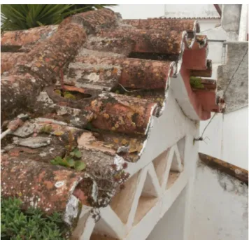
60). ... 22 FIG.2.5.BIOLOGICAL COLONIZATION OF ROOFING TILES FROM A HOUSE IN ALENTEJO REGION (SOUTH OF
PORTUGAL). ... 33
FIG. 2.6 BIOLOGICAL COLONIZATION OF GLAZED WALL TILES FROM THE SINTRA NATIONAL PALACE
(PORTUGAL). (A) COLONIZATION BY LICHENS. (B) COLONIZATION BY BRYOPHYTES THROUGH A FRACTURE OF THE CERAMIC BODY. ... 35 FIG. 2.7 GLAZED WALL TILES FROM PENA NATIONAL PALACE IN SINTRA (PORTUGAL) SHOWING A TILE
AFTER THE BIOFILM REMOVAL (ON THE LEFT) AND A TILE COVERED BY THE GREEN/BLACK BIOFILM (ON THE RIGHT). ... 43 FIG. 2.8 BIOLOGICAL COLONIZATION OF GLAZED WALL TILES FROM PRIVATE HOUSE IN THE ALENTEJO
DISTRICT (PORTUGAL) BY LICHENS AND A BLACK BIOFILM WITH DETAILS IN MACROPHOTOGRAPHY OF THE SPALLING AND STAINING OF THE GLAZE. ... 45
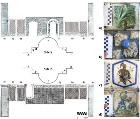
FIG.3.1SCHEME OF THE TRITON TUNNEL PASSAGEWAY WITH THE LOCATIONS OF THE PANELS (A-N) AND THEIR TILE MOTIFS (A-D).BAR REPRESENTS CM. ... 52
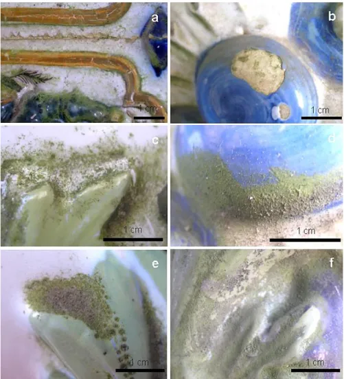
FIG.3.2DETAILED IMAGE OF THE DECAYED TILES.(A)FLAKING ON THE BORDER OF A TILE.(B)GROWTH OF THE BIOFILM ON THE CERAMIC BODY ON A LACUNA. (C-F) PREFERENTIAL GROWTH ON THE DEPRESSION AREAS WHERE GREEN BIOFILMS AND AIRBORNE PARTICLES ACCUMULATE. ... 53
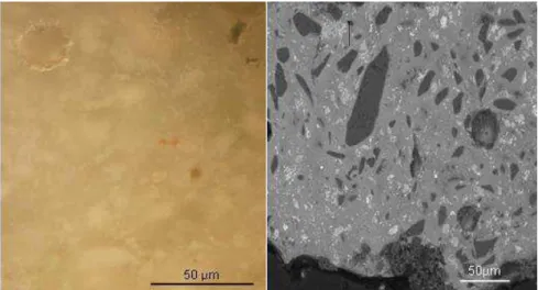
FIG.3.3WHITE GLAZE CROSS-SECTION OF SAMPLE W:(A) OBSERVED UNDER OPTICAL MICROSCOPE WITH WHITE TIN OXIDE CRYSTALS DISTRIBUTED ON THE GLASSY MATRIX AND (B) BACKSCATTERED ELECTRON IMAGE SHOWING SEVERAL MINERAL INCLUSIONS. ... 59
FIG. 3.4.DIGITAL IMAGE ANALYSIS OF TILE FROM PANEL E AND H.(A)ORIGINAL IMAGE OF ONE OF THE GLAZED TILES OF PANEL E.(B)FALSE-COLOR IMAGE OBTAINED FROM PCA BANDS.(C)BINARY IMAGE SHOWING THE MEASURED AREAS.(D)ORIGINAL IMAGE OF ONE GLAZED TILE OF PANEL H.(E)RESULT OF THE SUBSTRACTION OF PIXEL VALUES OF TWO FALSE-COLOR IMAGES FROM PCA BANDS. (F)
BINARY IMAGE SHOWING THE MEASURED AREAS. ... 61
FIG. 3.5 MICROGRAPHS OF THE BIOFILM SAMPLES OBSERVED BY LIGHT MICROSCOPE. (A) TREBOUXIA
CELLS SURROUNDING BY HYALINE HYPHAE OF FUNGI. (B) NON LICHENIZED SHORT FILAMENTS OF
DEVELOPING ON THE CHAIN OF ARTHROCONIDIA SCALE BARS =5ΜM (CROUS ET AL.,2012). .... 65
FIG. 3.7 CONFOCAL MICROGRAPHS OF BIOFILM-FORMING MICROORGANISMS COLLECTED FROM PENA
NATIONAL PALACE (SAMPLE SEMI-CIRCLE NORTH SIDE).(A)MAXIMUM INTENSITY PROJECTION OF A BIOFILM SCRAPED FROM TILES SHOWING SMALL QUANTITY OF INORGANIC MATERIALS.IT WAS MAINLY COMPOSED BY CLUSTERS OF THE GREEN ALGA TREBOUXIA SP. NOTE THE SINGLE, STELLATE CHLOROPLASTS SHOWING RED FLUORESCENCE DUE TO THE FLUORESCENCE OF CHLOROPHYLLS A AND B.CELLS SHOWED INTERNAL DIVISION OR WERE FILLED WITH AUTOSPORES.SOME AUTOSPORES WERE FREE.SMALL AMOUNTS OF EPS WERE SCATTERED AMONG ON THE SUBSTRATE.NEITHER THE CELLS NOR THE AUTOSPORES WERE SURROUNDED BY EPS.(B)3D EXTENDED FOCUS PROJECTIONS IN X-Y, X-Z AND X-Z VIEWS OF A TREBOUXIA SP. BIOFILM WITH SUBSTRATE SHOWING THE 3D VIEW INTO MICROSTRUCTURE OF TILES. CELLS WERE SCATTERED AMONG THE IRREGULARITIES OF THE SUBSTRATE. BIOFILM THICKNESS =26 ΜM. COLOR ALLOCATION: WHITE = REFLECTION FROM INORGANIC MATERIALS; RED = AUTOFLUORESCENCE OF CHLOROPHYLLS; BLUE = EXTRACELLULAR POLYMERIC SUBSTANCES (EPS) DYED WITH CON A-ALEXA 488. ... 67
FIG.3.8FLUORESCENCE PROPERTIES OF TREBOUXIA SP. FROM A BIOFILM FROM PENA NATIONAL PALACE
(NORTH SIDE).PSEUDOCOLOR CONFOCAL X-Y-Λ SINGLE SECTION CORRESPONDING TO THE EMISSION PEAK OF CHLOROPHYLLS A AND B.LIGHT COLORS REPRESENTED THE MAXIMUM FLUORESCENCE.(A) 2D PLOTS OF IN VIVO SPECTRAL PROFILES FROM LAMBDA SCANS (EXCITATION WAVELENGTH =488 NM. STEPS = 50, BAND-WIDTH = 20 NM) (B, C). MEAN FLUORESCENCE INTENSITY OF SINGLE VEGETATIVE CELLS (B) AND SPORES (C) PRESENT IN THE BIOFILM OF TREBOUXIA SP. ... 68 FIG. 3.9. FESEM IMAGES OF BIOFILM OVER GLAZE FRAGMENTS AT DIFFERENT MAGNIFICATIONS. (A)
FILAMENTOUS AND UNICELLULAR MICROORGANISMS CAN BE OBSERVED GROWING ON A FRACTURE OF THE GLAZE.(B)SURFACE OF THE GLAZE DENSELY COLONIZED.(C)COLONIZATION ON A FRACTURE OF THE GLAZE.(D)FILAMENTS OF MONILIFORM CELLS COMPATIBLE WITH NOSTOC SP.(E)FILAMENTOUS AND UNICELLULAR MICROORGANISMS GROWING INSIDE A PORE OR BUBBLE. (F) MICROORGANISMS GROWING IN CONTACT WITH THE SUBSTRATE. ... 69 FIG.4.1. ILLUSTRATION OF THE EXPERIMENTAL SET-UP.GLASS PETRI DISH ( =9 CM) WITH DISTILLED
WATER AT THE BOTTOM AND A NET TO SEPARATE THE WATER FROM THE GLAZED TILE SAMPLES. 76
FIG.4.2SCHEME WITH EXPERIMENTAL DESIGN WITH THE NUMBER OF REPLICATES OF PRISTINE AND AGED CONTROL AND INOCULATED SAMPLES ... 77 FIG.4.3.EXPERIMENTAL SET-UP FOR DETERMINATION OF WATER VAPOR PERMEABILITY,(A) TILE SAMPLE
EMBEDDED IN WAX OVER A PLASTIC CONTAINER AND (B) CHAMBER USED FOR THE EXPERIMENT WITH THE TILE SAMPLES INSIDE. ... 78
FIG. 4.4.DIFFRACTOGRAM OBTAINED FOR THE FIRED CERAMIC BODY WITH SIGNALIZATION OF THE MAIN MINERAL PHASES OF THE TILE MODELS. ... 82
FIG.4.5. µ-RAMAN SPECTRA OF (I) QUARTZ AND (II) CASSITERITE IDENTIFIED ON THE WHITE GLAZE OF THE TILE MODELS.LASER EXCITATION 632.8 NM;100× OBJECTIVE ULWD; LASER POWER WAS KEPT AT 1
MW. ... 83
FIG.4.6MICROSTRUCTURE AND SURFACE MORPHOLOGY OF REPRESENTATIVE SAMPLES OF THE PRISTINE AND AGED GLAZE TILE MODELS BEFORE INOCULATION, OBSERVED UNDER OPTICAL MICROSCOPY (A AND B) AND TWO BSE IMAGES, ONE GENERAL OVERVIEW (C AND D) AND DETAIL OF THE FISSURE (E AND F). ... 84
FIG. 4.8. WATER VAPOR PERMEABILITY OF PRISTINE (PRIST 1-3) AND AGED(AGED 1-3) TILE SAMPLES BEFORE INOCULATION WITH LINEAR TRENDLINES AND EQUATION WITH CAPILLARY COEFFICIENT VALUES. ... 86
FIG.4.9IMAGES OF THE TILE MODELS BEFORE AND AFTER INCUBATION EXPERIMENT.PETRI DISHES WITH
(A) PRISTINE TILE SAMPLES BEFORE INOCULATION,(B) AGED TILE SAMPLES BEFORE INOCULATION AND
(C) INOCULATED PRISTINE (LEFT) AND AGED (RIGHT) TILES SAMPLES AFTER 12 MONTHS INCUBATION. ... 87 FIG. 4.10.EXAMPLE OF THE APPLICATION OF DIGITAL IMAGE ANALYSIS IN A PRISTINE AND AN AGED TILE
MODEL COVERED BY THE GREEN BIOFILM. (A) ORIGINAL IMAGE OF ONE OF THE PRISTINE GLAZED TILES.(B)SEGMENTED IMAGE (BINARY) OF THE FIRST PRINCIPAL COMPONENT.(C)OUTLINES OF THE AREAS SELECTED FOR MEASURING OF THE FIRST PRINCIPAL COMPONENT OF THE PRISTINE GLAZED TILE.(D)ORIGINAL IMAGE OF ONE OF THE AGED GLAZED TILES.(E)SEGMENTED IMAGE (BINARY) OF THE FIRST PRINCIPAL COMPONENT. (F) OUTLINES OF THE AREAS SELECTED FOR MEASURING OF FIRST PRINCIPAL COMPONENT OF THE AGED GLAZED TILE. ... 88
FIG.4.11.AREA (IN MM2) COLONIZED BY THE PHOTOTROPHIC BIOFILMS IN AGED (BLUE) AND PRISTINE (RED) TILES OBTAINED BY DIGITAL IMAGE ANALYSIS. ... 89
FIG. 4.12.SEM IMAGES OF MAJOLICA TILE MODELS AFTER INOCULATION EXPERIMENTS.(A)GROWTH OF MICROORGANISMS WITHIN A FISSURE OF A PRISTINE MODEL GLAZE WITH EPS INDICATED BY ARROWS.
(B) COLONIZATION NEAR A FRACTURE OF THE AGED GLAZE WITH PENETRATION EPS INTO THE FRACTURE.(C) OVAL SHAPES PHOTOTROPHIC MICROORGANISM OVER A PRISTINE GLAZE SAMPLE (D)
AND FINGERPRINTS OF OVAL SHAPED CELL AFTER REMOVAL OF THE BIOFILM ON THE SAME PRISTINE SAMPLE (E)GROWTH OF MICROORGANISMS WITHIN A SURFACE FRACTURE OF A PRISTINE TILE. (F)
SURFACE OF THE CONTROL PRISTINE GLAZE AFTER EXPERIMENT. ... 91
FIG. 4.13.WATER UPTAKE BY CAPILLARITY OF THE PRISTINE AND AGED INOCULATED SAMPLES (APRIST AND AAGED) AND CONTROL SAMPLES (CPRIST AND CAGED) WITH LINEAR TRENDLINES AND EQUATION WITH CAPILLARY COEFFICIENT VALUES. ... 92
FIG.4.14.PIXE MAPPING OF THE GLAZE SURFACE ON THREE PRISTINE INOCULATED SAMPLES (1AP,2AP
AND 9AP) ON A NON-COLONIZED AREA AND ON A COLONIZED AREA AFTER BIOFILM REMOVAL. ... 94
FIG.5.1.SCHEME OF THE EXPERIMENTAL DESIGN SHOWING THE NUMBER OF REPLICATES OF PRISTINE AND AGED CONTROL AND INOCULATED SAMPLES. ... 104
FIG. 5.2.PETRI DISHES WITH PRISTINE AND AGED TILE MODELS, AT THREE DIFFERENT STAGES: BEFORE INOCULATION (BEFORE), AFTER INOCULATION (T=0) AND AFTER 12 MONTHS OF INCUBATION (T=12
MONTHS). ... 106
FIG. 5.3. EXAMPLE OF THE APPLICATION OF DIGITAL IMAGE ANALYSIS IN A PRISTINE AND AN AGED TILE MODEL COVERED BY THE FUNGUS. (A) ORIGINAL IMAGE OF THE PRISTINE GLAZED TILES; (B)
SEGMENTED IMAGE (BINARY) OF THE FIRST PRINCIPAL COMPONENT OF PRISTINE GLAZED TILE;(C)
OUTLINES OF THE AREAS SELECTED FOR MEASURING OF THE EXTENT OF FUNGAL COLONIZATION FROM FIRST PRINCIPAL COMPONENT OF THE PRISTINE GLAZED TILE; (D) ORIGINAL IMAGE OF THE AGED GLAZED TILES;(E)SEGMENTED IMAGE (BINARY) OF THE FIRST PRINCIPAL COMPONENT OF THE AGED GLAZED TILES AND (F)OUTLINES OF THE AREAS SELECTED FOR MEASURING EXTENT OF FUNGAL COLONIZATION FROM FIRST PRINCIPAL COMPONENT OF THE AGED GLAZED TILE. ... 107 FIG.5.4.AREA (IN MM2) COLONIZED BY THE FUNGUS IN AGED (BLUE) AND PRISTINE (RED) TILES OBTAINED
BY DIGITAL IMAGE ANALYSIS. ... 108
AND (D) CRYSTALS AND FUNGAL MICROCOLONIES ON THE SURFACE OF THE GLAZE OF AN AGED SAMPLE. ... 109
FIG.5.6.SEM IMAGES OF CONTROL GLAZED TILES AFTER 12 MONTHS INCUBATION.(A) CRYSTALS ON THE SURFACE OF THE GLAZE OF A PRISTINE CONTROL SAMPLE;(B) CRYSTALS ON THE SURFACE OF THE GLAZE OF AN AGED CONTROL SAMPLE;(C) HIGHER MAGNIFICATION OF THE CRYSTALS SHOWN IN (A); (D) HIGHER MAGNIFICATION OF THE CRYSTALS SHOWN IN (B);(E)EDS OF A CRYSTAL INDICATED AS SPECTRUM 1 IN (C); AND (F)EDS OF A CRYSTAL INDICATED AS SPECTRUM 2 IN (D). ... 110
FIG.5.7.SEM IMAGES OF INOCULATED GLAZED TILES AFTER 12 MONTHS INCUBATION.(A)VP-SEMBSE IMAGE OF A CRYSTAL ON THE SURFACE OF THE GLAZE OF A PRISTINE SAMPLE WITH FUNGAL HYPHAE COVERED BY A MUCILAGINOUS MATRIX;(B)EDS SPECTRUM OF AN AREA SIGNED AS SPECTRUM 1 IN
(A); (C) CRYSTALS ON THE SURFACE OF THE GLAZE OF AN PRISTINE SAMPLE CLOSE TO FUNGAL BIOMASS;(D)FUNGI ON THE GLAZE SURFACE ON AN AGED SAMPLE;(E) SURFACE OF A PRISTINE TILE SAMPLE WITH FUNGAL HYPHAE (F) AND CLEAN GLAZE SURFACE (G) WITH THE INTERFACE SIGNED WITH WHITE ARROW; AND (F) SURFACE OF AN AGED TILE SAMPLE WITH AN AREA COVERED BY FUNGAL HYPHAE (F) AND A CLEANED GLAZE SURFACE (G) WITH THE INTERFACE SIGNED WITH A WHITE ARROW.
... 111 FIG.5.8.WATER UPTAKE BY CAPILLARITY OF THE PRISTINE AND AGED INOCULATED SAMPLES (FPRIST AND
FAGED) AND CONTROL SAMPLES (CPRIST AND CAGED) WITH LINEAR TRENDLINES AND EQUATION WITH CAPILLARY COEFFICIENT VALUES. ... 112 FIG. 5.9. RAMAN SPECTRA AND IMAGE OF CRYSTALLINE COMPOUNDS IDENTIFIED OVER INOCULATED
SAMPLES (A)RAMAN SPECTRA OF OXALATE CRYSTALS OVER GLAZE TILE SAMPLES.(I)WEDDELLITE
(CAC2O4.2H2O) FOUND ON THE SURFACE OF INOCULATED PRISTINE GLAZES (II) WHELLITE DETECTED
ON THE SURFACE OF AN INOCULATED SAMPLE (III)WEDDELLITE (CAC2O4.2H2O) DETECTED ON THE
SURFACE OF AN AGED GLAZES AND (IV) PURE WHELLITE (CAC2O4.H2O) POWDER REFERENCE
SPECTRA WAS MADE IN SIMILAR CONDITIONS TO THE TILES SPECTRA.(B) MICROSCOPY IMAGE OFTHE CRYSTALS IDENTIFIED IN (A(II)) WITH A BROWN FUNGAL STRUCTURE. ... 113 FIG. 5.10. PIXE MAPPING ON THE GLAZE SURFACE OF THREE PRISTINE INOCULATED SAMPLES (14FP,
16FP AND 9FP), BOTH ON A NON-COLONIZED AND A COLONIZED AREA AFTER BIOFILM REMOVAL.116
FIG.6.1.IMAGES OF DIFFERENT DAMAGE AND PATHOLOGIES FOUND ON THE CASA DA PESCA GLAZED WALL TILES.(A) BROWN BIOLOGICAL COLONIZATION, LACUNAR AND DETACHMENT OF THE GLAZE;(B) SEVERE BIOLOGICAL COLONIZATION AND LACUNAE OF THE GLAZE; AND (C) BIOLOGICAL COLONIZATION,
LACUNAE OF TILES AND MECHANICAL DAMAGE DUE TO THEFT OR VADALISM. ... 122
FIG.6.2.GLAZED WALL TILES FROM THE STAIRS HANDRAILS IN THE CASA DA PESCA GARDEN:(A) DETAIL OF A GLAZED TILE ALMOST WITHOUT BIOFILM; (B) DETAIL OF A GLAZED TILE COVERED BY A BROWN BIOFILM; AND (C) COLONIZED TILE AREA SELECTED FOR THE EXPERIMENT BEFORE THE TREATMENTS
(AUGUST 2012). ... 124
FIG. 6.3. VPSEM MICROGRAPHS OF THE GLAZE: (A) SEM IMAGE OF A GLAZE CROSS-SECTION WITH INCLUSIONS;(B) FALSE COLORED IMAGE SHOWING THE DISTRIBUTION OF THE ELEMENTS K,FE, PB AND SN;(C)SE IMAGE WITH MAPPING OF SI,FE,PB AND SN;(D)X-RAY MICROANALYSIS SPECTRUM OBTAINED FROM A GLAZE SECTION. ... 130 FIG. 6.4.RAMAN SPECTRA OF (I) QUARTZ, (II) CASSITERITE AND (III) POTASSIUM FELDSPAR COLLECTED
FROM THE GLAZE CROSS-SECTIONS. ... 130
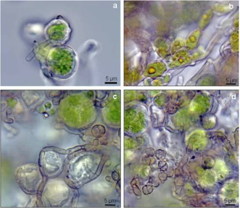
FIG. 6.5. MICROGRAPHS OF THE BIOFILM SAMPLES OBSERVED UNDER THE LIGHT MICROSCOPE. (A)
FIG. 6.6. SEM IMAGES OF THE BIOFILM OVER THE GLAZE: (A) ALGAE AND FUNGAL HYPHAE OVER THE SURFACE OF THE GLAZE; (B) ALGAL THALLI WITH THE CHARACTERISTIC MARGIN INDICATED WITH ARROW OVER THE GLAZE SURFACE;(C) ALGAE AND FUNGAL HYPHAE;(D) GLAZE SECTION (G) WITH ALGAE (A) AND GLAZE FRAGMENTS ATTACHED TO EPS (ARROWS); AND (E) SECTION DISPLAYING ALGAE (A) AND GLAZE (G). ... 132
FIG.6.7.PHOTOGRAPHIC DOCUMENTATION OF THE VISUAL EFFECT OF EACH TREATMENT APPLIED ON THE BROWNISH BIOFILMS ON GLAZED WALL TILES FROM CASA DA PESCA OVER 6 MONTHS. ... 134
FIG. 6.8. FLACKING-OFF AND DETACHED BIOFILM IN THE TIO2–TREATED AREA 6 MONTHS AFTER
TREATMENT. ... 135
FIG. 6.9. OPTICAL AND FLUORESCENCE MICROSCOPY IMAGES OF BIOFILM SAMPLES COLLECTED FOUR MONTHS AFTER THE BEGINNING OF THE EXPERIMENT:BIOFILM WITHOUT TREATMENT,TIO2-TREATED
BIOFILM, BIOTIN-TREATED BIOFILM, PREVENTOL-TREATED BIOFILM AND ALBILEX-TREATED BIOFILM.
... 135 FIG. 6.10. OPTICAL AND FLUORESCENCE MICROSCOPY IMAGES OF BIOFILM SAMPLES COLLECTED SIX
MONTHS AFTER THE BEGINNING OF THE EXPERIMENT: BIOFILM WITHOUT TREATMENT,TIO2-TREATED
BIOFILM, BIOTIN-TREATED BIOFILM, PREVENTOL-TREATED BIOFILM AND ALBILEX-TREATED BIOFILM.
... 136 FIG. 6.11.DGGE PROFILE OF THE BIOFILM SAMPLES COLLECTED FROM CASA DA PESCA GLAZED TILES
BEFORE AND FOUR MONTHS AFTER THE APPLICATION OF THE BIOCIDES. (A)DGGE PROFILES FOR CYANOBACTERIAL 16S RDNA. (B) DGGE PROFILES FOR FUNGAL ITS REGIONS. NUMBERS 1-3
INDICATE BANDS CORRESPONDING TO THE IDENTIFIED FUNGAL SPECIES DEVRIESIA MODESTA (1),
NEODEVRIESIACEAE SP. 2 (2) AND NEODEVRIESIACEAE SP. 1 (3).(C) DGGE PROFILES FOR EUKARYOTIC 18S RDNA.LANES C0 AND C4 CORRESPOND TO SAMPLES OF THE BIOFILM WITHOUT ANY TREATMENT COLLECTED ON THE FIRST DAY OF THE EXPERIMENT AND 4 MONTHS LATER, RESPECTIVELY. LANES T4, B4, P4 AND A4 CORRESPOND TO SAMPLES COLLECTED 4 MONTHS AFTERBIOCIDE APPLICATION ON AREAS TREATED WITH TIO2, BIOTIN, PREVENTOL AND ALBILEX,
RESPECTIVELY. ... 137 FIG. 6.12.PHYLOGENETIC TREE DERIVED FROM ITS1-5.8S-ITS2 REGIONS OF RRNA GENE SEQUENCES
SHOWING THE RELATIONSHIPS BETWEEN ISOLATED STRAINS AND OTU REPRESENTATIVE CLONES FROM BIOFILM SAMPLES COLLECTED ON CASA DA PESCA TILES (NAMES IN BOLD). THE CLOSEST RELATED SEQUENCES TO THE STRAINS ISOLATED BEFORE BIOCIDAL TREATMENTS WERE ALSO INCLUDED. ALL NODES OF THE TREE WERE ALSO RECOVERED USING MAXIMUM-LIKELIHOOD AND
MINIMUM-EVOLUTION TREEING ALGORITHMS.BAR,0.05 SUBSTITUTIONS PER NUCLEOTIDE POSITION.
... 145 FIG. 6.13. VP-SEM IMAGE OF THE GLAZES AFTER REMOVAL OF THE BIOFILMS FROM A NON-TREATED
COLONIZED GLAZE (NON-TREATED) AND TREATED GLAZES (TIO2,BIOTIN,PREVENTOL AND ALBILEX)
SURFACE OF MORPHOLOGY OF THE GLAZE AND DETAILS IN HIGHER MAGNIFICATION OF QUARTZ INCLUSIONS CAN BE OBSERVED. ... 146
FIG. 7.1.SCHEMATIC ILLUSTRATION OF TIO2 ELECTRONIC STRUCTURE CHARACTERIZED BY ITS VALENCE
(VB) AND CONDUCTION BAND (CB) ENERGY POSITIONS AND PHOTOCATALYTIC REACTIONS. .... 156
FIG.7.2.HYDROLYSIS AND CONDENSATION REACTIONS OF THE SOL-GEL METHOD. ... 157 FIG.7.3.SCHEME WITH EXPERIMENTAL DESIGN WITH THE NUMBER OF REPLICATES OF NON-TREATED AND
TIO2 TREATED SAMPLES. ... 159
FIG.7.4.EXAMPLE OF ANATASE RAMAN SPECTRA COLLECTED FROM THE THIN FILM DEPOSITED ON A 3TS SAMPLE. ... 161
FIG.7.5.SEM IMAGES OF THE CROSS-SECTION OF THE COATED SURFACES WITH ARROWS INDICATING THE
89.583NM, AND (D) SAMPLE 4WMT WITH Y=62.500NM. ... 162
FIG. 7.6.SCANNING ELECTRON MICROSCOPY IMAGES OF THE TIO2 COATED GLAZED SURFACES OF THE TILE SAMPLES: (A) SURFACE OF 1BWT SAMPLE, (B) SURFACE OF 3TST SAMPLE. (C) HIGHER MAGNIFICATION OF SURFACE OF TIO2 ON SAMPLE 1BWT SAMPLE, (D) HIGHER MAGNIFICATION OF SURFACE OF TIO2 ON SAMPLE 2WT SAMPLE,(E) HIGHER MAGNIFICATION OF SURFACE OF TIO2 ON SAMPLE 3TST SAMPLE AND (F) HIGHER MAGNIFICATION OF SURFACE OF TIO2 ON SAMPLE 4MWT.
... 163 FIG.7.7.PHOTOGRAPHIC DOCUMENTATION MADE DURING THE EXPERIMENT OF THE FUNGAL GROWTH ON
GLAZE TILES SAMPLES 1BW (UNCOATED); 1BWT (TIO2 COATED); 2W (UNCOATED); 2WT (TIO2
COATED);3TS(UNCOATED);3TST(TIO2 COATED);4MW(UNCOATED) AND 4MWT(TIO2 COATED).
... 164 FIG. 7.8. IRIDESCENT OF THE TILE SAMPLE 4MW AFTER THE APPLICATION OF THE TIO2 COATING
PHOTOGRAPHED IN AN OBLIQUE ANGLE. ... 165
FIG.7.9.IMAGE OF TILE SAMPLE FROM TILE 1BW(A) BEFORE AND (B) AFTER THE COATING. ... 165
FIG.7.10.COLORIMETRIC VARIATION BEFORE AND AFTER THE APPLICATION OF THE COATING EXPRESSED IN ∆E*,∆L*,∆A* AND ∆B*. ... 166
FIG.7.11.X-RAY DIFFRACTION PATTERNS OF THE CERAMIC BODY BEFORE (1-1BW,2-2GW,3-3TS,
4-4WM) AND AFTERHEAT-TREATMENT AT 450ºC. (1-1BWT, 2-2GWT, 3-3TST, 4 - 4WMT): A –
ALBITE, C- CALCITE, G- GEHLENITE, Q- QUARTZ, M – MULITE, W – WOLLASTONITE AND Ä – ÄKERMANITE. ... 167
FIG. 8.1 EXAMPLES OF ENVIRONMENTAL FACTORS INFLUENCING TILES BIOLOGICAL COLONIZATION (A)
NORTH-FACING TILE PANEL FROM CASA DA PESCA COVERED WITH A BROWN BIOFILM; (B) SOUTH
-FACING TILE PANEL PANELS FROM CASA DA PESCA WITHOUT VISIBLE BIOFILM.(C) TILES FROM FOUNTAIN LOCATED IN THE GARDEN OF MARQUÊS DE FRONTEIRA PALACE WITH PHOTOTROPHIC MICROORGANISMS, MOSSES AND VASCULAR PLANTS AND (D) TILE PANEL FROM CASA DA PESCA
LOCATED UNDER A RAIN GUTTER SHOWING A BLACK PATINA. ... 173 FIG.8.2TILES WITH DIFFERENT BIOLOGICAL COLONIZATION DUE TO PHYSICAL FEATURES OF THE TILES.(A)
TILE FROM PENA NATIONAL PALACE WITH DENSE COLONIZATION NEAR THE RELIEF;(B) TILE FROM
MARQUES DE FRONTEIRA PALACE SHOWING BIOLOGICAL COLONIZATION IN AREAS WHERE THE GLAZE HAS FALLEN... 175
FIG. 8.3 MICROBIAL COLONIZATION ON THE FILLING PASTE OF GLAZED TILESFROM MARQUES DE
xxix
Table List
Table List
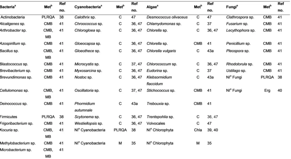
TABLE 2.1INVESTIGATED WORLDWIDE ARCHITECTURAL ASSETS ORGANIZED ACCORDING TO THE TYPOLOGY OF THE CERAMIC BUILDING MATERIAL (BRICK, ROOFING TILE AND GLAZED WALL TILE) WITH CORRESPONDING SUBSTRATE CHARACTERISTICS AND BIBLIOGRAPHIC REFERENCE. ... 18 TABLE 2.2 MICROORGANISMS REPORTED ON BRICKS AND ARCHITECTURAL SCULPTIRES TOGETHER WITH
THE IDENTIFIVATION METHOD AND REFERENCE NUMBER(TABLE 2.1.REF.NO.)... 24
TABLE 2.3.ORGANISMS REPORTED ON BRICKS AND ARCHITECTURAL SCULPTURES TOGETHER WITH THE IDENTIFICATION METHOD AND REFERENCE NUMBER (TABLE 2.1REF.NO.). ... 28
TABLE 2.4MICROORGANISMS REPORTED ON ROOFING TILES TOGETHER WITH THE IDENTIFICATIONMETHOS AND REFERENCE NUMBER(TABLE 2.1REF.NO.) ... 31
TABLE 2.5.ORGANISMS REPORTED ON ROOFING TILES TOGETHER WITH THE IDENTIFICATION METHOD AND REFERENCE NUMBER (TABLE 2.1REF.NO.). ... 34
TABLE 2.6MICROORGANISMS REPORTED ON GLAZED WALL TILES TOGETHER WITH THE IDENTIFICATION METHOD REFERENCE ... 37 TABLE 3.1. REFERENCE COMPOSITION AND ANALYTICAL RESULTS (WT.%) FOR CMOG C REFERENCE
GLASS (N=5). ... 55
TABLE 3.2.AVERAGE CHEMICAL COMPOSITION OF THE WHITE GLAZE AND STANDARD DEVIATION (STD) IN WT.%. ... 59
TABLE 3.3PHOTOTROPHIC MICROORGANISMS IDENTIFIED BY CULTURE METHODS ON GLAZED TILES FROM THE NORTH SIDE OF THE TRITON TUNNEL. ... 63
TABLE 3.4.FUNGI IDENTIFIED BY CULTURE METHODS ON GLAZED TILES FROM THE NORTH SIDE OF THE
TRITON TUNNEL. ... 64
TABLE 3.5PHYLOGENETIC AFFILIATIONS OF FIVE FUNGAL STRAINS (ISOLATES PP1 TO 5) ISOLATED FROM THE TILES. ... 64
TABLE 3.6 PHYLOGENETIC AFFILIATIONS OF MICROORGANISMS IDENTIFIED BY MOLECULAR BIOLOGY METHODS ON GLAZED TILES FROM THE NORTH SIDE OF THE TRITON TUNNEL. ... 66
TABLE 4.1COMPOSITION OF THE PRISTINE AND AGED GLAZE SURFACES IN OXIDE WEIGHT PERCENTAGE
(WT.%). ... 82 TABLE 4.2. PHYSICAL PARAMETERS ANALYSED IN PRISTINE (N=3) AND AGED (N=3) SAMPLES. ANOVA
AVERAGE AND STANDARD DEVIATION (±STD). ... 84
TABLE 4.3.CHARACTERISTICS OF THE BIOFILM ON THE PRISTINE AND AGED SAMPLES AFTER 12 MONTHS OF INCUBATION.BIOFILM COVERED AREA, OBTAINED BY DIGITAL IMAGE ANALYSIS (N=16); COLORIMETRIC PARAMETERS (L*, A* AND B*)(N=16) AND INTENSITY OF CHLA FLUORESCENCE MEASURED AT 684 NM
(N=3) AVERAGE VALUES ARE PRESENTED TOGETHER WITH THE STANDARD DEVIATION AND THE
ANOVA RESULTS. ... 88 TABLE 4.4. CHEMICAL COMPOSITION OF CONTROL AND INOCULATED SAMPLES AFTER 12 MONTHS
INOCUBATION ... 93
TABLE 4.5 SURFACE COMPOSITION OF THE COLONIZED SAMPLES ON AREAS PREVIOUSLY COVERED BY BIOFILM AND NON-COLONIZED AREAS... 95
TABLE 5.1AVERAGE AND STANDARD DEVIATION (±STD) OF WATER ABSORPTION CAPILLARY COEFFICIENT
xxx
IDENTIFIED BY Μ-RAMAN. ... 113
TABLE 5.3.CHEMICAL COMPOSITION IN OXIDES OF CONTROL AND INOCULATED SAMPLES AFTER 12 MONTHS EXPERIMENT IN WT.%(AVERAGE AND STANDARD DEVIATION). ... 114
TABLE 5.4.COMPOSITION OF THE COLONIZED PRISTINE SAMPLES ON AREAS PREVIOUSLY COVERED BY THE FUNGUS AND NON-COLONIZED AREAS IN OXIDE WEIGHT PERCENTAGE (WT%) AVERAGE OF A
750X750µM2.RESULTS ARE PRESENTED WITH ERROR VALUE. ... 114
TABLE 6.1AVERAGE CHEMICAL COMPOSITION AND STANDARD DEVIATION (STD) OF THE WHITE GLAZE IN WT.%. ... 131
TABLE 6.2.PHYLOGENETIC AFFILIATIONS OF EIGHT FUNGAL STRAINS (ISOLATES CP1 TO 8) ISOLATED FROM
CASA DA PESCA TILES. ... 133 TABLE 6.3.PHYLOGENETIC AFFILIATIONS OF THE OPERATIONAL TAXONOMIC UNITS (OTUS) OBTAINED FROM
THE 16S RDNA CYANOBACTERIAL SEQUENCES OF THE NON-TREATED BIOFILM SAMPLE (C0) AND
PREVENTOL-TREATED BIOFILM SAMPLE (P4) COLLECTED FROM CASA DA PESCA GLAZED TILES.140
TABLE 6.4.PHYLOGENETIC AFFILIATIONS OF THE OTUS OBTAINED FOR FUNGAL COMMUNITIES OF THE NON -TREATED BIOFILM SAMPLE (C0) AND PREVENTOL-TREATED BIOFILM SAMPLE (P4) COLLECTED FROM
CASA DA PESCA GLAZED TILES. ... 142
TABLE 6.5.PHYLOGENETIC AFFILIATIONS OF THE OTUS OBTAINED FROM 18S RDNA SEQUENCES OF THE NON-TREATED BIOFILM SAMPLE (C0) AND PREVENTOL-TREATED BIOFILM SAMPLE (P4) COLLECTED FROM CASA DA PESCA GLAZED TILES. ... 144
TABLE 7.1.DESCRIPTION OF THE TILE SAMPLES SELECTED FOR THE EXPERIMENT. ... 158
Abbreviations
µ-EDXRF Micro Energy Dispersive X-ray Spectroscopy µ-PIXE Micro Particle Induced X-ray Emision
µ-Raman Micro Raman spectroscopy
ANOVA Analysis of variance
APS Ammonium persulfate
BLAST Basic Local Alignment Search Tool
Chla Chlorophyll a
CLSM Confocal Laser Scanning Microscopy
cps counts per second
DGGE Denaturing Gradient Gel Electrophoresis
DMSO dimethyl-sulfoxide
DNA Deoxyribonucleic acid
EDTA Ethylene-diamine-tetra-acetic acid
FE-SEM Field Emission Scanning Electron Microscopy NCBI National Center for Biotechnology Information
OTU Operational taxonomic unit
PCA Principal components analysis
PCR Polymerase Chain Reaction
PDA Potato Dextrose Agar
Q Coeficient of water absoption by cappilary
QACs Quaternary Ammonium Compounds
Ra Roughness average
RNA Ribonucleic acid
ROS Reactive Oxygen Species
Rq Root-mean-squared roughness
Ry Total Roughness Height
SEM Scanning Electron Microscopy
SEM-EDS SEM with Energy Dispersive X-ray Spectroscopy SEM-SE SEM with Secondary Electrons
TAE Tris-Acetate-EDTA buffer
TEMED N’-tetramethylethylenediamine
VP-SEM Variable Pressure SEM
WDXRF Wavelength Dispersive X-ray Spectroscopy
wt. weight
XRD X-Ray Diffraction
General introduction
1.
Glazed wall tiles
1.1.
Glazed wall tiles, in Portuguese designated as “azulejos”, are ceramic plaques with at least the front face covered by a vitreous coating (glaze). Most of tiles are square-shaped, but they can have a variety of shapes, be flat or decorated with reliefs. Glazed tiles are architectural ceramic materials which are employed as a facing over another building material. The attachment of tiles to a surface is done by the use of a mortar in a process termed cladding and several purposes are associated with the selection of glazed tiles as a building material, such as the increase of impermeability, cleanability and mechanical resistance. In addition, the aesthetic function has particular relevance which explains the artistic and historic value of glazed wall tiles.
The production of traditional ceramic objects involves several steps, starting with the ceramic paste preparation, followed by the forming process, drying, ceramic body firing, glazing and second firing of the glaze.
The ceramic paste preparation consists in the selection, processing and mixture of the raw materials. These are often mixed, purified and grinded to obtain suitable characteristics for the forming process. In traditional ceramics, the paste is mainly composed of clay-based materials (or clay raw materials), which constitute the plastic component of the paste. Clay minerals are the basic components of clay raw materials, which are hydrated aluminosilicates organized in silicates or phyllosilicate sheets formed by tetrahedrons [SiO4]4- or octahedrons of several types, such as
[AlO3(OH)3]6- interconnected by OH- protons. The plasticity of clays is related to the morphology of
the plate-like clay mineral particles. When water is added to clays it acts as a lubricant allowing the particles to slide over the others letting the materials to be deformed and be able of retaining the shape. Konta (1995) and Andrade et al. (2011) reviewed in detail the structure, properties and application of clay minerals and clay raw materials.
Forming is the second step in ceramic tile manufacture and involves shaping the ceramic paste. This can be achieved by kneading of the paste with a roll and cutting it into slabs or using molds, particularly for high or low relief decoration of the surface.
After shaping, the tile slabs are air-dried in order to remove the excess of water. During this drying process, water evaporation implies shrinkage of the ceramic paste. Therefore, manufacture defects, such as deformation, cracking and even rupture might occur, influencing the quality of the final product.
For permanent hardening, the ceramic paste needs to be processed by thermal treatment or firing.During firing the ceramic paste suffers transformations, according to its composition and firing conditions (temperature, cycle and atmosphere) (Carter and Norton, 2013). The first reactions during firing are removal of free water (not removed by drying), combustion of organic matter, α- to β-quartz allotropic transformation, loss of OH− from the clay structure and carbonate decomposition. At temperatures above 800º C, most of the physicochemical transformations occur resulting in the neoformation of minerals phases and formation of grain boundaries through sintering. During the cooling stage, thermal contraction, crystallization and other phase transformations occur on the ceramic body.
Depending on the raw materials and firing conditions a wide variety of ceramic body typologies can be produced. These can be classified according to their porosity, color and composition (Ravaglioli and Krajewski, 1989). According to the literature, the most common tile ceramic bodies are earthenware during the 18-19th century (Borges et al., 1997; Carvalho et al., 2006; Pereira et al., 2011). These type of ceramics are opaque and non-vitrified, being fired at relatively low temperatures, varying between 960˚ and 1050˚ C (Tite, 2009; Dondi et al., 2014). According to Mimoso (2011) firing temperature could range between 800˚ and 1100˚ C, due to the difficulty in controlling temperature. The porosity of earthenware is generally characterized by high values, between 15 and 18%, because it is not vitrified (Ravaglioli and Krajewski, 1989). The porosity in these non-vitrified ceramics depends on voids between the grains and the pores created by gaseous liberation during firing which are not filled by a liquid glassy phase when temperature of glass formation (vitrification) is not achieved. Results of some studies concerning historic Portuguese tiles (16-19th century) have shown slightly high open porosity with values ranging between 22 and 28% (Santos et al., 2012).
In general, the chemical composition of earthenware used to produce Portuguese tiles result from the firing of calcareous rich clays. Pereira et al. (2011) characterized of blue and white majolica tiles from the 17 to 19th centuries being composed of: 40 to 44 wt. % of SiO2, 30-40 wt. %
of CaO, 12-15 wt. % of Al2O3, 1-2 wt. % of K2O and 5-7 wt. % of Fe2O3. Several studies described
the phases formed by firing of calcareous clays including calcium silicates, calcium aluminosilicates and anorthitic plagioclases (González-García et al., 1990; Trindade et al., 2009). In fact, the mineralogical phases of Portuguese majolica tiles are: quartz (SiO2), gehlenite (CaAl2SiO7),
diopside (CaMgSi2O6) and calcite (CaCO3) (Carvalho et al., 2006; Pereira et al., 2011). In terms of
After ceramic body completion, a vitreous coating (the glaze) is applied. Thus, glazed tiles are considered to be composite materials, due to the combination of these two materials: ceramic body and glaze (Vendrell-Saz, 2003; Carter and Norton, 2013).
Many glaze compositions have been utilized worldwide (Figueiredo et al., 2005; Coentro et al., 2012, 2014; Gulzar et al., 2012). This Chapter focuses particularly on the description of tin-opacified glazes. These glazes are composed of a lead-silicate glassy matrix with scattered quartz, feldspars and cassiterite (SnO2) crystals. Due to light scattering, these crystals, particularly
cassiterite, give opacity and a white color to the glaze (Tite, 2009).
Technically, majolica consists in the application of white opaque glaze over a fired ceramic body, on which the in-glaze pigments are painted before firing. Tite et al., (2015) confirmed recently that Egypt was the birth place of these glazes, circa 8th century A.D. However, in Europe, the continuous production of this type of glazes only started in the 13th century A.D. (Carter and Norton, 2013). The term Majolica appears only in this period, long after the beginning of the use of these glazes. This term was introduced to designate the Italian produced white glazed pottery which appeared due to the trade rout through the Spanish island of Maiorca (Carter and Norton, 2013). In the Portuguese context, the majolica technique in tile production only started to be used on the second half of the 16th century, probably brought by the Flemish and Italian artists that worked in the country during this period (Berendse et al., 1967). Subsequently, this was the main tile production technique during four centuries in Portugal.
The majolica glazes composition is usually a lead-alkali SiO2–PbO–R2O or high lead
silicate (SiO2-PbO) glaze (Tite et al., 1998; Doménech-Carbó and Doménech-Carbó, 2005; Tite,
2009). Considering that the composition varied through time the major elements on Portuguese glazed tiles are: 50-70 wt. % of SiO2, 6-10 wt. % of K2O, 1-2 wt. % of CaO, 3-10 wt. % of SnO2 and
13- 30 wt. % of PbO (Pereira et al., 2009, 2011; Coentro et al., 2012).
Glazed wall tiles as cultural heritage
1.2.
Glazed wall tiles are relevant cultural heritage assets due to their historic, artistic, and aesthetic value, as well as their distinguishable feature on the urban landscape. The variety of colors, glosses and iconography of these ceramic elements make their usage associated with an aesthetic intention. Thus, their application in claddings goes far beyond the practical properties, such as mechanical resistance, impermeability, durability and cleanability. Their application is spread worldwide, being particularly used in cultural heritage from the Iberian Peninsula, Italy, France, South America, South Asian and Western Asia countries (Berendse et al., 1967; Lemmen, 1993; Bandini et al., 2002) (see Chapter 2).
After their introduction in the Iberian Peninsula, glazed tiles soon became part of the Portuguese tradition and also an unique vehicle of art expression (Saporiti, 1992). The final decades of the 18th century attest the exceptional architectural integration and quality of paintings achieved by the Portuguese tilework during this period (Correia de Carvalho, 2012). During the 19th century, accompanying the industrial revolution, glaze wall tiles became commonly used on the façades of buildings. Nowadays, contemporary artists, architects and designers use glazed wall tiles as an artistic expression vehicle (Saporiti, 1992; Burlamaqui, 1996). For instance, national artists, such as Julio Resende (1917-2011), Querubim Lapa (1925- ) and Eduardo Nery (1938-2013) are associated to production of artistic glazed tiles (Burlamaqui, 1996). All the aforementioned examples show how the Portuguese tilework reflect the major artistic currents of each historical period. In fact, glazed wall tiles have been used more extensively and consistently in Portugal more than in other European countries. Portuguese tiles are for this reasons mentioned in many international books on tiles history, such as “Tiles 1,000 years of architectural decoration” by Lemmen (1993), “Les Metamorphoses de l´azur, l´art de lázulejos dans le monde latin” by Bandini et al. (2002) or “Tiles a general history “by Berendse et al. (1967).
Recently, in 2009, in the field of conservation and art history, a research unit dedicated to the investigation of tiles named Rede Temática em Estudos de Azulejaria e Cerâmica João Miguel dos Santos Simões (RTEACJMSS) (João Miguel dos Santos Simões Thematic Network on the Study of Tiles and Ceramics) at the Institute of the History of Art, Faculty of Letters, University of Lisbon (IHA-FLUL) was created with the aim of promoting the contact between research units dedicated to the investigation of tiles and ceramics (IHA-FLUL, n.d.). Together with the National Tile Museum (Museu Nacional do Azulejo) and other institutions several joint actions and projects have lately been conducted in order to explore and value Portuguese glazed tiles heritage.
The project ‘SOS Azulejo’ created by the Portuguese Judiciary Police Museum in 2007 to handle the increasing problem of theft, illegal trade and neglect affecting the glazed tile cultural heritage. This project has played a leading role in the creation of municipal legislation for safeguard of this patrimony and in the dissemination and educational actions (Sá, 2014).
Conferences and other dissemination actions regarding tiles as an important part of Portuguese cultural heritage have also arouse during the last years, such as AzTek - Conservação de azulejos históricos (2009) held in Lisbon or AZULEJAR 2012 (2012) held in Aveiro. The rising awareness of the importance to preserve the glazed tiles legacy is proven by the innumerous research projects and networks that were recently created for this purpose.
Deterioration of glazed wall tiles
1.3.
procedures (Raimondo et al., 2009) and also on the tiles conservation state. Production faults, such as surface and structural defects, may play also major role in the ability of ceramics to withstand deterioration processes (Vendrell-Saz, 2003; Kopar and Ducman, 2007; Mimoso et al., 2011; Pereira et al., 2011).
In glazed ceramics, the deterioration of the ceramic-glaze interface is a critical due to its composite nature (Fabbri, 2003; Vendrell-Saz, 2003; Kopar and Ducman, 2007; Mimoso et al., 2011). The characteristics of this interface area are dictated by the composition, compatibility between the two layers and the firing cycles (Kopar and Ducman, 2007; Mimoso et al., 2011).
Deterioration of ceramic tiles is also influenced by extrinsic factors, such as climatic and microclimatic conditions (affected by local urban geometry, building design or adjacent materials), atmospheric pollution and biological colonization (Silvestre and de Brito, 2009, 2011). But there are even more factors contributing to ceramic deterioration: the direct action of Man impelled by the economic situation or lack of knowledge and experience in the conservation field, which may affect material deterioration by direct destructive actions, lack of maintenance and inappropriate conservation interventions (Fabbri, 2003). For instance removal of glazed wall tiles from the wall during conservation and restorations interventions has been considered particularly damaging (Farinha and Tavares, 2003).
Physical deterioration of ceramic materials comprises mechanical damage and water related damage (diffusion, freeze-thaw cycles and soluble salts damage). The main factors causing mechanical damage are impact, scratching, abrasion, shrinking-expantion cycles and structural overload (Buys and Oakley, 1996; Silvestre and de Brito, 2009). Many of these mechanical damage with the exception of overload are caused by the action of external forces applied on the material that will result in its deterioration. Load damage is related to continuous forces, such as those imposed by the structural role of buildings. As a consequence, stress cracks, deformation and structural disintegration of the material may occur (Durbin, 2005; Kopar and Ducman, 2007; Yiu et al., 2007; Silvestre and de Brito, 2009, 2011)
increases the susceptibility to volume fluctuations due to water movement. For instance, in frost or salt crystallization high local tensile forces around the pores generate micro-cracking both in the ceramic body and the glaze (also known as crazing of the glaze)(Borges et al., 1997; Pereira et al., 2012) These forces can be exerted on the ceramic-glaze interface, resulting in fissures and scaling of the glaze, ensuing serious decay and material loss.
Concerning chemical deterioration of glazed wall tiles, the main damage is due to staining and corrosion. Contamination by external substances, such as metal corrosion products, biological colonization, soiling, or other colored substances, can cause the formation of stains. The aesthetic characteristics of the substrate are thus altered and the chemical stability can be affected. Characteristics of the substrate, like higher porosity and roughness, make the material more susceptible to this form of deterioration (Kopar and Ducman, 2007). Corrosion is caused by the chemical reaction of the substrate with any given substance causing its decay. Both the glaze and ceramic body can be chemically deteriorated, which is strongly related to the presence of water and its pH (Eppler, 1992).
Chemical corrosion affects the ceramic body particularly when water is mixed with atmospheric acids, formed from SO2, NOx, and CO2 (Ranogajec et al., 1997; Larbi, 2004).
Chemical reactions with the ceramic substrate can produce soluble compounds. This dissolution can result in alterations of the ceramic body, such as an increase in the capillarity porosity (Ranogajec et al., 1997; Larbi, 2004). The solubilization of compounds may weaken the structure and enhance the susceptibility to other types of deterioration. Water can also cause the hydration of mineral compounds resulting in volume fluctuations. Moreover, the remains of solubilized compounds in the ceramic body might act as a contamination by soluble salts as previously described.
Silicate based glazes are specially vulnerable by alkaline environments, since it enables the destruction of the silica network (Eppler, 1992; Hupa et al., 2005; Fröberg et al., 2007; Cannillo et al., 2009). In acidic environments, the reaction with water causes the leaching of ions, particularly alkalis and lead in the case of lead-silicate glazes (Blachere and Procedure, 1976; Wood and Blachere, 1978; Eppler, 1992; Hupa et al., 2005; Fröberg et al., 2007; Cannillo et al., 2009). Although being usually more resistant than the glassy phase, the crystalline phase can also be corroded. The corrosion rate depends on the chemical stability of the crystals (Eppler, 1992; Hupa et al., 2005; Fröberg et al., 2007; Cannillo et al., 2009). Differences in the ability to withstand corrosion of the crystalline and glassy phases of the glaze can lead to selective surface corrosion leading to the formation of pitting (Vandiver, 1992; Fröberg et al., 2007; Cannillo et al., 2009). The corrosion of glazes will cause the alteration of the gloss and can also contribute to mechanical changes of the glaze (Fabbri, 2003; Cannillo et al., 2009).
Figueiredo et al., 2005; Pereira et al., 2009, 2011; Coentro et al., 2012, 2014; El Nouhy, 2013; Gill et al., 2014), deterioration processes (Figueiredo et al., 2009; Pereira et al., 2012; Silva et al., 2013) and also conservation and treatment methodologies, such as consolidation (Vaz et al., 2008; Prudêncio et al., 2012), soluble salt removal (Borges et al., 1997; Pereira and Mimoso, 2012; Ottosen et al., 2014) and mortars (de Freitas et al., 2014).
The problem of understanding the deterioration of ceramic materials is compounded by the large range of ceramic building typologies, with different textural, compositional and physical characteristics, and by their varying weathering responses under different climatic and environmental conditions. The interactions between the numerous and synergistically acting factors leads to a dynamic and complex process of physical, chemical and biological deterioration. However, little research has been focused on the problem of microorganisms as agents of ceramic biodeterioration.
Biodeterioration of inorganic building materials
1.4.
Living organisms can interact with the surface of inorganic building materials on which they develop, altering their physical and chemical state (Gadd, 2010). This interaction is more frequent and severe when the materials are exposed outdoors. The process of transformation of a substance into new compounds by biochemical reactions or the action of microorganisms is defined as biodegradation. The term may involve a positive and beneficial connotation of the process (Pinna and Salvadori, 2008). However, transformations are considered harmful when microorganisms colonize anthropogenic materials, such as cultural heritage assets. Thus, the concept of biodeterioration or biological decay was defined as “any undesirable change in the properties of a material caused by living organisms” by Hueck in 1965 (Hueck, 1965). In the field of cultural heritage the decay and deterioration are perceived as damage, which are related the loss of aesthetical, physical, chemical or functional value (ICOMOS, 2008).
Biodeterioration of inorganic materials can result in physical, chemical and aesthetical damage (Kumar and Kumar, 1999; Warscheid and Braams, 2000). The damage is the result of complex mechanical, physical and chemical processes, which often occur simultaneously. The general mechanisms of biodeterioration of some inorganic building materials, such as stone, have been described by several authors (Sand, 1997; Kumar and Kumar, 1999; Warscheid and Braams, 2000; Pinna and Salvadori, 2008; Cuzman et al., 2011). However, little is known about glaze wall tiles biodeterioration (see Chapter 2).