Universidade do Algarve
Departamento de Ciências Biomédicas e Medicina
Phenotypic Study of Mutant
Embryos for the Gene ADTK1 in
Mice
Nídia Sofia Maltez Cunha
Tese Orientada por Professor Doutor José António Belo
Mestrado em Ciências Biomédicas
Biologia do Desenvolvimento Embrionário
Página II Dissertação de Candidatura ao Grau de Mestre em Ciências Biomédicas, Área de
Biologia do Desenvolvimento pela Universidade do Algarve
Master Thesis proposal in Biomedical Science, Developmental Biology by the University of Algarve
Página III As opiniões expressas nesta publicação são da exclusiva responsabilidade do seu
Autor.
The contents of this dissertation are of the exclusive responsibility of this Author. (Nídia Sofia Maltez Cunha)
Página IV Podemos nunca
vir a saber que resultados
produz a nossa acção.
Mas, se nada fizermos,
não haverá resultados.
Página V DEDICATION To my mother and father, and my brother. Thank you for your patience, love and support!
Página VI
Acknowledgments/Agradecimentos
Para começar, gostaria de agradecer em primeiro lugar ao meu orientador, Professor Doutor José António Belo, por me ter dado a oportunidade de realizar este trabalho, no seu laboratório, com a sua equipa fantástica e com o seu apoio. Por todos os esclarecimentos, conselhos, compreensão nos momentos de decisões difíceis e apoio prestado ao longo destes anos.
Queria também agradecer ao Centro de Biomedicina Molecular e Estrutural (CBME) pelo acolhimento e pelas óptimas condições de trabalho que nos oferece, por todo o apoio que me foi prestado e que possibilitou a elaboração deste trabalho.
Gostaria muito de agradecer a todos os meus colegas de laboratório, são uma equipa fantástica que muito me ajudou na realização de algumas experiências efectuadas para esta tese, com um bom espírito de equipa e trabalho e interajuda ainda me forneceram um ambiente muito agradável.
Um forte agradecimento à Marta por todo o ensino, ajuda e compreensão. Foi ela que me amparou nas situações mais difíceis de ultrapassar. Sem a sua ajuda não conseguiria escrever esta tese. À Carolina, “piriga” terrível, por toda a ajuda e ensinamentos, por todas as caminhadas ao biotério e pelas famosas limpezas de gaiolas. Ao Zé, ou melhor, ao “Tio Zé” pelas críticas construtivas constantes e pela leitura desta tese. Ao segundo tio, João Facucho por todas as ideias partilhadas pela manhã. À Marisa por todo o seu “tchanam”, é sem dúvida uma pessoa única. À Margaret por todos os conselhos e por ser muito querida comigo. À Rubi e ao João Furtado por toda a sua amizade e carrinho. A Sara e à Betty que sempre que as solicitei me ajudaram muito. As vizinhas do laboratório da frente, à Mónica e à Marinella por toda a amizade. À Lisa pelo trabalho que teve na construção genética do ratinho que foi o meu objecto de estudo. E por último aos meus “companheiros de guerra”, ao Fernando Cristo e ao João Baptista por todos os bons momentos que ultrapassamos juntos na UAlg onde nos conhecemos. Obrigado por todo o apoio, por todas as festas e todos os sorrisos ao longo destes anos. A todos desejo-lhe as maiores felicidades.
Página VII E por último agradeço às pessoas mais importantes, à minha família. Em primeiro lugar aos meus pais, os meus pilares de apoio. Sem vocês nunca teria chegado onde estou hoje, pois não sei se teria coragem de seguir em frente. À minha mãe por sempre ter acreditado, valorizado e apostado na sua filhota. Por todo o amor, carrinho e tempo que dedicou na minha formação. Ao meu pai por sempre ter vivido em prol da família que tanto ama. Por todo o apoio, compreensão, dedicação e sacrifícios. Ao maninho que tanto amo, por sempre me ter dado força. Com a sua presença torno-me mais forte e madura. Por todas as horas perdidas serem recompensadas com o seu crescimento. As minhas madrinhas, Leonor e Bia. À Leonor por toda a ajuda, conselhos e apoio nos momentos difíceis e de indecisões, apesar da distância está sempre presente. À Bia por ter estado constantemente envolvida no meu crescimento, sempre pronta a fazer as minhas vontadinhas desde pequena como que a avó que já não tenho. À restante família, pois a minha família é base de tudo o que sou e de todo o que consegui até hoje.
Ao Carlos por todo o seu amor, carrinho, compreensão e ajuda nos “painéis fotográficos”. Por todas as noites que me acompanhaste ao laboratório para eu não estar lá sozinha.
A todos os meus verdadeiros amigos, obrigado por todo o apoio em tudo o que preciso.
À Sofia por toda a compreensão, ajuda e “traduções”. Sem a tua paciência não conseguiria escrever esta tese.
Agradeço ainda a todas as restantes pessoas que não mencionei e que contribuíram para a conclusão desta tese de mestrado.
Página VIII
Abstract
A differential screening conducted to identify novel genes expressed in the anterior visceral endoderm (AVE) led to the isolation/identification of Adtk1, a gene coding for a protein with tyrosine and serine/threonine kinase domains. The analysis at birth of generated Adtk1 mouse mutants has been reported by our and other laboratories. Several defects were identified, namely the mutants were slightly smaller and had limbs with shortened long bones than wild-type littermates.
In the course of this thesis it was observed that inbreeding Adtk1 mutation into the C57Bl6 background resulted in embryonic death identified by genotyped conceptus, suggesting the importance of this gene along the embryonic development.
A more in-depth analysis of this mutation in mixed background (C57Bl6/DBA) revealed that Adtk1-/- animals also presented kidney defects. Furthermore, preliminary analysis of E9.5 Adtk1 KO embryos in a pure inbred background (C57Bl6) revealed that they display severe defects associated with neural tube closure.
Keywords: Adtk1, Bone Formation, Embryonic Development, Kidney, Mouse, Neural Tube, PCP.
Página IX
Resumo
Com o objectivo de caracterizar e melhor compreender o papel da endoderme visceral anterior ao longo do desenvolvimento embrionário de ratinho, tendo em conta os genes que nela são expressos, no nosso laboratório foi realizado um screening diferencial. No decorrer desse screening foram seleccionados genes que se encontram sobre expressos na região da endoderme visceral anterior. Vários novos genes foram identificados e entre estes encontrava-se o Adtk1 (Anterior Distal Tyrosine Kinase 1). O gene Adtk1 foi então isolado e caracterizado.
No nosso laboratório demonstrou-se que o gene Adtk1 está expresso desde muito cedo no desenvolvimento embrionário e permanece expresso durante toda a fase embrionária. Desde o início, a análise padrão de expressão sugeriu um papel importante para Adtk1 durante o desenvolvimento embrionário. Devido a esse facto foi feita uma inactivação deste gene em ratinho através de recombinação homóloga. Após a obtenção dos primeiros animais heterozigóticos para o gene Adtk1 realizaram-se os primeiros estudos preliminares, publicados em Gonçalves, et al (2011). Além do nosso laboratório também outros dois grupos de investigação realizaram o estudo do gene Adtk1, Imuta, et al (2009) e Kinoshita, et al (2009). Após a análise preliminar e as publicações o nosso laboratório continuou então o estudo do gene Adtk1.
O trabalho apresentado nesta tese concentrou-se na continuação do estudo do gene Adtk1. A fim de entender melhor o fenótipo obtido na ausência do gene Adtk1 durante o desenvolvimento de ratinho, vários tipos de experiencias foram realizadas e relatadas neste manuscrito.
O gene Adtk1 codifica uma proteína com domínio catalítico tirosina/serina/treonina cinase. Uma vez que este gene é sobre expresso na região da endoderme visceral anterior, região que contém tantos genes de particular interesse para os vários processos essenciais à embriogénese, seria de todo o interesse realizar-se uma análirealizar-se do fenótipo de ratinhos com o gene Adtk1 inactivado (KO), durante as várias fases do desenvolvimento embrionário.
Página X Para a análise do fenótipo de ratinhos Adtk1-/- cruzaram-se animais heterozigóticos, Adtk1+/-C57BL6. Contudo, não obtivemos qualquer ratinho KO na fase de recém-nascido. Tal facto levou-nos a suspeitar que uma morte intra-uterina poderia estar a ocorrer. Para testar esta hipótese começou-se a dissecar fêmeas durante o desenvolvimento embrionário, mas a suspeita da existência de morte intra-uterina começou a ser confirmada, através da presença de decíduas vazias (placenta, onde o embrião morto é reabsorvido pela mãe) em todas as ninhadas dissecadas. Devido a essa suspeita começamos a dissecar fêmeas ao longo de vários estádios do desenvolvimento embrionário. Começamos pelo E13.5, E12.5 e E10.5 na esperança de encontrar embriões Adtk1-/-,todavia a ausência destes embriões manteve-se. Assim sendo, continuámos a dissecar embriões a estádios menos desenvolvidos e observamos mais uma vez a existência de decíduas vazias em quase todas as fêmeas dissecadas. Isso indicava que o mais provável era os embriões estarem a morrer mais cedo do que o estádios analisados até ao momento. Fomos dissecando cada vez mais cedo e a E9.5 encontramos o primeiro embrião Adtk1-/-. Este embrião demonstrava más formações ao nível do fecho do tubo neural.
Análise preliminar de embriões E9.5 Adtk1-/- na estripe C57BL6 pura revelou defeitos graves associadas ao fecho do tubo neural. Estes resultados são ainda apoiados por experiencias realizadas utilizando Xenopus leavis onde a ausência dos genes ortólogos de Adtk1 durante o desenvolvimento induz defeitos na migração celular durante o fecho do tubo neural e gastrulação (M. Vitorino, não publicado). Assim sendo, é possível que o Adtk1 também esteja envolvido no fecho do tubo neural durante o desenvolvimento dos vertebrados.
Durante as dissecções a menores estádios de desenvolvimento foi encontrado um embrião Adtk1-/- a E8.5, em poucos embriões dissecados nesta fase. O único embrião Adtk1-/- encontrado não se revelou ser esclarecedor, uma vez que estava muito deformado, não nos permitiu identificar as diferentes estruturas que constituem
Página XI o embrião. No entanto, este é um resultado preliminar que precisa ser analisado através da dissecação de mais embriões nesta fase.
Posteriormente, procedeu-se a mais dissecações em estádios mais iniciais e até à data apenas dois embriões Adtk1-/- foram encontrados em E6.5. Tais embriões não parecem revelar uma grande diferença em comparação com os embriões do tipo selvagem no mesmo estádio. Assim, seria muito importante analisar as migrações ocorridas nestas etapas. Em Xenopus leavis, observamos que na ausência dos ortólogos de Adtk1, o fecho do blastóporo estava atrasado (Marta Vitorino, não publicado). Por isso, deve ser estudado se o mesmo tipo de problemas na migração de células está a ocorrer em embriões Adtk1-/ - de ratinho durante o desenvolvimento inicial, E5.5-E6.5, e avaliar se a adequada migração celular da endoderme visceral distal para originar a endoderme visceral anterior está a ocorrer em mutantes Adtk1.
Os estudos efectuados numa estirpe híbrida (C57BL6) indicaram-nos uma muito baixa percentagem de embriões Adtk1-/-. Como os recém-nascidos analisados por outro grupo de investigação tinham um fundo híbrido (estirpes CBA e C57BL6, nós na tentativa de obter recém-nascidos Adtk1-/- decidimos gerar um fundo híbrido. Para esse efeito, cruzamos ratinhos Adtk1+/- C57BL6 com ratinhos DBA do tipo selvagem. A partir deste cruzamento obtivemos a geração F1, Adtk1+/-C57BL6;DBA. Na idade adulta, os ratinhos heterozigoticos foram cruzados entre si resultando assim a geração F2. Esta geração foi recolhida logo após o nascimento e analisada em busca de ratinhos Adtk1-/- através de genotipagem.
Com um background misto obtivemos 2 recém-nascidos Adtk1-/-. Estudos preliminares utilizando estes ratinhos (C57Bl6/DBA), demonstraram que os rins de ratinhos Adtk1-/- apresentavam um córtex e uma zona glomérular com alterações estruturais e morfológicas em análise histológica. Como anteriormente já tínhamos determinado que Adtk1 é expresso nos rins do ratinho durante o desenvolvimento embrionário (de E12.5 até ao nascimento), os resultados obtidos no fundo misto, numa análise preliminar, sugerem que a expressão do gene Adtk1 poderá ser muito
Página XII importante para o desenvolvimento apropriado do rim. Todavia, o desenvolvimento do rim é um processo ainda não totalmente compreendido. Sabe-se que as moléculas de várias vias de sinalização estão envolvidas na formação do rim, no entanto, as suas posições relativas nas vias permanecem pouco conhecidas e alguns dos seus intermediários permanecem ainda por desvendar. Contudo, para melhor compreender a expressão do gene Adtk1 no decorrer do desenvolvimento embrionário renal, hibridações in situ para este gene e marcadores renais conhecidos foram feitas.
Dados apresentados por Imuta, et. al. (2009), mostraram que ratinhos Adtk1 -/-(Pkdcc) recém-nascidos apresentavam membros mais curtos do que os recém-nascidos do tipo selvagem. O esqueleto dos mutantes Adtk1 recolhidos após o nascimento foi analisado e também apresentava diferenças no comprimento do osso, embora mais ténues em comparação com as diferenças encontrada por Imuta et al (2009).
Podemos também afirmar que há vantagens e desvantagens em realizar a primeira análise de um ratinho KO sobre um fundo genético misto. No entanto, recentemente no nosso laboratório após esta mutação estar num fundo puro C57BL6, fomos capazes de demonstrar a importância deste gene ao longo do desenvolvimento embrionário, o que foi evidenciado pela morte embrionária nas dissecções genotipadas. Também mostramos os possíveis defeitos que ocorrem em estádios iniciais de desenvolvimento embrionário em estirpe pura.
No final desta tese de mestrado, algumas de nossas questões iniciais foram respondidas, enquanto outras permaneceram sem resposta. Neste momento, pensamos que ainda há muito trabalho a ser feito a partir destes dados preliminares.
Palavras-chave: Adtk1, Desenvolvimento Embrionário, Formação Óssea, PCP, Ratinho, Rins, Tubo Neural.
Página XIII
Abbreviations
3’ 3 prime 5’ 5 prime μL Microliter μm Micrometer aa Amino acid Ab AntibodyADE Anterior Definitive Endoderm
Adtk1 Antero Distal Tyrosine Kinase 1
AER Anterior Ectodermal Ridge
AP Anterior-Posterior
ASE Asymmetric Intronic Enhancer
AVE Anterior Visceral Endoderm
BMP Bone Morphogenetic Protein
bp Base pair
cDNA Coding Deoxyribonucleic Acid
CDS Coding Sequence
Cerl Cerberus-like
Chd Chordin
CNS Central Nervous System
DEPC Diethyl pyrocarbonate
Dhh Desert hedgehog
DIG Digoxigenin
Dkk Dickkopf
DLHP Dorsolateral hinge point
DMEM Dulbecco’s Modified Eagle Medium
DNA Deoxyribonucleic acid
Dpc Days post coitum
DV Dorso-Ventral
DVE Distal Visceral Endoderm
E Embryonic day
EGO Early Gastrula Organizer
EPI Epiblast
ER Endoplasmatic Reticulum
ES Embryonic stem
EtBr Ethidium bromide
FBS Fetal Bovine Serum
FD Faraday
FGF Fibroblast growth factor
Fw Forward
Fz Frizzled
h Hour
ICM Inner Cell Mass
Kb Kilo base
Ko Knock-out
JNK Jun-N terminal Kinase
LPM Lateral Plate Mesoderm
L-R Left-Right
LRP Low-density-lipoprotein
Receptor-related Protein
M Molar
MAP Mitogen-Activated Protein
MEF Mouse Embryonic Fibroblast
Mg Milligram
MGO Mid Gastrula Organizer
MHP Medium hinge point
min Minute
mL Milliliter
mRNA Messenger Ribonucleic Acid
NTD Neural tube defect
Oep One eye pinhead
ON Overnight
Otx2 Orthodenticle-related homeobox
PBS Phosphate-buffered saline
PCP Planar Cell Polarity
PCR Polymerase Chain Reaction
P-D Proximal-Distal PE Primitive Endoderm PFA Paraformaldehyde PKC Protein Kinase C p.m. PSM Presomitic Mesoderm Ptc Patched RA Retinoic acid Rev Reverse
RNA Ribonucleic acid
RT Reverse Transcription
RT Room Temperature
RTK Receptor tyrosine kinases
SDS Sodium dodecyl sulfate
SFRP Secreted Frizzled Related Protein
Shh Sonic hedgehog
SMART Simple Modular Architecture
Research Tool
Smo Smoothed
TE Trophoectoderm
TGF-β Transforming Growth Factor-Beta
UTR Untranslated Region
UV Ultraviolet
Página II
VE Visceral Endoderm
WISH Whole mount in situ hybridization
Wt Wild-type
Página i
Índice
Acknowledgments/Agradecimentos ... VI Abstract ... VIII Resumo ... IX Abbreviations ... XIII List of Figures ... v Introduction ... 11.1 Research in Developmental Biology ... 1
1.2 - Animal Models ... 2
1.2.1 – Mouse (Mus musculus) as Animal Model ... 2
1.3 - Mouse Embryogenesis ... 4
1.3.1 - Early Mouse Development ... 4
1.3.1.1 Fertilization, Pre-implantation and Implantation ... 5
1.3.1.2 Pre-implantation ... 6
1.3.1.3 Uterine Implantation ... 9
1.3.2 Gastrulation: Post-Implantion ... 9
1.3.3 Primitive Streak ... 11
1.3.4 The Node ... 12
1.3.5 Anterior Visceral Endoderm (AVE) ... 13
1.4 Kidney Development ... 15
1.4.1 Genes Involved in Renal Development ... 18
1.4.1.1 GDNF- Glial-Derived Neurotrophic Factor ... 18
1.4.1.2 Ret proto-oncogene (C-RET) ... 19
Página ii
1.4.1.4 Wnts signaling and Wnt4 ... 20
1.5 Neural Tube Defects ... 22
1.5.1Neurulation ... 22
1.5.2 Axial Skeletogenesis ... 24
1.5.3 Tail Bud Development ... 25
1.5.4 Genes involved in Developmental Mechanisms of Neural ... 25
1.5.4.1Tube Defects ... 25
1.5.4.2 Role of Planar Cell Polarity (PCP) signalling in the initiation of Neurulation.. 26
1.5.4.3 Regulation of Neural Plate Bending: Mechanisms of Spina Bifida ... 27
1.5.4.4 Molecular and cellular regulation of cranial neural tube closure ... 29
1.5.4.5 Neural crest emigration ... 30
1.6 The Adtk1 (Anterior Distal Tyrosine Kinase 1) Gene ... 31
1.7 Aim of this Project ... 33
2 Material and Methods ... 34
2.1 Strains, Embryos and Staging ... 34
2.1.1 Mice ... 34
2.1.2 Opening the abdominal cavity and locating Female Reprodutive Organs ... 34
2.1.3 Newborns Kidney Achievement... 35
2.1.4 Generation of other Background Mice ... 35
2.1.5 Preparation of Embryos and Kidneys ... 35
2.2 Genotyping ... 36
2.2.1 DNA extraction from the tail... 36
2.2.2 DNA extraction from mouse embryos or extraembryonic membranes ... 37
Página iii
2.2.4 Polymerase Chain Reaction (PCR) ... 38
2.3 Agarose gel electrophoresis for DNA and RNA ... 39
2.4 In Situ Hybridization (Whole Mount In Situ Hybridization-WISH) ... 39
2.4.1 Preparation of the anti-sense mRNA probe ... 40
2.4.1.1 Transformation of Competent E. coli ... 40
2.4.1.2Plasmid Amplification ... 40
2.4.1.3 Plasmid DNA Isolation and Purification ... 40
2.4.1.4 Plasmid Linearization ... 41
2.4.1.5 Anti-sense RNA probe Transcription ... 42
2.4.1.6 Anti-sense RNA probe clean up ... 42
2.4.2 Embryos or kidneys pre-treatments ... 43
2.4.3 Hybridization ... 43
2.4.4 Antibody Incubation ... 43
2.4.5 Immunological Detection ... 44
2.5 Histology ... 44
2.5.1 Paraffin embedding ... 44
2.5.2 Kidney Histology - Processes of Hematoxilin-Eosin ... 45
2.6 Skeletal Analysis ... 45
2.7 Photography ... 46
3 Results ... 47
3.1 Phenotype analyses in C57Black6 strain ... 47
3.1.1 Study Newborns Adtk1-/- in C57Black6 mouse ... 47
3.1.2 Analyze of Embryos in E9.5 Adtk1-/- C57Black6 ... 50
Página iv
3.1.4 Analyze of Embryos in E8.5 Adtk1-/- C57Black6 ... 56
3.1.5 Analyze of Embryos in E6.5 Adtk1-/- C57Black6 ... 57
3.2 Phenotype Analyses in Hybrid Background ... 58
3.2.1 Gene Adtk1 expression in Kidneys ... 61
3.2.2 Other Genes Involved in Kidney Development ... 62
3.2.3 Analysis of Adtk1-/- Mutants in the Hybrid Background C57Bl6/1stDBA ... 63
4 Discussion ... 66
4.1 Influence of Genetic Background on Mouse Phenotypes ... 66
4.2 Adtk1 involvement in PCP pathway ... 67
4.3 Adtk1 involvement in Gastrulation ... 68
4.4 Adtk1 involvement in Kidney Development ... 69
4.5 Adtk1 involvement in Bone Formation ... 70
5 Future Perspectives ... 72
Appendix ... 74
Página v
List of Figures
Figure 1: Different strains of Mus musculus. ... 3
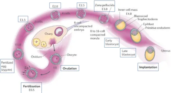
Figure 2: Mice life cycle. ... 4
Figure 3: First cleavages and pre-implantation in mouse embryo. ... 6
Figure 4: Pre-implantation and implantation in mice embryo development ... 7
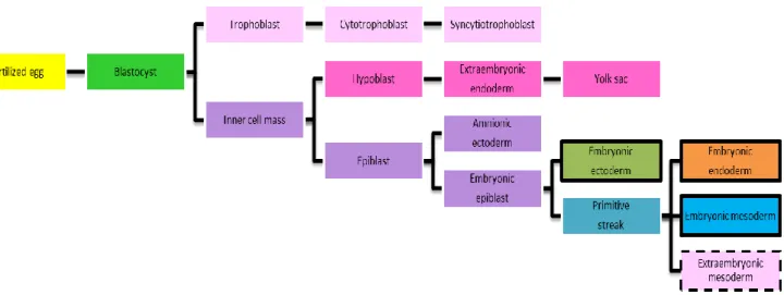
Figure 5: Schematic diagram demonstrating the tissue derivation in mouse. ... 8
Figure 6: Tissue contribution of the three germ layers. ... 10
Figure 7: Development of the mouse embryo layers from fertilization to gastrulation. rly prospective neurectoderm ... 11
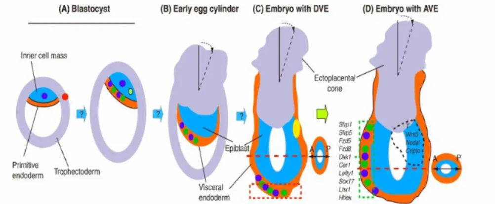
Figure 8: Schematic representation of the emergence of asymmetry during the peri-implantation development from blastocyst to right before gastrulation ... 14
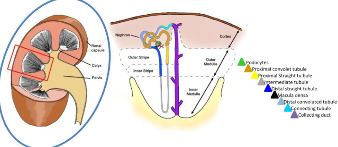
Figure 9: Illustration of the structure of the mammalian kidney. ... 15
Figure 10: Representation of the early even in mammalian kidney development ... 16
Figure 11: Schematic representation of the formation and patterning of nephrons .... 17
Figure 12: Schematic representation of the signals that promote or suppress the ureteric bud outgrowth. ... 21
Figure 13: Multisite closure of the neural tube in the mouse embryo ... 23
Figure 14: Mouse neural tube defects. ... 27
Figure 15: Molecular regulation of dorsolateral bending in mice neural tube closure. 29 Figure 16: Adtk1 KO induces neural tube defects. ... 50
Figure 17: In situ hybridization at E9.5 for Noggin. ... 52
Figure 18: Adtk1 absence disrupts neural tube closure at E9.5. ... 54
Figure 19: In situ hybridization for Fgf8 at E8.5 (A-A’’). ... 57
Figure 20: In situ hybridization for Fgf8 at E6.5. ... 58
Figure 21: In situ hybridization for different kidneys markers from E12 to E15.5 in wild-type mice. ... 61
Figure 22: Adtk1 null mutants present defects in kidney morphology ... 63
Figure 23: Kidney histology with hematoxilin-eosin. ... 64
Página 1
Introduction
1.1 Research in Developmental Biology
Developmental biology is the discipline that studies the process that occurs from having a single cell (the fertilized ovum) to complex cellular networks that give rise to the highest levels of complexity of the organism. It involves using complex tools such as gene induction and repression, tissue specialization, gene allocation and gene interactions.
Understanding the pathways on how this fundamental problem is solved is a topic of many projects in developmental biology. They include early pattern formation, the development of cell types and organ systems including the nervous system and the evolution of developmental mechanism, aging and senescence, and stem cells in developmental biology and medicine.
Many research groups are focused on the field of pattern formation, namely in the communication between cells and within cells. This is a central role in developmental biology research with major relations to quantitative and modeling approaches. The communication between cells is required and the respective (despite the fact that the form of an organism is already preprogrammed in its DNA) pathways are mediated by signaling molecules, which are released by signaling centers that organize other cells within a tissue or an organism. These signaling centers play a major role in embryogenesis and regeneration.
The origin of an individual organ during the development, organogenesis, also involves general rules that can be genetically dissected in various model systemsIt is a challenge to model these rules on a theoretical level and to understand on the molecular level how they contribute to form the many cell types that make up a complex organism.
Página 2 Developmental Biology is not only the Science that understands embryology and cell differentiation, it is also the basic science that understands human cancer, aging and the prerequisites for regenerative medicine.
1.2 - Animal Models
The major aim in genetic research is to understand the biological mechanisms of human development and disease. In many cases, however, there are both logistical and ethical issues involving studies in a human system. To overcome that barrier, a variety of animal models for genetic and functional studies relevant to human biology have been commonly used in the past years. Actually, technical developments that allow the manipulation of gene expression, coupled with the ongoing acquisition of genomic sequences, have enhanced the usefulness of animal models and assured their importance for the functional characterization of the human genome.[2]
1.2.1 – Mouse (Mus musculus) as Animal Model
The importance of mouse, Mus musculus, for the research in biomedical science is crucial because it is an excellent model organism for the study of genetics and development in vertebrates and it has a pivotal role in the study of mammalian development, physiology and biochemistry. At the turn of the twentieth century, genetic investigations of mouse mutations were initiated, shortly after the discovery of Mendel’s laws.[3] Its small size, modest cost, readily adaptation to laboratory conditions combined with a relatively short breeding cycle (approximately 8 weeks with a gestation time of 21 days and 4–6 weeks to maturity) make it an attractive model for both development and physiological studies, and very important for genetic analysis[1].
Probably the most important factor contributing to the use of mouse as a genetic model was the development of several, fully inbred, genetically homogenous mouse strains, such as the C57BL/6, DBA/2 and BALB/C lines (Figure 1).[4] The
Página 3 development of technologies to manipulate the mouse germ line by transgenesis or homologous recombination has made mouse once again the definitive system for studying mammalian gene function.[5] These genes alter the phenotypic consequences of mutations of other genes. Their importance has become obvious as investigators have learned that the phenotype of many engineered or spontaneous mutations can be influenced by genetic background.[6] This is of particular importance for mutations that are models of human diseases and the identification of modifiers that influence the severity or progression of a disease has potential as a target for therapeutic intervention. More than 700 mutant strains are also available for study and these strains involve almost every aspect of development and metabolism, such as coat colour, skeletal structure, hematology, endocrine and immune systems, neurological and behavioral characteristics, and viral, disease and tumor susceptibility or resistance. This gathering of developmental, physiological and genetic studies has turned it into the leading organism for the study of human disease, and in particular disease with an underlying genetic basis.
Figure 1: Different strains of Mus musculus. (Adapted from the journal Genome Research August 2004 Volume 14 Number 8)
Página 4 1.3 - Mouse Embryogenesis
1.3.1 - Early Mouse Development
In mammals, development begins with a sequence of mitosis followed by the separation of cells originating the embryo from cells providing support to the embryo itself during pregnancy.[7] Proliferation, differentiation, migration and apoptosis are the cellular processes underlying development.[7] These events occur according to a specific temporal and spatial program, driven by the architectural plan for each body structure.[8]
Embryonic development in the mouse begins at fertilization. Mouse mating usually takes place at night. When the plug is found the next morning, it is recorded as 0.5 d.p.c (0.5 days post-coitum, or E0.5, embryonic day 0.5) meaning that fertilization is occurred half a day previously. A litter of pups is born in the morning of 18-19.5 d.p.c. and traditionally, the gestation period in mice is considered to be 19.5 days (Figure 2).
Embryogenesis and fetal growth from fertilization to birth can be divided into six stages, each one featuring one or more special events (Figure 2). The seven stages
Figure 2: Mice life cycle. Embryogenesis and fetal growth from fertilization to birth can be divided in six stages: cleavage, implantation, gastrulation, turning, organogenesis and fetal growth and development. (Adapted from Principles of Development, Wolpert et al, 1998)
Página 5 include cleavage, implantation, gastrulation, turning, organogenesis, and fetal growth and development.[7]
1.3.1.1 Fertilization, Pre-implantation and Implantation
Fertilization is a multistep process. In mouse, male and female pronuclei, containing the haploid chromosomes of the sperm and egg, respectively, form during the one-cell zygote stage and migrate towards each other undergoing DNA replication at the same time. [7]First, sperm must bind to the egg protective membrane, the zona pellucida, which is a thick solid shell, made of glycoproteins.[7] In the process of binding to the egg membrane, the sperm releases special proteases that provide it with the ability to penetrate its way through the zona pellucida into the space that surrounds the egg membrane.[8] Although multiple sperm can make it into this space, only one can fuse with the egg.[8] Egg and sperm fusion causes fast electrochemical changes in the egg membrane that prevent the entry of additional sperm.[9] The fusion event also makes the newly fertilized egg to move down the pathway of animal development.[9]
The fertilized mouse egg is a small (approximately 80 µm in diameter) cell. It has polarity, because the polar bodies generated by the first and second meiotic divisions are sequentially extruded at the same site, where they remain tethered during ensuing cleavage (Figure 3). [10]
Fertilization of the egg inside the ampulla of the oviduct will activate to initiate pre-implantation development.[11] The control lineage segregation in the fertilized egg is essential to understand normal mammalian development.[12] Even though cleavage stages are prolonged, taking approximately 3 days for the egg to produce 16 cells, the zygotic genome is activated and maternal mRNA degraded at the two cell stage, within 24 hr of fertilization.[11]
Página 6
1.3.1.2 Pre-implantation
The initial pre-implantation events are controlled by maternal molecules. [13] Throughout the cleavage stage, all of the cells in the developing embryo are equivalent and totipotent, since they have not yet undergone differentiation and still retain the ability to produce every cell type present in the developing embryo and adult animal [1]. As a consequence of totipotency, cleavage stage embryos can be reduced to smaller groups of cells that each have the potential to develop into individual animals[1].
From third to eighth cell, cycles of pre-implantation development are shorter and take place as the embryo moves down the coiled oviduct to enter the uterine lumen, finally to hatch from the zona pellucida by enzymic activity and to adhere to the uterine endometrium and implant.[1]
These cleavages take around five days from fertilization and then the formation of the late blastocyst occurs, during which the fertilized egg undergoes 5 cleavages to reach a solid ball of 32 cells called the morula.[14] The morula cells make a two-lineage commitment to form the early blastocyst, which continues development to the late blastocyst stage prior to implantation.[14] At this stage the cells undergo a dramatic change in their behavior, through a mechanism called compaction, which consist on the increasing of contacts of the outer cells of the morula with one another by tight junctions and closing the inside of the sphere.[7] This phenomenon creates conditions
Figure 3: First cleavages and pre-implantation in mouse embryo. Left to right: 2-cell, 8-cell, early blastocyst, and late blastocyst. In the late blastocyst, cells have committed to the trophectoderm (future placenta) and inner cell mass (future body) lineages. (Adapted from Vernadeth B. Alarcon
http://www3.jabsom.hawaii.edu/Grad_DRB/faculty/alarcon.html (consulted in 4 of September of 2011)) 2-cell 8-cell early blastocyst late blastocyst
Página 7 to separate the trophoblast or trophoectoderm (the external cells of the morula) from the internal cells, the inner cell mass (ICM).[14]
The cells of the trophoblast secrete fluid into the morula and create a blastocoel and the ICM. This structure, ICM, becomes localized in one side of the ring of trophoblast cells, generating the structures named blastocyst.[15] The blastocyst is a spherical cyst, formed from the 32-cell stage (E3.5) and composed of an outer polarized epithelial monolayer, the trophectoderm, an inner cluster of nonpolarized cells, the ICM, and the blastocoel cavity, adjacent to the ICM (Figure 4).[15] The primitive endoderm will contribute to parietal and visceral endoderm. It differentiates on the surface of the ICM and includes one cell layer which is in contact with the blastocoel cavity.[8]
Figure 4: Pre-implantation and implantation in mice embryo development. After fertilization in the oviduct, the embryo experiences numerous sequences of mitotic cell division, finally forming a ball of cells named morula. The embryo enters the uterine lumen at the late morula stage, and transforms into a blastocyst that holds a cavity (named blastocoel) with two different cell populations, the inner cell mass (ICM) and the trophectoderm (the progenitor of trophoblast cells). Previously to implantation, the blastocyst goes from its outer shell (the zona pellucida) and differentiates to generate additional cell types — the epiblast and the primitive endoderm. At this stage, the trophectoderm connects to the uterine lining to beggin the process of implantation. E stands for embryonic day (Adapted from Wang and Dey 2006 [1])
Página 8 The trophoblast cells produce no embryonic structures, instead they form the chorion and the embryonic portion of the placenta. The ICM will give rise to the embryo and its associated yolk sac, allantois and amnion.[8] At the 64-cell stage, the ICM and the trophoblast cells have become separated cell layers.[9] The ICM actively supports the trophoblast, secreting proteins such as Fgf4 that make the trophoblast cells divide. [8]
The hypoblast (primitive endoderm) is an epithelial layer comprising about 20 cells that commences differentiation just prior to blastocyst implantation at 4–4.5 days post-fertilization from ICM cells adjacent to the blastocoelic cavity (Figure 4).[14] The remaining ICM cells at this time are known as the epiblast (primitive ectoderm).[16] The core of the ICM will develop into the epiblast, the progenitor tissue for the whole animal (Figure 5)[9].
Figure 5: Schematic diagram demonstrating the tissue derivation in mouse. The future extraembryonic tissues are represented in pink. The embryonic tissues are represented by the boxes limited in black. The extraembryonic mesoderm, represented by the box limited in black strips, may have two possible origins, the primitive streak or the yolk sac. Adapted from [1]
Página 9
1.3.1.3 Uterine Implantation
In order to proceed with development, the blastocyst must implant in the uterine wall. The mouse embryo implants, at stage 4.5, and during the immediate post-implantation (E5.0-E6.0) the embryonic tissue volume increases around 40 fold.[16]
Interactions between the late blastocyst and the uterine wall trigger the mural trophectoderm to differentiate into trophoblast giant cells and the polar trophectoderm to form the ectoplacental cone (Figure 4).[15]
The first 4.5 days of mouse development result in the formation of three mutually exclusive tissue lineages: the trophectoderm, the primitive endoderm and the epiblast (Figure 4).[15]
Implantation is completed by E5.5 and the embryo joins the mother in the uterine wall like a bean bud spouting in the soil. The epiblast elongates and an internal cavity is developed giving it a cup-shaped form (Figures 3, 4).[15] The development of the embryo progresses to the egg cylinder.[17];[7] The cavity formation is possibly the first apoptotic event in mouse development.[15]
Originally, the epiblast is a solid structure of cells. During early embryogenesis, signals trigger the cells in the center to die creating a hollow structure (Figure 4).[7] 1.3.2 Gastrulation: Post-Implantion
In the early post-implantation of mouse embryo there is a gastrulation stage, which involves changes in cell mobility, cell shape and cell adhesion.[1] This stage is a morphogenetic process that converts the epiblast into the three primary germ layers: endoderm, mesoderm and ectoderm, from which all the fetal tissues will develop. Together these layers form the basic body plan of the fetus, evident by about E8.5.[7] The ectoderm will generate skin and the nervous system, the mesoderm will give rise to the blood, bone and muscle and the endoderm will originate the respiratory and digestive tracts (figure 6).[18]
Página 10 The gastrulation begins approximately 6.5 days after fertilization where the epiblast cells lose their epithelial continuity at one point on the border of the cup and ingress to form a new tissue layer, the mesoderm (Figure 7).[18]
This site of new tissue production symbolizes the formation of the primitive streak and marks the beginning of gastrulation.[7] Along the next 12h the primitive streak elongates to reach the distal tip of the cylinder and therefore produces a line (or streak) of epiblast ingression and mesoderm production.[19] The place where the streak forms marks the posterior extreme of the embryo and the AP axis is now present, effectively running from the location where the streak formed to the distal tip of the cylinder and up to an anterior point on the other side of the border diametrically opposite the streak.[19]
Figure 6: Tissue contribution of the three germ layers. Schematic illustration which depicts the three germ layers and respective tissues that they will become in the future.
Página 11 The anterior extreme of the streak, that is close to the distal tip of the cylinder forms a specialized structure named the node. It is from the node that the most dorsal mesoderm in the embryo, the prechordal plate and notochord, are created. Definitive gut endoderm as well emerges from the epiblast in the vicinity of the node and intercalates into the existing visceral endoderm layer, in time fully replacing it. Together, midline mesoderm and endoderm derived from the node are known as axial mesendoderm.[11]
1.3.3 Primitive Streak
The epiblast is composed by pluripotent cells which will originate the primitive streak. The primitive streak designates the posterior end of the fetus and is created by the movement of epiblast cells inwards through a narrow band starting at the junction between the epiblast and extraembryonic ectoderm (Figure 7).[11] What initiates the
Figure 7: Development of the mouse embryo layers from fertilization to gastrulation. E 6.5, gastrulation begins and the primitive streak forms at the embryonic–extraembryonic junction. Mesoderm is produced in the streak. The origin of the streak indicates the posterior aspect of the future organism. By E7.5 the streak has elongated to the distal tip of the embryo and its anterior end has formed a specialized structure, the node. Axial mesendoderm derives from the node and moves anteriorly to underly prospective neurectoderm. The axes of the future organism are explicit at E6.5 and are revealed superimposed on an adult mouse. On the left are represented the future body axis of the mouse. (Adapted from Beddington 1999)
Página 12 movement in this particular place is at present unclear, nevertheless doubtless expression of genes (Lefty1 and Cerl1 for example) producing molecular asymmetry is involved.[11]
As the streak extends anteriorly, epiblast cells translocate into it and emerge as mesoderm. After reaching the distal tip of the egg cylinder, the anterior streak condenses into the ‘node’, and afterwards elongates anteriorly to form the notochord.[20] The length of the streak characterizes the extent of gastrulation and, since it is difficult to detect grossly, expression of Brachyury (T), a DNA-binding protein, is frequently used. Brachyury is thought to be related whit the migration of nascent mesoderm out of the streak.[20]
1.3.4 The Node
The node is a restricted area of the primitive streak which will give rise to the notochord and consequently has a vital role to play in patterning the midline axis of the mouse embryo.[21] It seems like a discrete morphological structure in the ventral midline at the rostral end of the late primitive streak at about E7.5.[21]
The concept of the organizer began with the finding of the induction in the eye and eventually increased into a unifying concept of early organization of the embryo with the classic studies of H. Spemann and H. Mangold in the 1920s, where transplantation of a small piece of non-pigmented dorsal region of a donor newt embryo to the ventral side of a pigmented host embryo caused the induction and recruitment of host cells to form a secondary axis consisting of the neural tube, notochord and somites.[1, 8]
In mice, the anterior tip of the primitive streak condenses into the node, or ‘organizer’.[22] The node forms axial mesendoderm, that includes mesoderm which will populate the midline of the embryo and the definitive gut endoderm [22] [23]. The function of the node in organizing the anterior half of the mouse embryo is an area of intense research [22, 23]. Nevertheless, its relevance as the anterior organizer has
Página 13 lately been challenged by the anterior visceral endoderm (AVE), which exerts its activity in head specification earlier than the formation of the node [24, 25].
1.3.5 Anterior Visceral Endoderm (AVE)
The mammalian embryo seems to have two signaling centers: one in the node, as previously mentioned, and one in the anterior visceral endoderm (AVE).[11]
A growing number of genes have been identified that are expressed in anterior visceral endoderm (AVE) prior or coincident with, the beginning of gastrulation in the mouse embryo.[23]
Though the first morphological signal of A-P asymmetry shows only with the appearance of the primitive streak at E6.5, evidence of a pre-existing molecular asymmetry in the visceral endoderm (VE), is already present at E5.5. The first sign of the existence of molecular asymmetry in the visceral endoderm derived from the expression of the VE-1 antigen, that is expressed in the anterior region of the visceral endoderm.[26] Expression of the VE-1 antigen4 and Otx2 [27, 28], Lim1 [25], goosecoid (gsc)[25], Hex[29] and cerberus-like 1 [25] genes in the AVE evidently precedes the beginning of gastrulation by half a day or more. They all seem to be co-expressed in a medial strip of visceral endoderm underlying almost the anterior third of the embryo, and their expression prior primitive-streak formation makes it impossible that localization of their transcripts is influenced by products of the streak.[11] Consequently, this strip of visceral endoderm is additionally patterned, being subdivided at the time of primitive-streak formation into at least two distinct domains.[11] The most anterior domain that corresponds to the site of future heart development, is evidenced by the beginning of Mrg1 expression.[30] Subsequently, Hesx1 is expressed in a population of AVE that overlies epiblast fated to form anterior CNS.[29] The expression of Mrg1 and Hesx1 in the AVE, while coincident with the onset of gastrulation, is as well improbable to be influenced by products of the primitive streak because their transcripts are sensed on the opposite side of the egg cylinder from the nascent streak, being disconnected from it by the proamniotic cavity.[30]
Página 14 Consequently, molecular description shows that the AVE has a single identity at least a day before overt gastrulation starts and that it obtains more complex pattern prior any interaction may have happened with mesoderm or endoderm produced during gastrulation.[11, 31]
The AVE is originated from the visceral endoderm (hypoblast) that migrates forward. As this region migrates, two antagonists of the Nodal protein are secreted, Lefty1 and Cerberus.[31] Lefty1 binds to the Nodal’s receptors and inhibits Nodal
binding, and Cerberus binds to Nodal itself. [25, 31, 32] Whereas the Nodal proteins inside the epiblast activate the expression of posterior genes that are needed for mesoderm formation, the AVE generated an anterior region where Nodal cannot act.[33] The AVE also starts expressing the anterior markers Otx2 and Wnt inhibitor Dickkopf.[34] Experiments of mutant mice indicate that the AVE induces anterior Figure 8: Schematic representation of the emergence of asymmetry during the peri-implantation development from blastocyst to right before gastrulation. A to D represent the early pre-gastrulation stages of mouse development, with asymmetric features listed for each stage. The tilting of the ectoplacental cone from the proximal-distal axis (unbroken line) is represented by the black broken line. Cells in the ICM, the primitive endoderm and the visceral endoderm which express β-catenin, Lefty1, Cer1 or Wnt3 are color coded. Many genes that are expressed in the AVE (D broken green rectangle) are also expressed previously in the distal visceral endoderm (DVE). Four examples of genes that are expressed in the posterior epiblast (D, broken black outline) are listed. In C (DVE) and D (AVE), a transverse section of the embryo is shown to illustrate the alignment of the prospective anterior-posterior axis first with the shorter and then with the longer diameter at the respective stage. (Adapted from Rossant and Tam 2009)
Página 15 specification by suppressing posterior patterning by Nodal and Wnt proteins.[33] Nevertheless, the AVE by itself cannot promote neural tissue, as the node can.[35]
After formation, the node will secrete Chordin; the head process and notochord will later and Noggin. These two BMP antagonists are not expressed in the AVE.[36] Whereas knockouts of both the chordin and the noggin gene do not disturb development, mice lacking both genes miss a forebrain, nose, and other facial structures.[36] It is possible that the AVE acts in the epiblast to restrict the Nodal signal, thus collaborating with the node-produced mesendoderm to induce the head-forming genes to be expressed in the anterior portion of the epiblast.[1]
1.4 Kidney Development
The intermediate mesoderm generates the urogenital system, in other words, the kidneys, the gonads, and their respective duct systems in mammalians.[37]
Podocytes
Proximal convolet tubule Proximal Straight tu bule Intermediate tubule Distal straight tubule Macula densa Distal convoluted tubule Connecting tubule Collecting duct
Figure 9: Illustration of the structure of the mammalian kidney. Each kidney is composed of a fibrous outer layer named the renal capsule, a peripheral layer named the cortex, and an inner layer named the medulla. The medulla is arranged in multiple pyramidal structures that together with overlying cortex comprise a renal lobe (red box). Urine drains from the tip of each pyramid (papilla) into minor and major calices which empty into the renal pelvis. The renal pelvis then transmits the urine to the bladder through the ureter. Nephrons are found within the cortex and medulla and have a characteristic structure that involves a glomerular blood filter containing podocytes and a tubular epithelium that loops down into the medulla. The tubule is subdivided into proximal, intermediate and distal segments (see color key) that are essential for the recovery and modification of the glomerular filtrate. (Adapted from Kidney of Mouse Development Stem book.org)
Página 16 Figure 10: Representation of the early even in mammalian kidney development. A: sagittal view of kidney components at embryonic E10.5. B: cross section of an E8.0 embryo. Nephrogenic tissues are specified from the intermediate mesoderm by signals from the surface ectoderm and somites. C: ureteric bud outgrowth and early patterning of the metanephric mesenchyme (MM). Gdnf expression by the MM promotes outgrowth and invasion of the ureteric bud at E11. Ureteric bud invasion rescues the MM from apoptosis and stimulates the condensation of the cap mesenchyme around its tip. (Adapted from Scott Boyle and Mark de Caestecker, 2006)
Mouse kidney development is also named nephrogenesis, proceeds through a series of three successive phases: the pronephros, mesonephrons and metanephros. The development starts with the formation of the pronephric duct (also known as the nephric or Wolffian duct; Figure 9). The nephric duct arises from the cranial portion of the intermediate mesoderm and grows caudally down the trunk to merge with the urogenital sinus (ventral portion of the cloaca) at E11 (Hoar, 1976). As it migrates, the nephric duct induces the generation of mesonephric nephrons from the adjacent intermediate mesoderm (known as the nephrogenic cord or nephrogenic mesenchyme).[37] Metanephros development is initiated at E10.5 at the caudal end of the nephric duct level with the hindlimb (27–28th somite).[37] Glial-derived growth factor (GDNF), secreted from a unique population of nephrogenic cells called the metanephric mesenchyme, promoves an elongates from the nephric duct named the ureteric bud (UB) that then invades the metanephric mesenchyme, giving rise to the collecting ducts, pelvis, and ureter. (see Figure 10 and 11) [37].
The UB origins a T-shaped bifurcation at E11.5, and then undergoes around 11 cycles of branching and elongation to generate the metanephric collecting duct system. [38]
Página 17 During this process, the UB tips are surrounded by a cap of metanephric mesenchyme, a subset of which form the nephron progenitors that proliferate, differentiate into glomerular and tubular epithelial cells, and fuse with the collecting duct. Previous studies demonstrated that reciprocal and inductive signaling between the UB and the metanephric mesenchyme are essential for starting and maintaining the cycles of UB branching and nephron induction that underlie the formation of the metanephros (Figure 11). [39] The permanent kidney of amniotes, the metanephros, is generated by some of the same components as the earlier, transient kidneys types.[40]
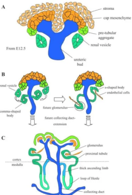
Figure 11: Schematic representation of the formation and patterning of nephrons. A: Early differentiation. The condensed cap mesenchyme becomes distinct from the surrounding stromal mesenchyme by E11.5. B: nephron elongation, fusion with the ureteric bud, and vacularization. Nephrogenesis proceeds with the formation of the comma- and S-shaped bodies and early fusion of the future distal segment with ureteric bud epithelium. Endothelial cells invade the glomerular cleft of the S-shaped body. C: patterning of nephron segments. A second temporally distinct phase of nephrogenesis occurs after E16.5 in the mouse in which there is increased growth and patterning of nephron segments associated with expansion of the renal cortex and patterning of the medullary region of the adult kidney. (Adapted from Scott Boyle and Mark de Caestecker, 2006)
Página 18
1.4.1 Genes Involved in Renal Development
More than 400 genes are known to be expressed during nephrogenesis.[40] The list includes transcription factors, growth factors, signaling molecules and extracellular matrix molecules, and data on them is catalogued in the Kidney Development Database, which is kept up to date and is available online over the Internet.
Several signaling pathways are involved in kidney development. In our context the kidney and its signaling pathways are very interesting because the development of kidneys in the Adtk1 knock-out mice (Adtk1-/-) seem to be affected by the absence of this gene. In this work particular attention was given to the relationship between Wnt4, Gremlin1, BMP4, C-RET and GDNF genes and Adtk1 since they are related to kidney development.
1.4.1.1 GDNF- Glial-Derived Neurotrophic Factor
Gdnf is expressed throughout the nephrogenic cord at E9.5 but turns out to be restricted to the region of the metanephric mesenchyme by E10.5. [41-43] GDNF signals over the Ret tyrosine kinase receptor, that is expressed by the nephric duct, organized with a membrane-tethered GFRa1 co-receptor. [44] [45] [46] GDNF signaling also implies cell surface heparan sulphate glycosaminoglycans that bind to GDNF and can have a role in ligand exhibition to the receptor.[47] Most of mouse embryos deficient in Gdnf, Ret, or Gfra1 fail to form a UB, even though the observation that a UB forms in a portion of the mutants shows that additional UB inducing signals must also exist. [48, 49] [50] [43] [51]
Several experiments using GDNF-soaked beads and transgenic overexpression of Gdnf have shown the importance of localizing GDNF-Ret signaling in order to prevent the formation of ectopic UBs.[52] [53] The maintenance and/or activation of Gdnf expression in the metanephric mesenchyme relay on a host of regulatory factors including transcription factors (Eya1, Six1, Sall1, Pax2, Hox11 proteins), secreted factors (Gdf11, Nephronectin) and FGF signaling. Inhibitors of Gdnf expression include
Página 19 Foxc1, a forkhead transcription factor, and the Robo2/Slit2 receptor/ligand pair best known for their chemorepellent role during neuron and axon migration [54].
1.4.1.2 Ret proto-oncogene (C-RET)
Additionally to promoting UB outgrowth, GDNF-Ret signaling also has a crucial role in subsequent UB branching, most likely by stimulating the cell migration and proliferation that characterizes the UB tip [55]. After UB invasion of the metanephric mesenchyme, Gdnf turns out to be expressed in the cap mesenchyme surrounding each UB tip.[42] [53] Expression of Ret becomes restricted to the UB tips and the continuance of this expression pattern relies upon retinoic acid signaling, consistent with earlier remarks linking vitamin A/retinol deficiency with renal abnormalities. [56, 57] Looking at the expression of retinoic acid receptors, retinoic acid is not thought to signal straight to the UB nevertheless is in its place hypothesized to stimulate the discharge of an unknown Ret-inducing factor from nearby stromal cells.[56, 57] If this factor stimulates Ret expression in the UB or performs via the metanephric mesenchyme is not established. The distance of Ret to the UB tip is relevant for branching morphogenesis as miss expression of c-ret throughout the ureteric epithelia has been demonstrated to inhibit UB growth and branching in transgenic mice, probably by acting as a ‘sink’ for GDNF ([58]). Ret null embryos do not have defects in nephric duct extension therefore other Gata3 targets must exist.[59]
1.4.1.3 Bone Morphogenetic Protein 4 (BMP4) and Gremlin1 BMP4 is implicated as a negative regulator of UB outgrowth. It is expressed in stromal cells enveloping the nephric duct before the outgrowth of the UB [60]. Even though Bmp4 null embryos die during early development, heterozygotes commonly display an ectopic UB.[61, 62] Organ culture experiments have shown that BMP4 may block the capacity of GDNF to promote ectopic budding from the nephric duct.[52] BMP4 activity can be inhibited after binding to the secreted BMP antagonist encoded by Gremlin1 (Grem1), that is expressed in an overlapping expression domain with Bmp4 in the early stages of UB outgrowth (Hsu et al., 1998; Michos et al., 2007; Michos
Página 20 et al., 2004). Grem1-deficient embryos exhibit blocked UB outgrowth and most of animals are born without metanephric kidneys. These defects are liberated after inactivation of one copy of the Bmp4 gene, consistent with the Grem1 null phenotype that is originated by excessive BMP4 signaling.[63] Expression of Gdnf in the Grem1 mutant metanephric mesenchyme is at the beginning normal but is downregulated progressively and lost by E11.75.[63] Despite the fact that these observations indicate that locally inhibiting BMP4 signaling around the nascent UB is necessary to maintain Gdnf expression, treatment of isolated metanephric mesenchyme with either BMP4 or Grem1 does not modify Gdnf expression.[63]
1.4.1.4 Wnts signaling and Wnt4
Wnt signaling via the canonical β-catenin pathway has been connected by several authors in UB branching.[64] Experiments with transgenic reporter mice have shown that β-catenin-induced genes are active in the UB during branching morphogenesis.[64] UB-specific inactivation of β-catenin stops branching at E12.5 resulting in renal aplasia or renal dysplasia.[65] Furthermore, this phenotype is related with reduced expression of Gdnf and Ret in the cap mesenchyme and UB tip, respectively. Several Wnts capable of signaling through the canonical β-catenin pathway are expressed in the developing metanephros, including Wnt6, Wnt7b, and Wnt9b in the collecting duct system, and Wnt4 in early nephron precursor.[66] Between these last ones, loss of Wnt4 or Wnt9b conducts to a disruption in UB branching following the T-stage, with the Wnt9b mutant phenotype being more aggressive than the Wnt4 mutant phenotype.[67] Verifying that a branching defect occurs after loss of Wnt4, that is expressed in the renal vesicle under the UB tip, indicates a feedback proccess in which nephrons induced in response to branching use Wnt4 to induce consequent UB branching.[67] In Wnt9b mutant embryos, expression of Wnt11 and Gdnf is downregulated before the morphological form of the branching defect.[68] Nevertheless, given the reciprocal nature of interactions happening between the cap mesenchyme and the UB tip and the presence of a GDNF-Ret
auto-Página 21 regulatory loop, it is not of our knowledge if the cellular target of Wnt9b activity is the cap mesenchyme or the UB.[68]
Wnt4 is expressed by the pre-tubular aggregates and is thought to work in an autocrine way to propagate the initial Wnt signal and complete the transition to the renal vesicle stage.[67] Both Wnt9b and Wnt4 perform signal via the canonical β-catenin pathway however can use different receptors and proceed in a linear pathway as Wnt9b is incapable to promote nephrogenesis in Wnt4 mutant mesenchyme in vitro.[67, 68] Gain-of-function experiments testing the effect of sustained β-catenin signaling in the metanephric mesenchyme indicate that even though Wnt signaling is crucial during the early phases of the nephrogenic program, it has to be attenuated at later stages so that nephron progenitors undergo a mesenchymal-to-epithelial transition.[37, 69]
Figure 12: Schematic representation of the signals that promote or suppress the ureteric bud outgrowth. The outgrowth and invasion of the ureteric bud (UB) from the nephric duct starts the metanephric kidney development. In a wild-type embryo, the Gdnf secretion from the metanephric mesenchyme promotes the receptor tyrosine kinase Ret, via the co-receptor Gfra1 and promotes the UB outgrowth and invasion. (Adapted from Gregory R. Dressler, 2009)
Página 22
1.5 Neural Tube Defects
Neural tube defects (NTDs) are congenital abnormalities of the central nervous system and axial skeleton that go from the fatal to the asymptomatic[70]. NTDs may be classified based on the embryonic event that is disturbed, and can result from a series of genetic defects and environmental influences.[70] Comprehending the genetic and embryonic basis of NTDs is showing the underlying developmental mechanisms, and allowing new approaches towards prevention of these severe birth defects.
To understand the embryonic origin of NTDs, it is needed to appreciate the normal developmental processes of (i) neurulation, by which the neural tube is composed, (ii) axial skeletogenesis, in which the skull and vertebral column differentiate and become modeled around the neural tube and (iii) tail bud development, that is responsible for formation of the entire body structure at post-lumbar levels of the body axis[70].
1.5.1Neurulation
Neurulation is the embryonic step that is responsible for the formation of the brain and spinal cord.[1] It starts with neural induction leading to the appearance of the neural plate, a condensed area of ectoderm in the dorsal midline.[1] The edges of the neural plate curve dorsally, starting at about 17–18 days post-fertilization, defining a longitudinal neural groove that expands with progressive elevation of the sides of the neural plate. The neural folds converge towards the midline and fuse, creating the neural tube.[1]
The initial closure event (Closure 1) occurs on E 8.5 in the mouse and is located at the future cervical/occipital boundary (Figure 13).[71] Fusion takes place from this level in rostral and caudal directions. Fusion is initiated separately, soon after this initial closure event, at two sites within the developing brain: Closure 2 is situated at the boundary between the midbrain and forebrain, while Closure 3 occurs at the extreme rostral end of the forebrain. Closure then continues in a bidirectional fashion,
Página 23 with completion of closure at ‘neuropores’ in the forebrain, hindbrain and low spinal region.[71]
This multisite closure process has been defined mainly in mouse embryos and was extrapolated to occur also in human development, on the basis of interpretation of NTD patterns in late fetuses.[72] Looking closer, mouse Closure 2 is currently recognized to be variable in its axial level among different genetic strains.[4] Consequently, Closure 2, even in the mouse, is not mandatory for brain formation. In contrast, Closure sites 1 and 3 are crucial for well succeeded neural tube closure.[72] Embryos in which Closure 1 fails develop craniorachischisis, where the neural tube is open from midbrain to low spine, while failure of Closure 3 leads to anencephaly with split face.[72] An aim for future investigation will be to determine the genetic variances that control the axial level of Closure 2, and to study if such genetic factors might explain the differing incidence of cranial NTDs.[72]
Figure 13: Multisite closure of the neural tube in the mouse embryo. Schematic summary of the successive initiation events of mouse neurulation (Closures 1, 2 and 3), the direction of spread of closure from the initiation sites (solid arrows) and the sites of completion of closure (anterior, hindbrain and posterior neuropores). The tail bud region (red shading) is the site of secondary neurulation which follows immediately after closure of the posterior neuropore. The main types of neural tube defect that result from failure of the different components of neurulation are indicated by the dotted arrows. Adapted from Copp AJ and Bernfield M (1994) Etiology and pathogenesis of human neural tube defects: Insights from mouse models. Current Opinion Pediatrics 6, 624–631.
Página 24 At E10.5 in mice, occurs the closure of the posterior neuropore in the upper sacral region, which marks the end of primary neurulation. Formation of the neural tube in the lower sacral and caudal regions happens by a different process that involves canalization of a solid cord of cells during tail bud development, without the closure of neural folds (secondary neurulation).[1]
1.5.2 Axial Skeletogenesis
After the gastrulation, the paraxial mesoderm flanking the closing occipital and spinal neural tube comes to be subdivided to form the segmentally organized, epithelial somites.[19] Both somites differentiate by the breakdown of their ventromedial wall to form the sclerotome, while their dorsolateral wall continues epithelial, comprising the dermomyotome.[8] Afterwards the dermomyotome undergoes an epithelio-mesenchymal transformation with cells delaminating from its dorsal and ventral edges to form the myotome. Axial skeletal development starts when the sclerotomal component of the mesodermal somites migrates to encompass the freshly closed neural tube, subsequently undergoing cartilaginous and bony differentiation to create the entire vertebrae.[73]
By contrast to spinal levels, in the cranial region, the skeletal structures are not formed completely from mesoderm. Fate mapping studies in birds first demonstrated that considerable portions of the skull and facial skeleton are resultant from cells of the neural crest, while related experiments in mouse reveal a larger contribution by the cranial mesoderm to the mammalian skull [74]. However, it is commonly agreed that the cranial skeleton of higher vertebrates is a composite of both mesoderm- and neural crest-derived cells.[73]
Closure of the cranial neural tube is crucial not only to main the brain development, but also for the formation of the skull vault. The embryonic brain works as a physical ‘template’ over which the skull becomes modeled. The absence of cranial neural tube closure, as in anencephaly, leads certainly to failure of formation of the skull vault, while the skull base is always present although invariably deformed. Later,