0099-2240/08/$08.00
⫹0 doi:10.1128/AEM.02419-07
Copyright © 2008, American Society for Microbiology. All Rights Reserved.
Listeria monocytogenes Biofilm-Associated Protein (BapL) May
Contribute to Surface Attachment of L. monocytogenes but Is
Absent from Many Field Isolates
䌤
Suzanne J. Jordan,
1Stefano Perni,
4Sarah Glenn,
1Isabel Fernandes,
2Manuela Barbosa,
2Manuela Sol,
2Rogerio P. Tenreiro,
3Lelia Chambel,
3Belarmino Barata,
3Isabel Zilhao,
3Timothy G. Aldsworth,
4Andreia Adriao,
5M. Leonor Faleiro,
5Gilbert Shama,
4and Peter W. Andrew
1*
Department of Infection, Immunity and Inflammation, University of Leicester, Leicester LE1 9HN, United Kingdom
1; INETI-DTIA,
EdificoS, Estrada do Paco Lumiar, 221649-038 Lisbon, Portugal
2; Instituto de Ciencia Aplicada e Tecnologia,
Campus da FCUL, Campo Grande, 1749-016 Lisbon, Portugal
3; Department of Chemical Engineering, University of
Loughborough, Loughborough LE11 3TU, United Kingdom
4; and Universidade do Algarve,
FERN, IBB-CBME, Campus de Gambelas, 8000-117 Faro, Portugal
5Received 26 October 2007/Accepted 20 May 2008
Listeria monocytogenes is a food-borne pathogen capable of adhering to a range of surfaces utilized within the
food industry, including stainless steel. The factors required for the attachment of this ubiquitous organism to
abiotic surfaces are still relatively unknown. In silico analysis of the L. monocytogenes EGD genome identified
a putative cell wall-anchored protein (Lmo0435 [BapL]), which had similarity to proteins involved in biofilm
formation by staphylococci. An insertion mutation was constructed in L. monocytogenes to determine the
influence of this protein on attachment to abiotic surfaces. The results show that the protein may contribute
to the surface adherence of strains that possess BapL, but it is not an essential requirement for all L.
monocytogenes strains. Several BapL-negative field isolates demonstrated an ability to adhere to abiotic
surfaces equivalent to that of BapL-positive strains. BapL is not required for the virulence of L. monocytogenes
in mice.
Listeria monocytogenes is a food-borne pathogen that causes
serious illness, including meningitis, septicemia, and stillbirth,
with a mortality rate of up to 30% (37). More recently, there
have been reports of listerial gastroenteritis following the
con-sumption of several different food types (16, 34). A number of
studies have demonstrated that this organism is able to persist
in the food-processing environment for several months and
even up to 10 years (23, 29). One of the major causes for
concern about L. monocytogenes in these environments is its
ability to attach to many different surfaces (2). Indeed, there is
recent evidence to show that listerial biofilms formed inside
the lumens of stainless steel tubes are able to withstand the
shears generated by high-Reynolds-number flows (31).
Bio-films, including those produced by L. monocytogenes, are more
resistant to detergents and disinfectants (33) and also are a
potential source of contamination within food-processing
plants; hence, they pose a risk to the maintenance of product
safety (27). Consequently, there is considerable interest in
de-termining the mechanisms of attachment and biofilm
forma-tion.
Our in silico analysis of the genome sequence of L.
mono-cytogenes identified an open reading frame (lmo0435) for a
protein with similarity to biofilm-associated proteins (Bap)
be-lieved to be important for the binding of staphylococci to
abiotic surfaces (10). This Bap protein also has been
impli-cated in the virulence of Staphylococcus aureus (10, 11). Thus,
the aim of the current study was to establish if this protein
(Lmo0435 [BapL]) of L. monocytogenes influenced biofilm
for-mation and virulence and to determine the prevalence of the
lmo0435 (bapL) gene within a selection of field isolates.
MATERIALS AND METHODS
Bacterial strains and plasmids.A list of the L. monocytogenes isolates and plasmids used in this study is given in Table 1. The strains were cultured in tryptone soya broth (TSB; Oxoid) or brain heart infusion agar (Oxoid) with shaking at 37°C unless otherwise stated. Escherichia coli JM109 used for the other cloning procedures was grown in Luria-Bertani broth (35) with shaking at 37°C. When required, agar (L11; Oxoid) at a concentration of 1.5% (wt/vol) was added to the broth to create a solid medium.
In silico analysis.Sequence data were submitted to BLASTP (1) available on the EGD genome sequence webpage (http://genolist.pasteur.fr/ListiList). Searches to determine the potential subcellular location and the functional regions of the query peptides were performed using PSORT-b (17) and Interpro-scan, respectively, on the ExPASy proteomics server. The EGD chromosome was viewed on ARTEMIS software version 6 (Sanger Centre, Cambridge, United Kingdom) to determine the location of the query sequences and the identity of the surrounding genes.
DNA manipulation and transformation. Standard DNA manipulation and electroporation protocols were used for E. coli as detailed by Sambrook et al. (35). Plasmid and chromosomal DNA was extracted from Listeria as outlined by Birnboim and Doly (3) and Dillard and Yother (12), respectively, with a few modifications. During the plasmid extraction, cell suspensions were incubated at 37°C for 15 min in the presence of 5 mg/ml lysozyme (Sigma) and 100 U/ml mutanolysin (Sigma) prior to alkaline lysis.
Competent cells of L. monocytogenes were prepared and electroporated as described by Park and Stewart (30). Following the 3-h recovery period, the cell
* Corresponding author. Mailing address: Department of Infection,
Immunity and Inflammation, University of Leicester, Leicester LE1
9HN, United Kingdom. Phone: 44 116 252 2941. Fax: 44 116 252 5030.
E-mail: pwa@le.ac.uk.
䌤
Published ahead of print on 30 May 2008.
5451
on July 16, 2019 by guest
http://aem.asm.org/
suspensions were plated onto brain heart infusion agar supplemented with the appropriate antibiotics.
Construction of the bapL mutant.A 919-bp fragment was amplified by PCR from a cell suspension of L. monocytogenes 10403s prepared according to the protocol detailed by Bubert et al. (5), using the primers sj007 and sj008 (Table 2). The fragment was cloned into a pGEM-T Easy vector (Promega) and excised with KpnI and HindIII for insertion into pAUL-A cut with the same enzymes. The resulting plasmid, pSJ004, was transformed into competent cells of L. mono-cytogenes 10403s as described above. The successful transformation was con-firmed by PCR using primers binding to the insert (sj007) and the vector (UR). Transformants harboring the plasmid were incubated in TSB at 40°C in the presence of 10 mg ml⫺1erythromycin prior to plating onto agar. The success of the pSJ004 insertion into the chromosome was determined by performing PCR on the chromosomal DNA of potential integrants using sj008 and UR (Table 2). The selected mutant was designated L. monocytogenes SJ78.
Attachment assay.A modified version of the protocol described previously (39) was used. Overnight cultures of the mutant and its parent were diluted 1:80 into microtiter wells (Greiner polystyrene, 655161) containing 200l fresh TSB supplemented with the appropriate antibiotics and prewarmed to 37°C. Only alternate wells of the microtiter tray were inoculated; the outer wells were not inoculated because this was found to be necessary to obtain a normal data set. These cultures were subsequently incubated at 37°C for 6 h to allow growth and attachment to occur.
The attached cells were washed with sterile phosphate-buffered saline (PBS) both prior to staining (with 0.1% [vol/vol] crystal violet) and in dye recovery (in an 80:20 [vol/vol] ethanol:acetone mixture). The eluted stain was quantified by
absorbance at 595 nm (MRX enzyme-linked immunosorbent assay plate reader; Dynatech Labs).
An estimate of the total number of cells for the mutant and wild type was done from representative wells in a duplicate tray, prepared as described above. Following agitation to remove attached cells from the well surface, 100-l sam-ples were taken for analysis. The samsam-ples were serially diluted in PBS and plated onto tryptone soy agar using the Miles and Misra technique (28). These plates were incubated overnight at 37°C prior to enumeration.
The attachment data were evaluated using the Tukey-Kramer test and Graph-Pad InStat version 3.0a for the Macintosh (GraphGraph-Pad Software, San Diego, CA [www.graphpad.com]).
Detachment assay.The detachment assay relies on the generation of quanti-fiable shear forces by the flow of a liquid between two parallel plates in order to remove cells attached to one of the plates. The shears are greatest at the center and decrease radially outwards. A radial flow chamber of the type originally described by Fowler and McKay (15) was used with a modified protocol to quantify the removal of attached L. monocytogenes to stainless steel discs. Listeria monocytogenes cultures were prepared by inoculating a single colony into 100 ml TSB and incubating the cultures statically at 30°C for 24 h. The cultures were aseptically poured into sterile petri dishes prior to the addition of a sterile stainless steel disc (diameter, 5 cm), which was immersed in each of the cultures for 2 h. Following their removal from the cell suspensions, the discs were mounted into a radial flow chamber (LH Engineering, Stoke Poges, United Kingdom) and exposed to a flow rate of 3 dm3/min of distilled water for 3 min.
The flow was delivered from a pressurized tank at 3 bars. After exposure to fluid shear flow, the discs were removed from the flow chamber, washed in PBS, fixed in 2% (vol/vol) glutaraldehyde (buffered with PBS) overnight, and washed three times in PBS for 15 min. The attached cells were stained with a live/dead stain (BacLite; Molecular Probes) and visualized with an epifluorescence microscope (Optiphot-2; Nikon). Starting from the center of the disc, the microscope stage was moved until the edge of the clearance zone was detected; this yielded the critical radius immediately beyond which the strength of the attachment of the cells just exceeded the removal forces generated by the flow. Knowledge of the critical radius enabled the shear force at that location to be calculated using the equation0(r)⫽ 3Q/rh
2
, where Q is the flow rate (m/s), h is the distance between the plates (m), is the fluid viscosity (Pa 䡠 s), and r is the radius (m) (15).
Four determinations of the critical radius (at an angular rotation of 90°) were recorded for each disc. For each strain, three independent cultures were used and three separate determinations of the critical radius were made.
In order to enumerate the number of cells attached, a second disc was pre-pared as above without exposure to the fluid flow. Twenty images of the biofilm were captured randomly across the disc surface with a digital camera (Coolpix 900; Nikon), and the cell count was done using Scion Image software (Scion Corporation, Frederick, MD).
PCR screen for the presence of the lmo0435 gene.Samples of the cell suspen-sion were prepared in 100l of 1⫻ PCR buffer (ABGene) as outlined before (5). The reactions were done in a total of 50l, containing 20 pmol of primers, 0.2 mM deoxynucleoside triphosphate mix (Bioline, United Kingdom), 1.5 mM MgCl2(ABGene), and 2.5 U Taq polymerase (ABGene). Following an initial
denaturation step of 5 min at 95°C, the PCR was performed with 35 cycles of 95°C for 1 min, annealing at an appropriate temperature (Table 2) for 1 min, followed by an extension for the required time at 72°C (Table 2) and a final cycle of 72°C for 5 min (DYAD PCR engine; MJ Research). Two PCR screens were performed using the primer pairs sj007/sj008 and sj009/sj010 (Table 2). Correct amplification was verified by electrophoresis of the products, together with the
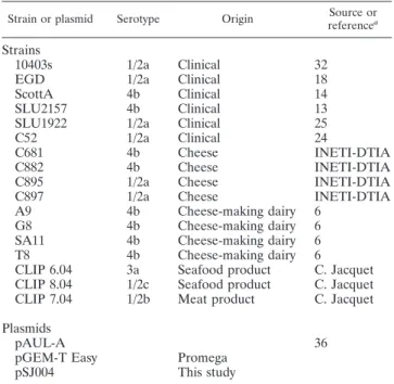
TABLE 1. Strains and plasmids
Strain or plasmid Serotype Origin Source or
referencea
Strains
10403s
1/2a
Clinical
32
EGD
1/2a
Clinical
18
ScottA
4b
Clinical
14
SLU2157
4b
Clinical
13
SLU1922
1/2a
Clinical
25
C52
1/2a
Clinical
24
C681
4b
Cheese
INETI-DTIA
C882
4b
Cheese
INETI-DTIA
C895
1/2a
Cheese
INETI-DTIA
C897
1/2a
Cheese
INETI-DTIA
A9
4b
Cheese-making dairy
6
G8
4b
Cheese-making dairy
6
SA11
4b
Cheese-making dairy
6
T8
4b
Cheese-making dairy
6
CLIP 6.04
3a
Seafood product
C. Jacquet
CLIP 8.04
1/2c
Seafood product
C. Jacquet
CLIP 7.04
1/2b
Meat product
C. Jacquet
Plasmids
pAUL-A
36
pGEM-T Easy
Promega
pSJ004
This study
TABLE 2. Details of PCR primers and conditions of use
Primer Sequence Source or
reference
Annealing temp (°C)
Extension time
sj007
TAGGTACCTGCTGGTACTTCTGGCAAG
This study
45.0
1 min
sj008
ATGAAGCTCACCTGCTACGTCCTCC
This study
45.0
1 min
sj009
TGCTCCAGCGAAAATCAA
This study
45.0
30 s
sj010
TGCTTCCCAGTAATACAACG
This study
45.0
30 s
UR
CAGGAAACAGCTATGAC
Promega
N/A
aN/A
Bact 1
CCAACAGAAGCTGCAAAACC
M. L. Faleiro
60.0
45 s
Lis1B
TTATACGCGACCGAAGCCAA
5
60.0
45 s
a
N/A, not applicable.
on July 16, 2019 by guest
http://aem.asm.org/
appropriate type of size ladder (NEB), through a 1.0 to 1.5% (wt/vol) agarose gel (Bioline) containing 1l of a 100-mg/ml ethidium bromide solution (Sigma).
Southern hybridization. Listerial chromosomal DNA was restricted with HindIII and transferred onto a nylon membrane (Hybond N⫹; Amersham Bio-sciences) by capillary transfer, and hybridization of the probe was done as detailed previously (35) with the following modifications: the membrane was incubated in a hybridization bottle containing Quikhyb solution (Stratagene) at 55°C for 30 min in a hybridization oven (Hybaid) prior to the addition of the radiolabeled probe.
The probe was created by PCR amplification with the primer pair sj009 and sj010, using colonies of L. monocytogenes 10403s as templates, as described in the previous section. Following purification using the Qiaquick PCR purification kit (Qiagen), the probe was sequenced prior to radiolabeling using the Ready-to-Go labeling kit (Pharmacia Biotech) as detailed in the manufacturer’s protocol. Immediately before its addition to the hybridization solution, the probe was boiled for 5 min and subsequently cooled on ice for 2 min.
After hybridization at 55°C overnight, the membrane was washed as before (35), apart from the third wash, which was done at 55°C. The membrane was exposed to X-ray film (Fuji Photo Film Company, Tokyo, Japan), which was developed in a Curix 60 film processor (Agfa Geveart).
Sequence analysis.Fragments were sequenced using the ABI Prism dye ter-minator cycle sequence ready reaction kit with AmpliTaq DNA polymerase FS, in conjunction with an Applied Biosystems 373 sequencing system, at the Uni-versity of Leicester, Leicester, United Kingdom.
Virulence assay.Passaged stocks of SJ78 and its isogenic parent were prepared using female MF1 outbred mice (Harlan Olac), as detailed before (39), and stored in single-use aliquots containing 10% (vol/vol) glycerol at⫺70°C. The thawed stocks were prepared for infection by centrifugation, followed by resus-pension in an appropriate volume of sterile PBS to obtain the required cell concentration. For virulence tests, groups of 10 mice were injected with approx-imately 5⫻ 105
viable bacteria in 100l into the tail vein. Determination of the dose administered was done by plating onto tryptone soy agar. Predetermined sets of five mice from each group were killed by cervical dislocation at 72 h postinfection. The livers and spleens were removed and homogenized, and the bacterial load was enumerated by colony counting (38).
RESULTS
In silico analysis.
A BLASTP search with Bap from S.
aureus against the EGD amino acid sequence (NC_003210)
revealed that Lmo0435 has 34% similarity to this protein.
Lmo0435 has 42% similarity to the Bap homolog in
Staphylo-coccus epidermidis (Bhp; AAK29746). We designated lmo0435
as bapL. Lmo0435 (BapL) is predicted to have 2,013 amino
acids, compared to 2,276 in Bap from S. aureus. These proteins
have the classic C-terminal LPXTG cell wall anchor domain of
a cell surface protein, as well as a signal peptide sequence. The
gene bapL and its upstream open reading frames have
tran-scriptional terminators, indicating that bapL is not
cotrans-cribed as part of an operon in L. monocytogenes.
Construction of an lmo0435 mutation in strain 10403s.
The
mutation of lmo0435 was prepared by constructing a
gene-specific mutagenesis vector, pSJ004, which was introduced into
10403s. The selection of potential integrants was by growth in
erythromycin at 40°C in liquid medium. The integrants were
subsequently quantified by plating onto solid medium. In order
to confirm the mutation, PCR was done on the chromosomal
DNA of the potential integrants using primers sj008 and UR.
PCR with successful integrants amplified a product of 950 bp
(data not shown). One recombinant mutant was selected and
named L. monocytogenes SJ78.
Attachment assay.
Figure 1 shows the results for the
attach-ment assays with the bapL mutant SJ78, its parent (10403s), a
dairy isolate (T8), and a cheese isolate (C897). The mutant
SJ78 showed a 50% reduction in attachment compared to
10403s (P
⬍ 0.001). This reduction in attachment was
inde-pendent of the growth of the mutant over the 6-h incubation
period, as the total numbers of planktonic cells enumerated for
the mutant and parent were the same (data not shown). C52
also showed a significantly lower level of attachment than that
of 10403s (P
⬍ 0.001), also without differences in the numbers
of planktonic cells (data not shown). The dairy and cheese
isolates (T8 and C897) attached as well as 10403s (P of
⬎0.05
for all comparisons). The planktonic cell counts for C897 and
T8 were also equivalent to 10403s at the end of the assay
period.
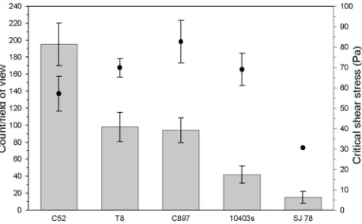
Detachment assay.
This assay was conducted to compare the
levels of adherence of L. monocytogenes cells already attached
to stainless steel discs. The radial flow device used here
en-sured that the discs were subjected to a constant liquid flow for
a fixed period of time, thus enabling the surface shear force as
a function of radius to be precisely determined. Locating the
radius at which the cells were detached from the discs gave the
critical shear force that marginally exceeded the forces of
at-tachment of the bacteria. The values of critical shear stress are
shown in Fig. 2. As can be seen, SJ78 required a shear
detach-ment force of 30 Pa, less than half (P
⬍ 0.01) of that required
to remove the isogenic parent 10403s (68 Pa). In order to
FIG. 1. Attachment of L. monocytogenes strains to polystyrene.
Data are the means
⫾ standard deviations of the results from six
experiments.
FIG. 2. Detachment of L. monocytogenes strains from stainless
steel. The points show the average critical shear stress zone of
clear-ance following the exposure of triplicate discs to a flow rate of 3
dm
3/min. The bar chart displays the mean number of cells per field of
view on the control discs. Data are the means
⫾ the standard
devia-tions of 12 readings for the zone of clearance and 60 fields of view for
the cell counts.
on July 16, 2019 by guest
http://aem.asm.org/
determine if the difference was also reflected in a reduced
ability to adhere to the stainless steel, counts were performed
on the number of attached cells after the 2-h contact time. The
bapL mutant had 50% less cells attached per field of view (P
⬍
0.01).
The cheese isolate C897 and dairy isolate T8 showed
signif-icantly greater adherence to stainless steel than 10403s (P
⬍
0.01), with at least twice the number of attached cells per field
of view. However, this did not correlate with the strength of
attachment quantified by the critical shear data; neither of the
two isolates required a significantly different (P
⬎ 0.05)
de-tachment shear force than 10403s.
C52, on the other hand, had a significantly higher number of
surface-attached cells than the other strains used in this assay
(P
⬍ 0.01). The strength of C52’s adherence, however, was
significantly less than that of the other wild-type isolates, as the
critical shear force was lower than that of 10403s, C897, and T8
(P
⬍ 0.01).
Virulence assay.
There were no significant differences (P
⬎
0.05) in the numbers of L. monocytogenes 10403s and the
mutant SJ78 recovered from the livers and spleens of mice 72 h
after infection (10403s, 9.09
⫾ 0.82 log
10CFU in the spleen
and 7.87
⫾ 0.86 log
10CFU in the liver; SJ78, 8.99
⫾ 0.51
log
10CFU in the spleen and 7.94
⫾ 0.45 log
10CFU in the liver;
n
⫽ 5).
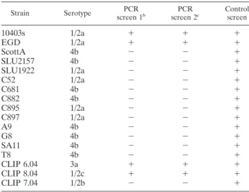
Presence of bapL (lmo0435) in L. monocytogenes strains.
Two different PCR assays were done to determine the
pres-ence of lmo0435 in a range of L. monocytogenes isolates from
different sources. In the first screen, using the primer pair sj007
and sj008, the expected product size was 919 bp. A second
screen used the primers sj009 and sj010, and the expected
product size was 343 bp. Positive control reactions were done
with primers Bact 1 and Lis1B (Table 2) for the iap gene.
Table 3 displays a summary of both of the PCR screens. All
17 isolates were positive for the control iap fragment, but only
four, EGD, 10403s, CLIP 6.04, and CLIP 8.04, possessed the
bapL gene. The amplified products for the four positive
iso-lates were of the expected size in both PCR screens. The
strains also were screened for the presence of bapL by
South-ern hybridization, using the PCR product amplified by sj009
and sj010 as the probe. This probe was to the central region of
bapL. Each of the isolates found positive by PCR showed a
single band of the same size after hybridization, whereas the
PCR-negative isolates were negative by hybridization (data not
shown).
DISCUSSION
The factors required for biofilm formation in L.
monocyto-genes are still relatively unknown, although some progress has
been made in this area through the screening of insertion
mutant libraries (19, 39) and proteomic studies (21, 40).
Sev-eral genes have been identified, including relA that encodes
(p)ppGpp synthetase (39), the superoxide dismutase gene
(sod), and the gene encoding 30s ribosomal protein S2 (rpsB)
(40), but because these do not code for surface proteins, they
will not directly determine attachment. The present study
uti-lized an in silico approach to identify the potential surface
proteins that may be involved in the attachment of this
organ-ism to surfaces. Data mining of the EGD chromosome
re-vealed that Bap, which plays a significant role in S. aureus
biofilm formation, shared similarity to the putative listerial
protein Lmo0435. A mutation was constructed in lmo0435 to
establish if this gene was required for the attachment of L.
monocytogenes to abiotic surfaces. Because we showed that it
could contribute to the attachment of L. monocytogenes 10403s
to abiotic surfaces (polystyrene and stainless steel), we
desig-nated lmo0435 as bapL.
The lmo0435 mutant of L. monocytogenes 10403s displayed a
significantly reduced level of attachment to polystyrene and
stainless steel surfaces than its isogenic parent, which was in
agreement with its predicted function. The presence of an
LPXTG motif, and a signal peptide sequence, strongly suggests
that Lmo0435 is outside the cell, anchored to the
peptidogly-can. In addition to its extracellular location, the protein has
polycystic kidney disease (PKD) repeat regions responsible for
the formation of an immunoglobulin G-like fold in the PKD1
protein. These regions are thought to be involved in cell
ad-hesion through the protein-protein interaction as well as the
protein-carbohydrate interactions by the PKD1 protein (22).
Many organisms, such as Pseudomonas spp. and
Staphylo-coccus spp., form biofilms that possess a thick extracellular
polysaccharide matrix surrounding the attached cells (7, 8). In
S. aureus and S. epidermidis, polysaccharide adhesin synthesis is
encoded by the intercellular adhesion (ica) locus (9, 20).
Strains with mutations in this operon are unable to form strong
biofilms on polystyrene (9); however, recent research has
shown that Bap expression is able to restore biofilm production
in an IcaA mutant (11). Microscopy images of L.
monocyto-genes suggest that attached cells produce a small quantity of
extracellular material (26 and our unpublished data), which
contains ruthenium-red-positive polysaccharides (4). In the
ab-sence of a thick extracellular polysaccharide, Lmo0435 may
serve as part of the adhesive matrix in L. monocytogenes.
Given the apparent role of bapL in the attachment of
10403s, it was surprising that a screen of dairy, food, and
TABLE 3. Summary of the PCR assays to detect the presence of
lmo0435 in the collection of L. monocytogenes isolates
aStrain Serotype PCR screen 1b PCR screen 2c Control screen
10403s
1/2a
⫹
⫹
⫹
EGD
1/2a
⫹
⫹
⫹
ScottA
4b
⫺
⫺
⫹
SLU2157
4b
⫺
⫺
⫹
SLU1922
1/2a
⫺
⫺
⫹
C52
1/2a
⫺
⫺
⫹
C681
4b
⫺
⫺
⫹
C882
4b
⫺
⫺
⫹
C895
1/2a
⫺
⫺
⫹
C897
1/2a
⫺
⫺
⫹
A9
4b
⫺
⫺
⫹
G8
4b
⫺
⫺
⫹
SA11
4b
⫺
⫺
⫹
T8
4b
⫺
⫺
⫹
CLIP 6.04
3a
⫹
⫹
⫹
CLIP 8.04
1/2c
⫹
⫹
⫹
CLIP 7.04
1/2b
⫺
⫺
⫹
a⫹ denotes presence and ⫺ denotes absence. bPCR with primers sj007/sj008.
cPCR with primers sj009/sj0010.
on July 16, 2019 by guest
http://aem.asm.org/
clinical isolates revealed that only 4 out of 17 possessed the
gene. It is particularly noteworthy that strains A9, G8, SA11,
and T8 do not have bapL, because these were isolated from
abiotic surfaces in cheese-making dairies. Furthermore, in the
laboratory assay of attachment and detachment, the
bapL-negative dairy and cheese strains tested (C897 and T8)
ad-hered significantly better than the bapL-positive 10403s and
EGD. In contrast, the bapL-negative C52 was significantly
worse in the attachment assays than did 10403s. Thus, at least
two mechanisms of persistent attachment to surfaces appear to
have evolved in L. monocytogenes: BapL dependent and BapL
independent. These mechanisms do not appear to be
distrib-uted in a serotype-specific manner. Although all of the 4b
isolates tested were bapL negative, both bapL-positive and
bapL-negative strains were found among the 1/2a isolates.
Where present, the bapL gene determines not only the ability
to attach to a surface but also the force necessary to remove
the cells already bound (see Fig. 2). There is not, however, a
general linkage between the number of L. monocytogenes cells
adhering to stainless steel and the force required to detach
them, because the bapL-negative strains, such as T8 and C897,
required higher shear stresses for removal than the
bapL-positive 10403s.
The results gained during this study indicate that factors
involved in the attachment of L. monocytogenes to surfaces can
be isolate specific and that caution should be used when trying
to extrapolate results from a single isolate. The strain 10403s is
frequently used for in vitro and in vivo studies, often as the
only object of study. Extrapolation from the data for 10403s in
this study would suggest that Lmo0435 was generally involved
in biofilm formation by L. monocytogenes, yet it was absent in
all of the cheese and dairy isolates tested. Surface attachment
by L. monocytogenes clearly is a multifactorial process, and
further work is required to determine the full repertoire of the
molecules involved.
ACKNOWLEDGMENTS
We thank C. Jacquet, D. Portnoy, and W. Tham for donations of L.
monocytogenes isolates and T. Chakraborty for the plasmid pAUL-A.
This work was funded by the European Union, as part of the
LMTOOCHE consortium (contract number QLKL1-CT-2002-02219).
REFERENCES
1. Altschul, S. F., T. L. Madden, A. A. Scha¨ffer, J. Zhang, Z. Zhang, W. Miller, and D. J. Lipman.1997. Gapped BLAST and PSI-BLAST: a new generation of protein database search programs. Nucleic Acids Res. 25:3389–3402. 2. Beresford, M. R., P. W. Andrew, and G. Shama. 2001. Listeria monocytogenes
adheres to many materials found in food-processing environments. J. Appl. Microbiol. 90:1000–1005.
3. Birnboim, H. C., and J. Doly. 1979. A rapid alkaline extraction procedure for screening recombinant plasmid DNA. Nucleic Acids Res. 7:1513–1523. 4. Borucki, M. K., J. D. Peppin, D. White, F. Loge, and D. R. Call. 2003.
Variation in biofilm formation among strains of Listeria monocytogenes. Appl. Environ. Microbiol. 69:7336–7342.
5. Bubert, A., J. Riebe, N. Schnitzler, A. Scho¨nberg, W. Goebel, and P. Schubert.
1997. Isolation of catalase-negative Listeria monocytogenes strains from listerio-sis patients and their rapid identification by anti-p60 antibodies and/or PCR. J. Clin. Microbiol. 35:179–183.
6. Chambel, L., M. Sol, I. Fernandes, M. Barbosa, I. Zilha˜o, B. Barata, S. Jordan, S. Perni, G. Shama, A. Adria˜o, L. Faleiro, T. Requena, C. Pela´ez, P. W. Andrew, and R. Tenreiro.2007. Occurrence and persistence of Listeria spp. in the environment of ewe and cow’s milk cheese dairies in Portugal unveiled by an integrated analysis of identification, typing and spatial-tem-poral mapping along production cycle. Int. J. Food Microbiol. 116:52–63. 7. Costerton, J. W., Z. Lewandowski, D. E. Caldwell, D. R. Korber, and H. M.
Lappin-Scott.1995. Microbial biofilms. Annu. Rev. Microbiol. 49:711–745. 8. Costerton, J. W., P. S. Stewart, and E. P. Greenberg. 1999. Bacterial biofilms:
a common cause of persistent infections. Science 284:1318–1322.
9. Cramton, S. E., C. Gerke, N. F. Schnell, W. W. Nichols, and F. Gotz. 1999. The intercellular adhesion (ica) locus is present in S. aureus and is required for biofilm formation. Infect. Immun. 67:5427–5433.
10. Cucarella, C., C. Solano, J. Valle, B. Amorena, I. Lasa, and J. R. Penades. 2001. Bap, a Staphylococcus aureus surface protein involved in biofilm for-mation. J. Bacteriol. 183:2888–2896.
11. Cucarella, C., M. A. Tormo, C. U´ beda, M. P. Trotonda, M. Monzo´n, C. Peris, B. Amorena, I´. Lasa, and J. R. Penade´s. 2004. Role of biofilm-associated
protein Bap in the pathogenesis of bovine Staphylococcus aureus. Infect. Immun. 72:2177–2185.
12. Dillard, J. P., and J. Yother. 1994. Genetic and molecular characterization of capsular polysaccharide biosynthesis in Streptococcus pneumoniae type 3. Mol. Microbiol. 12:959–972.
13. Ericsson, H., A. Eklow, M. L. Danielsson-Tham, S. Loncarevic, L. O.
Mentzing, I. Persson, H. Unnerstad, and W. Tham.1997. An outbreak of listeriosis suspected to have been caused by rainbow trout. J. Clin. Mi-crobiol. 35:2904–2907.
14. Fleming, D., S. Cochi, K. MacDonald, J. Brondum, P. Hayes, B. Plikaytis, M.
Holmes, A. Audurier, C. Broome, and A. Reingold.1985. Pasteurized milk as a vehicle of infection in an outbreak of listeriosis. N. Engl. J. Med. 312:404– 407.
15. Fowler, H. W., and A. J. McKay. 1980. Microbial adhesion to surfaces. Ellis Horwood, Chichester, United Kingdom.
16. Frye, D. M., R. Zweig, J. Sturgeon, M. Tormey, M. LeCavalier, I. Lee, L.
Lawani, and L. Mascola.2002. An outbreak of febrile gastroenteritis asso-ciated with delicatessen meat contaminated with Listeria monocytogenes. Clin. Infect. Dis. 35:943–949.
17. Gardy, J. L., C. Spencer, K. Wang, M. Ester, G. E. Tusnady, I. Simon, S.
Hua, K. deFays, C. Lambert, K. Nakai, and F. S. Brinkman. 2003. PSORT-B: improving protein subcellular localization prediction for Gram-negative bacteria. Nucleic Acids Res. 31:3613–3617.
18. Glaser, P., L. Frangeul, C. Buchrieser, C. Rusniok, A. Amend, F. Baquero,
P. Berche, H. Bloecker, P. Brandt, T. Chakraborty, A. Charbit, F. Chetouani, E. Couve, A. de Daruvar, P. Dehoux, E. Domann, G. Dominguez-Bernal, E. Duchaud, L. Durant, O. Dussurget, K. D. Entian, H. Fsihi, F. Garcia-Del Portillo, P. Garrido, L. Gautier, W. Goebel, N. Gomez-Lopez, T. Hain, J. Hauf, D. Jackson, L. M. Jones, U. Kaerst, J. Kreft, M. Kuhn, F. Kunst, G. Kurapkat, E. Madueno, A. Maitournam, J. M. Vicente, E. Ng, H. Nedjari, G. Nordsiek, S. Novella, B. de Pablos, J. C. Perez-Diaz, R. Purcell, B. Remmel, M. Rose, T. Schlueter, N. Simoes, A. Tierrez, J. A. Vazquez-Boland, H. Voss, J. Wehland, and P. Cossart.2001. Comparative genomics of Listeria species. Science 294:849–852.
19. Gorski, L., J. D. Palumbo, and R. E. Mandrell. 2003. Attachment of Listeria monocytogenes to radish tissue is dependent upon temperature and flagellar motility. Appl. Environ. Microbiol. 69:258–266.
20. Heilmann, C., O. Schweitzer, C. Gerke, N. Vanittanakom, D. Mack, and F.
Gotz.1996. Molecular basis of adhesion in the biofilm-forming Staphylococ-cus epidermidis. Mol. Microbiol. 20:1083–1091.
21. Helloin, E., L. Jansch, and L. Phan-Thanh. 2003. Carbon starvation survival of Listeria monocytogenes in planktonic state and in biofilm: a proteomic study. Proteomics 3:2052–2064.
22. Hughes, J., C. J. Ward, B. Peral, R. Aspinwall, K. Clark, J. L. S. Millan, V.
Gamble, and P. C. Harris.1995. The polycystic kidney disease 1 (PKD1) gene encodes a novel protein with multiple cell recognition domains. Nat. Genet. 10:151–160.
23. Jacquet, C., B. Catimel, V. Goulet, V. Lepoutre, P. Veit, P. Dehamout, and J.
Rocourt.1995. Typing Listeria monocytogenes during epidemiological inves-tigations of the French listeriosis outbreaks in 1992, 1993 and 1995, p. 161–176. In Proceedings of the XII International Symposium on the Prob-lems of Listeriosis. Promaco Conventions, Perth, Australia.
24. Jones, C. E., G. Shama, D. Jones, I. S. Roberts, and P. W. Andrew. 1997. Physiological and biochemical studies on psychrotolerance in Listeria mono-cytogenes. J. Appl. Microbiol. 83:31–35.
25. Loncarevic, S., M. L. Danielsson-Tham, L. Martenssen, A. Runehagen, and
W. Tham.1997. A case of foodborne listeriosis in Sweden. Lett. Appl. Microbiol. 24:65–68.
26. Marsh, E. J., H. Luo, and H. Wang. 2003. A three-tiered approach to differentiate Listeria monocytogenes biofilm-forming abilities. FEMS Micro-biol. Lett. 228:203–210.
27. Midelet, G., and B. Carpentier. 2002. Transfer of microorganisms, including Listeria monocytogenes, from various materials to beef. Appl. Environ. Mi-crobiol. 68:4015–4024.
28. Miles, A. A., and S. S. Misra. 1938. The estimation of the bactericidal power of blood. J. Hyg. 38:732–749.
29. Ojeniyi, B., J. Christensen, and M. Bisgaard. 2000. Comparative investiga-tion of Listeria monocytogenes isolated from a turkey processing plant, turkey products and from human cases of listeriosis in Denmark. Epidemiol. Infect.
125:303–308.
30. Park, S. F., and G. S. A. B. Stewart. 1990. High efficiency transformation of Listeria monocytogenes by electroporation of penicillin-treated cells. Gene
94:129–132.
on July 16, 2019 by guest
http://aem.asm.org/
31. Perni, S., S. J. Jordan, P. W. Andrew, and G. Shama. 2006. Biofilm development by Listeria innocua in turbulent flow regimes. Food Control 17:875–883. 32. Portnoy, D. A., P. S. Jacks, and D. J. Hinrichs. 1988. Role of haemolysin for the
intracellular growth of Listeria monocytogenes. J. Exp. Med. 167:1459–1471. 33. Robbins, J. B., C. W. Fisher, A. G. Moltz, and S. E. Martin. 2005.
Elimina-tion of Listeria monocytogenes biofilms by ozone, chlorine, and hydrogen peroxide. J. Food Prot. 68:494–498.
34. Salamina, G., E. Dalle, A. Niccolini, G. Poda, D. Cesaroni, M. Bucci, R. Fini,
M. Maldini, A. Schuchat, B. Swaminathan, W. Bibb, J. Rocourt, N. Binkin, and S. Salmaso.1996. A foodborne outbreak of gastroenteritis involving Listeria monocytogenes. Epidemiol. Infect. 117:429–436.
35. Sambrook, J., E. F. Fritsch, and T. Maniatis. 1989. Molecular cloning: a laboratory manual, 2nd ed. Cold Spring Harbor Laboratory, Cold Spring Harbor, NY.
36. Schaferkordt, S., and T. Chakraborty. 1995. Vector plasmid for mutagenesis and directional cloning in Listeria spp. BioTechniques 19:720–725. 37. Schlech, W. F. 2001. Foodborne listeriosis. Clin. Infect. Dis. 31:770–775. 38. Stephens, J. C., I. S. Roberts, D. Jones, and P. W. Andrew. 1991. Effect of
growth temperature on virulence of strains of Listeria monocytogenes in the mouse: evidence for a dose dependence. J. Appl. Bacteriol. 70:239–244. 39. Taylor, C. M., M. Beresford, H. A. S. Epton, D. C. Sigee, G. Shama, P. W.
Andrew, and I. S. Roberts.2002. Listeria monocytogenes relA and hpt mutants are impaired in surface-attached growth and virulence. J. Bacteriol. 184:621– 628.
40. Tremoulet, F., O. Duche, A. Namane, B. Martinie, The European Listeria
Genome Consortium, and J. C. Labadie.2002. Comparison of protein pat-terns of Listeria monocytogenes grown in biofilm or in planktonic mode by proteomic analysis. FEMS Microbiol. Lett. 210:25–31.