Pharmaceutical Sciences http://dx.doi.org/10.1590/s2175-97902017000300071 Ar
ti
∗Correspondence: C. A. F. Oliveira. Departamento de Engenharia de Alimentos, Faculdade de Zootecnia e Engenharia de Alimentos, Universidade de São Paulo. Av. Duque de Caxias – Norte, 225, CEP 13635-900, Pirassununga, SP, Brazil. Tel.: +55 19 3565-4173; Fax: +55 19 3565-4284. E-mail: [email protected]
Effect of peracetic acid on biofilms formed by
Listeria monocytogenes
strains isolated from a Brazilian
cheese processing plant
Sarah Hwa In Lee
1, Giovana Verginia Barancelli
2, Carlos Humberto Corassin
1, Roice Eliana
Rosim
1, Carolina Fernanda Sengling Cebin Coppa
1, Carlos Augusto Fernandes de Oliveira
1*1Department of Food Engineering, School of Animal Science and Food Engineering, University of São Paulo, Pirassununga,
SP, Brazil, 2Department of Agroindustry, Food and Nutrition, “Luiz de Queiroz” College of Agriculture, University of São
Paulo, Piracicaba, SP, Brazil.
This study aimed to investigate the efect of peracetic acid (PAA, 0.5%) on adherent cells of three strains of
Listeria monocytogenes strains belonging to serotypes 4b and 1/2b that had been previously isolated from the environment of a Brazilian cheese plant. The assays were conducted using polystyrene microplates and stainless steel coupons and the adhered cells were treated with PAA for 60, 120 and 180 s. On
stainless steel, bioilms were partially inactivated by PAA after 60 s and almost 100% of the cells were damaged within 180 s using epiluorescence microscopy with LIVE/DEAD® staining. On polystyrene microplates, PAA decreased (P<0.05) bioilm biomass produced by the three L. monocytogenes isolates
at 60 s, when compared with controls (no PAA treatment). However, PAA did not completely eliminate
L. monocytogenes cells on polystyrene microplates (decreasing 1.8-2.5 log cycles after treatment with
PAA for 180 s). The correct concentration and contact time of PAA is critical for eliminating bioilms
formed by L. monocytogenes on stainless steel surfaces, although further studies are needed for deining
eicient PAA treatments to remove adherent cells of this pathogen on plastic polymers.
Keywords: Peracetic acid (PAA)/ bioilm/efects. L. monocytogenes. Dairy plants/Brazil.
INTRODUCTION
Listeria monocytogenes is a rod-shaped,
non-spore pathogen that can be widely distributed in the environment, including soil, surface water used for agricultural purposes and food products (Casarin et al., 2014). The dairy industry is particularly susceptible to contamination by L. monocytogenes, and several cheese-associated Listeria outbreaks have been described worldwide (Johnsen et al., 2010; Koch et al., 2010).
The presence of L. monocytogenes in the environment of cheese processing plants can be a potential source of contamination, especially when the microorganism survives in niches that are diicult to sanitize or in places where moisture and food debris are present (Tompkin, 2002). Under these circumstances, the pathogen may
spread from the processing environment thus leading to the contamination of inal products through the ventilation system, dripping and splashing, or by workers (Kells, Gilmour, 2004).
Bioilms are large, complex, and organized bacterial ecosystems in which water channels are dispersed providing passages for nutrient, metabolite, and waste product exchange (Sauer, Rickard, Davies, 2007). L.
monocytogenes is one of the most important foodborne
pathogens that has the ability to adhere and produce bioilms on inert surfaces (Shi, Zhu, 2009; Takahashi et al., 2011). An important consequence of the protective
efect provided by L. monocytogenes bioilms is the lower
effectiveness of sanitizing agents against the bacterial cells, thus leading to a permanent source of contamination in the food processing facilities (Belessi et al., 2011).
Peracetic acid (PAA) is a common sanitizer used in the dairy industry (Ceragioli et al., 2010) and is normally
broad spectrum and is a strong oxidizing agent that is decomposed into safe waste products (Van der Veen, Abee, 2011). Several studies have demonstrated that PAA was eicient in removing adhered cells of L. monocytogenes
(Belessi et al., 2011; Ibusquiza, Herrera, Cabo, 2011;
Stoporth et al., 2002). Lee et al. (2016) also found that
PAA at 0.5% was able to inactivate bioilms formed by a
L. monocytogenes strain isolated from brine on stainless
steel, but was not efective for removing their adherent cells on polystyrene microplates. Thus, the present study aimed to evaluate the efect of PAA at diferent contact times on bioilms produced on polystyrene microplates and stainless steel coupons by L. monocytogenes strains previously isolated from the environment of a Brazilian cheese plant.
MATERIAL AND METHODS
L. monocytogenes Isolates
Three L. monocytogenes isolates, which were obtained from the loor of the pasteurization room (isolates A and B) and cooling chamber (isolate C) of a cheese processing plant located in the northeastern region of the state of São Paulo from October 2008 to September 2009, as described by Barancelli et al. (2011) were evaluated
in this study. The L. monocytogenes isolates A, B and C belonged to serotypes 4b, 1/2b and 1/2b, respectively. The isolates were maintained in tubes containing Tryptone Soya Broth (TSB; Oxoid, UK) broth with 15% glycerol at -80 °C until analyses for bioilm formation ability and PAA treatments.
Effect of Peracetic Acid on Biofilms Formed on Polystyrene Microplates
T h e e ff e c t o f PA A o n a d h e r e n t c e l l s o f L .
monocytogenes isolates A, B and C was evaluated
by determining the biofilm-forming index (BFI) on polystyrene microplates. Assays were conducted in 4 replicates, as originally described by Srey et al. (2014),
with modiications proposed by Lee et al. (2016). One
loop of each isolate stored in TSB with 15% glycerol was added to 5 mL of freshly prepared TSB, incubated at 37 °C for 24 h and diluted until reaching 0.5 on the McFarland scale (approximately 108 cells/mL). Triplicate aliquots (200 μL) of each TSB bacterial suspension were transferred into 3 wells of a lat bottomed, 96-well polystyrene microplate and incubated statically at 35 °C for 48 h. After incubation, the OD values of bacterial suspensions in the microplate wells were measured in a
microtiter plate reader (Labsystems, MultiSkan, USA) at 600 nm. Planktonic cells and the medium were removed, and each well was rinsed three times with 250 µL of phosphate bufer saline (PBS) to remove loosely attached cells. Then 250 µL of PAA (0.5%, pH: 2.3) (Dinâmica, Brazil) was added and allowed to react for 60, 120 and 180 s. The concentration of PAA (0.5%) used was the same as described by Lee et al. (2016), who showed that PAA was able to inactivate bioilms formed by a L. monocytogenes
strain isolated from brine on stainless steel. At the end of each treatment period, the disinfectant was removed and 250 µL of sodium thiosulfate 0.1 M (Chemco, Brazil) was added to each well for 5 min to stop the reaction. Instead of the disinfectant, PBS was used for treating the positive control (well with bacterial bioilm of each isolate tested not subjected to any disinfectant challenge) and negative control (well with non-inoculated TSB). Finally, wells were rinsed three times with 250 µL PBS.
Bioilms were ixed with 250 µL methanol (Synth, Brazil) for 15 min. Plates were dried in inverse position for 30 min, and then 250 µL crystal violet dye 0.1% (Synth, Brazil) was added and let set for for 15 min to stain the bioilm, positive control, and negative control wells. The stain was removed by pipetting, and the plate was rinsed with distilled water until the washing water was dye free and then air-dried for at least 2 h. The bound dye was re-solubilized in 95% ethanol (Synth, Brazil) for 30 min and transferred into a new plate. The OD of the dye solution was measured at 570 nm (OD570nm). Bioilm-removing eicacy at the diferent PAA contact times was compared using the BFI calculated with the following formula (Niu, Gilbert, 2004):
where OD570nm was obtained from the disinfected (treated) or positive control (bioilm treated with PBS) wells after staining, and the ODC570nm was obtained from the negative control wells (TSB wells treated with PBS) after staining. OD600nm was obtained from the disinfected or positive control wells, and ODC600nm was obtained from negative control wells after 48-h bioilm formation.
The efficiency of PAA against the biofilms was also evaluated by culturable cell counts in each well of the microplates, following the procedures recommended by Srey et al. (2014). Another set of triplicate aliquots of
µL of PBS. Each well was treated with PAA for 60, 120 and 180 s as mentioned above. After the PAA contact times, the sanitizer was removed from each well and 250 µL sodium thiosulfate 0.1 M (Chemco, Brazil) was placed into each well. Then, a sterile cotton swab was pressed to the bottom of the well and rotated 50 times clockwise and another 50 times counterclockwise (Srey et al., 2014).
Swabs were placed in test tubes containing sterilized PBS. Tubes were left to rest for 5 min, and then were vortexed for 30 s each. After that, the contents of the tubes were subjected to serial dilutions for spread plating on modiied Oxford Agar (Oxoid, UK). Plates were incubated at 37 °C for 48 h before counting. Results were expressed as colony forming units per well (CFU/well).
Effect of peracetic acid on biofilms formed on stainless steel
The ability to produce bioilms on stainless steel was evaluated by epiluorescence microscopy using calcoluor white dye (Sigma-Aldrich, Saint Louis, MO). Stainless steel coupons (1.0 x 1.0 cm) were placed in the bottom of wells of a 24-well lat-bottomed plastic microplate, and 2 mL of each TSB bacterial suspension (nearly 108 cells/ mL, or 0.5 in McFarland scale) was pipetted into a series of three wells. After incubation at 35 °C for 48 h without stirring, the stainless-steel coupons were removed from each well with sterile forceps, rinsed with sterile PBS bufer, and placed in sterile tubes. Right before treatment, 2.0% PAA was diluted to 0.5% with sterile distilled water. Contact times were 0, 60, 120 and 180 s. Two mL of PAA was added for each contact time and the reaction was stopped by adding 6 mL of sodium thiosulfate solution for 5 minutes. After the determined contact time, tubes were emptied, and stainless steel coupons were removed from each tube with sterile forceps, and inally placed on glass slides.
The viability of surface-bound bacteria was examined using the L7007 LIVE/DEAD® Baclight kit (Molecular Probes, US), which contains SYTO 9 and propidium iodide dyes that are usually applied to suspended bacteria. Therefore 30 µL of LIVE/DEAD® Baclight was applied directly to the adhered bacteria on stainless steel. After 10 minutes of incubation the efects of the PAA treatments on bioilms on stainless steel coupons were examined on an epiluorescence microscope (Nikon Eclipse N1-U 80i with camera software NIS Elements AR 4.13.01 64 bit, 4.0, Tokyo, Japan). All treatments were conducted in duplicate, and the comparison of the results was made with the negative control (bioilm formation without any treatment of each bacterium on stainless steel).
Statistical analysis
BFI values and colony counts (Log-transformed) obtained in the polystyrene microplate assays with PAA were analyzed by one-way analysis of variance using the Statistical Analysis System (SAS, 2002). The means for treatments showing signiicant diferences were compared using the Tukey test, considering P<0.05.
RESULTS AND DISCUSSION
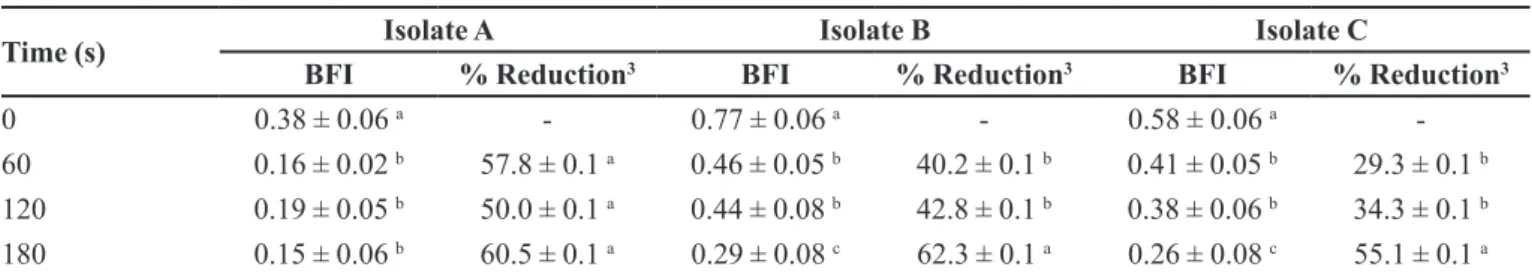
BFI values and their respective percentage reductions of biofilm-producing L. monocytogenes isolates on polystyrene microplates after treatment with PAA (0.5%) for diferent contact times are presented in Table I. BFI values of the three isolates decreased (P<0.05) after 60
s treatment, although there were no diferences (P>0.05)
between the treatment times for isolate A. Isolates B and C had lower BFI at 180 s, which was consistent with their higher percentage reductions (P<0.05) at this time when
compared with values obtained at 60 or 120 s. The isolate A belonged to serotype 4b, which is of great public health importance since this serotype is frequently involved in outbreaks of human listeriosis (Graves, Swaminathan, Hunter, 2007). Although in the present study the BFI values for isolate A (serotype 4b) were lower than for isolates B or C (serotype 1/2b) at all treatment times, the available data relating phylogenetic division, serotype, and bioilm formation remained inconclusive (Djordjevic, Wiedmann, Mclandsborough, 2002; Borucki et al., 2003). In a recent
report by Wang et al. (2016), the bioilm-formation ability
of serotype 1/2b was higher than serotype 1/2a, which is consistent with data presented in this study. However, the large variability of data reported in the literature indicates that biofilm formation of L. monocytogenes is strain dependent, since no clear correlation with serotypes could be established thus far (Doijad et al., 2015). Importantly,
no treatment was able to completely reduce the BFI value of the biofilm producers evaluated, with percentage reduction varying from 29.3 to 62.3%. Lee et al. (2016)
also reported that PAA was not effective for removing adherent cells of a L. monocytogenes strain isolated from brine on polystyrene microplates.
Bioilms of Listeria have been shown to be much more resistant to stress and to sanitizing agents than planktonic cells (Chavant, Gaillard-Martine, Hebraud, 2004). It is believed that bioilm formation enhances the capacity of foodborne bacteria to survive stressors that are commonly found in the food-processing environment (e.g. refrigeration, acidity, salinity, disinfection) (Giaouris
resistance of biofilms to antimicrobial agents are not completely understood, although restricted penetration into the bioilm, slow growth rate of bioilm organisms and induction of resistance mechanisms have been postulated (Donlan, Costerton, 2002). Van der Veen, Abee (2011) demonstrated that L. monocytogenes bioilms are more
resistant than planktonic cells to peracetic acid treatments, which corroborates the low effect of PAA treatment observed on culturable cells of L. monocytogenes isolated from cheese processing plants in Brazil.
The limited efect of PAA on the bioilms produced by the L. monocytogenes isolates on polystyrene microplates was confirmed by culturable cell counts, and is presented in Table II. Similar to the BFI variation after PAA treatment, counts were lower (P<0.05) when
compared with initial counts (5.5 ± 0.3 to 5.6 ± 0.6 log CFU/well), but decreased only 1.8-2.5 log cycles. It should be noted that the BFI method and the culturable cell count method may provide two completely diferent results. The BFI method enables an estimation of the biofilm mass composed of cells and extracellular polymeric substances (EPS) that attach to a surface. However, it does not provide information on the biological status of the cells. On the
other hand, culturable cell counts enable the evaluation of the bactericidal efect of the disinfectants on the bioilm (Srey et al., 2014). Destruction of existing bioilms can
involve removing the intact biofilm from surfaces via mechanical action for instance, or dissolving the bioilm structure by chemical disruption of EPS, for example. These mechanisms do not necessarily kill bacteria (Chan, Abedon, 2015).
On stainless steel, the bioilms formed by the three L.
monocytogenes isolates were afected by PAA (0.5%) after
60 s, with almost 100% cell damage within 180 s, as shown in Figure 1. Similar results were observed by Lee et al. (2016), indicating that PAA (0.5%) was able to inactivate the bioilm of L. monocytogenes formed on stainless steel. Our results are also in agreement with those reported by Ibusquiza, Herrera and Cabo (2011), who observed that PAA treatment for 10 min was efective in eliminating L.
monocytogenes bioilms. The authors considered that the
high oxidizing capacity and low molecular size of PAA are advantages for bioilm penetration.
In the present experiment, PAA was more efective against L. monocytogenes bioilms than other compounds
evaluated in previous reports, such as benzalkonium
TABLE I - Bioilm-forming index (BFI) and respective percentage reduction of bioilm-producing L. monocytogenes isolates (A-C1)
on polystyrene microplates, after treatments with peracetic acid (0.5 %) at diferent contact times2
Time (s) Isolate A Isolate B Isolate C
BFI % Reduction3 BFI % Reduction3 BFI % Reduction3
0 0.38 ± 0.06 a - 0.77 ± 0.06 a - 0.58 ± 0.06 a
-60 0.16 ± 0.02 b 57.8 ± 0.1 a 0.46 ± 0.05 b 40.2 ± 0.1 b 0.41 ± 0.05 b 29.3 ± 0.1 b
120 0.19 ± 0.05 b 50.0 ± 0.1 a 0.44 ± 0.08 b 42.8 ± 0.1 b 0.38 ± 0.06 b 34.3 ± 0.1 b
180 0.15 ± 0.06 b 60.5 ± 0.1 a 0.29 ± 0.08 c 62.3 ± 0.1 a 0.26 ± 0.08 c 55.1 ± 0.1 a
1 Isolates A, B and C were classiied as serotypes 4b, 1/2b and 1/2b, respectively. 2 Data are reported as mean ± standard deviation
of 4 replicates. 3 Relative to the initial BFI before treatment with peracetic acid (time: 0 s). a-c Values within each column with
diferent superscript letters difer signiicantly (P<0.05).
TABLE II - Culturable cell counts and respective reductions of bioilm-producing L. monocytogenes isolates (A-C1) on polystyrene
microplates, after treatments with peracetic acid (0.5%) at diferent contact times2
Time (s) A (Log CFU/well) B (Log CFU/well) C (Log CFU/well)
Count Reduction3 Count Reduction3 Count Reduction3
0 5.5 ± 0.3 a - 5.6 ± 0.4 a - 5.6 ± 0.6 a
-60 3.1 ± 0.3 b 2.3 ± 0.3 a 3.6 ± 0.7 b 2.1 ± 0.7 a 3.4 ± 0.8 b 2.2 ± 0.8 a
120 3.3 ± 0.2 b 2.1 ± 0.2 a 3.6 ± 0.7 b 2.1 ± 0.7 a 3.5 ± 0.4 b 2.4 ± 0.4 a
180 3.6 ± 0.5 b 1.8 ± 0.5 a 3.1 ± 0.2 b 2.5 ± 0.2 a 3.2 ± 0.4 b 2.4 ± 0.4 a
1 Isolates A, B and C were classiied as serotypes 4b, 1/2b and 1/2b, respectively. 2 Data are reported as mean ± standard deviation
of 4 replicates. 3 Relative to the initial BFI before treatment with peracetic acid (time: 0 s). a-c Values within each column with
FIGURE 1 - Epiluorescence photomicrographs of bioilms formed by bioilm-producing L. monocytogenes isolates on stainless steel surfaces for 48 hours at 35 ºC, after treatment with peracetic acid (0.5%, v/v) at 60, 120 and 180 s. Isolates A, B and C were
classiied as serotypes 4b, 1/2b and 1/2b, respectively. Bioilms were stained with bacterial viability kit LIVE/DEAD® BacLight (Molecular Probes, US), in which viable cells are luorescent green (Syto 9) and non-viable cells are luorescent red (propidium
iodide). Magniication: 1,000x. Bar = 10 µm.
chloride or nisin (Fatemi, Frank, 1999; Stoporth et al., 2002). Similarly, Belessi et al. (2011) also found a
signiicant reduction in the number of L. monocytogenes
attached cells with increasing contact times (from 1 to 6 min) with PAA (2.0%) on stainless steel coupons. The inhibitory efect of organic acids depends upon several factors, such as decreasing pH, the ratio of undissociated
forms of the acid entering into bacteria to inhibit metabolic activities, and chain length (Doores, 1993). It is also known that weak organic acids are lipophilic and penetrate the plasma membrane and acidifying the interior of the cell (Booth, Kroll, 1989).
to hydrophobic surfaces such as polystyrene and stainless steel, when compared to hydrophilic materials like glass (Donlan, Costerton, 2002). Pagedar, Singh and Batish (2010) observed that S. aureus cells had higher capacity to form biofilms on polystyrene than stainless steel, suggesting that hydrophobicity was an important factor in the formation of the bioilms, which is in agreement with the diferences in the PAA eiciency on polystyrene and stainless steel surfaces as observed in the present study. The survival of L. monocytogenes adhered cells after treatment with PAA at 0.5% (5,000 mg/L), which is nearly ten times over the concentrations normally used in food industries (300 – 700 mg/L), indicate the magnitude of risk posed by a potential failure in cleaning and disinfection procedures, including the possibility of L. monocytogenes
persistence in the food processing environment.
CONCLUSION
Although PAA (0.5%) was able to inactivate the three L. monocytogenes isolates on stainless steel, it only reduced 1.8-2.5 log cycles of culturable cells on polystyrene microplates. Further studies on the factors affecting the resistance of L. monocytogenes biofilms against sanitizers are needed for defining efficient treatments with organic acids such as PAA to remove adherent cells of this pathogen.
ACKNOWLEDGEMENTS
The authors thank the Conselho Nacional de Desenvolvimento Cientíico e Tecnológico (CNPq, Grant 309348/2013-7), for inancial support.
COMPETING INTERESTS
The authors declare that there is no conflict of interest.
REFERENCES
Barancelli GV, Camargo TM, Reis CMF, Porto E, Hofer E,
Oliveira CAF. Incidence of Listeria monocytogenes in cheese manufacturing plants from the northeast region of São Paulo,
Brazil. J Food Protect. 2011;74(5):816-819.
Belessi CEA, Gounadaki AS, Psomas AN, Skandamis PN. Efficiency of different sanitation methods on Listeria monocytogenes bioilms formed under various environmental
conditions. Int J Food Microbiol. 2011;145(Suppl 1):S46-S52.
Booth IR, Kroll RG. The preservation of foods by low pH. In: Gould GW. Mechanisms of action of food preservation procedures. London: Elsevier; 1989. p. 119-160.
Borucki MK, Peppin JD, White D, Loge F, Call DR. Variation
in bioilm formation among strains of Listeria monocytogenes.
Appl Environ Microbiol. 2003;69(12):7336-7342.
Casarin LS, Brandelli A, Casarin FO, Soave PA, Wanke CH,
Tondo EC. Adhesion of Salmonella Enteritidis and Listeria monocytogenes on stainless steel welds. Int J Food Microbiol.
2014;191:103-108.
Ceragioli M, Mols M, Moezelaar R, Ghelardi E, Senesi S, Abee
T. Comparative transcriptomic and phenotypic analysis of the responses of Bacillus cereus to various disinfectant treatments.
Appl Environ Microbiol. 2010;76(10):3352-3360.
Chan BK, Abedon ST. Bacteriophages and their enzymes in
bioilm control. Curr Pharm Design. 2015;21(1):85-99.
Chavant P, Gaillard-Martine B, Hebraud M. Antimicrobial
effects of sanitizers against planktonic and sessile Listeria monocytogenes cells according to the growth phase. FEMS
Microbiol Lett. 2004;236(2):241-248.
Djordjevic D, Wiedmann M, Mclandsborough LA. Microtiter
plate assay for assessment of Listeria monocytogenes bioilm
formation. Appl Environ Microbiol. 2002;68(6):2950-2958.
Doijad SP, Barbuddhe SB, Garg S, Poharkar KV, Kalorey DR, Kurkure NV, Rawool DB, Chakraborty T. Biofilm-forming
abilities of Listeria monocytogenes serotypes isolated from
diferent sources. PLoS One. 2015;10(9):e0137046.
Donlan RM, Costerton JM. Biofilms: survival mechanisms on clinically relevant microorganisms. Clin Microbiol Rev. 2002;15(2):167-193.
Doores S. Organic acids. In: Davidson PM, Branen AL, editors. Antimicrobials in foods. New York: Marcel Dekker; 1993. p.
95-136.
Fatemi P, Frank JF. Inactivation of Listeria monocytogenes/
Pseudomonas bioilms by peracid sanitizers. J Food Protect.
Giaouris E, Chorianopoulos N, Skandamis P, Nychas GJ.
Attachment and biofilm formation by Salmonella in food
processing environments. In: Mahmoud BSM. Salmonella: A
dangerous foodborne pathogen. Rijeka: Intech Open Access
Publisher; 2012. p. 157-180.
Graves LM, Swaminathan B, Hunter SB. Subtyping Listeria
monocytogenes. In: Ryser ET, Marth EH. Listeria, Listeriosis
and food safety. 3rd ed. Boca Raton: CRC Press; 2007. p.
283-304.
Ibusquiza PS, Herrera JJR, Cabo ML. Resistance to
benzalkonium chloride, peracetic acid and nisin during
formation of mature bioilms by Listeria monocytogenes. Food
Microbiol. 2011;28(3):418-425.
Johnsen BO, Lingaas E, Torfoss D, Strom EH, Nordoy I.
A large outbreak of Listeria monocytogenes infection with
short incubation period in a tertiary care hospital. J Infection. 2010;61(6):465-470.
Kells J, Gilmour A. Incidence of Listeria monocytogenes in
two milk processing environments, and assessment of Listeria monocytogenes blood agar for isolation. Int J Food Microbiol.
2004;91(2):167-174.
Koch J, Dworak R, Prager R, Becker B, Brockman S, Wicke A, Wichmann-Schauer H, Hof H, Werber D, Stark K. Large
listeriosis outbreak linked to cheese made from pasteurized milk,
Germany, 2006-2007. Foodborne Pathog Dis.
2010;7(12):1581-1584.
Lee SH, Cappato LP, Corassin CH, Cruz AG, Oliveira CAF.
Efect of peracetic acid on bioilms formed by Staphylococcus
aureus and Listeria monocytogenes isolated form dairy plants.
J Dairy Sci. 2016;99(3):2384-2390.
Niu C, Gilbert ES. Colorimetric method for identifying plant essential oil components that affect biofilm formation and
structure. Appl Environ Microbiol. 2004;70(12):6951-6956.
Pagedar A, Singh J, Batish VK. Surface hydrophobicity,
nutritional contents afect Staphylococcus aureus bioilms and
temperature influences its survival in performed biofilms. J
Basic Microb. 2010;50(Suppl 1):S98-S106.
Quarentei SS, Aquino S, Germano MIS, Germano PML. Princípios gerais de higienização. In: Germano PML, Germano MIS, editores. Higiene e vigilância sanitária de alimentos. 4ª ed.
Barueri, SP: Editora Manole; 2011. p. 631-667.
SAS Institute. User´s guide for SAS software navigator. 9th ed.
SAS Institute, Cary, NY, 2002.
Sauer K, Rickard A, Davies DG. Bioilms and biocomplexity. Microbe. 2007;2(7):347-355.
Shi X, Zhu X. Biofilm formation and food safety in food
industries. Trends Food Sci Technol. 2009;20(9):407-413.
Srey S, Park SY, Jahid IK, Oh SR, Han N, Zhang CY, Kim SH, Cho JI, Ha SD. Evaluation of the removal and destruction
effect of a chlorine and thiamine dilaurylsulfate combined treatment on L. monocytogenes bioilm. Foodborne Pathog Dis.
2014;11(8):658-663.
Stoporth JD, Samelis J, Sofos JN, Kendall PA, Smith GC.
Bioilm formation by acid-adapted and non-adapted Listeria
monocytogenes in fresh beef decontamination washings and
its subsequent inactivation with sanitizers. J Food Protect. 2002;65(11):1717-1727.
Takahashi H, Kuramoto S, Miya S, Kimura B. Desiccation
survival of Listeria monocytogenes and other potential
foodborne pathogens on stainless steel surfaces is afected by diferent food soils. Food Control. 2011;22(3-4):633-637.
Tompkin RB. Control of Listeria monocytogenes in the
food-processing environment. J Food Protect. 2002;65(4):709-725.
Van der Veen S, Abee T. Mixed species biofilms of Listeria
monocytogenes and Lactobacillus plantarum show enhanced
resistance to benzalkonium chloride and peracetic acid. Int J Food Microbiol. 2011;144(3):421-431.
Wang W, Zhou X, Suo Y, Deng X, Cheng M, Shi C, Xianming S. Prevalence, serotype diversity, bioilm-forming ability and
eradication of Listeria monocytogenes isolated from diverse
foods in Shanghai, China. Food Control. 2016 (in press).
Received for publication on 27th September 2016