UNIVERSIDADE DA BEIRA INTERIOR
Ciências da Saúde
Obesogens and Male Fertility: a Threat to Sertoli
Cell Function?
Ana Margarida Nunes Ferreira Ribeiro Cardoso
Dissertação para obtenção do Grau de Mestre em
Ciências Biomédicas
(2º
ciclo de estudos)
Orientador: Prof. Doutor José Eduardo Brites Cavaco (CICS–UBI)
Co-orientador: Prof. Doutor Carlos Pedro Fontes Oliveira (ICBAS-UP/I3S-UP)
Acknowledgments
First of all, I would like to thank to my supervisor Professor José Eduardo Cavaco and to my co-supervisor Professor Pedro Fontes Oliveira, for the opportunity they gave me to be a part of this project, for their recommendations and wise words. A special thanks to Professor José Eduardo Cavaco for his concern, enthusiasm and, above all, for his friendship.
I am especially grateful to my co-supervisor Luís Pedro Rato for his help in the laboratory and all the good advices. He was one of my strongest supporters and I have not words enough to express my thankfulness.
I would like to express my gratitude to Professor Marco G. Alves for all the suggestions that contributed to the success of this work and to Professor Ana Sousa for providing the precious TBT which was invaluable to its achievement. Besides, I would like to thank to Professor Rui A. Carvalho and Ivana Jarak for the NMR sample analysis.
I also would like to thank to Professor Sílvia Socorro for stimulating my interest in reproductive biology and to all the teachers that contributed to my academic formation. I gratefully acknowledge my friends from the course of Biomedical Sciences and all my colleagues here at CICS, especially from Lab058.
To all my family and other friends for their friendly support, I give my heartfelt thank you. Finally, I saved the most special acknowledgments for my parents, my brother and, mainly, for Bernardo, who were my company, support and encouragement during this work. I promise I will always make the possible and the impossible to keep you proud.
Resumo
Nas últimas décadas, diversos estudos têm evidenciado uma correlação inversa entre o aumento da esperança de vida e a qualidade espermática dos indivíduos residentes nos países desenvolvidos. Embora a etiologia desta tendência na fertilidade masculina ainda seja um assunto que suscite grande debate, os obesogénicos, compostos ambientais que predispõem para o ganho de peso, têm sido apontados como importantes causadores, sobretudo devido à sua ação enquanto desreguladores endócrinos.
Os obesogénicos podem ser encontrados praticamente em todo o lado, inclusive em dietas altamente calóricas ou no meio ambiente. O tributilestanho surge como o obesogénico modelo, sendo mesmo considerado um dos compostos mais tóxicos alguma vez introduzidos no ecossistema. Este apresenta caraterísticas lipofílicas e revela uma grande afinidade para se acumular em tecidos com elevado teor lipídico, como é o caso dos testículos. Uma vez armazenado nestes órgãos, o tributilestanho pode afetar a fisiologia e o próprio metabolismo testicular, função fulcral para a espermatogénese. A desregulação destas vias metabólicas pode estar na base molecular de efeitos reprodutivos adversos, como é o caso do aumento do
stress oxidativo testicular ou de defeitos espermáticos.
O adequado desenvolvimento das células germinativas é altamente dependente do suporte nutricional fornecido pelas células de Sertoli, cujo metabolismo revela características particulares. As células de Sertoli metabolizam a maioria da glucose a lactato que, por sua vez, constitui a principal fonte de energia das células germinativas em desenvolvimento. Assim, a regulação do metabolismo glicolítico das células de Sertoli tem um papel central no processo da espermatogénese. Curiosamente, já se evidenciou que estas células são um alvo preferencial para tóxicos ambientais capazes de alterar a sua estrutura e/ou função.
O objetivo deste estudo passou por avaliar o impacto do tributilestanho no metabolismo das células de Sertoli, com um foco particular no metabolismo glicolítico. Para tal, recorreu-se a três concentrações do composto: 0.1 nM, uma dose considerada subtóxica, mas para a qual o ganho de peso e, particularmente, a ativação do heterodímero do recetor de retinoide X/recetor ativado pelo proliferador de peroxissoma γ foram demonstrados; 10 nM, pertencente ao intervalo de concentrações descritas no soro e tecidos de alguns indivíduos; e 1000 nM, uma concentração a partir da qual foram demonstradosefeitos citotóxicos.
Os resultados obtidos demonstraram que a exposição à concentração mais elevada de tributilestanho (1000 nM) induz efeitos citotóxicos severos nas células de Sertoli de rato, reduzindo a sua proliferação para 28%, em comparação com o grupo controlo. Dada a
viii
tributilestanho, investigaram-se eventuais alterações na expressão de marcadores de células de Sertoli maduras, através da análise da inibina B e do recetor de androgénios. Para as concentrações utilizadas, não se observaram alterações significativas na expressão dos transcritos destes marcadores.
Contudo, em termos metabólicos, ambas as doses revelaram afetar as vias relacionadas com a glicólise e com a produção de lactato. De facto, a via glicolítica foi favorecida nas células de Sertoli expostas a 10 nM de tributilestanho, visto que o aumento do consumo de glucose e piruvato foi acompanhado por um aumento na produção de lactato. No entanto, não se verificou qualquer alteração na expressão dos transportadores de glucose 1, 2 e 3, ao passo que a expressão da enzima lactato desidrogenase se revelou diminuída em relação ao grupo controlo. Adicionalmente, constatou-se também que as células de Sertoli expostas a esta concentração de tributilestanho (10 nM) apresentavam uma maior expressão da isoforma 4 do transportador de monocarboxilatos, o que sugere um contributo para uma exportação de lactato mais elevada. Relativamente às células de Sertoli expostas à concentração mais baixa de tributilestanho (0.1 nM), estas não apresentaram diferenças no consumo da glucose, apesar de ter sido evidenciada uma diminuição da expressão dos transportadores de glucose 1 e 2 neste grupo experimental. De forma idêntica, também o consumo de piruvato foi significativamente inferior em comparação ao grupo de células expostas a 10 nM de tributilestanho. Não se verificaram alterações significativas na produção de lactato, facto que poderá ter resultado da diminuição da expressão da lactato desidrogenase. Paralelamente, verificou-se, em ambos os grupos, uma diminuição significativa dos níveis de alanina, o que favorece um aumento do estado redox citosólico, sujeitando as células a um possível ambiente oxidativo.
Em conclusão, este estudo destacou que o tributilestanho, para além de induzir efeitos citotóxicos significativos nas células de Sertoli de rato quando administrado numa dose elevada, promove diversas alterações numa das principais funções das células de Sertoli diferenciadas, o metabolismo glicolítico, podendo, desta forma, afetar a espermatogénese e consequentemente a fertilidade masculina.
Palavras-chave
Obesogénicos; Tributilestanho; Fertilidade Masculina; Células de Sertoli; Metabolismo Glicolítico.
Resumo Alargado
A chave para a sobrevivência do ser humano reside numa plena função reprodutiva. No entanto, nas últimas décadas, tem-se verificado um declínio na saúde reprodutiva masculina, tornando-se a infertilidade um problema que afeta milhões de casais em todo o mundo. Um terço dos casos de infertilidade têm sido atribuídos a problemas de causa masculina, sendo muitas vezes difícil estabelecer um diagnóstico claro para as anomalias observadas. De facto, é comum o homem apresentar uma saúde reprodutiva aparentemente normal, mas com alterações severas na qualidade e/ou quantidade do esperma, o que dificulta a escolha do tratamento a adotar. Desta forma, torna-se importante um maior compromisso entre a investigação e a clínica, no sentido de se escrutinar a etiologia associada a cada caso de infertilidade masculina.
Nos últimos anos, o crescimento acentuado das taxas de infertilidade masculina tem revelado particular incidência nos países desenvolvidos, onde o aumento da obesidade e de outras doenças metabólicas resultantes de hábitos alimentares erróneos e sedentarismo têm sido apontados como as principais causas. Isto é explicado em parte pela estreita relação entre a regulação metabólica sistémica e o sistema reprodutor masculino. Contudo, veio a perceber-se que o aumento exponencial nos casos de obesidade e infertilidade masculina não pode perceber-ser justificado unicamente através de hábitos diários, mas também por uma exposição permanente a fatores ambientais, como é o caso dos obesogénicos. A estes compostos é associada não só uma predisposição para o ganho de peso, mas especialmente uma tendência de atuação como potentes desreguladores endócrinos. Estes revelam-se capazes de: (1) aumentar o número de adipócitos e/ou o armazenamento de tóxicos nos mesmos; (2) alterar a quantidade de calorias armazenadas; e/ou (3) alterar os mecanismos moleculares através dos quais são regulados a saciedade e o apetite. Desta forma, para além de exacerbarem os efeitos dos hábitos diários, os obesogénicos induzem desregulações endócrinas que, por conseguinte, levam a severas disfunções metabólicas.
De entre as várias substâncias que predispõem os indivíduos para o aumento de peso, destaca-se o tributilestanho, o obesogénico modelo, considerado mesmo um dos compostos mais tóxicos alguma vez introduzidos de forma deliberada no ambiente. Inicialmente utilizado apenas como algicida e moluscicida na indústria da navegação, o tributilestanho chega-nos hoje não só através da cadeia alimentar, mas também pelo contacto com pó doméstico e produtos de preservação de madeiras. Efeitos biológicos adversos em diversas espécies têm vindo a ser atribuídos à contaminação por tributilestanho, nomeadamente ao nível do sistema reprodutor. Isto ocorre porque o tributilestanho tem características lipofílicas, acumulando-se facilmente em tecidos com elevado conteúdo lipídico, como é o caso dos testículos.
x
Nos mamíferos, os testículos são os elementos centrais do sistema reprodutor masculino, estando envolvidos na produção de espermatozóides, que determina a fertilidade masculina. A espermatogénese é o processo de expansão e desenvolvimento das células germinativas e, para que possa ocorrer de forma adequada, requer uma regulação eficaz das células de Sertoli. Estas células somáticas são fundamentais para o suporte da espermatogénese, através da formação da barreira hemato-testicular, que funciona simultaneamente como uma barreira anatómica, imunológica e fisiológica. De facto, para além de constituir um suporte físico para a espermatogénese, esta barreira permite igualmente o desenvolvimento de um ambiente imunológico adequado à ocorrência deste processo. Aliado a isto, as células de Sertoli são igualmente responsáveis por fornecer um apropriado suporte nutricional às células germinativas.
O metabolismo das células de Sertoli revela caraterísticas únicas, já que é através deste que a maioria da glucose produzida é convertida a lactato, a principal fonte de energia utilizada pelas células germinativas em desenvolvimento. Desta forma, a regulação do metabolismo glicolítico das células de Sertoli revela-se central para a espermatogénese e, consequentemente, para a fertilidade masculina. Este suporte metabólico fornecido pelas células de Sertoli pode, no entanto, ser facilmente perturbado por alterações metabólicas que ocorrem ao nível sistémico. Para além disso, estas células já foram identificadas como sendo um alvo preferencial dos tóxicos ambientais, que se revelam capazes de alterar a sua estrutura e/ou função.
Assim, dada a importância do metabolismo da glucose para a fertilidade masculina, o objetivo deste trabalho passou por avaliar o efeito do tributilestanho no metabolismo glicolítico de células de Sertoli obtidas a partir de culturas primárias de ratos de 20 dias. Para isso, testaram-se três concentrações de tributilestanho: 0.1 nM, uma dose que se encontra abaixo dos níveis fisiológicos, mas para a qual o ganho de peso e, particularmente, a ativação do heterodímero do recetor de retinoide X/recetor ativado pelo proliferador de peroxissoma γ já foram demonstrados; 10 nM, uma concentração que se encontra no intervalo dos níveis fisiológicos; e 1000 nM, uma concentração superior aos níveis fisiológicos e, inclusivamente, a partir da qual já foram evidenciados efeitos citotóxicos.
Os resultados obtidos mostraram que a exposição à concentração mais elevada de tributilestanho (1000 nM) induz severos efeitos citotóxicos nas células de Sertoli de rato, reduzindo a sua proliferação para 28%, em comparação com o grupo controlo. Dada a ausência de efeitos citotóxicos nos grupos expostos a concentrações mais baixas de tributilestanho, investigaram-se eventuais alterações na expressão de marcadores de células de Sertoli maduras, através da análise da inibina B e do recetor de androgénios. Para as concentrações utilizadas, não se observaram alterações significativas na expressão dos transcritos destes marcadores.
Contudo, em termos metabólicos, ambas as doses revelaram afetar as vias relacionadas com a glicólise e com a produção de lactato. De facto, a via glicolítica foi favorecida nas células de Sertoli expostas a 10 nM de tributilestanho, visto que o aumento do consumo de glucose e piruvato foi acompanhado por um aumento na produção de lactato e por uma diminuição da produção de alanina. No entanto não se verificou qualquer alteração na expressão dos transportadores de glucose 1, 2 e 3, ao passo que a expressão da enzima lactato desidrogenase estava diminuída em relação ao controlo. Adicionalmente, verificou-se também que as células de Sertoli expostas a esta concentração de tributilestanho (10 nM) apresentavam uma maior expressão da isoforma 4 do transportador de monocarboxilatos em relação ao grupo controlo o que sugere um contributo para uma exportação de lactato mais elevada. Relativamente ao grupo de células de Sertoli expostas à concentração mais baixa de tributilestanho (0.1 nM), estas não apresentaram diferenças significativas no consumo da glucose, apesar de ter sido evidenciada, neste grupo experimental, uma diminuição da expressão dos transportadores de glucose 1 e 2. De forma idêntica, também o consumo de piruvato foi significativamente inferior em comparação com o grupo de células expostas a 10 nM de tributilestanho. Não se verificaram alterações significativas na produção de lactato, facto que poderá ter resultado da diminuição da expressão da lactato desidrogenase. Paralelamente, verificou-se também uma diminuição significativa nos níveis de alanina em ambos os grupos, o que favorece um aumento do estado redox citosólico, sujeitando as células a um possível ambiente oxidativo.
Em conclusão, este estudo mostrou que o tributilestanho, para além de induzir efeitos citotóxicos significativos nas células de Sertoli de rato quando administrado numa dose elevada, também promove diversas alterações numa das principais funções das células de Sertoli diferenciadas, o metabolismo glicolítico, podendo desta forma afetar a espermatogénese e, consequentemente, a fertilidade masculina.
Abstract
In the last decades, several studies evidenced a negative correlation between life expectancy and sperm quality in developed countries. Although the etiology of this trend in male fertility remains a matter of debate, environmental compounds that predispose to weight gain, namely obesogens, are appointed as pivotal contributors due to their action as endocrine disruptors.
Obesogens can be found virtually everywhere, including in high-energy diets or in the surrounding environment. Tributyltin arises as the obesogen model, being considered one of the most toxic compounds ever introduced into the environment. Tributyltin presents lipophilic characteristics and high affinity to accumulate in tissues with high lipid contents, as is the case of testes. Once stored in these organs, tributyltin can affect testicular physiology and metabolism, which are crucial for spermatogenesis. Disruption of these tightly regulated metabolic pathways may be the molecular basis of adverse reproductive outcomes, such as increased oxidative stress or even sperm defects.
The appropriate development of germ cells is highly dependent on the nutritional support provided by Sertoli cells, which metabolism present some unique features. Sertoli cells metabolize glucose, being the majority of it converted to lactate, the main fuel for developing germ cells. Thus, the regulation of Sertoli cell glycolytic metabolism plays a central role on spermatogenesis. Interestingly, these cells were already proven to be a target for environmental toxicants, being these compounds able to alter their structure and/or function.
Herein, we evaluated the impact of tributyltin on Sertoli cell metabolism, with a particular focus on glycolytic metabolism. In order to achieve it, we selected 3 different concentrations of tributyltin: 0.1 nM, a subtoxic dose but to which weigh gain and retinoid X receptor-peroxisome proliferator-activated receptor γ activation were already described; 10 nM, a dose within the range of concentrations reported in serum and tissues of humans; and 1000 nM, a dose from which broad cytotoxic effects have already been evidenced.
Our results evidenced that the exposure to the highest concentration of tributyltin (1000 nM) induce severe cytotoxic effects in rat Sertoli cells, decreasing their proliferation to 28%, when compared with the control group. Since the lower concentrations of tributyltin (10 nM and 0.1 nM) did not induced cytotoxic effects, we investigated possible changes in Sertoli cells maturation markers through the analysis of inhibin B and androgens receptor. However, for the adopted concentrations, significant changes were not observed in the expression of these
xiv
However, both concentrations (10 and 0.1 nM) revealed to affect glycolysis and lactate production-related events. Indeed, the glycolytic pathway was favored in Sertoli cells exposed to tributyltin 10 nM, since the increased glucose and pyruvate consumption was followed by an increase in lactate production. However, the protein expression of glucose transporters 1, 2 and 3 remain unaltered, while the expression of lactate dehydrogenase was decreased when compared to control. In addition, Sertoli cells exposed to this concentration of tributyltin revealed also an increased expression of monocarboxylate transporter isoform 4 when compared to control, which had probably contributed to a higher lactate export.
Concerning to Sertoli cells exposed to the lowest concentration of tributyltin (0.1 nM), significant changes in glucose consumption were not observed, even though a decreased expression of glucose transporters 1 and 2 was evidenced in this experimental group. Similarly, also the pyruvate consumption was significantly lower when compared with the group of Sertoli cells exposed to tributyltin 10 nM. The absence of significant changes in lactate production may be due to the decreased lactate dehydrogenase expression. Besides, we also observed a significant decrease in alanine levels in both groups of Sertoli cells exposed to tributyltin, which favors a high cytosolic oxidative redox state, predisposing the cells to a possible oxidative environment.
In conclusion, this study showed that tributyltin, in addition to induce significant cytotoxic effects in rat Sertoli cells when administered at a high dose, also promotes several changes in one of the main functions of differentiated Sertoli cells, the glycolytic metabolism. In this regard, tributyltin may affect spermatogenesis and thus male fertility.
Keywords
Table of contents
Introduction ... 1
Testicular Anatomy and Histology ... 3
The Sertoli cell: morphology, function and metabolism ... 5
The obesogen hypothesis ... 8
Tributyltin as a threat for male fertility ... 11
Aims of the project ... 15
Materials and methods ... 19
Chemicals ... 21
Animals ... 21
Rat Sertoli Cell Primary Culture ... 21
Experimental Design ... 22
Cell Proliferation Assay ... 22
Total RNA Extraction ... 23
Reverse transcriptase quantitative polymerase chain reaction ... 23
Total Protein Extraction ... 24
Western Blot ... 24
Enzymatic Assays ... 25
Nuclear Magnetic Resonance Spectroscopy ... 25
Statistical analysis ... 26
Results ... 27
Exposure to TBT 1000 nM decreases the proliferation of primary cultures of rat Sertoli cells ... 29
Inhibin B and AR mRNA expression in rat Sertoli cells is not affected by exposure to TBT 10 nM and 0.1 nM ... 30
TBT 10 nM increase glucose consumption by cultured rat Sertoli cells ... 30
Exposure to TBT 0.1 nM decreases significantly the protein levels of both GLUT1 and GLUT2, but not GLUT3, in rat Sertoli cells ... 31
Exposure to TBT 10 nM increases PFK1 levels in rat Sertoli cells ... 33
Pyruvate consumption is increased in Sertoli cells exposed to TBT 10 nM ... 33
TBT modulates lactate production in primary cultures of rat Sertoli cells ... 34
Sertoli cells exhibit decreased alanine production after exposure to TBT 10 nM and 0.1 nM ... 36
xvi
Discussion... 37 Conclusions ... 45 References ... 49
List of Figures
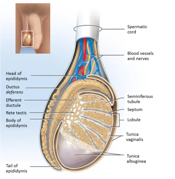
Figure 1: Schematic representation of the mammalian testis and epididymis.. ... 4
Figure 2: Schematic illustration of the seminiferous tubule, blood-testis-barrier (BTB),
spermatogenesis and interstitial tissue. ... 5
Figure 3: Schematic illustration of the glycolytic metabolism of Sertoli cells (SCs).. ... 8
Figure 4: Representative draft of tributyltin (TBT) structure. ... 11
Figure 5: Representative proton nuclear magnetic resonance (1H-NMR) spectrum
achieved for the insulin-transferrin-sodium selenite (ITS) supplement Dulbecco’s modified eagle medium Ham’s nutrient mixture F12 (DMEM:F12) showing the localization of H1-α-glucose and lactate peaks. ... 26
Figure 6: Effect of tributyltin (TBT) on rat Sertoli cells (SCs) survival after 6-hours
treatment. ... 29
Figure 7: Effect of tributyltin (TBT) on inhibin B and androgen receptor (AR) messenger
ribonucleic acid (mRNA) levels in rat Sertoli cells (SCs) after 6-hours treatment.. ... 30
Figure 8: Effect of tributyltin (TBT) on glucose and pyruvate consumption after 6-hours
treatment in primary cultures of rat Sertoli cells (SCs). ... 31
Figure 9: Effect of tributyltin (TBT) on glucose transporters (GLUTs) and
phosphofructokinase 1 (PFK1) after 6-hours treatment in primary cultures of rat Sertoli cells (SCs). ... 32
Figure 10: Effect of tributyltin (TBT) on lactate and alanine production after 6-hours
treatment in primary cultures of rat Sertoli cells (SCs). ... 34
Figure 11: Effect of exposure to tributyltin (TBT) on lactate dehydrogenase (LDH) and
monocarboxylate transporter 4 (MCT4) after 6-hours treatment in primary cultures of rat Sertoli cells (SCs). ... 35
List of Tables
Table 1: Summary of the proposed obesogens and their sources of exposure. ... 10
Table 2: Oligonucleotides and cycling conditions for quantitative real-time polymerase
chain reaction (qPCR) amplification of inhibin B, androgen receptor (AR) and β2-microglobulin (β2MG). ... 24
Abbreviations
1H-NMR Proton Nuclear Magnetic Resonance
AMPK 5’ Adenosine Monophosphate-Activated Protein Kinase
AR Androgen Receptor
β2MG β2-Microglobulin
BMI Body Mass Index
BTB Blood-Testis Barrier
CAMs Cell Adhesion Molecules
cDNA Complementary Deoxyribonucleic Acid
DMEM:F12 Dulbecco’s Modified Eagle Medium Ham’s Nutrient Mixture F12
dNTPs Deoxynucleotide Triphosphates
EDTA Ethylene Diamine Tetra Acetic Acid
EtOH Ethanol
FSH Follicle-Stimulating Hormone
GLUT1 Glucose Transporter 1
GLUT2 Glucose Transporter 2
GLUT3 Glucose Transporter 3
GLUT8 Glucose Transporter 8
GLUTs Glucose Transporters
GnRH Gonadotropin-Releasing Hormone
HBSS Hank’s Balanced Salts Solution
HED High-Energy Diets
HPT Hypothalamic-Pituitary-Testicular
ITS Insulin-Transferrin-Sodium Selenite
LCs Leydig Cells
LDH Lactate Dehydrogenase
LH Luteinizing Hormone
MCT4 Monocarboxylate Transporter 4
mRNA Messenger Ribonucleic Acid
PBS Phosphate-Buffered Saline
PFK Phosphofructokinase
PFK1 Phosphofructokinase 1
PPARу Peroxisome Proliferator-Activated Receptor Gamma
PVDF Polyvinylidene Difluoride
qPCR Quantitative Real-Time Polymerase Chain Reaction
RNA Ribonucleic Acid
RXR Retinoid X Receptor
SCs Sertoli Cells
SDS Sodium Dodecyl Sulfate
SEM Standard Error of the Mean
SRB Colorimetric Sulforhodamine B
T Testosterone
TBT Tributyltin
Testicular Anatomy and Histology
The mammalian testes are paired complex organs. They are divided into compartments, called testicular lobules, which are separated by the septum - fibrous inner extensions of the tunica albuginea (Figure 1) [1]. A human testis contains from 250 to 300 lobules, each one enclosing one to four highly coiled seminiferous tubules [2]. Seminiferous tubules are the testicular functional unities [3], containing the Sertoli cells (SCs) and the germ cells in different development stages. Surrounding each seminiferous tubule are contractile myoid cells that promote the movement of mature sperm and testicular fluids through the tubules [2]. The interstitial spaces between the seminiferous tubules contain all the blood and lymphatic vessels essential for the movement of hormones and nutrients into and out of the testes [4]. Besides, in this space we can find nerves, macrophages and also Leydig cells (LCs), which are responsible for the synthesis of sex steroid hormones [4]. The seminiferous tubules converge upon a plexus of channels, the rete testis, from which 15 to 20 ductuli efferents conduct spermatozoa to the epididymis [2].
Testes essentially perform two major functions: synthesis of steroid hormones, primarily testosterone (T), and formation of haploid germ cells, sperm [5]. The main hormonal control system of these functions is the hypothalamic-pituitary-testicular (HPT) axis, since it regulates the spermatogenic process through the interaction between the hypothalamus, pituitary and testes [6]. Hypothalamus releases the gonadotropin-releasing hormone (GnRH) into the hypophyseal-portal circulation, which stimulates gonadotrophic cells of the anterior pituitary to secrete the gonadotropins follicle-stimulating hormone (FSH) and luteinizing hormone (LH) [1, 7]. LH acts on the T-producing LCs, while FSH acts on SCs [7, 8]. Nevertheless, a chain of complex local interactions among the various testicular cell types such as germ, Sertoli, peritubular and LCs are involved in spermatogenesis control [7, 9]. Spermatogenesis is the process by which immature germ cells undergo division, differentiation and meiosis to originate spermatozoa. This process occurs in seminiferous tubules, through close association of germ cells with epithelial somatic cells, the SCs. SCs play a central role on the development of functional testes and, consequently, on the expression of a male phenotype [10, 11]. In fact, these cells influence testes formation in the embryo and spermatogenesis in the adult, by regulating the surrounding environment of the developing germ cells [8]. Within the seminiferous tubules, SCs extend from the basement membrane to the lumen, directly interacting with the developing germ cells (Figure 2) [10]. Adjacent SCs form tight junctional complexes, the basis of the formation of the blood–testis barrier (BTB), dividing the seminiferous epithelium into the basal compartment, where spermatogonia and spermatocytes are found, and the apical (or adluminal) compartment, containing different stages of meiotic spermatocytes, round spermatids, elongated spermatids and spermatozoa (Figure 2) [10, 12]. From Enrico Sertoli works, in 1865, came out the
4
concept that SCs act as “nurse cells” [13] since, in addition to physical support, these cells also provide nutrients and regulatory factors crucial to germ cell sustenance [10, 14].
Figure 1: Schematic representation of the mammalian testis and epididymis. The testis is
encapsulated by two layers: tunica vaginalis (the most outer tunic) and tunica albuginea. Extensions from tunica albuginea (septum) divide testis in lobules where the seminiferous tubules are located. Seminiferous tubules converge to the rete testis that is connected to the efferent ducts. The head of the epididymis is linked to the testis by several efferent ducts. Adapted from [1].
Developing germ cells form intimate associations with SCs and, at a given moment, 30 to 50 germ cells in various stages of development may be in contact with a single Sertoli cell [3]. Cell-to-cell interactions, not only between SCs and specific germ cells but also between adjacent SCs, are essential in the regulation of mammalian spermatogenesis [10, 15]. Indeed, SCs are able to adapt their production/secretion of proteins and factors involved in germ cell development to the changing needs of the germ cell, occurring in a stage-dependent manner [10]. Consequently, well-functioning SCs provide the developing germ cells with the appropriate nutrients, energy sources, hormones, and growth factors as well as protection
from harmful agents and the host’s own immune system [16]. Without the physical and metabolic support of the SCs, germ cell differentiation, meiosis and transformation into spermatozoa will be impossible to occur [17, 18].
Figure 2: Schematic illustration of the seminiferous tubule, blood-testis-barrier (BTB), spermatogenesis and interstitial tissue. The seminiferous epithelium is composed by
Sertoli cells (SCs) and different subtypes of developing germ cells. SCs reside on the basement membrane, under which are the lymphatic endothelium and the peritubular myoid cells. At the interstitial space are located the Leydig cells (LCs), which produce testosterone (T) in the presence of luteinizing hormone (LH). Between adjacent SCs, tight junctions are established, forming the BTB that divides the seminiferous epithelium into basal compartment, where spermatogonia and spermatocytes are found, and into adluminal compartment, containing different stages of meiotic spermatocytes, round spermatids, elongated spermatids and spermatozoa. Adapted from [19].
The Sertoli cell: morphology, function and metabolism
Differentiated SCs are the structural elements of the seminiferous epithelium, playing a main role in the regulation of spermatogenesis. Besides physically supporting spermatogenesis development, SCs also regulate the flow of nutrients, growth factors and other substances to
6
Within the undifferentiated fetal gonads, SCs are the first cells to differentiate, resulting in the seminiferous cord formation [11]. The germ cells are sequestered inside of these newly formed seminiferous tubules, becoming protected from undergoing meiosis [8]. SCs also influence testis formation in the embryo, since they ensure the regression of the Müllerian ducts via secretion of the anti-Müllerian hormone [21]. Such process requires the expression of specific genes on the Y-chromosome [8], namely the Sry, the male sex-determining gene expressed by SCs [16].
In mammals, at the time of puberty, SCs suffer a profound alteration on their morphology and function, becoming biochemical and morphologically distinct from the undifferentiated cells. SCs are columnar shaped with a large dimensions volume (from 2000 to 7000 m3 in mammals
[22]) that allows them to support a vast number of developing germ cells [10]. SCs exhibit prolonged cytoplasmic extensions surrounding germ cells. In most of species, the nucleus of SCs is located at the basal portion of the cytoplasm, presenting large dimensions (up to 850 m3) and an irregular shape [23].Another characteristic of their nucleus is the large nucleolus
with a three-partite structure [23]. Ultrastructure images from electron microscopy show that smooth endoplasmic reticulum is abundant on SCs, being associated with the junctional complexes established between SCs and germ cells. This smooth endoplasmic reticulum is organized into reservoirs of lipid droplets, which are involved in the metabolism of lipids or steroids [24].
In addition to creating the adequate microenvironment essential for a suitable development of germ cells into spermatozoa, the BTB also allows the formation of specific intratubular fluid, which is dependent on the function of SCs. In fact, functional BTB consists of three components: (1) an anatomical/physical barrier that prevents the entry of molecules and substances into the adluminal compartment of the seminiferous tubules; (2) an immunological barrier that limits the movement of immune cells of the immune system and regulates the levels of cytokines in the seminiferous epithelium; (3) a physiological barrier, since it contains transporters and channels in the apical and basolateral membranes that are highly dynamic and responsible to the needs of germ cells [25]. Nevertheless, this barrier is “permeable” enough to allow the migration of developing germ cells throughout the seminiferous epithelium, a crucial step for a functional spermatogenesis.
Mammalian spermatogenesis is characterized by continuous cellular differentiations with three main stages: (1) mitotic spermatogonial proliferation and differentiation, (2) meiotic phase and (3) spermiogenesis [26]. This process is highly dependent on SCs, being regulated by the HPT axis. Spermatogonial stem cells, which adhere to the basement membrane where the supporting SCs are also adherent, replicate mitotically not only to guarantee the germ cell line (spermatogonia type A), but also to give rise to spermatogonia type B [26]. Spermatogonia type B will enter meiotic prophase and differentiate into primary spermatocytes (Spermatocyte I). After crossing the BTB, these cells undergo the first division
of meiosis and form the haploid secondary spermatocytes (Spermatocyte II). The second meiotic division differentiates one Spermatocyte II into two equalized round spermatids. Thereafter, cell division stops and spermiogenesis starts to form elongated spermatids, which are finally released into the lumen of the tubule as immature spermatozoa, in a process called spermiation (Figure 2) [26].
Several modifications on SCs structure and function may affect BTB, unbalancing the metabolic cooperation established between these cells and the developing germ cells. Indeed, SCs metabolism and particularly glucose metabolism is pivotal for spermatogenesis and thus for male fertility. Robinson and Fritz [27] showed that cultured SCs are the main source of lactate in the testes, converting the majority of glucose into this metabolite. The rate-limiting step of lactate production is the membrane passage of glucose from the extracellular space, via specific glucose transporters (GLUTs) to SCs innerspace [28]. Four GLUTs (GLUT1, GLUT2, GLUT3 and GLUT8) have been identified in SCs to date [29-32]. However, GLUT8 has not been identified in SCs plasmatic membrane, which makes it unexpectable to be involved in the glucose uptake through plasma membrane [33]. Otherwise, GLUT1, GLUT2 and GLUT3 have been identified in the plasmatic membrane of SCs, allowing to assume their role as the primary responsible for glucose import in these cells. Once glucose enters in SCs cytoplasm, it suffers a series of multi-step reactions catalyzed by several enzymes. The first rate-limiting step in glycolytic metabolism is mediated by phosphofructokinase (PFK) that catalyzes the irreversible conversion of fructose-6-phosphate into fructose-1,6-bis-phosphate [34].Glucose is then converted to pyruvate and the glycolytic process is completed. The cytosolic pyruvate originated from glycolysis can follow three main distinct paths: (1) it can be converted to alanine by the action of alanine aminotransferase; (2) it can enter the tricarboxylic acid cycle; (3) or it can be converted to lactate by the action of lactate dehydrogenase (LDH). Indeed, LDH has a crucial role in providing lactate to developing germ cells, exporting it from SCs by monocarboxylate transporter isoform 4 (MCT4) (Figure 3) [35]. Additionally, alanine also has an important role, since it can be converted to pyruvate which may be used as a substrate by SCs for several biochemical pathways [36].
Since normal reproductive function is dependent on adequate nutritional state, it is expectable that metabolic disturbs promoted by environmental compounds may affect reproductive function as a result of increased adiposity and/or increased storage of fat-soluble toxicants [37]. Testicular tissue exhibits high lipid content and it easily retains these compounds Furthermore, the epididymal fat, adjacent to testicular tissue, also arises as another source of retention for fat-soluble toxicants, leading to testicular tissue permanently exposed to its effects. Recently, several data reported that the reproductive process is highly sensitive to subtle changes in hormonal levels, especially when induced by environmental
8
easily reach the testicular milieu and disrupt whole testicular physiology, compromising male reproductive potential. SCs are particularly susceptible to numerous toxic substances, which are capable of alter the structure and metabolism of these cells [39].
Figure 3: Schematic illustration of the glycolytic metabolism of Sertoli cells (SCs). In SCs, glucose
from interstitial space is taken through high-affinity glucose transporters (GLUTs), GLUT1, GLUT2 and GLUT3, which are present in the plasmatic membrane. In physiological conditions, the majority of glucose is converted to pyruvate which can follow three distinct paths. It can be converted to alanine by the action of alanine aminotransferase (represented as ALT); it can be converted into acetyl-CoA by the action of pyruvate dehydrogenase; or it can be converted to lactate by the action of lactate dehydrogenase (LDH). Acetyl-CoA enters the mitochondria to be used in the tricarboxylic acid (TCA) cycle, and/or can be converted into acetate. Both acetate and lactate are exported to the intratubular fluid by monocarboxylate transporter isoform 4 (MCT4). These substrates are then taken up by developing germ cells. Adapted from [40].
The obesogen hypothesis
Over the past three decades, obesity epidemics has become a public health concern with the prevalence rates of this disorder reaching 24% of individuals in developed countries [41, 42]. According to the World Health Organization (WHO), in 2014, nearly 2 billion adults were
classified as overweight or obese, conditions characterized by an excessive fat accumulation. Body mass index (BMI)
,
the ratio between weight and the square of the height, is the parameter typically used to evaluate these disorders. Indeed, WHO defines individuals with a BMI greater than or equal to 25 as overweight, and with a BMI greater than or equal to 30 as obese [41]. Obesity is mainly caused by the current lifestyle habits of developed countries, specially the overconsumption of high-energy diets (HED) and decreased physical activity. However, the daily increase on the number of obese people cannot be only explained by lifestyle habits, but also by a permanent exposure of environmental features that may exacerbate their effects, suggesting the environment as “another source” for the development of obesity [43].The idea that “something” on the environment is able to predispose to weight gain increased the complexity of obesity’s etiology, leading to the concept of “obesogens” [44, 45]. The term “obesogen” refers to chemical compounds present either in the environment as in foods, which are able to enhance adipogenesis by increasing the number of fat cells or the storage of fat-soluble toxicants into existing fat cells [37]. Since the increased release of chemical toxicants has been concurrent with the obesity epidemics happening for a few decades ago, cumulative noxious effects resulted from the exposure to these compounds, making the contact with them inevitable [43]. The obesogen hypothesis that has emerged in the recent years proposes that this exposure to obesogens can affect the biochemical pathways that control appetite and/or whole metabolic homeostasis and thus promote the development of obesity. Obesogens can be found virtually everywhere, including in HED or in the surrounding environment. Indeed, there is a wide range of compounds suspected of present obesogenic activity (Table 1). This is a matter of concern because obesity is associated with other comorbidities such as the decline of male reproductive health [46, 47].
Previous evidence had demonstrate a dose-response relationship between BMI and infertility in couples, since even a subtle increase in male weight was already suggested to affect fertility [48]. This may explain, in part, why total fertility rates in developed countries have significantly decreased, reaching the lowest values ever witnessed [49]. Among infertility cases, approximately 30–40% can be attributed to problems with the male partner [50]. Taking into account that today the time interval of men exposed to obesogens is high, this issue deserves special attention from all professionals of the reproductive area to understand how these toxics impact male fertility, especially those that predispose for metabolic disturbances.
One of the first studies demonstrating that obesogens could affect the reproductive capacity through “modulation” of the endocrine system dates from the latest 1960’s [51]. Since then, several reports evidenced that the major concern regarding obesogens is based on their
10
organs results from the high lipid content of these tissues, allowing these compounds to accumulate on testicular lipids and adversely affect whole testicular physiology, specially the formation of germ cells [52]. This happens because most of obesogens are lipophilic and exhibit numerous mechanisms of action, which allow them to disrupt the male reproductive function at either central and/or gonadal levels [53]. Several studies have shown that some obesogens affect testicular metabolism, which is highly dependent on glucose [27, 54-56]. However, it becomes necessary to deepen the knowledge on how these compounds may affect the testicular metabolism, since the disruption of the cooperation between testicular cells can lead to an arrest of spermatogenesis, and therefore compromise male fertility.
Table 1: Summary of the proposed obesogens and their sources of exposure. Legend: D -
2,4-dichlorophenoxyacetic; BPA – Bisphenol A; MSG – Monosodium Glutamate; PBDEs - Polybrominated Diphenyl Ethers; PCBs - Polychlorinated biphenyls; PFOA - Perfluorooctanoic acid; PVC - Polyvinyl chloride; TBT – Tributyltin.
Obesogen Main Sources Reference
2,4-D Herbicides [57]
Benzo[α]pyrene Residential wood burnings, cigarette smoke, charbroiled food, coal tar and automobile fume emissions [58]
BPA Food and drink packaging plastics, medical devices and thermal paper [59]
Chlorpyrifos Insecticides [60]
Diazinon Insecticides [61]
Diethylstilbestrol Cattle feed and medical treatments for breast and prostate cancers [62]
Fructose Fruit, vegetables and honey [63]
Genistein Soybeans and soy products, fava beans and coffee [64]
Lead Water, artificial turf and infant toys [65]
MSG Food additives and natural foods such as tomatoes and cheese [66]
Nicotine Tobacco, insecticides and nightshade plants [67]
Parathion Insecticides and acaricides [68]
PBDEs Flame retardant in building materials, electronics, furnishings, plasticizers and textiles [69]
PCBs Electric equipment, plasticizers, surface coatings, flame retardants, paints and carbonless copy paper [70]
PFOA Crawl and stain repellent on carpets, furniture, waterproof clothing, mattresses and microwavable food items [71]
Phthalates Plasticizers, PVC products, infant toys, detergents and personal care products [72]
Tributyltin as a threat for male fertility
Among the proposed compounds exhibiting obesogenic activity, tributyltin (TBT) is considered the obesogen model, being one of the most toxic substances ever deliberately introduced into the environment [74]. TBT belongs to the organotin family and presents three organic groups covalently bonded to a tin atom (Figure 4) [74]. TBT was discovered to be a particularly potent algaecide and molluscicide, becoming ubiquitous as the active component in marine antifouling ship paints since the mid-1960s [75]. This compound was extremely useful to the shipping industry, since the settlement of aquatic organisms on ships’ hulls led to the reduction of their maximum speed and to the upraise of the fuel and maintenance costs [75]. However, the action mechanism of these paints relies on the release of this biocide into the sea, resulting in a widespread environmental contamination of marine ecosystems.
A few years later, studies of adverse biological effects in a wide range of species were associated with TBT contamination of harbors and shipping lanes along European and North American coastlines [76]. As a consequence, severe restrictions have been adopted, including a global ban that entered into force since 2008 [77]. However, present and future restrictions will not immediately remove TBT and its degradation products from the marine environment, since these compounds are retained in the sediments where they persist [78]. In fact, TBT can remain in the ecosystem for tens of decades [79].Thus, despite its global ban, TBT acts as a long-term source of contamination being its presence in sediments a matter to be aware of.
Figure 4: Representative draft of tributyltin (TBT) structure. In the illustration is visible the central
tin atom covalently bounded to the three butyl (C4H9) radicals. Molecular formula:C12H28Sn.
Adapted from [80].
TBT enters the human food chain mainly through contaminated marine and freshwater species, but similarly through water consumption, being also present on polyvinyl chloride plastics which are into closer contact to this supplies [74]. Regulatory scientific advisory
12
human health risk assessments [81]. Moreover, reports from the 1990s evidenced individual dietary sources exposure as high as 375 g TBT/day in some demographic groups such as Japanese coastal fishing communities [82]. As expected, these levels have decline after the introduction of stricter environmental guidelines. However, TBT can still be found in considerable doses in seafood and vegetable market basket samples [83]. Besides, other “sources” of TBT exposure comes from contaminated household dust (in an estimated range for total organotins between 0.3–28 g/g) [84] and from wood preservatives [74].
Once in the body, TBT promotes adipogenesis by a covalent bound and posterior activation of the retinoid X receptor (RXR)-peroxisome proliferator-activated receptor gamma (PPARу) heterodimer, a master regulator of adipogenesis [85]. When activated, this heterodimer stimulates the peroxisome proliferator response element, which promotes the transcription of genes involved in adipogenesis. Thus, RXR-PPARу controls the differentiation of committed preadipocytes into mature fat cells, maintaining the stability of their differentiated state [86]. Studies using the murine 3T3-L1 preadipocyte cell line model confirm that TBT is an effective promoter of adipocyte differentiation at nanomolar concentrations, which is consistent with its role as RXR–PPARу ligands agonist [73]. Thus, TBT gives rise to mature adipocytes that are predisposed to acquire lipid droplets [87].
Interestingly, animal studies proposed that the increase in adiposity due to a TBT exposure is highest in males [73], suggesting that the effects of this obesogen may be gender-dependent. The first reports of the adverse reproductive effects induced by TBT came from marine gastropods, evidencing a dramatic rise in the incidence of imposex [88]. This phenomenon is one of the best documented examples of endocrine disruption in wildlife, being characterized by the masculinization process of hermaphroditic mollusks and associated with a reduction in fecundity [89]. Indeed, imposex was already reported in several TBT-exposed organisms, including bivalves [90] and fish [91] being in the majority of cases related to shell malformations (when applied) and mainly to sterility. Although TBT has been one of the best examples of imposex-inducer in a wide range of marine species [78],
t
he mechanism underlying its action is yet to be fully elucidated. In this regard, some data points the abnormal modulation of the RXR as the main contributor [92], since a significant increase in the transcription of RXR gene was observed at advanced stages of imposex [92].Even though the effects of TBT were firstly observed in the marine ecosystem, those findings led also to concerns on human health through the consumption of contaminated seafood. Indeed, food chain accumulation and bioaccumulation of TBT is of prime concern for mammals. In this regard, mammal gonadal depots are appointed as the largest contributors to the increased adipose mass and weight gain in TBT-exposed animals, making the impact of this obesogen on the reproductive potential a subject of great concern. Once stored in these
organs, TBT can affect testicular physiology and metabolism, which are crucial for spermatogenesis.
Several studies concerning toxics effects of TBT at testicular levels are based in the damages that this compound induce in testicular cells. The presence of PPARγ and RXR in mouse SCs was already described [93], suggesting that these cells may be a target for TBT. Indeed, TBT was recently reported to primarily affect SCs, which may lead to germ cell damage [39]. Besides, the cytotoxic effects of TBT on LCs isolated from 28 days-old Wistar rats was also assessed [94]. Data evidenced that an in vitro exposure in the range of 300–3000 nM of TBT reduced cell viability and affected both T production and redox balance on these cells. TBT also induces oxidative damage and cell death on Sertoli-germ cells co-culture from male Wistar rats [39]. The deleterious effects of TBT on male fertility were already observed in germ cells. Indeed, Si and collaborators [95] reported recently that Chinese Kun Ming mice exposed to 1, 10 and 100 µg of TBT/kg during postnatal period present a dose-dependent decrease on sperm count and motility, suggesting that even a perinatal TBT-exposure may cause long lasting alterations in male reproductive system. In addition, TBT has also been associated with testicular germ cell apoptosis, since apoptotic cells were found in the seminiferous tubules of 21-days old ICR mice after a 3-days oral administration of 25, 50 or 100 mg TBT/kg/day, when compared with the control [96].
The most recent evidences have suggested that cytotoxicity of TBT goes far beyond the structural damages in testicular cells [97]. Zuo and collaborators [98] observed that mice orally administrated with different concentrations of TBT (0.5, 5 and 50 µg/Kg), once every 3 days and during 60 days, elevated blood glucose levels. Despite those authors did not scrutinize the molecular mechanisms that lead to disruption of glucose metabolism, it is suggested that it may happen via GLUTs. According to Yamada and collaborators [97], TBT, at nanomolar levels, was enough to inhibit glucose uptake in human pluripotent embryonic carcinoma cell line. Those authors proposed that TBT reduced the cell surface-bound GLUT1. This decrease may be associated with reduced 5’ adenosine monophosphate-activated protein kinase (AMPK), which is involved in the translocation of GLUT1 for membrane surface and thus in an enhanced glucose uptake. Similarly to glucose metabolism, it was also reported that TBT elevated insulin levels, which generally indicates the occurrence of insulin resistance. These facts are of great physiologic relevance, since unbalanced glucose homeostasis and insulin resistance are two conditions that affect the metabolism of SCs [99, 100]. Thus it is possible that TBT may arise as a “candidate” to affect Sertoli cell metabolism (particularly glucose metabolism) with subsequent consequences for male reproductive health.
The metabolic cooperation established between testicular cells is a complex event and depends on the correct functioning of several metabolic pathways. All these events are affected by a myriad of elements that include environmental factors, such as obesogens. Testicular metabolism and particularly Sertoli cell metabolism, plays a crucial role on the normal occurrence of spermatogenesis. Recent advances have emphasized that the exposure to obesogens is an important contributor to the decline of male reproductive health. Thus, the increased evidences highlighting the dependence of germ cells on the appropriate metabolism of SCs and also that this metabolism arises as a target for obesogens, led us to develop this project.
The general aim of the research described in the present work was to disclose the association between TBT and male infertility, dissecting the toxic effects of this obesogen on Sertoli cell metabolism (particularly glucose metabolism) and the subsequent consequences for male reproductive health.
To achieve it, we firstly aimed to evaluate TBT effects on SCs survival and proliferation through a proliferation test. Then, we measured the messenger ribonucleic acid (mRNA) expression of the SCs markers androgen receptor (AR) and inhibin B, on the rat SCs exposed to non-cytotoxic concentrations of TBT. Besides, we also aimed to evaluate the consumption of specific substrates (glucose and pyruvate) by rat SCs, under the same conditions. We also analyzed how the exposure to this obesogen affected the expression of GLUTS 1, 2 and 3 and enzyme phosphofructokinase 1 (PFK1). To further disclose its effects, the lactate and alanine production was measured, as well as the respective expression and enzymatic activity of LDH and the expression of MCT4.
Chemicals
Dulbecco’s Modified Eagle Medium Ham’s Nutrient Mixture F12 (DMEM:F12) and Gentamicin were obtained from Biochrom GmbH (Berlin, Germany), while Insulin-Transferrin-Sodium Selenite (ITS) supplement, SuperSinal West Pico Chemiluminescent Substrate, Tween 20 and Maxima™ SYBR Green/Fluorescein Polymerase Chain Reaction (qPCR) Master Mix were obtained from Thermo Fisher Scientific (Waltham, USA). Bradford Reagent was obtained from Bio-Rad (Hercules, USA), TripleXtractor reagent was obtained from GRiSP (Portugal) and First-Strand cDNA Synthesis Kit plus Taq 2× Green Master Mix were obtained from NZYtech (Portugal). Polyclonal antibodies (GLUT1, GLUT2, GLUT3, MCT4 and PFK1) were obtained from Santa Cruz Biotechnology (Heidelberg, Germany), while monoclonal antibodies (LDH) were obtained from Abcam Plc (Cambridge, UK). All other chemicals were purchased from Sigma-Aldrich (Roedermark, Germany).
Animals
Twenty-four male Wistar rats (Rattus norvegicus) 20-days old from Charles River Laboratories (Barcelona, Spain) were used in the present study. Animals were housed in accredited animal colony (Health Sciences Research Center, University of Beira Interior) and maintained with food and water ad libitum in a constant room temperature (20 ± 2ºC) on a 12-hours cycle of artificial lighting. All animal experiments were performed according to the “Guide for the Care and Use of Laboratory Animals” published by the US National Institutes of Health (NIH Publication No. 85-23, revised 1996) and the European directives for the care and handling of laboratory animals (Directive 2010/63/EU). In accordance with the Portuguese law (Ordinance no. 1005/92 of 23 October), the research team requested a permission to perform this animal experimentation study to the Portuguese “Direcção Geral de Veterinária” (Portuguese Veterinarian and Food Department).
Rat Sertoli Cell Primary Culture
Animals were anesthetized and sacrificed by cervical dislocation. Testes were immediately excised in aseptic conditions and washed in ice cold Hank’s Balanced Salts Solution (HBSS) Ca2+/Mg2+ free, containing 10000 U/mL of penicillin, 10 mg/mL streptomycin and 25 μg/mL
amphotericin B (pH 7.4). Testes were decapsulated and the loosen tissue was washed in the same solution.
SCs were isolated by a method previously described by Oliveira and collaborators [35] with slight modifications. Briefly, the tissue from decapsulated testes was dispersed in a Petri dish containing glycine solution (HBSS plus 1 M glycine and 2 mM etilene diamine tetra acetic acid (EDTA); 0,002% (w/v) Soybean Trypsin Inhibitor; pH 7.2). The tubules were dispersed again in
22
temperature. The dispersed tubules were forced through a large-pore Pasteur pipette to uncoil them and further release the interstitial tissue/cells. The tubular pellet was then digested for 15-20 minutes at room temperature in HBSS containing collagenase type I and DNAse (250 U/mL). After digestion, the disaggregated seminiferous tubules were washed three times in HBSS by centrifuging the tubules suspension for 3 minutes at 300xg. The suspension was collected and resuspended in culture medium (1:1 mixture of DMEM:F12 supplemented with 10% fetal bovine serum, 15 mM HEPES, 50 U/mL penicillin, 50 µg/mL streptomycin, 0.5 µg/mL fungizone and 50 µg/mL gentamicin; pH 7.4). In order to disaggregate large SCs clusters, the cellular suspension was forced through a 20G needle. For cell culture, the concentration of the clusters on the cellular suspension obtained was adjusted to 5000 clusters/mL, plated on 100 mm2 culture plates (Thermo Fisher Scientific,
Waltham, USA), and incubated at 37ºC in a 5% CO2, 95% O2 atmosphere. The day of plating
was considered day 0 of culture and the cultures were left undisturbed until day 3.
Experimental Design
After exhibiting 80-90% of confluence, culture medium was replaced by serum-free medium (DMEM:F12 supplemented with insulin (10 mg), transferrin (5.5 mg) and selenium (5 µg) (ITS, Sigma) supplement; pH 7.4). In order to evaluate the effects of TBT on glycolytic metabolism, SCs were cultured in five groups: three in the presence of TBT and two in the absence of TBT (Vehicle and control groups). For the rat SCs exposed to the compound, we selected three concentrations of TBT: 0.1 nM, a subtoxic level; 10 nM, a physiological level; and 1000 nM, a level from which cytotoxic effects have already been reported. All concentrations were diluted in 0.025% ethanol (EtOH). On the other hand, in the vehicle group, SCs were cultured in the serum-free medium supplemented with ITS plus EtOH (0.025%), which allowed us to understand if the concentration of EtOH (0.025%) in which TBT was diluted has some effects on SCs. Besides, in the control group, SCs were cultured only in the serum-free medium supplemented with ITS. Treatments were performed at 37ºC during 6 hours in a 5% CO2, 95%
O2 atmosphere. 1 mL of culture media was collected at 0 hours and 6 hours of treatment and
stored at -80ºC until use. At the end of the treatment, cells were detached from the flask using a trypsin-EDTA solution and collected for protein and ribonucleic acid (RNA) extraction. Total number of cells per culture plate was determined with a Neubauer chamber through a 1:1 dilution of the cell suspension in Trypan Blue.
Cell Proliferation Assay
The effect of TBT on rat SCs proliferation was determined by the colorimetric sulforhodamine B (SRB) assay as previously described by Vichai & Kirtikara [101]. In brief, 2×10⁵ cells were
plated in each well of 24-well culture plates. Cells were left to grow until reaching 70–80% of confluence. Then, the seeding medium was replaced by ITS or ITS plus TBT (0.1 nM, 10 nM and 1000 nM) and cells were cultured for 6 hours. After treatment, cells were washed twice
in phosphate-buffered saline (PBS) solution and fixed overnight at -20ºC in a mixture of 1% acetic acid and 99% methanol. Afterwards, this mixture was discarded and the plate was left to dry at 37-40ºC. The fixed cells were then incubated with 0.05% (wt/vol) SRB in 1% acetic acid for 1 hour at 37ºC. The unbound dye was removed by washing with 1% acetic acid solution. Finally, bound dye was extracted with 10 mM Tris solution (pH 10). The absorbances were measured at 540 nm, in order to evaluate the effects of different TBT concentrations in the proliferation of SCs comparatively to control group.
Total RNA Extraction
Total RNA was extracted from isolated SCs by TripleXtractor (GRiSP, Portugal) according to the manufacturer’s instructions. Briefly, the cellular pellet was initially homogenized in 500 μL of TripleXtractor reagent. To ensure a complete dissociation of nucleoproteins complexes, samples were allowed to stand for 5 minutes at room temperature and then 100 μL of chloroform were added for phase separation. The samples were shaken vigorously for 15 seconds, allowed to stand for 5 minutes at room temperature and then centrifuged at 12000xg for 15 minutes at 4ºC. The colorless upper aqueous phase resulting from the centrifugation was transferred to a fresh tube and used to isolate total RNA. 250 μL of propan-2-ol were added to this aqueous phase to allow RNA precipitation. Afterwards, the mixture was centrifuged at 12000xg for 10 minutes at 4ºC and the RNA pellet was washed with 500 μL of 75% EtOH (in DEPC-H2O at -20ºC). This step was repeated after a centrifugation
at 7500xg for 5 minutes at 4ºC. After final centrifugation step, the supernatant was discarded, the RNA pellet was air-dried for 5 minutes and then dissolved by repeated pipetting in an appropriate volume of DEPC-H2O. RNA concentration and absorbance ratio
(A260/A280) were determined by spectrophotometry (NanophotometerTM, Implen, Germany).
Reverse transcriptase quantitative polymerase chain reaction
The complementary deoxyribonucleic acid (cDNA) synthesis was performed with the NZY First-Strand cDNA Synthesis Kit MB12501 (NZYtech, Portugal) according to the manufacturer’s instructions. Briefly, the reverse transcriptase reaction was performed in a final volume of 20 µL including 10 µL of NZYRT 2× Master Mix containing oligo(dT)18, random hexamers, MgCl2
and deoxynucleotide triphosphates (dNTPs), 2 µL of NZYRT Enzyme Mix containing NZY Reverse Transcriptase and NZY Ribonuclease Inhibitor, 1 µg of total RNA and an appropriated volume of DEPC-treated H2O. Reaction was carried out for 10 min at 25°C followed by 30 min
at 50°C, 5 min at 85°C and 20 min at 37°C.The resulting cDNA was stored at -20°C until use. qPCR was performed to determine AR and inhibin B mRNA expression levels. Specific primers were designed for the amplification of target genes and for β2-microglobulin (β2MG), which was used as housekeeping gene to normalize gene expression (Table 2). qPCR was carried out
24
optimized and the specificity of amplifications was determined by melting curves. qPCR amplifications were performed with 1 μL of synthesized cDNA in a 20 μL reaction containing 10 μL Maxima™ SYBR Green/Fluorescein qPCR Master Mix 2X (Thermo Fisher Scientific), 200 nM of forward and reverse primers specific for each gene(see Table 2 for details) andsterile nuclease-free water. Amplification conditions comprised 3 minutes of denaturation at 95°C, followed by 40 cycles at: 95°C for 30 seconds, a specific annealing temperature for 30 seconds and 72°C for 30 seconds. Samples were run in triplicate in each assay and expression values were normalized relatively to the β2MG gene using the formula 2−ΔΔCt, in accordance
with the mathematical model proposed by Pfaffl [102].
Table 2: Oligonucleotides and cycling conditions for quantitative real-time polymerase chain
reaction (qPCR) amplification of inhibin B, androgen receptor (AR) and β2-microglobulin (β2MG).
Gene Sequence (5’-3’) Annealing
Temperature (ºC) Accession Number
Inhibin B Forward: CAACATCACGCACGCTGTC 60ºC NM_080771.1 Reverse: GACGCCATCTGTCTCTGCAA AR Forward: TTTGGACAGTACCAGGGACC 60ºC NM_012502.1 Reverse: CTTCTGTTTCCCTTCCGCAG Β2MG Forward: ATGAGTATGCCTGCCGTGTG 60ºC NM_012512.2 Reverse:CAAACCTCCATGATGCTGCTTAC
Total Protein Extraction
SCs were homogenized in lysis buffer supplemented with 1% protease inhibitor cocktail, 1% sodium orthovanadate and 1% sodium fluoride, as described by our group [99]. The homogenate was allowed to stand 30 minutes on ice and the suspension was centrifuged at 14000xg for 30 minutes at 4ºC. The total protein concentration was quantified using the Bradford Protein Assay Kit II from Bio-Rad (Hercules, USA) according to the manufacturer’s instructions and the absorbances were measured by xMark Microplate Spectrophotometer from Bio-Rad (Hercules, USA).
Western Blot
Total protein extracted from rat SCs (25 µg) was mixed with the supplemented lysis buffer plus loading buffer (50% Glycerol (v/v), 20% Tris-HCl (v/v), 10% Sodium dodecyl sulfate (SDS) (w/v), 1.25% β-mercaptoethanol (v/v) and 0.05% bromophenol blue (v/v), pH=6.8). Samples