Interaction studies of Gla-rich protein
with bone morphogenetic proteins
Lúcia Alexandra Rosa dos Santos
Dissertação para a obtenção do grau de
Mestre em Ciências Biomédicas
Trabalho efectuado sob a orientação de:
Prof.ª Doutora Dina Costa Simes
Doutora Marta Rafael
Interaction studies of Gla-rich protein
with bone morphogenetic proteins
Lúcia Alexandra Rosa dos Santos
Dissertação para a obtenção do grau de
Mestre em Ciências Biomédicas
Trabalho efectuado sob a orientação de:
Prof.ª Doutora Dina Costa Simes
Doutora Marta Rafael
“Interaction studies of Gla-rich protein (GRP) with bone morphogenetic proteins”
Declaração de autoria de trabalho
Declaro ser a autora deste trabalho, que é original e inédito. Autores e trabalhos consultados estão devidamente citados no texto e constam da listagem de referências incluída.
________________________ (Lúcia Santos)
© Lúcia Santos. A Universidade do Algarve tem o direito, perpétuo e sem limites geográficos, de arquivar e publicitar este trabalho através de exemplares impressos reproduzidos em papel ou de forma digital, ou por qualquer outro meio conhecido ou que venha a ser inventado, de o divulgar através de repositórios científicos e de admitir a sua cópia e distribuição com objetivos educacionais ou de investigação, não comerciais, desde que seja dado crédito ao autor e editor.
i
Agradecimentos
Quero começar por agradecer à professora Dina Simes por me ter aceitado no seu grupo de trabalho, pela sua orientação e pela oportunidade que me deu de expandir os meus conhecimentos.
Quero agradecer à Marta Rafael pela paciência e dedicação que teve comigo. Muito do que aprendi durante este ano devo-o a ti! Obrigada por me ajudares a crescer um pouco mais.
Gostaria também de agradecer à Carla Viegas, pela forma como me recebeu e pela sua ajuda que foi fundamental para a realização deste trabalho.
Um muito obrigado a todos os que fazem e fizeram parte deste grupo. Sofia Cavaco, Rúben Costa, Sofia Santos e Inês Matias Luís obrigado por me terem recebido tão bem, pelos conselhos e pela boa disposição!
Agradeço às minhas colegas de curso, Raquel, Ana Rita e Catarina, pelos momentos de descontração e por ouvirem os meus desabafos. Às minhas amigas, Rita, Verónica, Raquel, Andreia, Jaqueline e Lucianna que me ouviram tantas vezes a falar da minha tese mesmo sem perceberem nada do que dizia! Ao Luís por me ter apoiado sempre e ter tornado tudo muito mais fácil.
Aos meus pais e avós obrigada por acreditarem em mim e apoiarem sempre as minhas escolhas. Espero ter-vos deixado orgulhosos!
ii
Abstract
Cardiovascular disease is one of the main causes of death worldwide. Vascular calcification is a risk factor that strongly contributes to disease progression and to which the vitamin-K dependent family of proteins (VKDPs) appear to play a major role. Gla-rich protein, or GRP, was the last member of the VKDPs to be identified and has been associated with ectopic calcification in tissues such as skin, vasculature and cartilage in cases of dermatomyositis and pseudoxanthoma elasticum, chronic kidney disease and osteoarthritis, respectively, suggesting a possible role in the development and/or regulation of pathological calcification. Matrix Gla protein (MGP), another VKDP, is a recognized inhibitor of soft tissue calcification. Although its inhibitory mechanism is still not completely understood, distinct studies reported the binding of MGP to bone morphogenetic proteins (BMPs), known bone formation promoters in both skeletal and vascular tissue, antagonizing its function. However, these mechanisms of action are not enough to explain the numerous reported cases of calcification in humans leading us to hypothesize GRP as one of the missing regulators of calcification in soft tissues. Considering the reported data on MGP-BMP-2 interaction, and since an effect of BMP-2 on GRP expression has been previously demonstrated, we have focused in understanding the importance of GRP in calcification inhibition via interaction with MGP and BMP-2, either as a duplet or as a part of a larger protein complex. To further investigate these possibilities, we have engineered HEK293T cells to overexpress GRP and MGP and used their conditioned media in addition to recombinant BMP-2. Our immunoprecipitation assays demonstrate, for the first time, an interaction between GRP and BMP-2, supporting our hypothesis of GRP acting as a regulator of ectopic calcification via an interaction with BMP-2. Although our novel data indicate that GRP-BMP-2 interaction could be determining to vascular calcification, further functional studies will soon be performed to prove this hypothesis.
Keywords: Vitamin K dependent proteins (VKDPs), Gla-rich protein (GRP), matrix Gla protein (MGP), bone morphogenetic protein-2 (BMP-2), vascular calcification.
iii
Resumo
A doença cardiovascular é uma das principais causas de morte no mundo. A calcificação vascular é um factor de risco que contribui para a sua progressão e para o qual as proteínas dependentes da vitamina K (VKDPs) parecem ter um papel determinante. A proteína rica em Glas (GRP), a última das VKDPs a ser identificada tem sido associada a vários casos de calcificação ectópica na pele, vasculatura e cartilagem, em casos de dermatomiose e pseudoxanthoma elasticum, doença crónica do rim e osteoartrite, respectivamente, sugerindo um possível papel na regulação da calcificação patológica. A proteína Gla da matriz (MGP), outra VKDP, é reconhecida como um inibidor da calcificação dos tecidos moles. Embora o seu mecanismo de inibição não seja completamente conhecido, diversos estudos mostram a sua ligação a proteínas morfogenéticas do osso (BMPs), conhecidas por promoverem a formação de osso nos tecidos esquelético e vascular, antagonizando a sua função. Contudo, estas proteínas e os seus mecanismos de acção não são suficientes para explicar os inúmeros casos de calcificação ectópica em humanos levando-nos a propor a GRP como um dos reguladores adicionais envolvidos na inibição da calcificação. Considerando a interacção MGP-BMP-2 e o efeito da BMP-2 na expressão da GRP em condrócitos de ratinho, focámo-nos em investigar a importância da GRP na inibição da calcificação através do estudo da interacção com a MGP e BMP-2, quer como um dupleto ou como parte de um complexo proteico. Para este efeito, sobre-expressámos a GRP e MGP em células HEK293T e o seu meio condicionado, ao qual se adicionou BMP-2 recombinante, foi usado em ensaios de imunoprecipitação. Os nossos resultados demonstram, pela primeira vez, uma interação entre a GRP e BMP-2, apoiando a nossa hipótese de que a GRP actua como um regulador da calcificação ectópica e que esta poderá ocorrer através da interacção com a BMP-2. Neste momento, estamos a desenvolver estudos funcionais que julgamos ser determinantes para provar a nossa hipótese original que foi fortemente reforçada com os resultados deste trabalho.
Palavras-chave: Proteínas dependentes da vitamina K (VKDPs), proteína rica em Glas (GRP), proteína Gla da matriz (MGP), proteína morfogenética do osso-2 (BMP-2), calcificação vascular.
iv
Resumo alargado
Uma dieta pobre em micronutrientes é um problema que afecta não só países pobres como também os países mais desenvolvidos, conduzindo ao aparecimento de diversas doenças tais como anomalias ósseas, calcificação arterial ou doença cardiovascular, osteoartrite, doença crónica do rim (CKD) e cancro.
A vitamina K é um micronutriente cuja principal função é actuar como um co-factor para a γ-glutamil carboxilase (GGCX), enzima responsável pela conversão dos ácidos glutâmicos (Glu) em ácidos glutâmicos γ-carboxilados (Gla). Estes resíduos apresentam elevada afinidade para o cálcio, essencial para as diversas funções biológicas da família de proteínas dependentes da vitamina K (VKDP). Durante a reacção de γ-glutamil carboxilação, a forma reduzida da vitamina K (KH2) é convertida na sua forma oxidada (KO) que, por sua vez, pode ser reciclada pela enzima epóxido reductase da vitamina K (VKOR), promovendo o início de um novo ciclo. No entanto, esta enzima pode ser inibida, no fígado, por antagonistas da vitamina K (VKA), como é exemplo a varfarina, utilizada de forma generalizada como anticoagulante no tratamento de problemas tromboembólicos arteriais ou venosos.
As VKDPs são uma família de proteínas constituída por 16 membros, dos quais os mais conhecidos e melhor caracterizados são os factores de coagulação, cuja função é essencial à sobrevivência. No entanto, existem outros membros desta família, como são exemplo a osteocalcina (OC), proteína Gla da matriz (MGP) e a proteína rica em Glas (GRP), descritas como tendo um papel na remodelação óssea e prevenção da calcificação vascular e uma associação a diversas doenças relacionadas com a calcificação anormal de tecidos.
A MGP, isolada pela primeira vez a partir do osso bovino, é reconhecida pela sua capacidade de inibir a calcificação de tecidos moles. A atribuição desta função à MGP tornou-se clara após estudos em ratinhos knock out (KO) para este gene. A calcificação massiva das artérias observada nestes animais é acompanhada por uma transição fenotípica das células vasculares do músculo liso (VSMCs) para células do tipo osteo-condrocítico. O mesmo fenótipo é também observado em ratinhos tratados com varfarina, apontando assim para a importância da γ-carboxilação da MGP na inibição
v
da calcificação vascular. Embora, o mecanismo pelo qual a MGP exerce a sua função ainda não seja completamente conhecido, são apontadas duas principais hipóteses: i) ligação da proteína a iões ou cristais de cálcio que se encontram em excesso nos tecidos moles levando à sua libertação para a circulação; ii) ligação da MGP a proteínas conhecidas pelo seu papel na osteo/condrogénese, como as proteínas morfogenéticas do osso (BMPs), antagonizando a função das mesmas. Na verdade, vários estudos realizados confirmam a interacção entre a MGP e as BMPs 2, 4 e 7.
A MGP tem sido associada a patologias humanas como são exemplo as
pseudoxanthoma elasticum (PXE) e esclerose sistémica (Ssc), caracterizadas pela
mineralização das fibras elásticas e calcificação da pele, respectivamente. Outro exemplo é a síndrome de Keutel, uma doença autossómica recessiva caracterizada por mutações no gene da MGP. Esta síndrome é caracterizada por uma calcificação anormal da cartilagem, estenose periférica pulmonar e hipoplasia facial leve mas, ao contrário do que acontece em ratinhos KO, os doentes vivem até à idade adulta e não têm calcificação arterial severa o que sugere o possível envolvimento de outros reguladores da calcificação neste processo.
A GRP, inicialmente purificada da cartilagem calcificada do esturjão (Acipenser
nacarii), foi a última das VKDPs a ser identificada. A principal característica estrutural
desta proteína é o seu elevado número de resíduos Gla encontrados na sua forma matura (15 em humanos) que lhe confere a capacidade de ligação ao cálcio. A GRP tem sido associada com a calcificação ectópica, tendo já sido demonstrado em humano calcificação da pele, tecidos vascular e cartilagíneo em casos de dermatomyositis,
pseudoxanthoma elasticum (PXE), doença crónica do rim, cancro e osteoartrite.
A calcificação vascular é uma doença que afecta a população desde há mais de 5 milhares de anos, que por si só é um factor determinante e independente de risco para a morte por doença cardiovascular. Existem dois tipos de calcificação vascular dependendo da sua localização: i) calcificação da camada íntima dos vasos ou calcificação aterosclerótica e ii) calcificação da camada média dos vasos ou esclerose de Monckeberg. A primeira é relacionada com factores de risco como hipertensão, inflamação ou dislipidémia, enquanto a segunda está muito mais associada com o envelhecimento, CKD e diabetes mellitus.
vi
Durante muito tempo a calcificação vascular foi considerada uma consequência inevitável do envelhecimento, mas actualmente é considerada um processo activo que implica diferentes mecanismos moleculares de inibição onde as VKDPs podem desempenhar um papel fundamental.
Os cristais de cálcio e fosfato são abundantes na circulação, podendo a sua deposição ser evitada devido à presença de inibidores específicos, como a MGP. Para esta proteína, foi demonstrado que o impedimento da deposição destes cristais depende da presença de resíduos Gla na sua estrutura. Quando o fornecimento de vitamina K é inadequado, ou quando são administrados antagonistas da vitamina K, a proteína MGP torna-se inactiva conduzindo a casos de calcificação ectópica. No entanto, existem indicações de que esta proteína não actua de forma independente: a presença da MGP num complexo com a fetuína-A e minerais de cálcio e fosfato foi descrito por diversos estudos, que sugerem um papel na prevenção da calcificação vascular através do impedimento da deposição de minerais, libertando-os para a circulação.
Como acontece durante a osteogénese e condrogénese, as VSMCs libertam vesículas da matriz (MVs) que servem como local para a deposição de cristais de cálcio e fosfato promovendo a calcificação da matriz extracelular. Em condições normais, estas MVs possuem inibidores da calcificação, como a fetuina-A e MGP, que impedem este processo de deposição de cristais. Contudo, quando a quantidade de cálcio é elevada, como descrito em patologias associadas à calcificação vascular, estas MVs estão aptas a calcificar. Nos locais de calcificação vascular, as VSMCs podem sofrer uma alteração de fenótipo para células do tipo osteocondrocítico. Esta transição pode ser despoletada, por exemplo, pela BMP-2 que promove o aumento da expressão de factores osteogénicos essenciais para a transdiferenciação celular, tais como o
Core-binding factor subunit alpha-1/Runt-related transcription factor 2 (Cbfa/Runx2) e SRY-related HMG box transcription factor 9 (Sox9). Como mencionado anteriormente, a
MGP pode prevenir esta transição através da sua interacção com a BMP-2, impedindo a sua ligação aos receptores, bloqueando assim a transcrição normalmente promovida pela BMP-2.
vii
Embora os estudos feitos indiquem a MGP como um inibidor da calcificação vascular, quer através da sua presença num complexo com a fetuína-A e minerais de cálcio e fosfato, quer através da sua ligação às BMPs, estes mecanismos não são suficientes para explicar o fenótipo associado a casos em que esta proteína está ausente. A presença de outros possíveis inibidores, sugerida recorrentemente na literatura, aliada aos conhecimentos que temos hoje levou-nos a propor a hipótese da GRP desempenhar esse papel. Deste modo, e considerando uma possível relevância da GRP no mecanismo de inibição da calcificação vascular, assim como o facto de tratamentos com BMP-2 em condrócitos de ratinho resultarem numa diminuição da transcrição da GRP, investigámos a possibilidade de regulação via interacção com a BMP-2. Assim, para estudar uma possível interacção entre GRP e BMP-2, começámos por sobre-expressar a GRP humana em células HEK293T tratadas com vitamina K. O mesmo desenho experimental foi utilizado para a produção de MGP humana, com o objectivo de ser usada como controlo positivo da interacção da BMP-2. Os resultados obtidos mostram a presença de GRP no meio condicionado ao contrário do que acontece com a MGP que fica maioritariamente retida no interior da célula e/ou na matriz extracelular e portanto pouco disponível no meio condicionado. Neste caso, é necessário proceder a optimizações do procedimento. Aos meios condicionados obtidos das HEK293T sobre-expressando GRP e MGP foi adicionada BMP-2 recombinante e para testar a interacção entre as proteínas usaram-se técnicas de immunoprecipitação. Os resultados indicam uma ligação entre a GRP e BMP-2, reforçando o potencial papel da GRP na regulação da calcificação ectópica, através dum mecanismo semelhante ao utilizado pela MGP. Estudos funcionais com vista a esclarecer estas questões de forma mais aprofundada estão neste momento a ser desenvolvidos.
Dado o envolvimento da GRP em diversas patologias onde a calcificação ectópica representa um risco determinante para a sua progressão, conhecer o seu mecanismo de acção será essencial para estabelecer esta proteína como um potencial alvo para o diagnóstico e até tratamento e/ou prevenção destas doenças. A co-localização destas três proteínas – GRP, MGP e BMP-2 – e a comparação entre tecidos patológicos e
viii
saudáveis trará com certeza novas perspectivas para o mecanismo de acção da GRP e ajudará na avaliação do seu potencial como biomarcador.
ix
Abbreviations
α-SMA Alpha smooth muscle actin aa Aminoacid
ALK1 Activin-like kinase receptor 1 ALP Alkaline phosphatase
BAEC Bovine aortic endothelial cells BMPs Bone morphogenetic proteins bp Base pair
BSA Bovine serum albumin
Cbfa1 Core-binding factor subunit alpha-1 CBB Coomassie brilliant blue
cDNA Complementary deoxyribonucleic acid CDS Complete coding sequence
cGRP Carboxylated Gla-rich protein CKD Chronic kidney disease C-SMAD Common mediator SMAD
DMEM Dulbecco’s modified eagle medium DNA Desoxyrebonucleic acid
E.Coli Escherichia coli ECM Extracellular matrix
EDTA Ethylene diamine tetra acetic acid ER Endoplasmatic reticulum
FBS Fetal bovine serum FMC Fetuin-mineral complex Gas6 Growth arrest-specific 6 GGCX γ-glutamyl carboxylase Gla γ-carboxylated glutamic acid Glu Glutamic acid
x
GRP Gla-rich protein HA Hydroxyapatite
HEK Human embryonic kidney cells HDL High density lipoprotein IP Immunoprecipitation I-SMAD Inhibitory SMAD
KH2 Vitamin K hydroquinone KO Vitamin K epoxide LDL Low density lipoprotein MGP Matrix Gla protein MK Menaquinone
mRNA Messenger ribonucleic acid MVs Matrix vesicles
MMLV-RT Moloney-murine virus reverse transcriptase NAD(P)H Nicotinamide adenine dinucleotide phosphate OA Osteoarthritis
OC Osteocalcin O/N Overnight
PAGE Polyacrylamide gel electrophoresis PBS Phosphate buffer saline
PBST Phosphate buffer saline tween20 Pro Proline
PXE Pseudoxanthoma elasticum
qPCR Quantitative polymerase chain reaction rBMP Recombinant BMP
RNA Ribonucleic acid
TAM TYRO3, AXL, and MER receptors tRFP Total red fluorescent protein R-SMAD Restricted SMAD
xi
RT-PCR Reverse transcriptase polymerase chain reaction RT Room temperature
RUNX2 Runt-related transcription factor 2 SDS Sodium dodecyl sulphate
Ssc Systemic sclerosis SMC Smooth muscle cell
SMADs SMA and MAD from homologous proteins in C. elegans and D.
melanogaster
SOX9 SRY-related HMG box transcription factor 9 TGFα Transforming growth factor-alfa
TGFβ Transforming growth factor-beta tRFP Total red fluorescent protein ucGRP Undercarboxylated Gla-rich protein
UCMA Upper zone of growth plate and cartilage matrix associated ucMGP Undercarboxylated matrix Gla protein
VEGF Vascular endothelium growth factor VKA Vitamin K antagonist
VKDP Vitamin K dependent protein VKOR Vitamin K epoxide reductase VSMC Vascular smooth muscle cell
Index
Abstract ... ii Resumo ... iii Resumo alargado ... iv Abbreviations ... ix 1. Introduction ... 11.1. The relevance of a micronutrient: Vitamin K ... 1
1.2. VKDPs and the importance of vitamin K recycling ... 2
1.3. Matrix Gla protein and Gla-rich protein: complementary partners (in health and disease)? 5 1.3.1. Matrix Gla protein ... 5
1.3.1.1. Protein and gene structural characterization ... 5
1.3.1.2. MGP, the calcification inhibitor ... 6
1.3.1.3. MGP-related disorders ... 7
1.3.2. Gla-rich protein ... 8
1.3.2.1. Protein and gene structural characterization ... 8
1.3.2.2. Novel protein: old function? ... 10
1.4. Bone Morphogenetic Proteins ... 11
1.4.1. Cellular signalling... 12
1.4.2. BMP-2 ... 13
1.4.3. BMP’s relation with VKDPs ... 13
1.5. Vascular calcification ... 14
1.5.1. Vascular calcification and VKDPs ... 15
1.5.2. Vascular calcification and matrix vesicles ... 16
2. Objectives ... 18
3. Methods ... 19
3.1. Cell culture ... 19
3.2. Human GRP cloning into expression vectors ... 19
3.3. Production of human recombinant proteins in HEK293T cells ... 20
3.4. Genomic DNA extraction ... 21
3.5. RT-qPCR ... 21
3.6. Purification of recombinant human GRP through affinity chromatography ... 21
3.7. Immunoprecipitation ... 22
3.7.1. Immunoprecipitation using magnetic beads... 22
3.7.2. Immunoprecipitation using protein A sepharose beads ... 23
3.8. Electrophoretic fractioning and immunodetection of proteins... 23
3.9. Total protein content staining ... 24
3.10. Human BMP-2 cDNA amplification ... 24
3.11. Immunocytochemistry ... 25
4. Results ... 26
4.1. HEK293T cells express and secrete GRP-mKate2 fusion protein into extracellular media ... 26
4.3. Human MGP recombinant protein is majorly retained intracellularly or in the matrix of
HEK293T cells ... 30
4.4. Identification of a novel BMP-2 alternative transcript... 31
4.5. GRP co-immunoprecipitates with BMP-2... 32 4.6. Co-localization of GRP with MGP... 34 5. Discussion ... 36 6. Perspectives ... 41 7. References ... 42 Annexes ... 49
Index of figures
FIGURE 1.1-THE VITAMIN K CYCLE.GAMMA-GLUTAMYL CARBOXYLASE (GGCX) CONVERTS GLUTAMATE (GLU) INTO Γ
-CARBOXYLATED GLUTAMATE (GLA) RESIDUES THROUGH CONVERSION OF REDUCED VITAMIN K(KH2) TO VITAMIN K
EPOXIDE (KO).IN ORDER TO BE REUSED,KO IS RECYCLED BY VITAMIN K EPOXIDE REDUCTASE (VKOR) WHICH CAN BE INHIBITED BY WARFARIN.ADAPTED FROM WILLEMS ET AL (2014)8. ... 2
FIGURE 1.2-VITAMIN K DEPENDENT PROTEINS (VKDPS) GENERAL STRUCTURE AND PROCESSING.A)GENERAL STRUCTURE OF A VKDP.VKDPS ARE COMPOSED BY A SIGNAL PEPTIDE (SP), A PROPEPTIDE (PRO) THAT CONTAINS THE GGCX
RECOGNITION SITE AND A MATURE PROTEIN (MP) INCLUDING THE GLA DOMAIN.B)VKDPS CELLULAR PROCESSING:SP IS REQUIRED FOR PROTEIN TRANSLOCATION INTO THE ENDOPLASMATIC RETICULUM (ER) WHERE VKDPS BIND TO GGCX
AND Γ-CARBOXYLATION OCCURS.AFTER CARBOXYLATION,VKDPS ARE RELEASED INTO THE GOLGI, WHERE THE PRO IS FURTHER PROCESSED BY FURIN.MATURE VKDPS (MP) ARE THEN RELEASED TO THE EXTRACELLULAR MATRIX WHERE THEY CAN EXERT THEIR SPECIFIC FUNCTION.ADAPTED FROM BRISTOL ET AL (1996)18 ... 4
FIGURE 1.3-STRUCTURAL ORGANIZATION OF MGP AT GENE AND PROTEIN LEVELS.A)GENE STRUCTURE OF MGP.BOXES REPRESENT CODING EXONS AND ORANGE BOXES REPRESENT EXONS CODING FOR THE GLA DOMAIN.B)ILLUSTRATION OF
MGP PROTEIN STRUCTURE:SP, SIGNAL PEPTIDE;MP, MATURE PROTEIN;P, SERINE PHOSPHORYLATION;GGCX, Γ
-GLUTAMYL CARBOXYLASE RECOGNITION SITE; AXXF, PROTEOLYTIC CLEAVAGE SITE IN MGP; GLA, GLA DOMAIN. ADAPTED FROM VIEGAS ET AL (2008)28. ... 6
FIGURE 1.4-GRP PROTEIN STRUCTURE AND SPLICE VARIANTS.A)PROTEIN STRUCTURE OF GRP.SP, SIGNAL PEPTIDE;PRO,
PROPEPTIDE; GGCX, Γ-GLUTAMYL CARBOXYLASE RECOGNITION SITE; AXXF, PROTEOLYTIC CLEAVAGE SITE; RXXR,
FURIN-LIKE CLEAVAGE SITE;MP, MATURE PROTEIN.B)GRP-SPLICE VARIANTS.BOXES REPRESENTS CODING EXONS AND RED BOXES REPRESENTS ABSENT EXONS IN EACH VARIANT.ADAPTED FROM RAFAEL ET AL (2014)45. ... 9
FIGURE 1.5-BONE MORPHOGENETIC PROTEINS (BMPS) SIGNALLING.BMPS BINDS TO TYPE I RECEPTOR (BMPRI) THAT BECAME PHOSPHORYLATED BY TYPE II RECEPTOR (BMPRII) WHICH RECRUITS SMADS.AFTER PHOSPHORYLATION OF RESTRICTED SMADS (R-SMADS) THESE PROTEINS RECRUIT COMMON MEDIATOR SMADS (C-SMADS) TO FORM A COMPLEX THAT MIGRATES TO THE NUCLEUS AND PROMOTES TRANSCRIPTION OF SPECIFIC TARGET GENES. THE SIGNALLING CAN BE INHIBITED BY INHIBITORY SMADS (I-SMADS), WHICH COMPETE WITH R-SMADS TO BINDING TYPE I
RECEPTORS.ADAPTED FROM YAMAGUCHI ET AL (2000)53. ... 12
FIGURE 1.6-DIFFERENT TYPES OF VASCULAR CALCIFICATION IN THE ARTERIAL VESSEL WALL.A)HEALTHY VESSEL IN THE ABSENCE OF CALCIFICATION.B)INTIMAL CALCIFICATION WITH ACCUMULATION OF CALCIUM DEPOSITS IN INTIMA.C)
MEDIAL CALCIFICATION WITH CALCIUM DEPOSITS ALONG THE TUNICA MEDIA.ADAPTED FROM WILLEMS ET AL (2014)8.
... 15 FIGURE 3.1-PLASMID CONSTRUCTIONS USED TO OVEREXPRESS GRP AND GRP-MKAKE2 IN VITRO.A) PMKATE2-C-GRPF1 (F1 STANDS FOR ALTERNATIVE TRANSCRIPT F1). B) PMKATE2-N-GRPF1. CYTOMEGALOVIRUS PROMOTER; GRP CODING SEQUENCE; MKATE2 CODINGSEQUENCE;KANAMYCIN/NEOMYCIN RESISTANCE GENE. ... 20
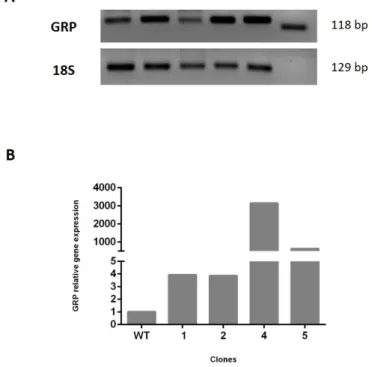
FIGURE 4.1-GENE EXPRESSION LEVELS OF HEK293T CLONES OVEREXPRESSING GRP.EXPRESSION LEVELS OF GRP GENE IN
HEK293T STABLE CLONES (NUMBERED 1-5) WERE OBTAINED BY (A)RT-PCR AND QUANTIFIED BY (B) QPCR;18S
WAS USED AS A HOUSEKEEPING TO NORMALIZE EXPRESSION. ... 26
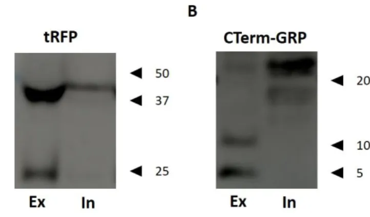
FIGURE 4.2-ANALYSIS OF GRP PROTEIN LEVELS IN TRANSIENTLY TRANSFECTED HEK293T CELLS.HEK293T CELLS WERE TRANSIENTLY TRANSFECTED WITH PMKATE2-N-GRP OR PMKATE2-C-GRP PLASMIDS AND CONDITIONED EXTRACELLULAR (EX) AND INTRACELLULAR (IN) MEDIA WERE ANALYSED BY SDS-PAGE FOLLOWED BY WESTERN BLOT. GRP-MKATE2 WAS IMMUNODETECTED WITH (A) TRFP AND GRP WITHOUT MKATE2 TAG WAS IMMUNODETECTED WITH (B)CTERM-GRP.RELEVANT MOLECULAR MASS MARKERS (KDA) ARE INDICATED ON THE RIGHT SIDE OF THE PANELS. ... 27
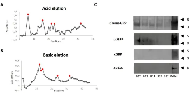
FIGURE 4.3-GRP-MKATE2 PURIFICATION THROUGH AFFINITY CHROMATOGRAPHY.CONDITIONED MEDIA FROM HEK293T
CELLS TRANSFECTED WITH PMKATE2-N-GRP WAS COLLECTED AND FRACTIONED THROUGH AN AFFINITY COLUMN WITH
CTERM-GRP ANTIBODY. (A) ACID AND (B) BASIC ELUTION WAS PERFORMED TO FRACTION AFFINITY-BOUNDED PROTEINS AND PROTEIN CONTENT OF EACH FRACTION COLLECTED WAS DETERMINED BY SPECTROPHOTOMETRY AT 280. (C)FRACTIONS EXHIBITING HIGHER ABSORBANCE LEVELS (RED POINTS) AND CONDITIONED MEDIA INSOLUBLE FRACTION
-GRP BY WESTERN BLOT. THE Γ-CARBOXYLATION STATUS OF PURIFIED GRP WAS INVESTIGATED USING UNDERCARBOXYLATED (UCGRP) AND CARBOXYLATED (CGRP)ANTIBODIES, RESPECTIVELY; THE PRESENCE OF ANNEXIN 6
(ANXA6) WAS ALSO INVESTIGATED.RELEVANT MOLECULAR MASS MARKERS (KDA) ARE INDICATED ON THE RIGHT SIDE OF THE PANELS. ... 29 FIGURE 4.4-GRP PURIFICATION THROUGH AFFINITY CHROMATOGRAPHYY.CONDITIONED MEDIA FROM HEK293T CELLS
TRANSFECTED WITH PMKATE2-C-GRP CONSTRUCT WERE COLLECTED AND FRACTIONED THROUGH AN AFFINITY COLUMN WITH THE CTERM-GRP ANTIBODY. (A) ACID AND (B) BASIC ELUTION WAS PERFORMED TO FRACTION AFFINITY
-BOUNDED PROTEINS AND PROTEIN CONTENT OF EACH FRACTION COLLECTED WAS DETERMINED BY SPECTROPHOTOMETRY AT 280. FRACTIONS EXHIBITING HIGHER ABSORBANCE LEVELS (RED POINTS) WERE SIZE -SEPARATED AND THE PRESENCE OF PROTEIN OF INTEREST WAS INVESTIGATED BY WESTERN BLOT USING THE CTERM-GRP ALTHOUGH NO POSITIVE SIGNAL WAS OBTAINED (DATA NOT SHOWN). ... 30 FIGURE 4.5-MGP OVEREXPRESSION IN HEK293T CELLS. PN-FLAG-MGP WERE TRANSIENTLY EXPRESSED IN HEK293T
CELLS AND INTRACELLULAR (IN) AND EXTRACELLULAR (EX) CONDITIONED MEDIA WERE ANALYSED BY SDS-PAGE AND WESTERN BLOT.MGP WAS IMMUNODETECTED WITH M2 AN ANTIBODY SPECIFIC FOR F TAG.RELEVANT MOLECULAR MASS (KDA) IS INDICATED ON THE LEFT SIDE OF THE PANEL. ... 31 FIGURE 4.6 - BMP-2 ALTERNATIVE TRANSCRIPT IDENTIFICATION. A FRAGMENT OF APPROXIMATELY 1000 BP
CORRESPONDING TO A NOVEL ALTERNATIVE TRANSCRIPT (GENBANK IDENTIFICATION NUMBER 4670498) WAS IDENTIFIED IN CARTILAGE OF AN OA PATIENT.RELEVANT MOLECULAR MASS (BP) IS INDICATED ON THE LEFT SIDE OF THE PANEL. ... 31
FIGURE 4.7-CO-IMMUNOPRECIPITATION OF GRP AND BMP-2.CONDITIONED MEDIA FROM CELLS TRANSFECTED WITH PMKATE2-N-GRPF1 AND PN-MGP-FLAG TO WHICH 100 NG/ML OF RECOMBINANT BMP-2 WAS ADDED, WERE USED TO PERFORM IMMUNOPRECIPITATION ASSAYS.A)50 NG OF RECOMBINANT BMP-2 WAS ANALYSED BY SDS-PAGE AND WESTERN BLOT TO DETERMINE THE APPROPRIATE AMOUNT FOR DETECTION AND ELECTROPHORETIC MIGRATION PATTERN. IMMUNODETECTION WAS PERFORMED USING BMP-2/4 ANTIBODY. GRP-MKATE2 WAS CAPTURED WITH TRFP ANTIBODY AND THE CO-IMMUNOPRECIPITATED PROTEINS WERE ANALYSED BY WESTERN BLOT USING BMP-2/4 ANTIBODY. BMP-2 WAS CAPTURED WITH CORRESPONDING ANTIBODY AND THE IMMUNOPRECIPITATED PROTEINS ANALYSED BY WESTERN BLOT USING B) CTERM-GRP AND C) TRFP ANTIBODIES.
RELEVANT MOLECULAR MASS (KDA) MARKERS ARE INDICATED ON THE LEFT SIDE OF PANELS. ... 32 FIGURE 4.8-PUTATIVE PROTEOLYTIC CLEAVAGE OF GRP-MKATE2.(A)PUTATIVE PROTEOLYTIC CLEAVAGE SITE IN
GRP-MKATE2 FUSION PROTEIN AND THE RESULTING FRAGMENTS.(B)OUR HYPOTHESIS OF FRAGMENTS’ RECOGNITION USING
CTERM-GRP AND TRFP ANTIBODIES, ACCORDING TO PUTATIVE PROTEOLYTIC CLEAVAGE. ... 33
FIGURE 4.9–CO-IP AND SDS-PAGE ANALYSIS FOR PROTEINS IDENTIFICATION.IMMUNOPRECIPITATED PROTEINS WERE SIZE-SEPARATED THROUGH SDS-PAGE AND GEL WAS STAINED WITH CBBG-250.BANDS OF INTEREST WERE CUT AND ANALYSED BY LC-MS/MS.IDENTIFICATION RESULTS CONFIRM THE BAND BETWEEN 50 AND 37 KDA AS GRP-MKATE2 WHEREAS THE BAND AT 25 KDA WAS IDENTIFIED AS MKATE2. RELEVANT MOLECULAR MASS (KDA) MARKERS ARE INDICATED ON THE LEFT SIDE OF PANEL. ... 34 FIGURE 4.10-GRP CO-LOCALIZES WITH MGP IN CO-TRANSFECTED HEK293T CELLS.HEK293T CELLS WERE TRANSIENTLY
CO-TRANSFECTED WITH PMKATE2-C-GRP AND PN-FLAG-MGP OR TRANSFECTED WITH PMKATE2-C-GRP ONLY.(A) GRP DETECTION WAS ACHIEVED USING THE CTERM-GRP ANTIBODY AND SECONDARY ANTI-RABBIT-ALEXA 488
(GREEN), WHILE MGP WAS DETECTED WITH FLAG-TAG M2 ANTIBODY AND ANTI-MOUSE-ALEXA 594(RED).(B)GRP
(GREEN) CO-LOCALIZES WITH PAN-CADHERIN (RED) INDICATING ITS PRESENCE IN CELLULAR MEMBRANE.SCALE BAR REPRESENTS 20 µM. ... 35
FIGURE 6.1 - PROPOSED MODEL OF GRP AND BMP-2 INTERACTION. OUR RESULTS DEMONSTRATE AN INTERACTION BETWEEN GRP AND BMP-2.IN A FUTURE WORK WE ARE INTERESTED IN (A) IDENTIFY ADDITIONAL INTERACTING PARTNERS AND (B) CLARIFY THE ROLE OF MGP IN THIS INTERACTION. ... 41
Introduction
1
1. Introduction
1.1.
The relevance of a micronutrient: Vitamin K
The intake of micronutrients lower than recommended is widespread, not only in poor but also in developed countries, due to unbalanced diets rich in calories while deficient in micronutrients 1. Micronutrients insufficiency leads to their unbalanced
usage: short-term survival functions have priority over to those that are less essential
1. Consequently, persistent deficiency of micronutrients may cause age-related
diseases, such as bone abnormalities, osteoarthritis, chronic kidney disease (CKD), cancer and cardiovascular disease 1.
Vitamin K is a micronutrient which major function is to act as a cofactor for the γ-glutamyl carboxylase (GGCX), an essential enzyme for the activation of vitamin K-dependent proteins (VKDPs) biological function (Fig. 1.1) 2,3. VKDPs, initially described
when studying coagulation cascade, namely coagulation factors II, VII, IX and X, are critical for short-term survival as shown by mice knockouts 1,2,4,5. Beyond coagulation
factors, there are other VKDPs, such as matrix Gla protein (MGP) and osteocalcin (OC), that although less critical for survival, when vitamin K supply is inadequate have long-term consequences and may lead to age-associated conditions 1.
Discovered by the Danish scientist Henrik Dam in 1930s, vitamin K belongs to a family of lipid-soluble vitamins that includes a common 2-methyl-1,4-napthoquinone ring 2,3,7. Depending on the structure of the substituted ring derivative, it can be
classified in two biological active forms, vitamin K1 (or phylloquinone) and vitamin K2 (or menaquinone, MK) that can be obtained from diet 8. While vitamin K1, the major
form obtained by diet, is found in high concentrations in green leafy vegetables, vitamin K2 is found in fermented food such as cheese and Japanese natto, but can also be produced by intestinal bacteria in distal colon 3,5,6. There is also a third form, the
synthetic vitamin K3 (or menadione), that needs to be converted into vitamin K2 to exhibit vitamin K activity 1,2,5.
Dietary vitamin K is emulsified by bile salts to form micelles which are taken up by intestinal cells and incorporated into chylomicrons entering the blood stream and to
Introduction
2 be transported to different target tissues 4. In spite of both forms being absorbed by
small intestine, vitamin K1 is incorporated in lipoproteins enriched in triacylglicerol while vitamin K2 is transported by low and high-density lipoproteins (LDL and HDL, respectively) 10. Different tissue distribution is directly related to distinct transport
mechanisms: whereas the vitamin K1 is mostly found in liver, vitamin K2 is distributed systemically through extra-hepatic tissues, and can actually be more active than vitamin K1 in these tissues 1,9.
Figure 1.1 - The vitamin K cycle. Gamma-glutamyl carboxylase (GGCX) converts glutamate (Glu) into γ-carboxylated glutamate (Gla) residues through conversion of reduced vitamin K (KH2) to vitamin K epoxide (KO). In order to be reused, KO is recycled by vitamin K epoxide reductase (VKOR) which can be inhibited by warfarin. Adapted from Willems et al (2014)8.
1.2.
VKDPs and the importance of vitamin K recycling
Vitamin K dependent proteins become functional after being γ-carboxylated by the γ-glutamyl carboxylase (GGCX) enzyme that is localized in the endoplasmatic reticulum (ER) membrane 11. This enzyme is responsible for the conversion of glutamic acid (Glu)
into γ-carboxylated (Gla) residues which exhibit high affinity for calcium ions and calcium crystals, a property being intimately related to VKDPs specific functions 11. The
reduced form of vitamin K (KH2) is used by the GGCX as a co-factor for γ-glutamyl carboxylation 3. During this reaction, KH2 is converted into vitamin K epoxide (KO) that
can be recycled by the vitamin K epoxide reductase (VKOR) enzyme promoting the beginning of new cycle (Fig. 1.1) 12. This recycling enzyme can be inhibited in liver by
vitamin K antagonists (VKA), such as 4-OH coumarin analogs (e.g. warfarin) widely used in anticoagulant therapies for the treatment of arterial and venous thromboembolic
Introduction
3 disorders (Fig.1.1) 3,14,15. In these cases, and when vitamin K concentrations are
increased, an alternative pathway occurring in liver, independent of coumarins, can be used to provide the γ-carboxylation cofactor. This pathway uses NAD(P)H dehydrogenases (DT-diaphorases) to reduce vitamin K 13.
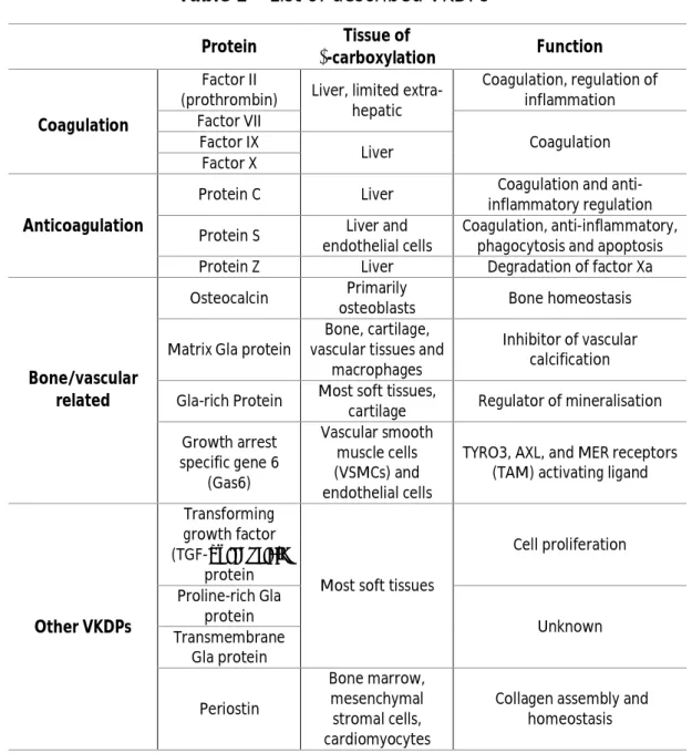
Vitamin K dependent proteins constitute a family of 16 described members – Table 1 9. The most well-known VKDPs are coagulation factors such as prothrombin (factor
II), factors VII, IX and X, and anticoagulation factors such as proteins C, S and Z, that are synthesized mostly in liver with the exception of protein S 10,14,15.
Table 1 – List of described VKDPs
1, 9Protein Tissue of
γ-carboxylation Function
Coagulation
Factor II
(prothrombin) Liver, limited extra-hepatic Coagulation, regulation of inflammation Factor VII Coagulation Factor IX Liver Factor X Anticoagulation
Protein C Liver Coagulation and anti-inflammatory regulation Protein S Liver and
endothelial cells
Coagulation, anti-inflammatory, phagocytosis and apoptosis Protein Z Liver Degradation of factor Xa
Bone/vascular related
Osteocalcin Primarily
osteoblasts Bone homeostasis Matrix Gla protein
Bone, cartilage, vascular tissues and
macrophages
Inhibitor of vascular calcification Gla-rich Protein Most soft tissues,
cartilage Regulator of mineralisation Growth arrest specific gene 6 (Gas6) Vascular smooth muscle cells (VSMCs) and endothelial cells
TYRO3, AXL, and MER receptors (TAM) activating ligand
Other VKDPs
Transforming growth factor (TGF-α) inducible
protein
Most soft tissues
Cell proliferation Proline-rich Gla protein Unknown Transmembrane Gla protein Periostin Bone marrow, mesenchymal stromal cells, cardiomyocytes
Collagen assembly and homeostasis
Introduction
4 Other members of this family of proteins were identified and shown to be synthesized in extra-hepatic tissues, with widespread physiologic activities such as regulation of bone turnover, prevention of vascular calcification and apoptosis 10,14.
Osteocalcin, MGP, GRP and growth arrest specific gene 6 protein (Gas6) can be included in this group of proteins, exhibiting highly specific physiologic functions – Table 1 10,14.
VKDPs exhibit a general structural homology: a signal peptide, a propeptide and a mature protein that contains the Gla domain (Fig 1.2A) 13,16. The signal peptide is
required for protein translocation into the ER where VKDPs bind to carboxylase 16. The
propeptide contains the GGCX recognition site being shown to play an important role in the γ-carboxylation of these proteins 13,16,17. After carboxylation, all known VKDPs
are released from the ER and directed to the Golgi, where the propeptide is further processed by the endopeptidase furin (with the exception of MGP, Fig. 1.2B) 13.
Figure 1.2 - Vitamin K dependent proteins (VKDPs) general structure and processing. A) General structure of a VKDP. VKDPs are composed by a signal peptide (SP), a propeptide (Pro) that contains the GGCX recognition site and a mature protein (MP) including the Gla domain. B) VKDPs cellular processing: SP is required for protein translocation into the endoplasmatic reticulum (ER) where VKDPs bind to GGCX and γ-carboxylation occurs. After carboxylation, VKDPs are released into the Golgi, where the Pro is further processed by furin. Mature VKDPs (MP) are then released to the extracellular matrix where they can exert their specific function. Adapted from Bristol et al (1996) 18
Introduction
5
1.3.
Matrix Gla protein and Gla-rich protein: complementary
partners (in health and disease)?
1.3.1. Matrix Gla protein
Matrix Gla protein was identified for the first time in 1983 in bovine bone by Price and co-workers 19. It was the second Gla protein to be isolated from bone and
demonstrated to be strongly associated with collagenous bone matrix 19. Despite its
high accumulation in extracellular matrix (ECM) of bone, this protein is also synthetized in other tissues such as cartilage, lung, kidney, heart, arterial vessel wall and in calcified atherosclerotic lesions 19–22. MGP was also identified in rat, shark,
chicken, xenopus and human, either as protein or as mRNA 23–26.
1.3.1.1. Protein and gene structural characterization
MGP is a small secreted VKDP that can undergo two types of post-translational modifications, γ-carboxylation and phosphorylation 27. The human mature protein
consists of 89-aa, with a molecular weight of 14 kDa; five out of the nine Glu residues can be γ-carboxylated while three of its five serine residues can be phosphorylated 27.
The human MGP gene is located on chromosome 12 (p13.1 - p12.3) and consists of four exons and three large introns with a total length of 3.9 kb (Fig. 1.3A) 27.
Matrix Gla protein share common features with other VKDPs, such as a signal peptide, a mature protein containing the Gla domain, a AXXF motif and a RXXR furin-like cleavage site (Fig. 1.3B) 16. In MGP the GGCX recognition site, although included in
its propeptide, is not cleaved after the γ-carboxylation reaction and therefore is integrated in the mature protein, a feature that is unique among VKDPs 2,27.
Introduction
6 Figure 1.3 - Structural organization of MGP at gene and protein levels. A) Gene structure of MGP. Boxes represent coding exons and orange boxes represent exons coding for the Gla domain. B) Illustration of MGP protein structure: SP, signal peptide; MP, mature protein; P, serine phosphorylation; GGCX, γ-glutamyl carboxylase recognition site; AXXF, proteolytic cleavage site in MGP; Gla, gla domain. Adapted from Viegas et al (2008) 28.
1.3.1.2. MGP, the calcification inhibitor
The function of MGP as an inhibitor of soft tissue calcification just became clear after studies in MGP null-mice 29. These animals died six to eight weeks after birth due
to an extensive calcification that caused rupture of the thoracic and/or abdominal aorta 29. Moreover, the authors demonstrated a phenotypic transition in vascular
smooth muscle cells (VSMCs) to chondrocyte-like cells 27,28. A phenotype similar to
MGP null-mice was obtained after treatments with warfarin, with rats developing massive calcification of their cartilage, arteries and heart valves 29,30, demonstrating
that the capacity of MGP to inhibit calcification is dependent of its γ-carboxylation status 31.
The precise molecular mechanism by which MGP exerts its function is not yet completely understood but has been proposed to be through binding of calcium ions or calcium crystals present in excess in soft tissues and clearance into circulation 32.
Supporting this hypothesis is the fact that MGP mRNA is expressed in many tissues while the protein usually accumulates in sites of ectopic calcification and circulates in plasma 32. To confirm these facts Roy and Nishimoto showed that MGP binds to
increasing concentrations of hydroxyapatite (HA), further suggesting a binding site for HA in MGP. Moreover, Hackeng and colleagues demonstrated a Ca2+ binding to MGP
and consequently a possible conformational change in protein’s structure 26,32. It was
also suggested that MGP could antagonize proteins known to have a role in chondrogenesis and bone formation, such as bone morphogenetic proteins (BMPs), an issue that will be further discussed in this section 34.
Introduction
7 Because MGP is poorly soluble in its mature secreted form, it was though that this protein circulates either in an aggregated form or bound to higher molecular weight chaperones 32. Price and co-workers were the firsts to identify MGP as part of a
protein-mineral complex composed by hydroxyapatite, fetuin-A and other proteins in serum of rats treated with bisphosphonate etidronate, an inhibitor of bone mineralization 35. Nishimoto and colleagues, showed that MGP binds to vitronectin
which is a multifunctional plasma and ECM protein with a role in cell adhesion, complement activation, coagulation and fibrinolysis 36. This interaction was proved to
occur in vitro by colocalization and immunolocalization studies and may be a mechanism to anchor MGP to the ECM that could modify its activity 36. This group has
also identified a binding site for vitronectin in a region flanking MGP C-terminus 36.
1.3.1.3. MGP-related disorders
Matrix Gla protein has been associated to several human disorders, such as
pseudoxanthoma elasticum (PXE) and systemic sclerosis (Ssc) characterized by elastic
fibres mineralization and skin calcification, respectively 36,37. Two different studies
investigating MGP protein accumulation, both in dermis sections from PXE and skin biopsies from Ssc patients, respectively, have shown that MGP is more abundant in calcified tissues of these patients when compared to healthy controls 36,37. MGP has
also been associated in cases of vascular calcification. For example, a study demonstrates, by immunohistochemistry, that undercarboxylated MGP (ucMGP) is strongly associated with intimal and medial calcification 39.
The Keutel syndrome, a human autosomal recessive disorder is characterized by mutations on MGP gene predicting the production of a non-functional protein 40. This
syndrome is characterized by abnormal cartilage calcification, peripheral pulmonary stenosis and mild facial hypoplasia 40. Unlike the mouse model, Keutel patients survive
until adulthood and lack severe arterial calcification suggesting the involvement of alternative calcification regulators 40.
Introduction
8
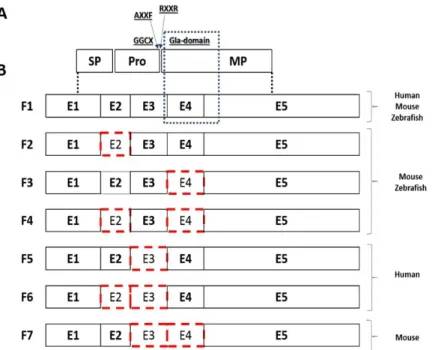
1.3.2. Gla-rich protein
Gla-rich protein (GRP), also known as upper zone of growth plate and cartilage matrix associated protein (UCMA41,42), firstly isolated from the calcified cartilage of
Adriatic sturgeon (Acipenser nacarii), was the latest VKDP to be discovered 28. This
protein is highly conserved amongst vertebrates and exhibits two paralogs, GRP1 and GRP2, in teleost fishes, whose existence might be explained due to a genome duplication event 28,43.
1.3.2.1. Protein and gene structural characterization
GRP is a small secreted protein, negatively charged although highly insoluble at neutral pH 28. The protein exhibits a VKDP general protein structure, as previously
mentioned in section 1.1.1 (Fig. 1.4A) 28. Sturgeon mature GRP, purified from calcified
cartilage, was shown to be a 74-aa long protein, exhibiting a molecular weight of 10.2 kDa where all its Glu residues were experimentally shown to be γ-carboxylated 28. Its
gene structure is also highly conserved amongst species, comprising four introns and five exons 28. Exons 3, 4 and 5, all containing putative Gla residues, code for the mature
protein but exon 4, by exhibting the larger number of Gla residues, is considered to be the functional core of the protein 28.
Although its highly conserved gene structure, GRP alternative transcription appears to be species-specific (Fig. 1.4B). In murine embryo chondrocytes, during development, four distinct transcripts of GRP gene were reported, as well as in the two zebrafish paralogs 40,41. In both species, GRP-F1 is the longest and most abundant
transcript; the other three, GRP-F2, GRP-F3 and GRP-F4, lack exon 2, exon 4 or both, respectively 44. Overexpression of mouse GRP isoforms, in HeLa cells, evidenced that
GRP-F1 and GRP-F3 are secreted proteins that were localized, and detected by immunofluorescence, in the Golgi apparatus 44. On the contrary, GRP-F2 and GRP-F4
isoforms are not secreted, once they lost exon 2 they lack the signal peptide, and aggregate in a structure similar to the aggresome 44. Since exon 4 comprises most of
the Gla residues, these transcripts give rise to two isoforms missing 60% of the putative Gla residues most possibly affecting protein function 44.
Introduction
9 Recently, Rafael and colleagues identified in human osteoarthritis (OA) cartilage two novel alternatively spliced variants of human GRP gene, GRP-F5 and GRP-F6, and a new alternative transcript in cartilage from wild-type murine femurs, GRP-F7 45. The
two novel human transcripts, both GRP-F5 and GRP-F6, lack exon 3 leading to the loss of furin-like cleavage site for propeptide processing and GGCX recognition site 45.
GRP-F6 also lacks exon 2 leading to the translation of a protein with shortening signal peptide length 45. Using an overexpression system in HEK293T cells, it was
demonstrated that the new human protein isoforms, GRP-F5 and GRP-F6 are both secreted but GRP-F6 is secreted later than GRP-F5 since this transcript have a shorter signal peptide, pointing to a lower secretion potential and efficiency during secretory process 45.
In terms of tissue distribution, at the transcriptional level, human GRP-F1 alternative spice variant is ubiquitously expressed in foetal and adult tissues whereas GRP-F5 and F6 are mostly present in foetal tissues 45. These data suggest that the
biological function of splice variants should be further evaluated, specially throughout development 45.
Figure 1.4 - GRP protein structure and splice variants. A) Protein structure of GRP. SP, signal peptide; Pro, propeptide; GGCX, γ-glutamyl carboxylase recognition site; AXXF, proteolytic cleavage site; RXXR, furin-like cleavage site; MP, mature protein. B) GRP-splice variants. Boxes represents coding exons and red boxes represents absent exons in each variant. Adapted from Rafael et al (2014)45.
Introduction
10
1.3.2.2. Novel protein: old function?
The main characteristic of GRP is its high number of Gla residues found in its mature protein form (15 putative Gla residues in humans) conferring it the highest Gla residues/protein size ratio 46. This feature has been conserved over 450 million years
of evolution, with an impressive conservation degree in number and position of Glu residues as well as in its binding site for GGCX 28. This high density of Gla residues
indicates a high calcium binding capacity for GRP, a function that has been extensively shown for other VKDPs 46. Nevertheless, the Gla capacity to bind calcium can confer
different functions to distinct proteins, whether it is related to the coagulation cascade or involved in physiological or pathological mineralization of tissues, such as in case of vascular calcification 14.
Gla-rich protein was found to be associated with ectopic calcification in human skin, vascular and cartilaginous tissues in cases of dermatomyositis, PXE, CKD, cancer and OA 46,47.Although experimental data demonstrating GRP degree of carboxylation
in humans is still not available, the use of conformational antibodies, specific to carboxylated GRP (cGRP) and undercarboxylated GRP (ucGRP) protein forms, respectively, indicates a different pattern of association of these two forms to healthy or pathological conditions 47. Two studies demonstrated a preferential association of
ucGRP with pathological situations, in cases of osteoarthritis and skin and breast cancers, while cGRP is more prevalent in corresponding healthy tissues 45,47. It was also
demonstrated the colocalization of cGRP and ucGRP at sites of microcalcifications, suggesting that both forms have affinity to bind calcium, while the colocalization of both antibodies indicates an incomplete γ-carboxylation in healthy conditions 47.
Different studies indicate that the role of GRP remains to be further clarified. Although at first GRP expression cannot be confirmed in no other tissues than in mice cartilage, later it was shown that GRP is expressed in rat and human skeletal tissue and that is accumulated in soft tissues, such as skin and the vascular system 43,45,46.
Eitzinger and colleagues show that GRP-deficient mice have a normal development where no skeletal abnormalities or calcification of cartilage and bone is identified, indicating that this protein is not essential for normal cartilage development and
Introduction
11 endochondral ossification 48. Nevertheless, the authors point to a possible phenotype
related to calcification and bone turnover during pathological conditions and/or later in development 48. In contrast, in zebrafish, the knockdown of GRP1 leads to severe
growth retardation and abnormal skeletal development and a similar phenotype occurs when γ-carboxylation is inhibited by warfarin 49. These results show that GRP1
and the γ-carboxylation of its Glu residues are crucial for zebrafish skeletal development 49. These contradictory data may be partially explained by the finding
that GRP1, in zebrafish, is earlier expressed than its murine orthologue 48.
1.4.
Bone Morphogenetic Proteins
Bone morphogenetic proteins (BMPs) are multi-functional growth factors that belong to the transforming growth factor-beta (TGF-β) superfamily of proteins 50.
These molecules were discovered by Dr. Marshal Urist in the 1960s and since then twenty BMP family members have been identified 50.
BMPs are synthesized as large precursors consisting of a i) signal peptide, which directs the protein to the secretory pathway; ii) a prodomain, that mediates proper folding and iii) the mature peptide 50,51,52. After signal peptide cleavage, prodomain
undergoes glycosylation and dimerization followed by proteolytical cleavage of the mature protein that becomes active 50. Active BMPs are composed by 50-100 amino
acids among which seven cysteines, where six are used to form intramolecular disulphide bonds while the seventh forms a covalent disulphide bond with another monomer and is secreted as a dimer 51.
Although primarily known for their capacity to induce ectopic bone formation, BMPs have a role in a variety of cell functions 50. These proteins are well known to act
in mesenchymal stem cells differentiation and consequently leading to bone and cartilage formation, but are also involved in other diverse processes such as embyogenesis, organogenesis and tooth morphogenesis, cell proliferation and differentiation, apoptosis, chemotaxis, glucose homeostasis and modulation of iron homeostasis 51,52.
Introduction
12
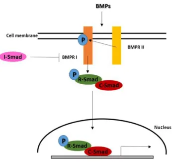
1.4.1. Cellular signalling
BMPs binds to membrane type I and type II serine/threonine kinases receptors that are required for cellular signalling (Fig. 1.5) 50. This binding results in type I
receptor phosphorylation promoted by type II receptor which recruits Smads proteins (fusion of names SMA and MAD from homologous proteins in C. elegans and D.
melanogaster, respectively), that are part of the signalling cascade 50. After
phosphorylation of restricted Smads (R-Smads) these proteins recruit common mediator Smads (C-Smads) to form a complex that migrates to the nucleus and promotes transcription of specific target genes 50,53. The signalling can be regulated at
four levels: extracellular, in the membrane, intracellular and in the nucleus 50.
Extracellularly, the presence of antagonists with high affinity to BMPs receptors prevent its binding 50. Receptors oligomerization can determine the specificity of
signalling pathway activation 50. In the cytoplasm, the signal transmitted can be
modulated by inhibitory Smads (I-Smads), which compete with R-Smads for binding type I receptors, or Smurf1 50,53. Finally, in the nucleus, the activation of specific target
genes and their transcription can be inhibited by co-repressors 50.
Figure 1.5 - Bone morphogenetic proteins (BMPs) signalling. BMPs binds to type I receptor (BMPR I) that became phosphorylated by type II receptor (BMPR II) which recruits Smads. After phosphorylation of restricted Smads (R-Smads) these proteins recruit common mediator Smads (C-Smads) to form a complex that migrates to the nucleus and promotes transcription of specific target genes. The signalling can be inhibited by inhibitory Smads (I-Smads), which compete with R-Smads to binding type I receptors. Adapted from Yamaguchi et al (2000) 53.
Introduction
13
1.4.2. BMP-2
One of the most important known functions of BMP-2 is in bone and cartilage formation 54. These skeletal tissues are composed by several mesenchymal cell types,
such as osteoblasts and chondrocytes, that differentiate in response to Core-binding factor subunit alpha-1/Runt-related transcription factor 2 (Cbfa/Runx2) and SRY-related HMG box transcription factor 9 (Sox9), respectively 53,55. Regulation of
osteoblast differentiation is coordinated by various local factors in a paracrine and/or autocrine manner 53. It was demonstrated that BMP-2, BMP-4 and BMP-7, are
responsible for osteoblast and chondrocytes differentiation 53,54,56. In particular, BMP-2
was shown to promote the expression of osteoblast phenotypic markers alkaline phosphatase (ALP), collagen type I and OC by increasing the expression of the transcription factor Cbfa/Runx2 50,53.
1.4.3. BMP’s relation with VKDPs
When Urist and colleagues first discovered BMPs, they almost immediately realise the tight relation between BMP and MGP, ie, BMPs and a regulator counterpart. Since then, several studies were performed to prove this interaction and to understand how it occurs in vivo 57. Since these proteins are both synthesized by smooth muscle cells
(SMCs), Wallin and co-workers suggested that MGP may neutralize the bone promoting effect of BMP-2 in the vascular wall by forming a complex between the two proteins and, although not proving a regulatory mechanism for MGP, their data confirms the binding between MGP and BMP-2 34. Later, the same group, showed that
Ca2+ and γ-carboxylation of MGP is important for optimal BMP-2 binding to MGP and
further demonstrated, by immunofluorescence in glandular tissues, the existence of MGP-BMP-2 in vivo 34,58. Boström and colleagues, by overexpressing MGP in a
mesenchymal cell line with the capacity to differentiate in different lineages and by adding recombinant BMP-2 (rBMP-2), showed that MGP modulates the capacity of BMP-2 to induce differentiation 59. It was suggest that MGP binding to BMP-2 probably
Introduction
14 studies indicates that MGP inhibits BMP-2 signalling by preventing receptor binding and Smad1 activation 60.
Once BMP-4 is closely related with BMP-2, Yao and colleagues hypothesized that the inhibitory effect MGP is not limited to BMP-2 but could also include BMP-4 61.
Immunoprecipitation studies confirmed an interaction between the two proteins 61. It
was also proved that, as in the case of MGP and BMP-2 interaction, calcium ions and Gla residues were important for the binding between MGP and BMP-4, but proline 64 (Pro64), a conserved residue in human MGP sequence, was also critical for binding and inhibition of BMP-4 62.
It was proposed a model of a regulatory pathway where MGP and 2 and BMP-4 are involved 61,63. BMP-2 and BMP-4 induce the expression of activin-like kinase
receptor 1 (ALK1), a transforming growth factor-β (TGF-β) type I receptor associated with angiogenesis, that when stimulated induces the expression of MGP and vascular endothelial growth factor (VEGF) 61,63. MGP provides both a positive feedback, by
enhancing the ALK1 signaling, and a negative feedback, by binding and inhibiting BMP-2 and BMP-4 that in turn, declines ALK1 expression and activity 61,63. This pathway is
central for vascular development, disease and homeostasis 63. BMP-7 were also
implicated in this regulatory circuit and Yao and colleagues demonstrated that MGP binds and inhibits BMP-7 and BMP-4 in a similar manner 64.
However, MGP is not the only VKDP that is related with BMP-2. Surmann-Schmitt and colleagues have shown that GRP’s mRNA and protein levels are down-regulated in murine chondrocytes treated with rBMP-2. Their results suggest a potential regulatory effect of BMP factors on GRP expression, although without mechanistic any data.
1.5.
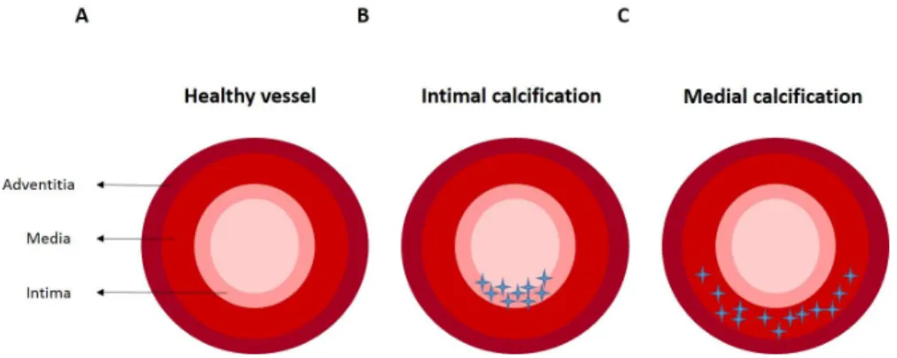
Vascular calcification
Vascular calcification is a disease described as being present over 5000 years ago, related with increased cardiovascular mortality and morbidity, and considered as a strong and independent risk factor for cardiovascular death 8,9. Calcification is mainly
Introduction
15 and subsequent altered hemodynamics, contributing to the development of aortic stenosis, cardiac hypertrophy, hypertension, heart failure, and myocardial ischemia 8.
Within the vessel wall there are two different sites of calcification development: intimal and medial calcification 65. Intimal or atherosclerotic calcification (Fig. 1.6B) is
caused by a combination of risk-factors such as hypertension, inflammation, or dyslipidemia, and is characterized, in early stage, by a disperse punctate form and, as the disease progresses, by aggregates of calcium phosphate crystals deposits producing larger patchy stippled crystals and associating with the necrotic region of an atheroma 8,66. Medial calcification (Fig.1.6C), also known as Monckeberg’s sclerosis, is
characterized by amorphous mineral deposits along the elastin fibres of the tunica media which in an early stage are linear deposits that become circumferential rings of mineral as the disease proceeds 8,66. This type of calcification is highly associated with
aging, CKD and diabetes mellitus 8.
Figure 1.6 - Different types of vascular calcification in the arterial vessel wall. A) Healthy vessel in the absence of calcification. B) Intimal calcification with accumulation of calcium deposits in intima. C) Medial calcification with calcium deposits along the tunica media. Adapted from Willems et al (2014)8.
1.5.1. Vascular calcification and VKDPs
For decades, vascular calcification development was considered as an inevitable consequence of aging and disease, but it is now considered to be an active process involving different molecular mechanisms where clearly a great importance has been given to VKDPs 8,65,67. Human body fluids are supersaturated with calcium and
phosphate but spontaneous mineralization does not normally occur due to a tight control by calcification inhibitors, namely MGP and eventually GRP 8,66. In the case of
Introduction
16 MGP, it was demonstrated that this protein depends on its Gla residues to bind calcium/phosphate crystals, but when vitamin K supply is inadequate or vitamin K antagonists are administered, the proteins become undercarboxylated, inactive, resulting in arterial calcification 39. This hypothesis was proved by two independent
studies that demonstrated the presence of poorly carboxylated MGP in calcified vasculature of aging rats and rats treated with warfarin, and a study in humans showing deposition of ucMGP in arteries from patients with atherosclerosis and Monckeberg’s sclerosis 31,39,58.
Fetuin-A, an extracellular calcium-regulatory protein, is also a potent inhibitor of calcium-phosphate precipitation 68. Fetuin-A and MGP form a high molecular mass
complex with calcium-phosphate mineral, named fetuin-mineral complex (FMC), that is considered to have a role in prevention of vascular calcification avoiding mineral growth and targeting it for clearance from blood 69,70. MGP γ-carboxylation appears to
be essential for the presence of this protein in the complex 8.
1.5.2. Vascular calcification and matrix vesicles
As it occurs during osteogenesis and chondrogenesis, VSMCs release particular membrane structures, known as matrix vesicles (MVs), that serve as the initial nidus for calcium phosphate crystal deposition which promotes the onset of extracellular matrix calcification 8,71. Under normal conditions, MVs are loaded of inhibitory proteins
such as fetuin-A and MGP that block hydroxyapatite nidus 8,71. However, in a
calcium-rich environment, reported in pathologies associated with increased vascular calcification, these membrane structures are able to calcify. At calcification sites, VSMCs can undergo phenotypic transition to osteoblast/chondrocyte-like cells, upregulating the expression of mineralization-regulating proteins, such as BMPs
8,66,71,72. BMP-2 is a crucial factor in this transition promoting the upregulation of
osteogenic key factors as Cbfa1/Runx2 gene, essential for osteoblast differentiation and bone development, that consequently leads to an osteogenic phenotype 8,73. As
Introduction
17 interaction with BMP-2 avoiding its binding to the receptors and consequently blocking gene transcription 59,60.
Objectives
18
2. Objectives
Matrix Gla protein is an established inhibitor of soft tissue calcification and, although still not completely understood, it is known that its inhibitory mechanism is regulated by antagonizing BMPs through direct protein-protein interaction. However, these proteins and their mechanisms of action are not enough to explain the several cases of pathological calcification. Gla-rich protein, the newest VKPD, has also been recently associated with ectopic calcification related diseases. This association, together with results that report a potential regulatory effect of BMP factors on GRP expression in murine chondrocytes, lead us to hypothesize this protein as one of the missing regulators involved in the inhibition of ectopic calcification. In this way, the major goal of this work is the study of the possible interaction between GRP and BMPs. To achieve our objectives we established a system of production of human recombinant GRP and MGP in HEK293T cells and used their conditioned media to perform interaction studies between these proteins and BMPs.
Aiming at the better characterization of GRP biological role, it is also our goal to produce and optimize purification methods to isolate human recombinant γ-carboxylated protein. The success of this objective will further enable us, in a near future, to prove our hypothesis on GRP participation on calcification-related events through the development of functional assays.