Received: November 4, 2011 Accepted: April 20, 2012
Conflict of Interests: The authors state that there are no financial and personal conflicts of interest that could have inappropriately influenced their work.
Copyright: © 2011 Pereira et al.; licensee EDIPUCRS. This is an Open Access article distributed under the terms of the Creative Commons Attribution-Noncommercial-No Derivative Works 3.0 Unported License.
Orthokeratinized odontogenic cyst: A report of
two cases in the mandible
Cisto odontogênico ortoqueratinizado: relato de dois casos
em mandíbula
Francisco de Assis Caldas Pereira a
Manuela Torres Andion Vidal a
Paulo Sérgio Flores Campos b
Alberto de Aguiar Pires Valença Neto c
Lúcio Costa Safira Andrade c
Alex Fernandes c
Jean Nunes dos Santos a
a Laboratory of Oral Surgical Pathology, School of
Dentistry, Federal University of Bahia, Salvador, BA, Brazil
b Division of Oral and Maxillofacial Radiology,
School of Dentistry, Federal University of Bahia, Salvador, BA, Brazil
c Division of Oral and Maxillofacial Surgery, School
of Dentistry, Federal University of Bahia, Salvador, BA, Brazil
Correspondence:
Jean Nunes dos Santos
Faculdade de Odontologia – UFBA Avenida Araujo Pinho, 62 – Canela Salvador, BA – Brasil
40110-150
E-mail: [email protected]
Abstract
Purpose: To describe the behavior clinical and pathological features of two cases of odontogenic cyst orthokeratinized.
Case description: In case 1, a 17-year-old woman presented hard and painless swelling in the left posterior mandible. Radiographically, a radiolucent area with a radiopaque was observed. A clinical diagnosis of ameloblastoma or odontogenic keratocystic tumor was established. The histopathological examination revealed an orthokeratinized odontogenic cyst. In case 2, a 23-year-old woman presented a radiolucent lesion surrounded by radiopaque halo symmetrically distributed on both partially erupted lower third molars. A clinical diagnosis of bilateral dentigerous cyst (DC) was rended. The histopathological examination showed bilateral orthokeratinized odontogenic cyst.
Conclusion: Orthokeratinized odontogenic cyst should be considered in the differential diagnosis of lesions occurring in the jawbones associated with an impacted tooth, particularly those cases simulating dentigerous cyst. In addition, we observed the importance of radiographs taken prior to orthodontic treatment as an important tool in the diagnosis of oral pathologies. Performing routine radiograph is of high clinical value, especially before orthodontic treatment.
Key words: Odontogenic cysts; jaw cysts; nonodontogenic cysts
Resumo
Objetivo: Descrever os aspectos clinicopatológicos de dois casos de cisto odontogênico ortoqueratinizado e a compará-los a relatos anteriores.
Relato do caso: No caso 1, uma mulher de 17 anos de idade, apresentou edema endurecido e indolor na mandíbula posterior esquerda. Radiograficamente, uma área radiolúcida com uma radiopacidade foi observada. O diagnóstico clínico de ameloblastoma ou tumor odontogênico ceratocístico foi estabelecido. O exame histopatológico revelou um cisto odontogênico ortoqueratinizado. No caso 2, uma mulher de 23 anos apresentou duas lesões radiolúcidas circundadas por halos radiopacos simetricamente distribuídas, associada aos terceiros molares inferiores irrompidos. Um diagnóstico clínico de cisto dentígero bilateral (DC) foi estabelecido. O exame histopatológico mostrou ser um cisto odontogênico ortoqueratinizado.
Conclusão: Cisto odontogênico ortoqueratinizado deve ser considerado no diagnóstico diferencial das lesões que ocorrem na mandíbula associado com dentes impactados, especialmente aqueles casos que simulem o cisto dentígero. Além disso, observamos a importância de tomadas radiográficas antes do tratamento ortodôntico no diagnóstico de patologias bucais.
Introduction
The term Odontogenic Keratocystic (OK) was irst
introduced by Philipsen in 1956 to describe a particular
pathological entity characterized by a ibrous cystic wall
lined by keratinized epithelium. Thus, all cystic lesions of the jaws that had aspects of odontogenic keratocysts were considered, regardless of the clinical and other histological
features (1-4). Currently, signiicant differences between
keratinized cystic lesions are recognized and orthokeratinized odontogenic cyst (OOC) is no longer part of the spectrum of odontogenic keratocyst (3,5-7).
Orthokeratinized odontogenic cyst (OOC) is a relatively rare lesion of the jaws, mainly located in the posterior segment of the mandible (1,3). Clinically, it presents no tendency to relapse due to low local aggressiveness, and is not associated with the nevoid basal cell carcinoma syndrome (NBCCS) (2,4,6). Radiographically they are presented as an unilocular radiolucent lesions with well
deined margins (2,3). Histologically, it is represented by a ibrous cystic wall lined by an orthokeratinized epithelium
with thick keratinization, displaying a well-developed
stratum granulosum and a deined layer of basal cells
showing no tendency to palisade (1-5). The authors report two cases of OOC in young adults, comparing them with previous studies.
Case description
Case report 1
A 17-year-old woman was referred to the School of Dentistry of Federal University of Bahia, in Salvador, BA, Brazil, due to a hard and painless swelling in the left posterior mandible. The patient had noticed this swelling in the region about six months before. On palpation, the region corresponding to the lesion was located in the premolar region, extending into the molar region, and wascovered by normal mucosa. Radiographically, a radiolucent area with a radiopaque halo causing divergence between the roots of the premolars was found (Fig. 1). Thus, a clinical diagnosis of ameloblastoma or odontogenic keratocystic tumor was established. Initially, an incisional biopsy was scheduled, but because of a great cleavage plane presented in the surgery, the lesion was completely enucleated. The specimen was sent for histopathological examination, that revealed a orthokeratinized odontogenic cyst, measuring 2,5×1,5×0,6
cm. This case also showed calciication and epithelial remnants scattered in the ibrous wall (Fig. 2). After surgery,
the patient was followed up for a period of ten months, observing signs of bone healing at the injury site (Fig. 3).
Case report 2
A 23-year-old woman was referred to School of Dentistry of Federal University of Bahia for orthodontic treatment.
However, on a routine panoramic radiography, radiolucent
lesions surrounded by radiopaque halo were symmetrically found to be distributed on both partially erupted lower
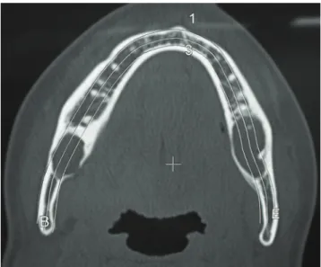
third molars. Computerized tomography (CT), axial view and bone window, showed unilocular lesions bilaterally in the posterior region of the mandible. The lesion on the right displayed a slightly irregular contour and not always corticated margins. To the left, the lesion exhibited regular contour and living cortical outline. Interestingly, despite extensive thinning of cortical bone, the lesions promoted a mild buccolingual distention (Fig. 4). On CT dental arch
Fig. 2. Photomicrography of case 1 showing calcification scattered in the fibrous cystic wall.
Fig. 1. Panoramic radiograph displaying a cystic lesion provoking divergence of the lower left premolar roots.
reconstruction, bone window, to the right side we observed a partially erupted third molar in a horizontal position and slightly downwards to anterior, revealing unicystic injury associated with its crown. This lesion, which extended to the base of the mandible, showed irregular and occasionally corticated margins (above mentioned characteristics) and did not cause tooth displacement or root resorption. On the left, a partially erupted third molar, angled to mesial, showed unicystic injury associated with its crown, extending to the base of the mandible, exhibiting regular contour and corticated margins, and not causing displacement or important dental root resorption (Fig. 5). On physical examination, there were no signs of swelling in that region, and the mucosa and cortical bone remained intact. A clinical diagnosis of bilateral dentigerous cyst (DC) was rended. Afterwards, a biopsy was performed in both lesions, and specimens were sent for histopathological examination that showed bilateral orthokeratinized odontogenic cysts (Fig. 6), measuring 1,5×1,0×05 cm and 1,3×0,8×0,3 cm each. Both lesions were enucleatedfollowed by third molars removals. Thepatient did not show evidence of recurrence for a period of 27 months.
Fig. 5. Computerized tomography, dental arch reconstruction, bone window, exhibiting both lesions.
Fig. 4. Computerized tomography, axial view and bone window, showing unilocular lesions bilaterally in the posterior region of the mandible.
Discussion
Originally, the orthokeratinized odontogenic cyst (OOC) was described as a variant of odontogenic keratocyst (OK).
However, both lesions show biological characteristics which are suficiently distinct to recognize them as speciic
pathological entities (2-4,6,7). Thus, OOC representsa very particular developmental odontogenic cyst (2), whereas the OK was recently included in the group of odontogenic tumors
by the World Health Organization, named as keratocystic
odontogenic tumor (KOT) (5,6,8-10).
Odontogenic cysts with orthokeratinized epithelium are uncommon. According to Li et al. (2), the incidence varies from 5.2 to 16.8% between series, including cases previously diagnosed as odontogenic keratocysts. They represent approximately 11% of all cystic lesions of the
jaws (8,10). It is dificult to determine the prevalence of
these lesions, asthe criteria used to characterize them are not standardized among different studies (1,2,10).
Dong et al. (11)in a recent review showed that females were more frequently affected than males (ratio 2.59:1). Both cases presented here affected young adult female;
this inding is consistent with previous reports that show
a predilection for young adults (1-3). However, it differs from other reports that show nosigniicant sex predilection (1,3). Li et al. (2) have shown greater preference for males.
According to previous studies, OOC more often affects the mandible, especially its posterior region, as observed in
this study (1-3). However, in a particular caseof this study,
there was a rare bilateral involvement of orthokeratinized odontogenic cyst in the posterior mandible associated with the crowns of partially erupted third molars, whose initial clinical diagnosis was bilateral dentigerous cyst. According to several authors, OOCs are commonly associated with impacted teeth, and are generally diagnosed as dentigerous cysts (DCs) (3,4,7,11). The dentigerous cyst is a classic form of developmental odontogenic cyst, affecting more frequently young patients,and generally arises as a single and totally asymptomatic small lesion, similar to what is seen in OOC (1,12). In contrast, the KOT displays a locally
aggressive behavior associated with signiicant rates of
recurrence, as well as the NBCCS (4,10,15). Despite these differences, Li et al (2) observed that nearly half the cases of OOC in their study had previously been diagnosed as ameloblastoma or odontogenic keratocyst tumor.The former diagnosis was attributed to one case of this study.
According Li et al. (2) and Vuhahula et al. (3), OOCs associated with impacted teeth may represent true dentigerous cysts with orthokeratinization, due to the pluripotency of odontogenic cystic epithelium, which is capable of inducing proliferation of orthokeratinized epithelium on the surface of a DC.Furthermore, orthokeratinized epithelial lining
can be seen in other cysts affecting the maxillofacial region as dermoid/epidermoid cysts, even odontogenic keratocyst (3,13).
Histopathologically, the cases of this study fulill the morphological criteria that deine the COO (2-4,7,10). The
epithelial lining of both of orthokeratinized odontogenic cyst was thin and uniform epithelium, and the basal layer was less developed, with its cells displaying a cuboidal aspect without evidence of palisade, or nuclear polarization and hyperchromatism, which are characteristics of the odontogenic keratocystic tumor (3,4,7,8). Li et al. (2) also described the presence of odontogenic epithelial
remnants in OOC. However, we have not found previous reports about the presence of dystrophic calciication in this
cyst as seen on case 1.
Radiographies are constantly required prior to ortho- dontic therapy, as they provide a comprehensive view of the jaw bones, with minimal distortion and overlapping images (14,15). According Bondermark et al. (15), pretreat- ment orthodontic radiographs represent an important source for detecting abnormalities and diseases in the maxillomandibular region. In the present study, unilocular radiolucencies, associated with bilaterally impacted third molars were diagnosed from radiographic examinations performed with orthodontic purpose. Therefore, OOC should be taken into account in the differential diagnosis of radiolucencies associated with an impacted tooth. It is important to state that although unilocular radiolucencies
with well deined margins represent a classical radiographic
pattern, multilocular radiolucencies also may be found less frequently (11).
KOT is locally invasive and should be treated radically, through peripheral ostectomy, chemical curettage or block resection (8,10).In a recent review, Madras and Lapointe (8) observed that the use of more aggressive therapeutic modalities, such as block resection, are associated with lower recurrence rates. In contrast, the OOC is treated in a conservative manner, performing only the enucleation of the lesion, as observed in both cases of this study. This form of treatment shows very satisfactory results, observing a relapse rate of only 2.2% of all cases (2,5,7,10).
1. Da Silva MJ, de Sousa SO, Corrêa L, Carvalhosa AA, De Araújo VC. Immunohistochemical study of the orthokeratinized odontogenic cyst: a comparison with the odontogenic keratocyst. Oral Surg Oral Med Oral Pathol Oral Radiol Endod 2002;94:732-7. 2. Li TJ, Kitano M, Chen XM, Itoh T, Kawashima K, Sugihara K, Nozoe E, Mimura T.
Orthokeratinized odontogenic cyst: a clinicopathological and immunocytochemical study of 15 cases. Histopathology 1998;32:242-51.
3. Vuhahula E, Nikai H, Ijuhin N, Ogawa I, Takata T, Koseki T, Tanimoto K. Jaw cysts with orthokeratinization: analysis of 12 cases. J Oral Pathol Med 1993; 22:35-40.
4. Rangiani A, Motahhary P. Evaluation of bax and bcl-2 expression in odontogenic keratocysts and orthokeratinized odontogenic cysts: A comparison of two cysts. Oral Oncol 2009;45:41-4.
5. Fregnani ER, da Cruz Perez DE, Soares FA, Alves FA. Synchronous ameloblastoma and orthokeratinized odontogenic cyst of the mandible. J Oral Pathol Med 2006 35:573-5. 6. González-Alva P, Tanaka A, Oku Y, Yoshizawa D, Itoh S, Sakashita H ET AL. Keratocystic
odontogenic tumor: a retrospective study of 183 cases. J Oral Sci 2008;50:205-12. 7. Thosaporn W, Iamaroon A, Pongsiriwet S, Ng KH. A comparative study of epithelial
cell proliferation between the odontogenic keratocyst, orthokeratinized odontogenic cyst, dentigerous cyst, and ameloblastoma. Oral Dis 2004;10:22-6.
8. Madras J, Lapointe H. Keratocystic odontogenic tumour: reclassification of the odontogenic keratocyst from cyst to tumour. J Can Dent Assoc 2008;74:165-165h.
9. World Health Organization Classification of Tumours. Pathology and genetics of head and neck tumours. Lyon: IARC Press; 2005.
10. Onuki M, Saito A, Hosokawa S, Ohnuki T, Hayakawa H, Seta S, Muramatsu T, Furusawa M. A case of orthokeratinized odontogenic cyst suspected to be a radicular cyst. Bull Tokyo Dent Coll 2009;50:31-5.
11. Dong Q, Pan S, Sun LS, Li TJ. Orthokeratinized odontogenic cyst: a clinicopathologic study of 61 cases. Arch Pathol Lab Med 2010;134:271-5.
12. Buyukkurt MC, Omezli MM, Miloglu O. Dentigerous cyst associated with an ectopic tooth in the maxillary sinus: a report of 3 cases and review of the literature. Oral Surg Oral Med Oral Pathol Oral Radiol Endod 2010;109:67-71.
13. Ide F, Saito I, Horie N, Shimoyama T. Orthokeratinized odontogenic cyst of the mandible with heterotopic cartilage. Head Neck Pathol 2009;3:150-52.
14. Puricelli E. Panorametry: suggestion of a method for mandibular measurements on panoramic radiographs. Head Face Med 2009;5:19.
15. Bondemark L, Jeppsson M, Lindh-Ingildsen L, Rangne K. Incidental findings of pathology and abnormality in pretreatment orthodontic panoramic radiographs. Angle Orthod 2006;76:98-102.