©Revista Brasileira de Fisioterapia
O
RIGINALA
RTICLEMetabolic and clinical comparative analysis
of treadmill six-minute walking test and
cardiopulmonary exercise testing in obese
and eutrophic women*
Análise clínica e metabólica comparativa entre o teste de caminhada de seis
minutos e o teste de exercício cardiopulmonar em mulheres obesas e eutróficas
Luciana Di Thommazo-Luporini1, Soraia P. Jürgensen1, Viviane Castello-Simões1, Aparecida M. Catai1, Ross Arena2, Audrey Borghi-Silva1
Abstract
Background: Impaired exercise tolerance is directly linked to decreased functional capacity as a consequence of obesity. Objectives: To analyze and compare the cardiopulmonary, metabolic, and perceptual responses during a cardiopulmonary exercise test (CPX) and a treadmill six-minute walking test (tread6MWT) in obese and eutrophic women. Methods: Twenty-nine female participants, aged 20–45 years were included. Fourteen were allocated to the obese group and 15 to the eutrophic group. Anthropometric measurements and body composition assessment were performed. Results: In both tests, obese women presented with significantly higher absolute oxygen uptake, minute ventilation, and systolic and diastolic blood pressure; they also presented with lower speed, distance walked, and oxygen uptakecorrected by the weightcompared to eutrophics. During the maximal exercise test, perceived dyspnea was greater and the respiratory exchange ratio was lower in obese subjects compared to eutrophics. During the submaximal test, carbon dioxide production, tidal volume, and heart rate were higher in obese subjects compared to eutrophic women. When analyzing possible correlations between the CPX and the tread6MWT at peak, there was a strong correlation for the variable heart rate and a moderate correlation for the variable oxygen uptake. The heart rate obtained in the submaximal test was able to predict the one obtained in the maximal test. Bland-Altman plots demonstrated the agreement between both tests to identify metabolic and physiological parameters at peak exercise. Conclusions:
The six-minute walking test induced ventilatory, metabolic, and cardiovascular responses in agreement with the maximal testing. Thus, the six-minute walking test proves to be important for functional evaluation in the physical therapy routine.
Keywords: obesity; exercise test; physical fitness; physical therapy.
Resumo
Contextualização: A reduzida tolerância ao exercício está relacionada à diminuída capacidade funcional consequente da obesidade.
Objetivos: Analisar e comparar respostas cardiopulmonares, metabólicas e subjetivas durante um teste de esforço cardiopulmonar e um teste de caminhada de seis minutos na esteira em mulheres obesas e eutróficas. Métodos: Foram incluídas 29 mulheres com idades entre 20 e 45 anos. Catorze voluntárias foram alocadas no grupo de obesas e 15, no grupo de eutróficas. Foram realizadas medidas antropométricas e de composição corporal. Resultados: Em ambos os testes, as obesas apresentaram maiores valores de consumo absoluto de oxigênio, ventilação-minuto e pressão arterial sistólica e diastólica; ainda apresentaram menor velocidade de caminhada, distância percorrida e consumo de oxigênio relativo, quando comparadas com as eutróficas. Durante o teste máximo de exercício, a dispneia percebida foi maior e o quociente respiratório menor nas obesas em relação às eutróficas. Durante o teste submáximo, produção de dióxido de carbono, volume corrente e frequência cardíaca foram maiores nas obesas, comparadas às eutróficas. Houve forte correlação entre a frequência cardíaca e moderada correlação entre o consumo de oxigênio no pico dos testes. A frequência cardíaca obtida no teste submáximo aplicado foi capaz de predizer a frequência cardíaca obtida no teste máximo.
1Physical Therapy Department, Nucleus of Research in Physical Exercise, Universidade Federal de São Carlos (UFSCar), São Carlos, SP, Brazil
2Department of Orthopaedics and Rehabilitation, Physical Therapy Program and Department of Internal Medicine, Division of Cardiology, University of New Mexico School of Medicine,
Albu-querque, New Mexico, United States of America
*L. Di Thommazo-Luporini, S. Pilon Jürgensen, V. Castello, C. Negrão Dias, R. Luís Luporini, J. Carlos Bonjorno-Júnior, C. Ricardo de Oliveira, A. Maria Catai, A. Borghi-Silva. Comparative analysis of cardiopulmonary and clinical responses to six minute walking test and maximal exercising test in obese women. Eur Respir J. 2011 Sept;38(Suppl55):862s-863s.
Introduction
Obesity-induced limitations of the cardiopulmonary and metabolic systems, commonly resulting in exertional dyspnea, collectively contribute to the limitations in
func-tional capacity frequently observed in obese individuals1.
In addition, the sedentary lifestyle often adopted by these individuals further compounds and contributes to im-paired exercise tolerance.
Cardiopulmonary exercise testing (CPX) is consid-ered the “gold standard” method for comprehensively as-sessing the response to aerobic exercise. The value of CPX data is manifold, including evaluation of exercise tolerance, quantification of impairment and disability levels, deter-mination of the physiological mechanism(s) for exercise
intolerance, and exercise prescription2. However, given the
increasing recognition of the value of functional capac-ity assessments in numerous populations in conjunction with acknowledgement of the difficulty in broadly applying CPX, there is a need for valid and reliable exercise testing assessments that can be implemented in a time- and
cost-efficient manner3. The six-minute walking test (6MWT) is a
well-established functional assessment4 that may provide
a reasonable estimation of functional capacity in obese
patients5,6, even with associated diseases7. However, more
work is needed in this area to validate initial findings. Some authors have found a significant correlation
between 6MWT distance and peak oxygen uptake (VO2)
measured by CPX in patients with advanced heart failure8.
However, no previous study has evaluated the clinical significance of the 6MWT exercise measures comparable
to CPX results in obese women. As VO2 peak is the “gold
standard” measure of aerobic capacity, it seems worthwhile to determine if cardiopulmonary, metabolic, and perceptual responses to the 6MWT are in concordance with CPX out-comes in obese women. When the population undergoing functional assessment requires closer monitoring of physi-ological variables, performing the 6MWT on a treadmill (tread6MWT) may be particularly attractive. This approach has been previously applied in healthy subjects and in
certain patient populations9-11, not including female obese
patients. Therefore, the aim of this study was to analyze and compare the cardiopulmonary, metabolic, and perceptual responses to CPX and tread6MWT in obese and eutrophic women. The ultimate goal was to apply a submaximal test that could be easily used in a physical therapy clinical set-ting as part of the routine evaluation of obese women. We hypothesized that the tread6MWT is related to CPX in obese women and that it would elicit cardiopulmonary and metabolic responses in agreement with CPX responses.
Method
Design and study population
The current investigation is an observational, cross-sectional, comparative study. Subjects were sedentary females, from 20 to 45 years of age, allocated to an obese
group (OG): BMI≥30 kg.m-2 and weight stable for the past
one year; or eutrophic group (EG): 18.5≤BMI≤24.9 kg.m-2.
Exclusion criteria were: pregnancy; currently smoking or abstinence from smoking less than one year prior to study initiation; alcohol or drug addiction; presence of diabetes; uncontrolled hypertension; diagnosis of cardiopulmo-nary disease, such as corocardiopulmo-nary artery disease; chronic obstructive pulmonary disease or asthma; neurological or orthopedic dysfunctions; and/or the use of β-blockers. The experimental procedures were performed respecting
a minimum resting period of 48h: (a) 1st Visit: clinical and
physical therapy evaluations (anamnesis, anthropometric measures, regular physical activity pattern investigation, pulmonary function evaluation, bioelectrical impedance
analysis); (b) 2nd Visit: CPX on a treadmill; and (c) 3rd Visit:
tread6MWT performance. Approval by the institutional Ethics Committee at Universidade Federal de São Carlos (UFSCar), São Carlos, SP, Brazil (Approval 230/2009) and written informed consent from all subjects were obtained before the study initiation.
Os gráficos de Bland-Altman demonstraram concordância entre os testes para identificar parâmetros metabólicos e fisiológicos no pico do exercício.
Conclusão: O teste de caminhada de seis minutos induziu respostas ventilatórias, metabólicas e cardiovasculares concordantes com as do teste máximo, provando ser importante na rotina de avaliação funcional fisioterápica de mulheres obesas.
Palavras-chave: obesidade; teste de esforço; aptidão física; fisioterapia.
Measures
Physical therapy evaluations
Anthropometric data (body weight, height, and BMI) was
evaluated according to methodology previously described12.
Regular physical activity patterns were collected by the modi-ied Baecke questionnaire for epidemiological studies, which
was previously validated in Portuguese13. Occupation, sports
activities, and leisure habits were quantiied to assess the physical activity patterns.
Foot-to-foot bioelectrical impedance analysis was assessed with the Tanita body composition analyzer (model TBF-310; Tanita Corp., Tokyo, Japan), which calculates fat mass, fat-free mass, percentage of fat. his set has been used previously to
assess obese women14. Subjects were measured in the morning
in bathing suits without shoes or any kind of metal in contact with their bodies. he subjects were advised to be in absolute fast for at least four hours as well as to urinate prior to the as-sessment of body composition.
Spirometric tests were performed using the ergospirometry
system (Oxycon Mobile®, Mijnhardt/Jäger, Würzburg, German)
with low measurement carried out using a calibrated pneu-motachograph. he subjects completed at least three accept-able maximal forced and slow expiratory maneuvers according
to the recommendations of the American horacic Society15.
Experimental protocols
All of the experimental procedures described above were performed during the afternoon to avoid the inluence of circa-dian changes, in a climate-controlled room with temperature between 22–24ºC and relative air humidity between 40–60%. he subjects received orientation on the experimental protocols and were instructed to abstain from cafeine, stimulants and alcoholic beverages during the 24 h preceding the tests, avoid strenuous physical activities for 24 h before the experiment, to have a good night’s sleep, and to ingest a light meal at least 2h prior to the exercise tests. To further standardize the assessment and eliminate potential confounders, the subjects performed the tests during the follicular phase of their menstrual cycle.
Cardiopulmonary exercise testing
Symptom-limited CPX was performed on a treadmill (Mas-ter ATL, Inbramed, Porto Alegre, RS, Brazil). he exercise test consisted of: a) 4-min rest at standing position on the treadmill;
b) incremental phase according to the Bruce ramp protocol12; c)
3-min recovering period. All the subjects were actively encour-aged by the investigators in a standardized fashion throughout the test to walk and/or run to the limit of tolerance. Heart rate (HR), arterial blood pressure (ABP) measured using a standard
cuf sphygmomanometer (Diasyst®, São Paulo, Brazil) and
per-ceived exertion by Borg scale16 were measured at each stage of
exercise protocol, and throughout the recovering period. Ventilatory expired gases were continuously measured dur-ing CPX and analyzed breath-by-breath usdur-ing an
ergoespiro-metric system (Oxycon Mobile®, Mijnhardt/Jäger, Würzburg,
German), which was calibrated before each test according to manufacturer speciications. Patients were also monitored using a thoracic MC5 lead (cardiac monitor Ecaix TC500, São Paulo, SP, Brazil). Test termination criteria followed American horacic
Society recommendations2. Two qualiied physical therapists
with physician supervision conducted each exercise test.
Aerobic capacity was evaluated using VO2 data obtained at
the peak of exercise. Metabolic and ventilatory data were pro-cessed and calculated in mobile averages, every eight breath-ing cycles. he average value the last 15 s was deined as the
subject’s peak VO2 and peak respiratory exchange ratio (RER).
Visual analyses of breathing and metabolic responses were made by three duly trained observers in order to determine ventilatory anaerobic threshold (VAT). he V-slope method was used to determine VAT through consensus of three
expe-rienced reviewers17.
Treadmill Six-Minute Walking Test
he tread6MWT was performed on a treadmill with zero
inclination and subject-controlled speed18. Subjects began at
a speed of 3 km.h-1. hey were instructed and encouraged to
walk as far as possible, according to American horacic
So-ciety guidelines4. In addition, they were allowed to increase or
decrease the speed of the treadmill at any time, according to symptoms of fatigue or dyspnea. Heart rate and perceived
exer-tion by the Borg scale16 were measured every three minutes and
at the exercise peak. he subjects’ ABP was measured at rest in the standing position on the treadmill as well as at the exercise peak and throughout their recovery period. he treadmill’s data panel showing speed and distance covered was not visible to the subject; only the start, stop, and speed up or slow down buttons were accessible to them. he subjects were allowed to interrupt the test if they had symptoms such as tachycardia or any dis-comfort that would make it impossible for them to continue the test. Two tests were completed with a minimum interval of one hour in order to reduce learning efects. Only the outcomes from the second tread6MWT were considered for statistical analysis.
Statistical analysis
Universidad Autónoma de Barcelona, Barcelona, Spain). he target sample size was calculated to be four in each group con-sidering a 5% type I error, a 2-sided test, and an 80% power to
detect a signiicant diference of VO2=12.9 mL.kg
-1.min-1 at the
maximal metabolic rate in obese and normal weight subjects19.
he analysis was performed with MedCalc statistical software, Version 11.4.4.0 (MedCalc Software, Mariakerke, Belgium). Data are presented as mean±SD after testing for normal distribution (Shapiro-Wilk test) and as median (minimum, maximum) for categorical variables. he Student unpaired
t-test was used for the comparison of continuous
demo-graphic and anthropometric data, bioelectrical impedance indexes, lung function, symptom data, and CPX and tread-6MWT outcomes between the OG and the EG. he Student
paired t-test was used to compare CPX and tread6MWT
out-comes in each studied group. Fisher’s exact test was used to compare the medication intake between groups. The
re-lationship between relative VO2 (mL.kg
-1.min-1) and BMI in
CPX and the tread 6MWT as well as the correlation between
HRpeak and VO2 (mL.min
-1) were assessed using Pearson’s
correlation. The stepwise regression was analyzed
consid-ering HRpeak in both tests. Moreover, the absolute limits of
agreement between the metabolic and cardiovascular vari-ables assessed by CPX and the tread6MWT were evaluated
by the Bland-Altman analysis20. The probability of a type I
error was set at 5% (p<0.05).
Results
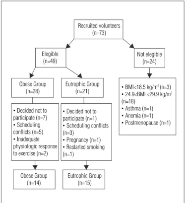
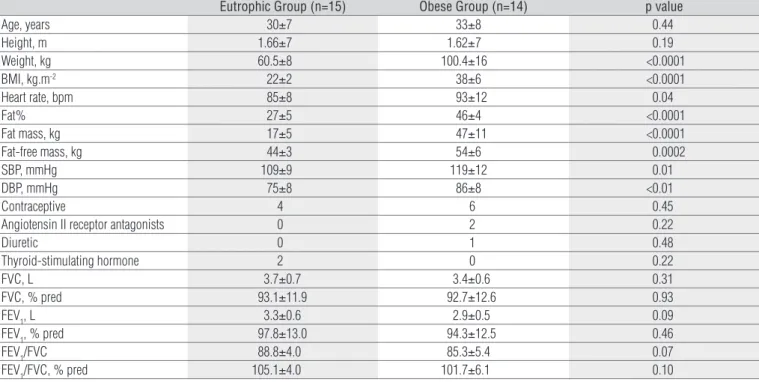
Seventy-three subjects were recruited. Figure 1 illustrates the attrition rate of each group. Table 1 lists demographic and anthropometric data, cardiovascular parameters at rest, bioelectrical impedance indexes, current medica-tions, and lung function of both groups. Significant differ-ences between groups were found regarding weight, BMI, HR, systolic and diastolic ABP (SBP and DBP, respectively), and body composition data. There were no differences in medication intake between groups. Three obese subjects presented with controlled hypertension and two eutroph-ics presented with controlled hypothyroidism. Data from spirometry confirmed exclusion criteria and no difference was found between the OG and the EG (Table 1). All women were considered to be sedentary according to Baecke ques-tionnaire results, with a total score at or below 8: seven obese women had scores below 6 while the other half had scores between 6 and 8; four eutrophics had scores below 6, and eleven had scores between 6 and 8. Nevertheless, there were no significant differences between groups (p=0.26).
Exercise-related physiological and subjective
responses
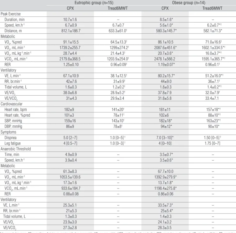
Table 2 shows CPX and tread6MWT results. All patients were able to successfully complete both tests without compli-cations, except two obese subjects that presented inadequate ABP response to exercise during CPX and were excluded from the inal sample. he test duration of CPX as well as the speed reached and the distance covered in both tests were signii-cantly lower in the OG compared to the EG.
As expected, in both tests, obese women had higher
absolute values for VO2 (p<0.0001) compared to
eutroph-ics but, when VO2 were corrected by weight, they were
significantly lower (p=0.02) in the OG. Moreover, minute ventilation, SBP, and DBP were higher in the OG. There
were no differences in RR, leg fatigue, VE/VO2 or VE/VCO2
between groups.
In relation to the maximal test, peak RER was lower and dyspnea symptoms were signiicantly higher in the OG com-pared to the EG. here were no diferences in
percent-pre-dicted peak VO2, tidal volume, HR, and the percent-predicted
HR (%HR) between groups. Regarding the tread6MWT, VCO2,
tidal volume, HR, and %HR were all higher in the OG com-pared to the EG.
Table 2 also shows the comparison between integra-tive responses to CPX vs. tread6MWT. CPX induced a higher Recruited volunteers
(n=73)
Elegible (n=49)
Obese Group (n=28)
Eutrophic Group (n=21)
Eutrophic Group (n=15) Obese Group
(n=14)
• BMI<18.5 kg/m2 (n=3)
• 24.9<BMI <29.9 kg/m2 (n=18)
• Asthma (n=1)
• Anemia (n=1)
• Postmenopause (n=1)
• Decided not to participate (n=1)
• Scheduling conflicts (n=3)
• Pregnancy (n=1)
• Restarted smoking (n=1)
• Decided not to participate (n=7)
• Scheduling conflicts (n=5)
• Inadequate physiologic response to exercise (n=2)
Not elegible (n=24)
increment in the majority of the test outcomes for the OG,
except distance walked, VE/VCO2, DBP, and perceived leg
fa-tigue. In contrast, CPX induced a higher increment in most of
the variables of the EG, except walking speed and VE/VO2 ratio.
At VAT during CPX, markedly lower values were found in the OG compared to the EG for time, treadmill speed,
and relative peak VO2 as were higher absolute values for
VO2, VCO2, VE, and RR. There were no differences between
groups in relation to the other metabolic and ventilatory parameters at VAT.
Considering all the subjects, absolute VO2 peak (L.min
-1)
during CPX was modestly correlated (r= 0.53) to the same
pa-rameter of the tread6MWT (Figure 2A). In addition, a strong
positive correlation (r=0.77) was found between HR
peak during
CPX and the tread6MWT in the OG (Figure 2B). Figure 2C shows a stepwise regression analysis (y=-66.9299+1.2324x)
be-tween HRpeak in both tests (r
2=0.58; p=0.001).
Agreement between CPX and the tread6MWT
Analysis of agreement between both exercise tests applied to assess functional capacity was carried out by a Bland-Altman plot. Considering the metabolic and ventilatory parameters reached in the CPX as the ‘‘gold standard,’’ we chose CPX-peak
VO2 vs.tread6MWT-peak VO2 and CPX-VE vs. tread6MWT-VE
to plot as primary variables of interest.
Two additional secondary variables from CPX were se-lected as well: CPX-SBP vs. tread6MWT-SBP and CPX-peak HR vs. tread6MWT-peak HR. As reported in Figures 3A and 3B,
the mean of the diferences to identify the relative VO2 and the
VE by CPX and the tread6MWT was 6.0±5.6 mL.kg-1.min-1 and
29.0±16.9 L.min-1, respectively. Similarly, the agreement of both
tests to identify SBP and HR at the peak of exercise calculated by the Bland-Altman method found a mean diference between the tests of 17.5±19.4 mmHg and 32.9±19.4 beats per minute (Figures 3C and 3D), respectively. herefore, there was agree-ment between the tests in all performed analysis.
Discussion
In this study, we demonstrated that the submaximal tread-6MWT promoted cardiorespiratory and metabolic responses in agreement with CPX in obese women, thus showing the advantage of lower cardiopulmonary and metabolic stress as well as lower perceived dyspnea during the test. herefore, it is
possible to predict the HRpeak of the symptom-limited CPX by
means of the HRpeak reached in the tread6MWT. Additionally,
the exercise tests demonstrated a lower functional capacity in obese women in comparison with eutrophics, expressed by metabolic, ventilatory, and cardiovascular variables and dysp-nea perception, as well as speed and walking distance. Eutrophic Group (n=15) Obese Group (n=14) p value
Age, years 30±7 33±8 0.44
Height, m 1.66±7 1.62±7 0.19
Weight, kg 60.5±8 100.4±16 <0.0001
BMI, kg.m-2 22±2 38±6 <0.0001
Heart rate, bpm 85±8 93±12 0.04
Fat% 27±5 46±4 <0.0001
Fat mass, kg 17±5 47±11 <0.0001
Fat-free mass, kg 44±3 54±6 0.0002
SBP, mmHg 109±9 119±12 0.01
DBP, mmHg 75±8 86±8 <0.01
Contraceptive 4 6 0.45
Angiotensin II receptor antagonists 0 2 0.22
Diuretic 0 1 0.48
Thyroid-stimulating hormone 2 0 0.22
FVC, L 3.7±0.7 3.4±0.6 0.31
FVC, % pred 93.1±11.9 92.7±12.6 0.93
FEV1, L 3.3±0.6 2.9±0.5 0.09
FEV1, % pred 97.8±13.0 94.3±12.5 0.46
FEV1/FVC 88.8±4.0 85.3±5.4 0.07
FEV1/FVC, % pred 105.1±4.0 101.7±6.1 0.10
Table 1. Demographic, anthropometric, and body composition data; baseline cardiovascular variables; current medications; and lung function of each group.
Data presented as mean±SD. BMI: body mass index; Fat%: percentage of total body fat mass; FVC: forced vital capacity; FEV1: forced expiratory volume in one second. (Student unpaired t-test or Fisher’s exact
Comparison between CPX and tread6MWT
outcomes
Because the 6MWT is better tolerated than CPX by indi-viduals with disabilities and because it has been used to
evalu-ate functional capacity in several populations21, we compared
exercise responses between tests. It is well known that CPX is considered the “gold standard” to assess the integrative exercise response since it is a progressive exercise to the tolerance limit
that combines the assessment of ECG, hemodynamic,
subjec-tive symptoms and ventilatory expired gas analysis measures2.
Another advantage of the CPX is that it is applied on an er-gometer, which allows for the collection of the aforementioned variables while controlling for workload titration. However, the time required, the need for specially trained staf, and the cost of the equipment limits the widespread applicability of CPX.
Interestingly, although the 6MWT is usually applied in a cor-ridor to obtain a more secure monitoring of the physiological
Eutrophic group (n=15) Obese group (n=14)
CPX Tread6MWT CPX Tread6MWT
Peak Exercise
Duration, min 10.7±1.6 – 8.5±1.6* –
Speed, km.h-1 6.7±0.9 6.7±0.7 5.6±1.0* 6.2±0.7*†
Distance, m 812.1±186.7 633.3±61.0† 580.3±145.7* 582.1±71.3*
Metabolic
VO2, %pred 91.1±15.5 64.5±13.3† 86.1±10.5 71.0±16.6†
VO2, mL.min-1 1739.2±255.7 1299±274.2† 2087.6±451.6* 1602.1±334.5*†
VO2, mL.kg-1.min-1 28.7±4.4 21.4±4.3† 20.7±3.6* 16.0±3.7*†
VCO2, mL.min-1 2179.8±368.5 1203.9±254.0† 2478.1±566.2 1595.1±365.7*†
RER 1.25±0.10 0.96±0.09† 1.19±0.07* 0.96±0.1†
Ventilatory
VE, L.min-1 67.1±10.9 38.1±12.5† 80.2±15.7* 51.2±16.0*†
RR, br.min-1 42±7.6 31±9.9† 44±9.0 36±7.1†
Tidal volume, L 1.6±0.3 1.2±0.2† 1.8±0.3 1.4±0.2*†
VE/VO2 38.0±6.8 28.9±5.2† 37.8±7.9 32.0±7.9†
VE/VCO2 31±4.3 29.9±3.4 31.8±5.8 33.4±7.1
Cardiovascular
Heart rate, bpm 182±9 141±20† 181±11 157±18*†
Heart rate, %pred 101±3 78±11† 102±6 88±10*†
SBP, mmHg 159±16 143±10† 182±18* 163±23*†
DBP, mmHg 86±9 78±8† 94±12* 90±10*
Symptoms
Dispnea 5.0 [2–7] 1.0 [0–5] † 7.0 [3–10]* 1.50 [0–5] †
Leg fatigue 4 [0.5–7] 1.0 [0–3] † 4 [0–10] 1.75 [0–7]
Anaerobic Threshold
Time, min 4.9±0.9 – 3.5±0.7* –
Speed, km.h-1 3.9±0.4 – 3.5±0.6* –
Metabolic
VO2, %pred 61.3±8.3 – 67.7±10.0 –
VO2, mL.min-1 1053.5±139.6 – 1392.9±279.9* –
VO2, mL.kg-1.min-1 17.3±1.6 – 13.7±1.8* –
VCO2, mL.min-1 933.6±184.7 – 1198.4±275.8* –
RER 0.88±0.08 – 0.86±0.06 –
Ventilatory
VE, L.min-1 25.3±5.1 – 33.5±7.3* –
RR, br.min-1 21±5.3 – 25±5.4* –
Tidal volume, L 1.3±0.3 – 1.4±0.3 –
VE/VO2 23.9±3.0 – 24.1±3.2 –
VE/VCO2 27.3±2.8 – 28.3±3.5 –
Table 2. Maximal cardiopulmonary exercise test (CPX) and treadmill six-minute walking test (tread6MWT) data at the peak of the tests and at the anaerobic threshold.
Data presented as mean±SD or median ± [minimum, maximum] when appropriate. VO2: oxygen uptake; VCO2: carbon dioxide production; RER: respiratory exchange rate; VE: minute ventilation; RR: respiratory rate; SBP: systolic blood pressure; DBP: diastolic blood pressure. *Significant differences comparing obese versus eutrophic volunteers (Student unpaired t-test, p<0.05); †Significant differences comparing CPX
parameters, it can also be applied on a treadmill11. his was one
of the reasons for choosing the tread6MWT, the other reason being to collect ventilatory expired gas variables with the same equipment used during CPX for comparative purposes.
Gui-marães, Carvalho and Bocchi18 applied the tread6MWT with
ventilatory expired gas monitoring in heart failure patients and found it to be a feasible approach to functional assessment in this population. Despite the previous application of the tread-6MWT in healthy subjects and certain patient populations, we acknowledge that tread6MWT distance results are not
inter-changeable with corridor tests4,11.
Some studies have shown correlation and regression analy-sis to complement Bland-Altman method-comparison plots of
agreement22. In this way, besides the demonstrated agreement
between CPX and the tread6MWT through Bland-Altman
analysis, the linear regression showed the HRpeak in CPX could be
predicted by means of HRpeak reached in the tread6MWT. hus,
although CPX and the tread6MWT measure diferent aspects of exercise tolerance, the second test becomes an adjunct method in physical therapy evaluation when CPX is not available.
It is well known that obese individuals consume a greater amount of oxygen compared to eutrophics at the same
exter-nal work load due to their increased body mass4. In our results,
the OG showed a decreased work capacity compared to the EG since the former reached VAT earlier and had a lower CPX
duration with higher VO2 absolute values. his metabolic
be-havior may be consequence of a lower tolerability to exercise as a result of the increase in arterial lactate with subsequent metabolic acidosis in obese individuals compared to normal weight individuals.
Hulens et al.23 applied a maximal exercise test on a cycle
ergometer to a large sample of obese and lean women. hey demonstrated that exercise capacity in the obese population
was decreased as evidenced by lower relative VO2, VE, HR,
and RER compared to the lean subjects. Other authors24 also
demonstrated reduced physical itness and functional capacity during CPX in obese women compared to normal weight and overweight women. hese indings are in accordance with the results of the present study and corroborate the deleterious impact that excess body weight has on physical function and,
ultimately, prognosis25.
Comparing the walking distance in both tests, the OG cov-ered a shorter distance than the EG. However, the OG covcov-ered a greater distance during the tread6MWT compared to the CPX, which may be attributed to the self-selection of walking speed and lower physical requirements. Regarding the submaximal test, several studies have demonstrated shorter 6MWT tances covered by obese individuals, with an increase in
dis-tances after weight loss and/or aerobic training6,12.
Although pulmonary function evaluated by spirometry remains relatively normal compared to predicted values and no relevant impairment in pulmonary gas exchange occurs in
obese women during exercise1, they present with a blunted
hyperventilation response during intense exercise25,26. Our
results conirmed these previous indings since our obese women did not elevate their respiratory rate proportionally
to the signiicantly higher absolute VO2 peak compared to
the EG as demonstrated in Table 2. his inadequate behavior could be related to the perceived dyspnea by the OG during CPX exercise peak and to the oxygen cost of breathing in
Figure 2. (A) Correlation between peak VO2 in both tests in studied groups; (B) Correlation between Peak HR in both tests – tread6MWT and CPX – in obese
women; (C) Linear regression considering HRpeak in both tests in obese women.
1000 2200
A)
B)
2000 1800 1600 1400 1200 1000 800
r=0.53 p=0.003
r=0.77 p=0.001
r2=0.58
p=0.001 1500
CPX VO2 (mL. min -1)
CPX HRpeak (bpm)
tread6MWT VO
2
(mL. min
-1)
tread6MWT HR
peak
(bpm)
2000 2500 3000
160 110 120 130 140 150 160 170 180 190
tread6MWT HR
peak
(bpm)
110 120 130 140 150 160 170 180 190
170 180 190 200
CPX HRpeak (bpm)
160 170 180 190 200
C)
obese individuals, which can be almost threefold higher (at
3.45 mL of oxygen per liter of ventilation) than eutrophics27.
VE/VCO2 ratio relects the ventilatory eiciency but we found
no diference between groups. In contrast with our indings,
others authors28 have described a decreased mean
ventila-tory eiciency, which increased after a 12-week functional exercise program in obese women, although not to sedentary healthy levels. Future studies are necessary to better investi-gate ventilatory eiciency in obesity, even at diferent levels
of this disease, including VE/VO2 slope during maximal and
submaximal exercise tests.
In our study, obese women had higher SBP and DBP com-pared to eutrophics at rest and during both exercise tests. How-ever, the consequences of obesity on cardiac function remain unclear. Some authors have described alterations in systolic
or diastolicfunction29, with diastolic abnormalities
representa-tive of the early cardiac consequences of obesity30, while other
researchers have pointed out normal cardiac function in obese
individuals31. Similarly to our results, Séres et al.32 found higher
ABP values in morbidly obese individuals than in controls at rest and during a symptom-limited CPX.
Clinical implications
The high energy output required to move total body mass
leads obese women to have reduced exercise capacity32.
Moreover, dyspnea during exercise is a common complaint
in this population1 which could limit their performance in a
maximal symptom-limited exercise test. For this reason, we decided to comprehensively assess the response to exercise to confirm a submaximal test (i.e. tread6MWT) could be used as a reasonable method to evaluate functional capacity with ventilatory, metabolic, cardiovascular, and perceptual responses in agreement with CPX. In addition, HR responses of the tread6MWT applied in the present study can predict
Figure 3. Bland-Altman plots show agreement of means difference: (A) VO2 (mL.kg-1.min-1); (B) VE (L.min-1); (C) SBP (mmHg); (D) HR
peak (bpm).
BIAS=6.0(mL.Kg-1.min-1); SD=5.6 (mL.Kg-1.min-1) BIAS=29.0(L/min); SD=16.9 (L/min)
BIAS=17.5 (mmHg); SD=19.4 (mmHg) BIAS=32.9 (bpm); SD=19.4 (bpm)
Average of CPX and tread6MWT (mL.Kg-1 .min-1) Average of CPX and tread6MWT (L/min)
Average of CPX and tread6MWT (bpm)
CPX - tread6MWT (mL.Kg
-1 .min -1)
CPX - tread6MWT (mmHg)
+1.96 SD 17.0
Mean 6.0
-1.96 SD -5.0
+1.96 SD 62.0
Mean 29.0
-1.96 SD -4.2
10 15 20 25 30 35
-10
-30 -20 -10 0 10 20 30 40 50 60 -5 0 5 10 15 20
A) B)
C) D)
+1.96 SD 55.5
Mean 17.5
-1.96 SD -20.5
120 140 160 180 200 220 140 150 160 170 180 190 200
Average of CPX and tread6MWT (mmHg)
CPX - tread6MWT (L/min)
CPX - tread6MWT (bpm)
-10
-30 40 50 60 70 80 90 100
0 10 20 30 40 50 60 70
+1.96 SD 70.9
Mean 32.9
-1.96 SD -5.0 -10
0 10 20 30 40 50 60 70 80
maximal HR responses. That result is very important given that the submaximal test allows the physical therapist to prescribe the intensity of rehabilitation programs in the obese population without submitting those patients to maximal stress testing.
hus, ABP and HR measures are variables easily assessed during the tread6MWT and, as the Bland-Altman plot has shown, they have a good level of agreement between CPX and the submaximal test, a inding which supports the clinical util-ity of the 6MWT in this patient population.
Study limitations
Some limitations of this study should be mentioned. We evaluated only the female obese population due to their adherence to the protocol and their availability to schedule visits. However, more studies are necessary to assess obese men. Although the 6MWT is usually applied in a corridor, we reproduced it on a treadmill to apply the same ergometer used in CPX.
Conclusions
he tread6MWT promoted ventilatory, metabolic, and cardiovascular responses in agreement with CPX. Additionally, it may prove to be an adequate submaximal exercise test for functional evaluation of obese women in the physical therapy routine without imposing maximal stress or a perceived dysp-nea level as high as the one imposed by symptom-limited CPX.
Acknowledgements
he Fundação de Amparo à Pesquisa do Estado de São Paulo (FAPESP 2009/01842-0) São Paulo, SP, Brazil and Co-ordenação de Aperfeiçoamento de Pessoal de Nível Superior (CAPES), Brasília, DF, Brazil for funding this study. he Vilmar Baldissera of the Laboratory of Exercise Physiology, Biology Department, at UFSCar, for kindly lending us the body com-position analyzer. More importantly, however, we are indebted to the participants for their cooperation throughout the study.
References
1. Zavorsky GS. Cardiopulmonary aspects of obesity in women. Obstet Gynecol Clin North Am. 2009;36(2):267-84.
2. American Thoracic Society; American College of Chest Physicians. ATS/ACCP Statement on cardiopulmonary exercise testing. Am J Respir Crit Care Med. 2003;167(2): 211-77.
3. Pires SR, Oliveira AC, Parreira VF, Britto RR. Teste de caminhada de seis minutos em diferentes faixas etárias e índices de massa corporal. Rev Bras Fisioter. 2007;11(2):147-151.
4. ATS Committee on Proficiency Standards for Clinical Pulmonary Function Laboratories. ATS statement: guidelines for the six-minute walk test. Am J Respir Crit Care Med. 2002;166(1): 111-7.
5. Perecin JC, Domingos-Benício NC, Gastaldi AC, Sousa TC, Cravo SLD, Sologuren MJJ. Teste de caminhada de seis minutos em adultos eutróficos e obesos. Rev Bras Fisioter. 2003;7(3):245-51.
6. Maniscalco M, Zedda A, Giardiello C, Faraone S, Cerbone MR, Cristiano S, et al. Effect of bariatric surgery on the six-minute walk test in severe uncomplicated obesity. Obes Surg. 2006;16(7):836-41.
7. Vasconcelos KSS, Dias JMD, Dias RC. Relationship between pain intensity and functional capacity of obese individuals with knee osteoarthritis. Rev Bras Fisioter. 2006;10(2):213-8.
8. Cahalin LP, Mathier MA, Semigran MJ, Dec GW, DiSalvo TG. The six-minute walk test predicts peak oxygen uptake and survival in patients with advanced heart failure. Chest. 1996;110(2): 325-32.
9. Camargo VM, Martins Bdo C, Jardim C, Fernandes CJ, Hovnanian A, Souza R. Validation of a treadmill six-minute walk test protocol for the evaluation of patients with pulmonary arterial hypertension. J Bras Pneumol. 2009;35(5):423-30.
10. Prochaczek F, Winiarska H, Krzyzowska M, Brandt JS, Swida KR, Szczurek ZW, et al. Six-minute walk test on a special treadmill: Primary results in healthy volunteers. Cardiol J. 2007;14(5): 447-52.
11. Stevens D, Elpern E, Sharma K, Szidon P, Ankin M, Kesten S. Comparison of hallway and treadmill six-minute walk tests. Am J Respir Crit Care Med. 1999;160(5 Pt 1):1540-3.
12. Castello V, Simões RP, Bassi D, Catai AM, Arena R, Borghi-Silva A. Impact of aerobic exercise training on heart rate variability and functional capacity in obese women after gastric bypass surgery. Obes Surg. 2011;21(11):1739-49.
13. Florindo AA, Latorre MRD, Jaime PC, Tanaka T, Zerbini CAF. Metodologia para a avaliação da atividade física habitual em homens com 50 anos ou mais. Rev Saúde Pública. 2004;38(2):307-14.
14. Minderico CS, Silva AM, Keller K, Branco TL, Martins SS, Palmeira AL, et al. Usefulness of different techniques for measuring body composition changes during weight loss in overweight and obese women. Br J Nutr. 2008;99(2):432-41.
15. Lung function testing: selection of reference values and interpretative strategies. American Thoracic Society. Am Rev Respir Dis. 1991;144(5):1202-18.
16. Borg GA. Psychophysical bases of perceived exertion. Med Sci Sports Exerc. 1982;14(5):377-81.
17. Beaver WL, Wasserman K, Whipp BJ. A new method for detecting anaerobic threshold by gas exchange. J Appl Physiol. 1986;60(6):2020-7.
18. Guimarães GV, Carvalho VO, Bocchi EA. Reproducibility of the self-controlled six-minute walking test in heart failure patients. Clinics (Sao Paulo). 2008;63(2):201-6.
19. Browning RC, Kram R. Energetic cost and preferred speed of walking in obese vs. normal weight women. Obes Res. 2005;13(5):891-9.
20. Bland JM, Altman DG. Statistical methods for assessing agreement between two methods of clinical measurement. Lancet. 1986;1(8476):307-10.
21. Miyamoto S, Nagaya N, Satoh T, Kyotani S, Sakamaki F, Fujita M, et al. Clinical correlates and prognostic significance of six-minute walk test in patients with primary pulmonary hypertension. Comparison with cardiopulmonary exercise testing. Am J Respir Crit Care Med. 2000;161(2 Pt 1): 487-92.
23. Hulens M, Vansant G, Lysens R, Claessens AL, Muls E. Exercise capacity in lean versus obese women. Scand J Med Sci Sports. 2001;11(5):305-9.
24. Fornitano LD, Godoy MF. Exercise testing in individuals with morbid obesity. Obes Surg. 2010;20(5):583-8.
25. Zavorsky GS, Murias JM, Kim do J, Gow J, Christou NV. Poor compensatory hyperventilation in morbidly obese women at peak exercise. Respir Physiol Neurobiol. 2007;159(2): 187-95.
26. Zavorsky GS, Hoffman SL. Pulmonary gas exchange in the morbidly obese. Obes Rev. 2008;9(4):326-39.
27. Babb TG. Mechanical ventilatory constraints in aging, lung disease, and obesity: perspectives and brief review. Med Sci Sports Exerc. 1999;31(1 Suppl): S12-22.
28. Castres I, Lemaitre F, Tardif C, Beuret-Blanquart F, Tourny-Chollet C. Dynamic cardiorespiratory changes in obese women. J Sports Med Phys Fitness. 2011;51(2):283-91.
29. Pascual M, Pascual DA, Soria F, Vicente T, Hernández AM, Tébar FJ, et al. Effects of isolated obesity on systolic and diastolic left ventricular function. Heart. 2003;89(10):1152-6.
30. Scaglione R, Dichiara MA, Indovina A, Lipari R, Ganguzza A, Parrinello G, et al. Left ventricular diastolic and systolic function in normotensive obese subjects: influence of degree and duration of obesity. Eur Heart J. 1992;13(6):738-42.
31. Chockalingam A, Linden MA, Dellsperger KC, Thomas TR. Correlation of normal diastolic cardiac function with VO2 in the metabolic syndrome. Prev Cardiol. 2009;12(3):163-8.