Quim. Nova, Vol. 33, No. 2, 292-294, 2010
Artigo
*e-mail: mary@dqoi.ufc.br
ANTIFUNGAL IRIDOIDS, TRITERPENES AND PHENOL COMPOUNDS FROM Alibertia myrciifolia SPRUNGE EX. SCHUM
João Henrique S. Luciano,Mary Anne S. Lima* e Edilberto Rocha Silveira
Departamento de Química Orgânica e Inorgânica, Centro de Ciências, Universidade Federal do Ceará, CP 12200, 60451-970 Fortaleza - CE, Brasil
Ilka Maria Vasconcelos e Georgia Sampaio Fernandes
Departamento de Bioquímica e Biologia Molecular, Universidade Federal do Ceará, 60451-970 Fortaleza - CE, Brasil Elnatan Bezerra de Souza
Coordenação de Biologia, Universidade do Vale do Acaraú, 62040-370 Sobral - CE, Brasil
Recebido em 12/2/09; aceito em 30/7/09; publicado na web em 21/1/10
Thenew iridoid glucoside 10-O-vanilloyl-geniposidic acid has been isolated from the aerial parts of Alibertia myrciifolia along with hydroxyhopanone, 3α,22-dihydroxyhopane, ursolic acid, luteolin-3´,4´-dimethyl ether, caffeic acid and geniposidic acid. The structures of the isolated compounds were determined by means of mass spectrometry and nuclear magnetic resonance spectral analyses. The antifungal activities of the iridoids 10-O-vanilloyl-geniposidic acid and geniposidic acid were evaluated against the phytopathogenic fungi strains Colletotrichum gloeosporioides, Fusarium solani and Aspergillus niger.
Keywords: Alibertia myrciifolia; triterpenes; iridoids.
INTRODUCTION
Species of Rubiaceae have widespread occurrence in Brazil and are well known by their economic and therapeutic importance; however, plants belonging to the genus Alibertia have been limited to a few spe-cies. Previous reports reveal these species as rich sources of iridoids, lavonoids, caffeic acid and its esters, and pentacyclic triterpenes.1,2
Alibertia myrciifolia Sprunge ex. Schum (Rubiaceae) is a small tree growing abundantly in the Brazilian Northeast region, specially in Araripe´s Plateau, where is known as “café bravo” (port. lit.: wild coffee). In our previous work we have isolated D-mannitol, sco-poletin, pomolic acid, methyl esters of ursolic and oleanolic acids and the cytotoxic lavonoids corymbosin, lethedocin, apometzgerin, acacetin and apigenin.2
In continuation of our chemical investigation of the aerial parts of this plant, the present study deals with the isolation and structural determination of the new iridoid 10-O-vanniloyl-geniposidic acid (1) along with the known geniposidic acid (2),3 hydroxyhopanone
(3),4 3α,22-dihydroxyhopane (4),4 ursolic acid (5),5
luteolin-3´,4´-dimethyl ether (6)6 and caffeic acid (7).7 The structures of the known
compounds were established by 1H and 13C NMR spectral data and
comparison with those reported in the literature. The antifungal ac-tivity of both iridoids 1 and 2 was evaluated against Colletotrichum gloeosporioides, Fusarium solani and Aspergillus niger.
EXPERIMENTAL
General procedures
IR spectra were recorded using a Perkin Elmer 1000 FT-IR spec-trophotometer. Optical rotations were measured on a Perkin Elmer 341 polarimeter. The mass spectra were obtained on a Shimadzu QP 5000 DI-50 mass spectrometer by electron impact ionization (70 eV).
1H and 13C NMR spectra were recorded on a Bruker Avance DRX-500
(500 MHz for 1H and 125 MHz for 13C). Chromatography columns
were carried out on silica gel 60 (Merck, 230-400 mesh), Waters RP-18 and Sephadex LH-20, and silica gel 60 (Merck, 60 F254, 0.2 mm) was used for analytical TLC. All compounds were visualized on TLC by spraying with vanillin/perchloric acid/EtOH followed by heating.
Plant material
Aerial parts of Alibertia myrciifolia Spruce ex K. Schum. were collected at Chapada do Araripe, Crato, Ceará State. A voucher specimen (31016) has been identiied by Dr. E. B. de Souza of the Universidade Estadual do Vale do Acaraú, Sobral, Ceará State, and deposited at the Herbário Prisco Bezerra (EAC), Departamento de Biologia, Universidade Federal do Ceará, Fortaleza, Ceará, Brazil.
Extraction and isolation
The air dried aerial parts (790.0 g) of A. myrciifolia were pulveri-zed and extracted with hexane at room temperature. The solvent was removed under reduced pressure yielding a greenish resinous extract (6.6 g). The marc obtained after hexane extraction was extracted with EtOH to yield a dark brown solid (67.0 g), after evaporation under vacuum. The hexane extract (6.6 g) was coarsely fractionated on a silica gel column by elution with hexane, CHCl3, EtOAc and MeOH. Chromatography of the CHCl3 fraction (1.4 g) on silica gel using he-xane/EtOAc mixtures with increasing polarity yielded 31 fractions (10 mL). The sub-fraction F-22 yielded β-sistosterol (25.0 mg). Successive lash chromatography of the EtOAc fraction (3.2 g) using 0-100% hexane/EtOAc mixture yielded 23 fractions (20 mL), that were pooled together in 10 fractions after TLC analysis. Flash chromatography of the EtOAc sub-fraction F-2 (119.5 mg) using hexane/EtOAc 85:15 mixture yielded hydroxyhopanone (3) (41.0 mg). Chromatography of the EtOAc sub-fraction F(4-5) (331.4 mg) using hexane/EtOAc 9:1 mixture yielded 3α,22-dihydroxyhopane (4) (28.2 mg).
Antifungal iridoids, triterpenes and phenol compounds from Alibertia myrciifolia 293 Vol. 33, No. 2
LH-20 to give 12 sub-fractions (50 mL), which were combined in 5 sub-fractions according to TLC analysis. From the CHCl3 sub-fraction (8-9) luteolin-3´,4´-dimethyl ether (6) (1.5 mg) was obtained. The EtOAc fraction (7.6 g) was further puriied over Sephadex LH-20 by elution with MeOH to afford 20 sub-fractions (50 mL), which were combined in eight sub-fractions according to TLC. Flash chromatography of the EtOAc sub-fraction F(7-11) (4.7 g) using CHCl3/EtOAc mixtures with increasing polarity, afforded ursolic acid (5) (9.1 mg). The EtOAc F(12-13) sub-fraction (180.0 mg) was further puriied by lash chromatography after elution with a mixture of CH2Cl2:MeOH:AcOH (88:10:2) to yield caffeic acid (7) (16.4 mg). The n-BuOH fraction (9.4 g) was re-chromatographed on Sephadex LH-20, to give 11 sub-fractions (50 mL) which were combined in 5 according to TLC analysis. Chromatography of the n-BuOH sub-fraction F(7-8) (5.8 g) using Waters RP-18 column and the MeOH:H2O (1:1) as eluent yielded geniposidic acid (2) (10.5 mg). By the same procedure, the n-BuOH F(9-11) (544.5 mg) sub-fraction was rechromatographed on a Waters RP18 column to afford 10-O-vanilloyl geniposidic acid (1) (3.5 mg).
10-O-Vanilloyl geniposidic acid (1): yellow resin [α]20
D + 23.7º (CHCl3; c 0,1). IR (ilm) νmax /cm-1: 3382, 2920, 2855, 1692, 1645,
1589, 1551, 1537, 1450, 1402, 1353, 1282, 1219, 1183, 1107, 1077, 1042, 908, 880. EI-MS, m/z 524 (M+10 %) 503 (0,5%), 484 (0,5%), 471 (0,5%), 445 (0,5%), 430 (0,5%), 413 (0,5%), 385 (0,5%), 368 (0,5%), 342 (0,5%), 318 (1%), 290 (1%), 274 (3%), 260 (1%), 245 (2%), 230 (7%), 202 (10%), 168 (47%), 153 (33%), 124 (79%), 109 (100%), 97 (20%), 81 (53%), 60 (65%), 44 (98%). 1H and 13C NMR
data are given in Table 1.
Antifungal assay
The ilamentous fungus Colletotrichum gloeosporioides was ob-tained from the Departamento de Biologia, Universidade Federal do Ceará, Brazil, whereas Fusarium solani (URM 3708)and Aspergillus niger (URM 3292)were provided by the Departamento de Micologia, Universidade Federal Rural de Pernambuco, Brazil.
The assay for antifungal activity toward Colletotrichum gloe-osporioides, Fusarium solani and Aspergillus niger was carried out in 100 x 15 mm Petri plates containing 25 mL of potato dextrose agar. After the mycelial colony had developed, sterile ilter paper disks (0.6 cm in diameter, Whatman) were placed at a distance of 0.5 cm away from the rim of the mycelial colony. Aliquots (75 µL) of the iridoids 1 and 2 (solutions of 122.5 µg of 1 and 150 µg of 2 in 0.1% dimethyl sulfoxide) were added to the disks. Nistatin (2 µg) and DMSO were used as positive and negative control, respec-tively. The plates were incubated at 27 °C for 72 h until mycelial growth had enveloped the disks containing the negative control and had formed crescents halos of inhibition around disks containing samples with antifungal activity.8
RESULTS AND DISCUSSION
Compound 1 was obtained as a yellowish resin after isolation and puriication. The IR spectrum showed absorption bands at 3382 cm-1 for hydroxyl; at 1645 and 1692 cm-1 for conjugated carbonyl
groups, and at 1402 cm-1 for C-O-H, respectively. The low resolution
EIMS of 1 showed a molecular ion at m/z 524, in agreement with the molecular formula C24H28O13.
The 1H NMR spectrum of 1 showed a very similar feature to
those described for geniposidic acid (2), by the signals of two olei-nic hydrogens at δ 7.23 (d, J = 0.9 Hz, H-3) and 5.86 (s, H-7), two diastereotopic methylene groups at δ 4.99 (d, J = 14.0 Hz, H-10eq)
and 4.87 (d, J = 14.0 Hz, H-10ax), and 2.89 (m, H-6) and 2.12 (m, H-6), and the β-D-glucopyranosyl moiety at δ 4.71 (d, J = 7.7 Hz, H-1´), 3.38 (m, H-5´), 3.33 (m, H-4´), 3.29 (m, H-3´), 3.25 (m, H-2´), 3.66 (dd, J = 12.1, 5.3 Hz, H-6´´) and 3.85 (d, J = 12.1 Hz, H-6´´). In addition, were also observed one methoxyl at δ 3.81 (s, OCH3-3´´) and three aromatic hydrogens on an AMX spin system at δ 7.48 (dd, J = 8.4, 2.1 Hz, H-6’’), 7.40 (d, J = 2.1 Hz) and 6.59 (d, J = 8.4 Hz) relative to the presence of an additional vanilloyl moiety.
The unequivocal NMR assignments of compound 1 shown in Table 1 was established by analysis from COSY, HMQC, HMBC and NOESY experiments. The site of the vanilloyl ester linkage at C-10 was conirmed by the 1H,1H scalar correlations observed from the
COSY analysis, that revealed the correlations between the methylene group at δ 2.89 and 2.12 (2H-6) with the methine hydrogen at δ 3.37 (H-5) and the oleinic at δ 5.86 (H-7), which showed long-range cou-pling with the allylic methylene at δ 4.99 and 4.87 (2H-10). Although
Table 1. NMR assignments for compound 1
Carbon δH δC
HMBC
2J C,H
3J C,H
1 5.15 d (7.3) 98.0 H-9 H-3, H-1
3 7.23 d (0.9) 148.4 H-1
4 119.1 H-3, H-5
5 3.37 m 37.7 H-1, H-3, H-7
6 2.12 m
2.89 m 40.4 H-7
7 5.86 s 130.9 H-9
8 140.5 H-9, H-10 H-6
9 2.77 t (7.3) 48.4 H-7
10 4.87 d (14.0)
4.99 d (14.0) 63.8
11 176.0 H-3
1´ 4.71 d (7.7) 100.7 H-1
2´ 3.25 m 75.1
3´ 3.29 m 78.4 H-2’, H-4’
4´ 3.33 m 71.7 H-3’
5´ 3.38 m 78.1 H-6’
6´ 3.66 dd (2.2; 12.2) 3.85 d (2.2) 62.9
1´´ 115.5 H-5´´
2´´ 7.40 d (2.1) 113.4 H-6´´
3´´ 151.5 H-2” H-5”, -OCH3
4´´ 163.4 H-2´´, H-6´´
5´´ 6.59 d (8.4) 118.4
6´´ 7.48 dd (2.1; 8.4) 126.5 H-2”
7´´ 169.4 H-2”, H-6”
-OCH3 3.81 s 56.3
1H and 13C NMR spectra were acquired in CD
3OD at 500 and 125 MHz
Luciano et al.
294 Quim. Nova
no correlation of the oleinic hydrogen at δ5.86 (H-7) with the carbon of the methylene group at C-10 was observed in the HMBC spectrum, cross-peak correlations of this hydrogen were found with the methine carbons at δ 48.4 (C-9) and 37.7 (C-5) and with the methylene at δ 40.4 (C-6), and the exact location of the double bond between the carbons atoms C-7 and C-8 was deduced. The relative stereochemis-try of 1 was established by the NOESY experiment and revealed by the diagnostic NOE cross-peak between the hydrogen H-5 at δ 3.30 with the H-9 at δ 2.77, that in turn showed correlation with the H-1 at δ5.15, in agreement with the relative stereochemistry related to the geniposidic acid.3 Therefore, the structure of compound 1 was
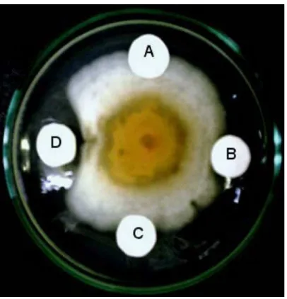
elucidated to be the new iridoid 10-O-vanilloyl-geniposidic acid. Geniposidic acid (2), but not 10-O-vanniloyl geniposidic acid (1), was inhibitory to the mycelial mass development of C. gloeosporio-ides at 150 µg/mL (Figure 1S, supplementary material). In contrast, both compounds were inactive toward A. niger and F. solani. SUPPLEMENTARY MATERIAL
The fungal growth inhibitory assay of compounds 1 and 2, and
1H NMR, 13C NMR, COSY, HMQC, HMBC, IR and EIMS spectra
of compound 1 are available free of charge at http:quimicanova sbq. org.br, as PDF ile.
ACKNOWLEDGMENTS
The authors are grateful to CNPQ/CAPES/PRONEX/FUNCAP for the fellowships and inancial support.
REFERENCES
1. Olea, R. S. G.; Roque, N. F.; Bolzani, V. S.; J. Braz. Chem. Soc.1997, 8, 257; Silva, V. C.; Bolzani, V. S.; Young, M. C. M.; Lopes, M. N.; J. Braz. Chem. Soc.2007, 18, 1405; Brochini, C. B.; Martins, D.; Roque, N. F.; Bolzani, V. S.; Phytochemistry1994, 36, 1293; Young, M. C. M.; Braga, M. R.; Dietrich, S. M. C.; Gottlieb, H. E.; Trevisan, L. M. V.; Phytochemistry 1992, 31, 3433.
2. Luciano, J. H. S.; Lima, M. A. S.; Souza, E. B.; Silveira, E. R.; Biochem. Syst. Ecol. 2004, 32, 1227; Militão, G. C. G.; Pessoa, C.; Costa-Lotufo, L. V.; Moraes, M. E. A.; Moraes, M. O.; Luciano, J. H. S.; Lima, M. A. S.; Silveira, E. R.; Pharm. Biol.2005, 43, 480.
3. Bitar, H. E.; Nguyen, V. H.; Gramain, A.; Sévenet, T.; Bodo, B.; Tetra-hedron Lett. 2004, 45, 515.
4. Tanaka, R.; Matsunaga, S.; Phytochemistry 1992, 31, 3535.
5. Ahmad, V. U.; Rahman, A.; Handbook of Natural Products Data. Pen-tacyclic Ttriterpenoids, Elsevier: New York, 1994.
6. Stevens, J. F.; Wollenweber, E.; Ivancic, M.; Hsu, V. L.; Sundberg, S.; Deinzer, M. L.; Phytochemistry 1999, 51, 771.
7. Bolzani, V. S.; Trevisan, L. M.; Young, M. C. M.; Phytochemistry 1991, 30, 2089.
Quim. Nova, Vol. 33, No. 2, S1-S7, 2010
Material Suplementar
*e-mail: mary@dqoi.ufc.br
ANTIFUNGAL IRIDOIDS, TRITERPENES AND PHENOL COMPOUNDS FROM Alibertia myrciifolia SPRUNGE EX. SCHUM
João Henrique S. Luciano,Mary Anne S. Lima* e Edilberto Rocha Silveira
Departamento de Química Orgânica e Inorgânica, Centro de Ciências, Universidade Federal do Ceará, CP 12200, 60451-970 Fortaleza - CE, Brasil
Ilka Maria Vasconcelos e Georgia Sampaio Fernandes
Departamento de Bioquímica e Biologia Molecular, Universidade Federal do Ceará, 60451-970 Fortaleza - CE, Brasil
Elnatan Bezerra de Souza
Coordenação de Biologia, Universidade do Vale do Acaraú, 62040-370 Sobral - CE, Brasil
Figure 1S. Fungal growth inhibitory assay. (A) 0,1% DMSO as negative control; (B) 150 µg geniposidic acid (2); (C) 112,5 µg 10-O-vanniloyl geniposidic
Luciano et al.
S2 Quim. Nova
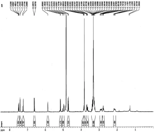
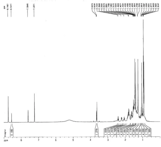
Figure 2S.1H NMR spectrum (300 MHz, CD
3OD) of compound 1
Figure 3S.13C-BB NMR spectrum (75 MHz, CD
Antifungal iridoids, triterpenes and phenol compounds from Alibertia myrciifolia S3
Vol. 33, No. 2
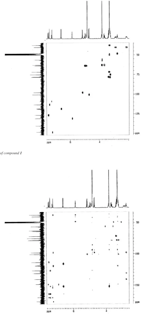
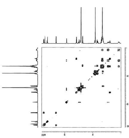
Figure 4S. HMQC spectrum of compound 1
Luciano et al.
S4 Quim. Nova
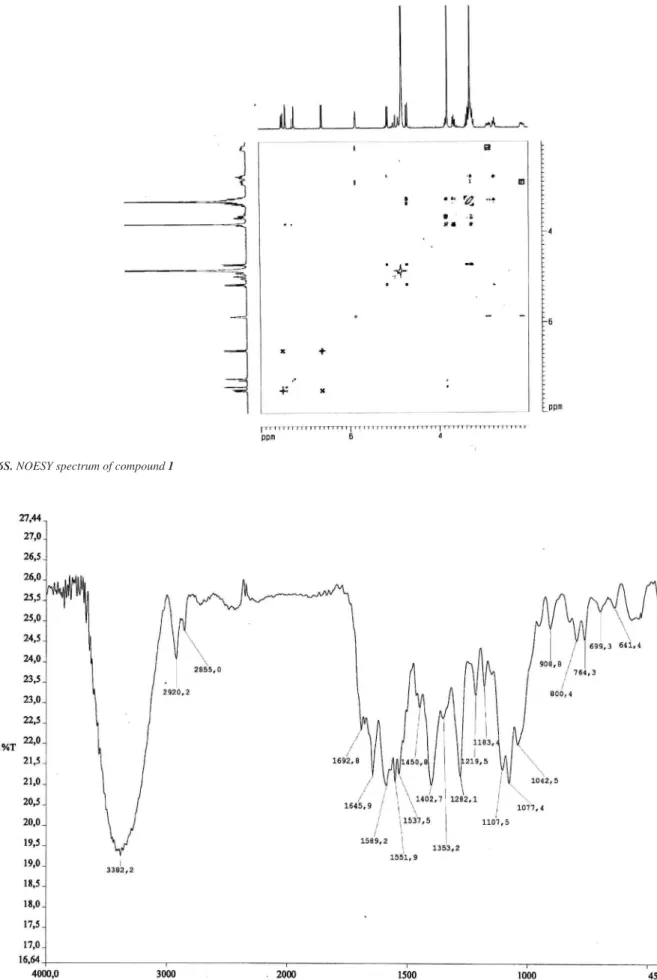
Figure 6S. NOESY spectrum of compound 1
Antifungal iridoids, triterpenes and phenol compounds from Alibertia myrciifolia S5
Vol. 33, No. 2
Figure 8S. Mass spectrum of compound 1 (electronic impact - 70 eV)
Luciano et al.
S6 Quim. Nova
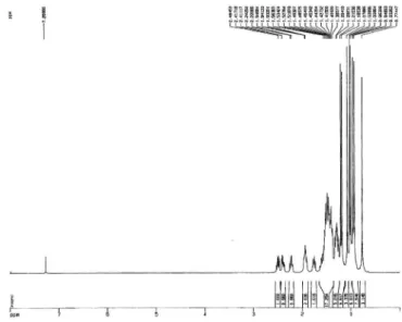
Figure 10S.1H NMR spectrum (500 MHz, CDCl
3) of compound 3
Figure 11S.13C-BB NMR spectrum (125 MHz, CDCl
Antifungal iridoids, triterpenes and phenol compounds from Alibertia myrciifolia S7
Vol. 33, No. 2
Figure 12S.1H NMR spectrum (500 MHz, C
5D5N) of compound 4
Figure 13S.13C-BB NMR spectrum (125 MHz, C