Short Report
Printed in Brazil - ©2017 Sociedade Brasileira de Química0103 - 5053 $6.00+0.00
*e-mail: [email protected]
A New Prenylisoflavone from the Antifungal Extract of Leaves of
Vatairea guianensis
Aubl.
Ronilson F. Souza,*,a Geilson A. da Silva,b Alberto C. Arruda,b Milton N. da Silva,b Alberdan S. Santos,b Daniella P. A. Grisólia,c Moises B. Silva,c Claudio G. Salgadoc and
Mara Silvia P. Arrudab
aDepartamento de Ciências Naturais, Universidade do Estado do Pará,
68860-000 Salvaterra-PA, Brazil
bPrograma de Pós-Graduação em Química, Instituto de Ciências Exatas e Naturais,
Universidade Federal do Pará, 66075-970 Belém-PA, Brazil
cLaboratório de Dermato-Imunologia UEPA/UFPA/MC (Universidade do Estado do Pará/
Universidade Federal do Pará/Centro de Referência e Treinamento em Dermatologia Sanitária do Estado do Pará “Dr. Marcello Candia”), 67200-000 Marituba-PA, Brazil
A new compound, 5,7,3’-trihydroxy-4’-methoxy-8-prenylisoflavone, was isolated from the leaves of Vatairea guianensis Aubl. (Fabaceae), together with two known isoflavones lupiwighteone and 5,7,4’-trihydroxy-3’-methoxy-8-prenylisoflavone. All isolated compounds were characterized based on infrared (IR), UV, 1H and 13C nuclear magnetic resonance (NMR), including 2D NMR
analyses and high resolution mass spectrometry. The ethanolic extract from V. guianensis leaves displayed activity against Candida dubliniensis, C. albicans and C. krusei. However, the EtOAc fraction from that extract exhibited more significant activity than the ethanolic extract, showing antifungal activity for all fungi species investigated. The major compound 5,7,3’-trihydroxy-4’-methoxy-8-prenylisoflavone isolated from that EtOAc fraction was also active against
C. parapsilosis and C. dubliniensis.
Keywords: antifungal activity, Vatairea guianensis, Fabaceae,
5,7,3’-trihydroxy-4’-methoxy-8-prenylisoflavone
Introduction
Many plants from Brazilian biomes such as the Cerrado, Atlantic Forest and Amazon Forest have been used as natural medicines by local communities for treating tropical diseases such as fungal and bacterial infections.1,2 The population makes use of these traditional medicines through medical prescriptions, self-medication, home remedies and other means.3 Among them Vatairea guianensis Aubl. (Fabaceae), a plant native to the Amazon, popularly known as “fava de impingem” (ringworm bean) is used in folk medicine for treating mycoses.4,5
The genus Candida has at least 15 distinct species that cause human disease, such as C. kefyr, C. tropicalis, C. lusitaniae, C. dubliniensis, C. guilliermondii and C. rugosa, but the five most common pathogens are:
C. glabrata, C. tropicalis, C. parapsilosis, C. krusei and the most common and well-studied of the disease-causing species in that genus is C. albicans.6 Candidiasis may present in forms ranging from superficial skin lesions7 to disseminated infections and in recent decades, there has been a substantial increase in the occurrence of invasive fungal infections (IFIs) caused by C. albicans,8 which can naturally colonize the skin, genital, and/or intestinal mucosa in up to 70% of healthy individuals9 and is the most common bloodstream pathogen in the USA, responsible for 50% of the cases.6
azole resistance and of greatest concern is the evolution of multidrug-resistant organisms refractory to several different classes of antifungal agents, especially among common Candida species.11
Given the increasing occurrence of fungal infections in humans along with increasing resistance to available medicines, it is important to identify new antifungal compounds that will enable development of new medicines.12
Previous phytochemical investigations involving various parts of V. guianensis have shown compounds belonging to the anthraquinone, triterpene and isoflavone classes.13-16 The present paper reports the isolation and structural characterization by spectroscopic and spectrometric methods of a new compound, named 5,7,3’-trihydroxy-4’-methoxy-8-prenylisoflavone (3), along with other two known isoflavones lupiwighteone (1) and 5,7,4’-trihydroxy-3’-methoxy-8-prenylisoflavone (2) (Figure 1). The ethanolic extract, hexanic, EtOAc and MeOH/H2O fractions and compounds 1 and 3 were evaluated for their antifungal activities against certain pathogenic fungi but, due to insufficient quantities, the antifungal testing of compound 2
was not carried out.
Experimental
General experimental procedures
The melting point (mp) was determined on a Quimis Q340S23 melting point analyzer. UV spectra were obtained from HPLC equipped with DAD Prominence SPDM-20A
(Shimadzu, Tokyo, Japan). Infrared (IR) spectra were recorded on a Shimadzu Corporation IR Prestige 21 spectrometer (Tokyo, Japan) with KBr pellets. Nuclear magnetic resonance (NMR) spectra, including 1D and 2D experiments (see Supplementary Information), were recorded on a Varian Mercury-300 spectrometer (Palo Alto, CA, USA), operating at 300 MHz at 1H and 75 MHz at 13C, using acetone-d
6, CD3OD or a mixture (CDCl3 and a few drops CD3OD) as deuterosolvents (0.6 mL). Mass spectrometry (MS) analysis was performed on a XEVO G2-SQ-TOF mass spectrometer (Waters Corp., Milford, MA, USA) equipped with a lockspray source where an internal reference compound (leucine-enkephalin) was introduced simultaneously with the analyte for accurate mass measurements. Electrospray mass spectra data were recorded in a positive ionization mode for a mass range from m/z 50 to 1000 with a scan time of 0.1 s. The source temperature was set to 150 °C with a cone gas flow of 20 L h-1. The desolvation gas flow was set to 600 L h-1 at a temperature of 250 °C. The capillary was set at 3.5 kV with cone voltage at 20 V. MassLynx software (Waters, Milford, MA, USA) was used for system control and data acquisition. High-performance liquid chromatography (HPLC) was carried out in a semi-preparative LC-8A Shimadzu system with a SPD-10AV Shimadzu UV detector (Tokyo, Japan), using a Phenomenex Gemini C18 column (250 × 10 mm, 5 µm), isocratic system of 50% acetonitrile-water and a flow rate of 4.7 mL min-1. Detection was performed at 254 and 282 nm. All solvents were filtered through a 0.45 mm nylon membrane filter prior to analysis. Open column chromatography was run using silica gel 60 (70-230 Mesh, Macherey-Nagel, Düren, Germany). Thin layer chromatography (TLC) was performed on precoated silica gel aluminium sheets (Macherey-Nagel, Düren, Germany) by detection with a spraying reagent (vanillin/sulfuric acid/EtOH solution) followed by heating at 100 °C and with NP-PEG reagent (diphenylborinic acid aminoethyl ester-polyethylene glycol) for flavonoid detection.
Plant material
Leaves of V. guianensis were collected in November 2010 in the city of Belém, state of Pará, Brazil. Identification was performed by Manoel R. Cordeiro from Embrapa Amazônia Oriental, Pará, Brazil, and a voucher specimen (IAN - 187050) has been deposited in the herbarium at Embrapa Amazônia Oriental.
Extractions and isolation
Obtaining the ethanol extract from leaves of V. guianensis and fractionating it were carried out according Figure 1. Chemical structures of isoflavones 1-3 isolated from the leaves
to procedures reported by Souza et al.13 Briefly, dried and powdered leaves (1.0 kg) were subjected to extraction with ethanol by maceration at room temperature. The solvent was removed under vacuum, furnishing a residue (180.0 g). The crude ethanol residue (50.0 g) was dissolved in 500 mL MeOH/H2O mixture (9:1), then partitioned three times with hexane (3 × 500 mL), ethyl acetate (3 × 500 mL) and the remaining hydroalcoholic phase. The solutions obtained were dried to provide three fractions: a hexane fraction (7.5 g), an EtOAc fraction (22.0 g) and a remaining MeOH/H2O fraction (20.0 g). The EtOAc fraction (10.0 g) was subjected to silica gel column chromatography with gradient elution of hexane-EtOAc (9:1, 1:1 and 0:10) and EtOAc-MeOH (9:1, 8:2 and 0:10), to obtain six fractions (Fr.1-Fr.6), respectively. The Fr-2 fraction (1.0 g) eluted with hexane:EtOAc (1:1) was sonicated in 4.8 mL of acetonitrile for 1 min. Next, 1.2 mL of H2O was added and sonicated again for 1 min. The solution was subjected to solid phase extraction (SPE) in a C18 cartridge (Phenomenex, 1 g of stationary phase / 6 mL). After evaporation, the residue (about 100 mg) was submitted to semi-preparative reversed phase HPLC (250 × 10 mm Phenomenex Gemini C18, 50% acetonitrile-water, flow rate 4.7 mL min-1, 254 nm) to yield 1 (10 mg), 2 (3 mg) and 3 (9 mg), which showed chromatographic peak retention times of 17.3, 18.8 and 20.0 min, respectively. These compounds were identified by NMR and mass spectrometry methods, and by comparison with available reported data.
5,7,3’-Trihydroxy-4’-methoxy-8-prenyl-isoflavone (3)
Pale yellow solid; soluble in the solvents: acetone, ethyl acetate, methanol, pyridine and DMSO; mp 110-113 °C; UV λ / nm (acetonitrile-water) 239, 265; IR (KBr) νmax / cm−1 3363, 2966, 1656, 1514, 1425, 1273, 1037, 837; 1H and 13C NMR spectral data, see Table 1; HRESITOF-MS
(electrospray ionization-high resolution time-of-flight mass spectrometry) m/z 369.1352 [M + H]+ (calcd. for [M + H]+, C21H20O6 + H+, 369.1338).
In vitro antifungal activity
The minimal inhibitory concentrations (MICs) were determined by broth microdilution methods based on the Clinical and Laboratory Standards Institute (CLSI) reference protocol M38-A2.17 All the test strains were subcultured on Sabouraud dextrose agar (SDA) (Sigma-Aldrich, Saint Louis, MO, USA), incubated for 24-72 hours at temperature 30 °C, and their inocula were prepared according to procedures reported by Daboit et al.18 That involved scraping across the surface of the fungal colonies with a sterile pipette and sterile saline solution (0.85%),
containing 0.05% Tween 40. After standing for 3-5 min, at room temperature for deposition of larger particles, the concentration of spores in the supernatant was adjusted spectrophotometrically (530 nm) to a per cent transmission in the 80-82 range for Candida albicans (INGOS 40175), Candida krusei (ATCC 6258), Candida parapsilosis (ATCC 22019) and Candida dubliniensis (CBS 7987). These suspensions were diluted to 1:50 in a synthetic RPMI-1640 medium buffered with morpholinopropanisulfonic acid (MOPS; Sigma®, Saint Louis, MO, USA).
The extracts, fractions and pure compounds were primarily dissolved in dimethyl sulfoxide (DMSO) and diluted in sodium bicarbonate-free RPMI 1640 medium (Sigma®, Saint Louis, MO, USA) buffered with
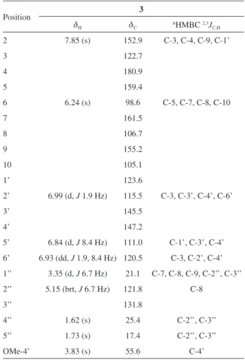
165 mmol L-1 MOPS, pH 7.0, and supplemented with 4 mmol L-1 L-glutamine. The final DMSO concentration was maintained as less than 1%. Concentrations ranged Table 1. The 1H and 13C NMR chemical shifts (d
Η in ppm) and HMBC
correlations of compound 3 in CDCl3 and a few drops of CD3ODa
Position 3
dH dC bHMBC 2,3JC,H
2 7.85 (s) 152.9 C-3, C-4, C-9, C-1’
3 122.7
4 180.9
5 159.4
6 6.24 (s) 98.6 C-5, C-7, C-8, C-10
7 161.5
8 106.7
9 155.2
10 105.1
1’ 123.6
2’ 6.99 (d, J 1.9 Hz) 115.5 C-3, C-3’, C-4’, C-6’
3’ 145.5
4’ 147.2
5’ 6.84 (d, J 8.4 Hz) 111.0 C-1’, C-3’, C-4’ 6’ 6.93 (dd, J 1.9, 8.4 Hz) 120.5 C-3, C-2’, C-4’ 1’’ 3.35 (d, J 6.7 Hz) 21.1 C-7, C-8, C-9, C-2’’, C-3’’ 2’’ 5.15 (brt, J 6.7 Hz) 121.8 C-8
3’’ 131.8
4’’ 1.62 (s) 25.4 C-2’’, C-3’’
5’’ 1.73 (s) 17.4 C-2’’, C-3’’
OMe-4’ 3.83 (s) 55.6 C-4’
from 1024 to 0.125 µg mL-1 for extracts and fractions; for pure compounds they ranged from 256 to 0.125 µg mL-1, and the assay was carried out as reported previously.19 The MIC was determined as the lowest concentration of the crude extract, fractions and the pure compound that inhibited the complete growth of the organisms.
Minimum fungicidal concentrations (MFCs) were established following the incubation time determined for the MIC. Thirty µL from each well with complete growth inhibition were inoculated onto SDA plates and incubated at 30 °C for up to 72 h. The MFC was defined as the lowest concentration of the extracts, fractions and pure compounds, in which there was either no growth or a growth up to seven colonies, which corresponds to a 99.9% kill;20 fluconazole was used as the standard antibiotic. All experiments were conducted in triplicate.
Results and Discussion
Compound 1 was identified as lupiwighteone by comparing its spectral data with those reported in literature.21,22 This substance is being reported for the first time for the genus Vatairea and presents important anti-inflammatory activity according to Paoletti et al.,23 as well as being considered a very potent phytoestrogen.24
Compound 2 was identified as 5,7,4’-trihydroxy-3’-methoxy-8-prenylisoflavone. This substance has been reported only once for the plant Wyethia mollis;25 however, the data from HRESITOF-MS, 1H and 13C NMR are being reported for the first time in this paper.
Compound 3 was obtained as a pale yellow solid, with the molecular formula C21H20O6, based on the [M + H]+ peak at m/z 369.1352 (calcd. for [M + H]+, C
21H20O6 + H+, 369.1338) in the HRESITOF-MS, and confirmed by 1H and 13C NMR experiments (Table 1). The 1H NMR signal at dH-2 7.85 and 13C NMR signals at dC-2 152.9, dC-3 122.7 and dC-4 180.9, were typical of isoflavones.26 Additionally,
the 1H NMR spectrum exhibited signals in the aromatic region at dH 6.84 (1H, d, J 8.4 Hz), 6.93 (1H, dd, J 1.9 and 8.4 Hz) and 6.99 (1H, d, J 1.9 Hz), which indicated an ABX spin system of a 1,3,4-trisubstituted phenyl group, as well as one singlet at dH 6.24 assigned to a pentasubstituted benzene ring. The signal singlet at dH 3.83 indicated the presence of an OMe group connected to an aromatic ring. All couplings were confirmed through analysis of the 1H-1H correlation spectroscopy (COSY) spectrum. Besides the signals related to C-ring carbons, the 13C NMR spectrum of 3 exhibited another 21 signals attributed to twenty-one carbons with the aid of the heteronuclear correlation spectroscopy (HETCOR) and heteronuclear multiple bond correlation (HMBC) experiments.
The C-prenyl group [dH 3.35 (d, J 6.7 Hz, H-1’’), 5.15 (brt, J 6.7 Hz, H-2’’), 1.62 (s, Me-4’’) and 1.73 (s, Me-5’’)] was confirmed as attached at C-8 by the 3J
C,H correlations in the HMBC experiments (Table 1) between the signals at dH 7.85 (H-2) and 3.35 (H-1’’) with the signal at dC 155.2 (C-9). The location of the OH and OMe groups at C-3’ and C-4’ of the B-ring, respectively, was sustained by combining the HMBC cross-peaks from H-6’ (dH 6.93) and OMe (dH 3.83) to C-4’ (dC 147.2) with the nuclear Overhauser effect (NOE) observed in the NOE difference spectra, which revealed spatial interactions between H-5’ and OMe-4’. Therefore, 3 was characterized as 5,7,3’-trihydroxy-4’-methoxy-8-prenylisoflavone. This compound is a new natural product.
In evaluating the inhibition of fungal growth by V. guianensis, the extracts that presented MICs ≤ 800 µg mL-1 and the pure compounds with MICs ≤ 250 µg mL-1 were defined as active, these values being based on the work of Stein et al.27 The results of the MICs obtained in this study are shown in Table 2.
The most significant result of the minimum inhibitory concentration for the EtOH extract from leaves was 32 µg mL-1 for C. dubliniensis. For the strains of fungi
Table 2. Minimal inhibitory concentration (MIC) and minimum fungicidal concentration (MFC) of crude extract, fractions and constituents isolated from
V. guianensis against pathogenic microorganisms
Sample MIC (MFC) / (µg mL
-1)
C. parapsilosis C. albicans C. krusei C. dubliniensis
EtOH extract > 1024 (−) 128 (512) 128 (−) 32 (1024)
Hexanic fraction 64 (−) 1024 (1024) 512 (512) 64 (−)
EtOAc fraction 8 (32) 16 (32) 8 (32) 8 (16)
MeOH/H2O fraction > 1024 (−) 1024 (−) 1024 (−) 1024 (−)
1 > 256 (−) > 256 (−) > 256 (−) 32 (−)
3 32 (−) > 256 (−) > 256 (−) 8 (−)
Fluconazole 64 (−) − 64 (−) 2 (−)
C. albicans and C. krusei the EtOH extract was also active showing MIC of 128 µg mL-1, although it was inactive for C. parapsilosis (MIC > 1024 µg mL-1) (Table 2).
The EtOAc fraction showed promising fungicide activity against all fungi, with MIC values in the range of 8-16 µg mL-1 and MFC with values in the range of 16-32 µg mL-1 (Table 2). The major compound (3) showed fungistatic activity against C. parapsilosis (MIC = 32 µg mL-1) and a more significant result against C. dubliniensis (MIC = 8 µg mL-1), whereas the compound lupiwighteone (1) was active only against C. dubliniensis (MIC = 32 µg mL-1).
Conclusions
Phytochemical investigation of the antifungal fraction (EtOAc) from V. guianensis leaves resulted in the isolation of a new isoflavone (3) besides other two known isoflavones. These compounds are in agreement with the typical chemical profile of plants of the Vatairea genus and Fabaceae family. The results of evaluating the antifungal activity of V. guianensis indicate the potential of this species, but it is necessary to expand the studies to provide scientific support to popular use of this plant in the treatment of skin infections caused by fungi.
Supplementary Information
Supplementary information, including 1H and 13C NMR, DEPT, COSY, HETCOR, and HMBC spectra, as well as mass spectra (Figures S1-S9), is available free of charge at http://jbcs.sbq.org.br as a PDF file.
Acknowledgments
The authors are grateful to Coordenação de Aperfeiçoamento de Pessoal de Nível Superior (CAPES), to CAPES PROAMAZONIA, and to Conselho Nacional de Desenvolvimento Científico e Tecnológico (CNPq) for financial support.
References
1. Alves, T. M. A.; Silva, A. F.; Brandão, M.; Grandi, T. S. M.; Smânia, E. F.; Smânia Jr., A.; Zani, C. L.; Mem. Inst. Oswaldo Cruz 2000, 9, 367.
2. Duarte, M. C. T.; Figueira, G. M.; Sartoratto, A.; Rehder, V. L. G.; Delarmelina, C.; J. Ethnopharmacol. 2005, 97, 305. 3. Robinson, M. M.; Zhang, X.; http://www.who.int/medicines/
areas/policy/world_medicines_situation/en/, accessed in August 2016.
4. Lima, H. C.; Arq.Jard. Bot. Rio de Janeiro 1982, 26, 173.
5. Santos, J. F. L.; Pagani, E.; Ramos, J.; Rodrigues, E.;
J. Ethnopharmacol. 2012, 142, 503.
6. McCarty, T. P.; Pappas, P. G.; Infect. Dis. Clin. North Am. 2016,
30, 124.
7. Kashem, S. W.; Kaplan, D. H.; Trends Immunol. 2016, 37, 440. 8. Garey, K. W.; Aitken, S. L.; Dima-Ala, A.; Beyda, N. D.; Kuper,
K.; Xie, Y.; Koo, H. L.; Am. J. Med. Sci. 2015, 349, 320. 9. Perlroth, J.; Choi, B.; Spellberg, B.; Med. Mycol. 2007, 45, 346. 10. Miyazaki, T.; Kohno, S.; Expert Rev. Anti-Infect. Ther. 2015,
13, 1183.
11. Cowen, L. E.; Sanglard, D.; Howard, S. J.; Rogers, P. D.; Perlin, D. S.; Cold Spring Harb. Perspect. Med. 2015, 5, a019752. 12. Mahlo, S. M.; McGaw, L. J.; Eloff, J. N.; Crop. Prot. 2010, 29,
1529.
13. Souza, R. F.; Marinho, V. H. S.; Silva, G. A.; Costa-Jr, L. M.; Silva, J. K. R.; Bastos, G. N. T.; Arruda, A. C.; Silva, M. N.; Arruda, M. S. P.; J. Braz. Chem. Soc. 2013, 24, 1857. 14. Souza, R. F.; Silva, J. K. R.; Silva, G. A.; Arruda, A. C.; Silva,
M. N.; Arruda, M. S. P.; Rev. Virtual Quim. 2015, 7, 1893. 15. Piedade, L. R.; Wolter Filho, W.; Acta Amaz. 1988, 18, 185. 16. Ottobelli, I.; Facundo, V. A.; Zuliani, J.; Luz, C. C.; Brasil, H. O.
B.; Militão, J. S. L. T.; Braz-Filho, R.; Acta Amaz. 2011, 3, 393. 17. CLSI Document M38-A2; Reference Method for Broth Dilution Antifungal Susceptibility Testing of Filamentous Fungi. Approved Standard, 3rd ed.; Clinical and Laboratory Standards
Institute: Wayne, Pennsylvania, USA, 2008.
18. Daboit, T. C.; Magagnin, C. M.; Heidrich, D.; Antochevis, L. C.; Vigolo, S.; Meirelles, L. C.; Alves, K.; Scroferneker, M. L.;
Mycoses 2014, 57, 116.
19. Mahlo, S. M.; McGaw, L. J.; Eloff, J. N.; J. Ethnopharmacol.
2013, 148, 909.
20. Pfaller, M. A.; Sheehan, D. J.; Rex, J. H.; Clin. Microbiol. Rev.
2004, 17, 280.
21. Al-Maharik, N.; Botting, N. P.; Tetrahedron 2003, 59, 4177. 22. Hashidoko, Y.; Tahara, S.; Mizutani, J.; Agric. Biol. Chem. 1986,
50, 1797.
23. Paoletti, T.; Fallarini, S.; Gugliesi, F.; Minassi, A.; Appendino, G.; Lombardi, G.; Eur. J. Pharmacol. 2009, 620, 120. 24. Wätjen, W.; Weber, N.; Lou, Y. J.; Wang, Z. Q.; Chovolou, Y.;
Kampkötter, A.; Kahl, R.; Proksch, P.; Food Chem. Toxicol.
2007, 45, 119.
25. Bohm, B. A.; Choy, J. B.; Lee, A. Y. M.; Phytochemistry1989,
28, 501.
26. Bandeira, P. N.; Farias, S. S.; Lemos, T. L. G.; Braz-Filho, R.; Santos, H. S.; Albuquerque, M. R. J. R.; Costa, S. M. O.; J. Braz. Chem. Soc. 2011, 22, 372.
27. Stein, A. C.; Alvarez, S.; Avancini, C.; Zacchino, S.; Poser, G. V.; J. Ethnopharmacol.2006, 107, 95.