Artigo
*e-mail: [email protected]
INTERACTIONS OF CHITOSAN/GENIPIN HYDROGELS DURING DRUG DELIVERY: A QSPR APPROACH
Nancy L. Delgadillo-Armendariza, Norma A. Rangel-Vazqueza,*, Edgar A. Marquez-Brazonb,c and Blanca Rojas-De Gascueb
aDivisión de Estudios de Posgrado e Investigación, Instituto Tecnológico de Aguascalientes, Ave. López Mateos #1801 Ote. Col. Fracc. Bona Gens CP 20256, Aguascalientes, Ags. México
bUniversidad de Oriente, Instituto de Investigaciones en Biomedicina y Ciencias Aplicadas, “Dra. Susan Tai”, IIBCA-UDO, Apdo. Postal 245, Cumaná, Estado Sucre, Venezuela
cUniversidad de Oriente, Departamento de Química, Núcleo de Sucre, Apdo. Postal 245, Cumaná, Estado Sucre, Venezuela
Recebido em 10/04/2014; aceito em 27/06/2014; publicado na web em 05/09/2014
A hydrogel comprised of chitosan crosslinked using the low-toxicity crosslinker genipin was prepared, and the absorption of glibenclamide by the hydrogel was investigated. Optimized structures and their molecular electrostatic potentials were calculated using the AM1 method, and the results were used to evaluate the molecular interactions between the three compounds. The quantitative structure-property relationship model was also used to estimate the activity of the chemicals on the basis their molecular structures. In addition, theoretical Fourier transform infrared spectra were calculated to analyze the intermolecular interactions in the proposed system. Finally, the hydrophilicity of the hydrogel and its influence on the absorption process were also estimated.
Keywords:chitosan; genipin; glibenclamide; FTIR; QSPR modeling.
INTRODUCTION
The use of natural polymers (e.g., gelatin, chitosan, and silk proteins) for the development of hydrogels is gaining in importance owing to their inherent biocompatibility.1 Hydrogels are high-water content materials prepared from crosslinked polymers that can provide sustained, local delivery of a variety of therapeutic agents. Use of the natural polymer chitosan as a scaffold material for hydrogels has been intensively investigated because of the polymer’s biocompatibility, low toxicity, and biodegradability. Therefore, the advanced develop-ment of chitosan hydrogels has led to new drug delivery systems that release their payloads in response to varying environmental stimuli.2,3 Chitosan (see Figure 1), the product of the N-deacetylation of chitin, has also received particular attention because it is produced as a waste product during crustacean (shrimp and crab) processing.4-8 In addition, it offers other advantages; chitosan can not only be used to control the release of active agents but also be prepared without the use of hazardous organic solvents because it is soluble in aqueous acidic solutions. Given the above-mentioned properties, chitosan is extensively used for the development of drug delivery systems.
Chitosan has been effectively used in drug delivery applications as hydrogel systems, drug conjugates, biodegradable release syste-ms, and other components.9,10 Chitosan-based systems are used for the delivery of proteins/peptides, growth factors, anti-inflammatory
drugs, and antibiotics, as well as, in gene therapy and bioimaging applications.9,11,12
The crosslinking of chitosan is considered a relevant approach for improving control of drug delivery. Among other reagents, glu-taraldehyde and tripolyphosphate have been widely used to crosslink this biodegradable polymer. However, there are concerns over the toxicity of most of the investigated crosslinking agents, especially glutaraldehyde, which may reduce the biocompatibility of chitosan delivery systems. Therefore, there is a need for the development of crosslinking agents with low cytotoxicity.1,13
Genipin, which is a natural, water-soluble, and bifunctional cross-linker 14 (Figure 2) obtained from the fruits of Gardenia jasminoides1, was selected for the study as a possible biocompatiblecrosslinker for chitosan. Genipin is an excellent natural crosslinker for proteins, collagen, gelatin, and chitosan. It has a low acute toxicity with an in vivo LD50 of 382 mg/kg in mice and is 10,000 times less toxic than glutaraldehyde and degrades more slowly than glutaraldehyde and many other commonly used synthetic crosslinking reagents. Furthermore, genipin is also used as a regulating agent for drug delivery, as the raw material for gardenia blue pigment preparation, and as an intermediate for alkaloid synthesis.1,15-21
Glibenclamide (see Figure 3), also known as glyburide, is an antidiabetic drug in the class of medications known as sulfonylureas, which are closely related to sulfa drugs. Sulfonylureas bind to ATP-dependent K+ channels in beta cells of the pancreas, depolarizing them and stimulating the release of Ca2+, which in turn stimulates insulin production.22,23
Figure 1. Chitosan structure (red: oxygen, white: hydrogen, blue: carbon and purple: nitrogen atoms)
In this study, Fourier transform infrared (FTIR) spectroscopy was used to analyze the intermolecular interactions in the proposed system. As one of the ways to verify the obtained results, we apply the theoretical calculations by means of the computational chemistry tools.24 Using the quantitative structure-property relationship (QSPR) model/method, we estimate the activity of the chemicals on the basis of only their molecular structure. Sorption of organic chemicals in soils or sediments is usually described by sorption coefficients.25
In addition, the log P value of the hydrogel was determined as a measure of its hydrophobicity.26 The log P value of a com-pound, which is the logarithm of its partition coefficient between n-octanol and water (coctanol/cwater), is a well-established measure of the compound’s hydrophilicity. Low hydrophilicities, and therefore high log P values, cause poor absorption or permeation. It has been shown that compounds must have a log P value less than 5 in order to have a reasonable probability of being well absorbed.27 Typically, the log P value of a substance at pH 7.4 is considered as an index of the compound behavior in plasma.28
Finally, the molecular electrostatic potential (MESP) was inves-tigated using the Austin Method 1 (AM1) calculations. This method provides information about the regions in which intermolecular inte-ractions occur between compounds.29 The electrostatic potential is the energy of the interaction of a point positive charge (an electrophile) with the nuclei and electrons of a molecule. Negative electrostatic potentials indicate areas that are prone to electrophilic attack. The electrostatic potential can be mapped onto the electron density by using color to represent the value of the potential.30
EXPERIMENTAL
The AM1 method was initially used to optimize the structures of the compounds investigated in the present study because it generates lower-energy structures, even when the initial structures are far away from the minimum structures. The Polak-Ribiere algorithm was used for mapping the energy barriers of the conformational transitions. For each structure, 5715 iterations, a level convergence of 0.001 kcal/ mol/Å, and a line search of 0.1 were performed.
Structural parameters
The optimized structural parameters were used for the vibrational wavenumber calculations with AM1 method in order to characterize all the stationary points as minima. The structural parameters were calculated by selecting the Constrain bond and length options from the Build menu for the two methods of analysis.
FTIR
An infrared spectrum is commonly obtained by passing infrared electromagnetic radiation through a sample that possesses a perma-nent or induced dipole moment and determining what fractions of the incident radiation are absorbed at particular energies. The energy
of each peak in an absorption spectrum corresponds to the frequency of the vibration of part of the molecule, thus allowing qualitative identification of certain bond types in the sample.
The FTIR spectra were obtained by first selecting from the Compute menu, vibrational and rotational spectrum options; after this step, using the vibrational spectrum option, FTIR spectrum pattern is obtained for the two methods of analysis. The results of the analyses for the optimized structures for chitosan, genipin, and glibenclamide obtained using the AM1 method are listed in Tables 1–3, respectively.
Figure 3. Glibenclamide structure (red: oxygen, white: hydrogen, blue: carbon, yellow: sulfur and purple: nitrogen atoms)
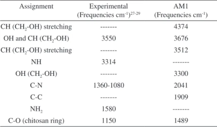
Table 1. FTIR results of chitosan
Assignment Experimental (Frequencies cm-1)27-29
AM1 (Frequencies cm-1)
CH (CH2-OH) stretching --- 4374
OH and CH (CH2-OH) 3550 3676 CH (CH2-OH) stretching --- 3512
NH 3314
---OH (CH2-OH) --- 3300
C-N 1360-1080 2041
C-C --- 1909
NH2 1580
---C-O (chitosan ring) 1150 1489
Table 2. FTIR results of genipin
Assignment Experimental (Frequencies cm-1)30-32
AM1 (Frequencies cm-1)
OH stretching --- 6052 CH2 asymmetric
stretching
--- 4694, 4530
CH stretching (ring) 3745 4091, 3885
C=C 3398, 3245 3354
CH2 (scissoring) 3100 3039
C–C, C–O 2520 2680, 2571
C–C, C–O, CH 1681, 1622,1105 1663, 1038
CH3, CH2 1443 1405
C–O–C 830, 835 818
C–O–C (out of plane) 773 755
Table 3. FTIR results of glibenclamide
Assignment Experimental (Frequencies cm-1)33-35
AM1 (Frequencies cm-1)
NH asymmetric stretching
--- 5583
CH asymmetric stretching
--- 5007
CH2 stretching --- 4511
CH3 (O-CH3) 3591 4028
NH (amide) 3367, 3311, 1713 3314
CH=CH 3035 3529
C=C (ring) 1591 2830
C=C, S=O 2412 2474
C=O 1652, 1618 1720, 1715
S=O2 1341-1332, 1158 1316 C–C, C–N, C–O 1995, 1334, 1154, 1090,
1018, 924, 793
Electrostatic potential
After obtaining the Gibbs free energy for the optimized geome-tries using the AM1 method, two-dimensional contour diagrams of the electrostatic potentials surrounding the three molecules, their total electron densities and spin densities, their molecular orbitals, and the electron densities of the individual orbitals were plotted.
HyperChem software displays the electrostatic potential as a contour plot when the appropriate option in the Contour Plot dialog box is selected. Atomic charges indicate where large negative values (sites for electrophilic attack) are likely to occur. However, the largest negative value for the electrostatic potential is not necessarily adjacent to the atom with the largest negative charge.31
RESULTS AND DISCUSSION
Structural parameters
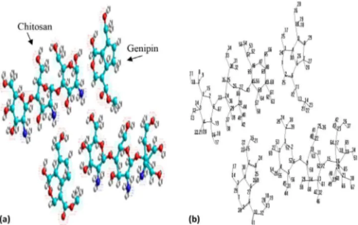
The thermodynamic data obtained are listed in Table 4. The Gibbs free energy value (−947.998 kcal/mol) indicates that the crosslinking reaction involves crosslinking of two free amino groups in chitosan with one molecule of genipin (see Figure 4).17,18 Nucleophilic attack by the primary amine occurs at the carbon atom in genipin, while the secondary amine attacks the aldehyde group.21
QSPR methodology is one of the most powerful tools for descri-bing the relationship between the biological activity and physicoche-mical characteristics of a molecule. Diffusion is also influenced by the nature of the crosslinker. In addition, the porosity, water uptake, and swelling of hydrogels are influenced by the chemical nature and length of the bridging units of the crosslinker.
Swelling and diffusion in hydrogels are also influenced by the hydrophilicity of the crosslinked polymer.32 Therefore, the Log P value for chitosan/genipin was calculated and found to be −22.15. This value represents the hydrophilic property of the substance and is
considered to be an indicator of the ability of the hydrogel to absorb molecules.33 In hydrogels formed by chitosan crosslinked with itself, release is largely controlled by the crosslinking density; consequently, the higher the crosslinking density, the lower the release rate.
However, other system parameters, such as drug concentration, often play a major role. To our knowledge, there are no examples of hydrogels formed by chitosan crosslinked with itself that exhibit pH-sensitive swelling.
Indeed, the numerous inter-chain interactions formed by cross-linking inhibit swelling because most of the amino groups of chitosan react with the crosslinker. Such systems do not present a release profile that can be further modulated after administration; for example, drug release cannot be targeted in the gastro-intestinal tract, which limits their range of application.5,32
Table 4 shows the thermodynamic results of glibenclamide absorption process; here the Gibbs free energy value −243.08 kcal/ mol is spontaneous because of the formation of hydrogen bonds between the carbonyl groups of glibenclamide and NH groups of chitosan. Nucleophilic attack of the amino group of chitosan at the carbonyl of the genipin leads to formation of a stable bond (amide), and an oxygen atom in genipin is replaced by a nitrogen atom from chitosan (see Figure 5).34
Figure 5 shows that the title system (chitosan-genipin/gliben-clamide) is not planar, and based on observations, deformation of the ring structure depends on the nature of the substituents (OH, NH2, and CH2-CH2). Figure 5 shows the results of the computational analysis to determine the optimized geometries for chitosan/genipin and chitosan (genipin)/glibenclamide. Their molecular structures as well as the numbering of their atoms are also shown in the figure.
The optimized structural parameters for chitosan, chitosan crosslinked with genipin, and the chitosan/genipin hydrogel with absorbed glibenclamide are listed in Tables 5–7, respectively, in ac-cordance with the atom numbering scheme given in Figures 4and 5. The title system (chitosan-genipin/glibenclamide) is not planar, and based on observations, deformation of the ring structure depends on the nature of the substituents (OH, NH2, and CH2-CH2). Therefore, we could compare the calculation results given in Tables 5–7 with experimental data. As discussed in the previous literature, several authors have explained the changes in frequency or bond length of the C–H bond upon substitution due to a change in the charge distribution on the carbon atom of the ring.31 The substituents may either be electron withdrawing (Cl, Br, F) or electron donating (CH3, C2H5).
The carbon atoms are bonded to hydrogen atoms via σ bonds, and substitution of a hydrogen atom for a halogen atom on the benzene Figure 4. Structure of chitosan/genipin using computational chemistry
Table 4. QSPR properties results
Property Chitosan/Genipin Chitosan/Genipin-Glibenclamide ∆G (kcal/mol) - 947.998 - 243.08
Mass (amu) 1455.43 2442.44
Surface area (Å2) 1798.11 2910.84
Volume (Å3) 3507.66 5860.58
Log P - 22.15 - 46.33
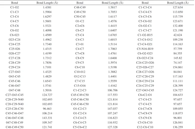
Table 5. Structural parameters calculated for chitosan crosslinking with genipin using AM1 method
Bond Bond Length (Å) Bond Bond Length (Å) Bond Bond Length (Å)
C1-O2 1.4381 C48-C49 1.5817 C1-C3-C4 127.634
C1-C3 1.5904 C49-C50 1.7053 C3-C4-C5 113.850
C3-C4 1.6297 C50-C45 1.6117 C4-C5-C6 115.739
C4-C5 1.5601 O1-C2 1.4576 C5-C6-O2 123.671
C5-C6 1.5321 C2=C6 1.4415 C6-O2-C1 132.400
C6-O2 1.4098 C6-C5 1.6407 C1-C3-C7 121.190
C6-023 1.4589 C5-C4 1.6765 C1-O2-H15 42.624
O23-C24 1.4962 C4-C3 1.6015 C3-C4-O12 109.238
C24-C25 1.7340 C3-01 1.5114 C3-C4-O21 114.163
C25-026 1.4315 C4-C7 1.7063 C5-N16-H19 57.759
O26-C27 1.4199 C7=C8 1.4761 C6-O2-O23 84.355
C27-C28 1.7312 C8-C9 1.6468 C6-O23-C24 145.82
C28-C29 1.7856 C9-C5 1.5974 C24-C25-O26 74.147
C29-C24 1.7101 C6-C10 1.5198 C25-026-C27 154.861
C27-O43 1.4325 C10-012 1.3882 C28-C27-O26 84.041
O43-C45 1.4829 O12-C13 1.4401 C27-C28-C29 117.163
C45-C46 1.6543 C7-C15 1.5559 C28-C29-C24 111.111
C46-O47 1.5741 C15-O16 1.4162 C24-C25-C29 128.399
O47-C48 1.5016 C1-C2-C3 106.706 C27-O43-C45 124.727
C27-O43-C45 124.727 C45-C49-C50 117.553 C6=C2-O1 104.633
C28-C29-N40 102.693 C45-C46-C50 121.814 C7-C4-C5 97.657
C28-C29-N40 102.693 C45-C46-C50 121.814 C7-C4-C5 97.657
C25-C26-C30 96.463 O1-C2-C3 147.037 C4-C7=C8 109.055
C45-C46-O47 106.214 O1-C2-C4 107.247 C7=C8-C9 118.073
C46-O47-C48 143.331 C3-C4-C5 116.823 C5-C9-C8 96.801
047-C48-C49 109.347 C6-C4-C5 116.932 C5-C6-C10 126.041
C48-C49-C50 121.741 C5-C6=C2 127.328 C12-C6-C10 136.259
Table 6. Bond length calculated for chitosan (crosslinking with genipin)/
glibenclamide using AM1 method Bond Bond
Length (Å) Bond
Bond
Length (Å) Bond
Bond Length (Å) C1-C3 1.5904 C4-C3 1.6290 O26-C27 1.4198 C1-O2 1.4392 C6-O23 1.4606 C27-C28 1.7307 O2-C6 1.4106 O23-C24 1.4979 C28-C29 1.7857 C6-C5 1.5326 C24-C25 1.7343 C29-C24 1.7105 C5-C4 1.5599 C25-O26 1.4315 C27-043 1.4325 043-C45 1.4828 C10-N12 1.3458 C6-C10 1.5195 C45-C46 1.6548 N12-C14 1.4551 C7-C15 1.5556 C46-O47 1.5737 C14-C15 1.6129 C31-C32 1.6139 O47-C48 1.5008 C15-C16 1.5797 C32-C33 1.5749 C48-C49 1.5816 C16-C17 1.5147 C33-C34 1.6242 C49-C50 1.7053 C17=C21 1.3415 C34-C29 1.7610 C50-C45 1.6717 C21-C20 1.4668 C26-O27 1.2279 O1-C3 1.5116 C20=C19 1.3411 S22-O23 1.5798 C3-C4 1.6016 C19-C18 1.4664 S22-O24 1.5794 C4-C5 1.6762 C18=C16 1.3563 C10-O11 1.2316 C5-C6 1.6404 C20-S22 1.7650 N28-C29 1.4657 C6=C2 1.4415 S22-N25 1.7177 C29-C30 1.5879 C2-O1 1.4579 N25-C26 1.3478 C30-C31 1.6051 C4-C7 1.7066 C26-N28 1.3436 C9-C5 1.5969 C7=C8 1.4762 C8-C9 1.6465
ring reduces the electronic density at the carbon atom owing to an induction effect. The ring carbon atoms exhibit a larger attraction for the valence electron cloud of the remaining hydrogen atoms, resulting
in an increase in the C–H force constant and a decrease in the cor-responding bond lengths. The reverse holds true for substitution with electron donating groups.
The actual change in the C–H bond lengths is influenced by the combined effects of the inductive–mesmeric interaction and the electric dipole field of the polar substituent. The calculated geomet-ric parameters can be used as the foundation for calculating other parameters for the compound.35
FTIR
The calculated FTIR results for chitosan/genipin and chitosan (crosslinked with genipin)/glibenclamide are presented in Table 8. The second column indicates the absorption bands at 2320 and 1097 cm−1, which clearly appeared after crosslinking with genipin. The peak at 1097 cm−1 represents the C–N stretching vibration for the tertiary nitrogen atom formed from the reaction of the lysine with genipin, while the band at 2320 cm−1 was assigned to C–C, C–H, C–O, and C–N bonds that formed following crosslinking. Moreover, the adsorption band that appeared at 1236 cm−1 (amide III) represents a mixed CO–N/N–H vibration mode, and the peak at 813 cm−1 is a characteristic absorption band for the C–H stretching vibration associated with the heterocyclic ring of the product.
is indicated by the growth of the absorption band at 1630 cm−1 in the FTIR spectrum of the hydrogel.
As a consequence of these two main reactions, the intensity of the absorption band for the amino groups at 3597 cm−1 is drastically reduced in the spectrum of the hydrogel.36-40
The third column in Table 8 lists the absorption bands that appe-ared after the absorption of glibenclamide by the chitosan/genipin crosslinked hydrogel. These peaks include a characteristic amide band at 3407 cm−1 and peaks for SO and SO
2 stretching vibrations at 2683 and 1369 cm−1, respectively. The site of interaction on glibenclamide is likely the C=O group, which would also affect the NH vibration.38
The absorption peaks observed at 2412 and 793 cm−1 are associated with C−C, CH, C−O, and OH bending vibrations, while the absorption bands at 1955, 1334, 1090, and 924 cm−1 are due to the C−C and C−O bonds in the chitosan crosslinked with genipin.41 The new broad peak that appeared near 1415 cm−1 after crosslinking with genipin is asso-ciated with the ring-stretching vibrations of the heterocyclic amine. In addition, the C–N stretching of amide III at 1233 cm−1 shifted to 1260 cm−1 after crosslinking with genipin, while the peak at 1740 cm−1, which corresponds to the carboxylic groups in the electrospun chitosan fibers, disappeared in the genipin-crosslinked chitosan.39
Electrostatic potential
The MESP values (Figure 6) ranged from −0.075 to 0.607 eV and 0.065 to 0.718 eV for the crosslinking of chitosan with genipin and glibenclamide absorption by the crosslinked hydrogel, respectively.
Table 7. Angle calculated for chitosan (crosslinking with genipin)/glibenclamide using AM1 method
Bond Angle (°) Bond Angle (°) Bond Angle (°)
C1-C3-C4 127.626 C45-O47-C46 106.193 C6-C10-O12 136.273
C5-C3-C4 113.831 C45-C51-C46 135.376 C10-O12-C13 146.014
C6-C5-C4 115.859 C45-O47-C48 143.381 C9-C5-C6 124.608
O2-C5-C6 123.559 C49-O47-C48 109.344 C1=C2-C6 119.726
C1-O2-C6 132.357 C49-C50-C48 121.732 C2-C6=C5 119.633
C1-C3-O2 106.768 C49-C50-C45 117.545 C6=C5-C4 121.828
C1-C3-C7 121.067 C46-C50-C45 121.806 C5-C4=C3 118.351
O12-C3-C4 109.22 C50-C49-N61 106.1840 C4=C3-C1 119.103
O21-C3-C4 114.184 O1-C3-C4 107.2720 C1-C3-O8 121.394
C5-C6-N16 147.176 C3-C4-C5 116.783 C3-O8-C9 133.702
C6-O2-O23 84.6643 C4-C5-C6 116.972 C5-C4-C10 117.572
C24-O23-C25 109.381 C5-C6=C2 127.341 C3-C1=C2 121.358
C24-C25-O26 74.1901 C6=C2-O1 104.610 C4-C10-N12 120.165
C25-C26-C27 154.839 C2-O1-C3 147.022 C4-C10=O11 120.411
C28-C26-C27 84.0271 O1-C3-O14 132.731 O11=C10-N12 119.425
C27-C28-C29 117.165 C4-C5-C7 97.6437 C10-N12-C14 124.203
C24-C28-C29 111.158 C4-C7=C8 109.065 N12-C14-C15 137.288
C24-C29-O37 102.782 C7=C8-C9 118.035 C14-C15-C16 145.455
C27-C28-N40 140.123 C8-C9-C5 96.8362 C15-C16=C18 118.951
C27-C28-O43 84.0492 C9-C5-C4 118.42 C15-C16-C17 127.566
C25-C30-O26 96.3597 C4-C7-C15 133.485 C16-C17=C21 123.344
C27-O43-C45 124.622 C7-C15-O16 126.308 C17=C21-C20 120.522
C45-O43-C46 132.147 C5-C6-C10 125.969 C21-C20=C19 117.485
C20=C19-C18 121.978 C18-=C16-C17 113.483 C33-C34-C29 126.013
C19-C18=C16 123.188 C28-C29-C30 121.280 N28-C26=O27 122.574
C20-S22-N25 128.431 C29-C30-C31 131.325 N25-C26=O27 124.259
S22-N25-C26 132.168 C30-C31-C32 122.219 O24=S22-N25 97.0564
N25-C26-N28 113.167 C31-C32-C33 113.669 O23=S22=O24 40.1588
C26-N28-C29 130.906 C32-C33-C34 122.978 C20-S22=O23 94.3538
C19=C20-S22 117.351
Table 8. FTIR results using computational chemistry Assignments / Wavenumber
(cm-1)
Chitosan/Genipin Chitosan(Genipin)-Glibenclamide N–H and CH stretching 3859 4412
OH stretching --- 4412
CH2 scissoring (genipin) 3597 3460 NH (glibenclamide) --- 3407 CH2 scissoring
(gliben-clamide)
--- 3298 C=C (glibenclamide) --- 2884 CH=CH, C=O, S=O
(gliben-clamide)
--- 2683 NH2 asym stretching and OH
stretching
3458, 3295 ---C−C, CH, C−O and OH
(ab-sorption of glibenclamide)
--- 2412, 793 C–O, C–C, C–H (genipin) 3089, 1441, 721 2320 C–C, C–O, C–N (absorption
of glibenclamide)
2884, 2683, 1952 1955, 1334, 1090, 924 C–C, C–H, C–O, C–N
(chi-tosan and genipin)
2320
---S=O2 --- 1369
C=O (Genipin-amide group) 1630 1715
CO–N, N–H 1236 1235
C–O, C–N, C–C (chitosan and genipin)
The negative regions appeared near the OH groups (C–OH bonds) in the crosslinked chitosan.
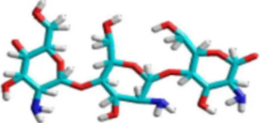
The absorption of glibenclamide by the chitosan crosslinked with genipin mainly involved the formation of hydrogen bonds between the CH2OH and CH groups. Figure 7 shows the structure of the genipin--crosslinked chitosan with absorbed glibenclamide.
CONCLUSIONS
Genipin, a natural crosslinking agent, reacts with compounds containing primary amine groups, such as chitosan, to form cova-lently crosslinked networks. In the case of chitosan, genipin reacts with the free amino groups present in the glucosamine units. The results of this study support the relevance of genipin as a valuable natural, nontoxic, crosslinking agent for controlled drug release in drug delivery systems based on chitosan.
The negative log P values for the crosslinked chitosan indicated that absorption properties of the hydrogel are dominated by its hydrophilic character and that absorption plays an important role in both glibenclamide absorption partitioning and receptor-binding. It
was also shown that genipin changes the hydrophilicity of chitosan. FTIR results revealed that secondary amide linkages are formed via the reaction of genipin ester groups with chitosan amino groups, yielding a polymeric network structure. The MESP values indicated that the nucleophilic and electrophilic regions mainly involved the NH/C–OH and C=O bonds, respectively.
REFERENCES
1. Thakur, G.; Mitra, A.; Rousseau, D.; Basak, A.; Sarkar, S.; Pal, K.; J. Mater. Sci.: Mater. Med. 2011,22, 115.
2. Bhattarai, N.; Gunn, J.; Zhang, M.; Adv. Drug Delivery Rev.2010, 62, 83.
3. Muzzarelli, R. A.; Carbohydr. Polym. 2009, 77, 1. 4. Wu, C. S.; Polymer2005,46, 147
5. Berger, J.; Reist, M.; Mayer, J. M.; Felt, O.; Peppas, N. A.; Gurny, R.;
Eur. J. Pharm. Biopharm.2004, 57, 19.
6. Diaconu, M.; Tache, A.; Victoria, S. A.; Gatea, F.; Litescu, S.; Radu,
G.; UPB Scientific Bulletin, Series B: Chemistry and Materials Science
2010,72, 115.
7. Patel, M.; Ravi, R.; Patel, J.; Patel, K.; J. Pharm. Pharm. Sci. 2010,13,
536.
8. Sailaja, A. K.; Amareshwar, P.; Chakravarty, P.; J Pharm. Biol. Chem. Sci. 2010, 1, 474.
9. Dash, M.; Chiellini, F.; Ottenbrite, R. M.; Chiellini, E.; Prog. Polym. Sci. 2011, 36, 981.
10. Grolik, M.; Szczubiałka, K.; Wowra, B.; Dobrowolski, D.; Orzechowska-Wylęgała, B.: Wylęgała, E.; Nowakowska, M.; J. Mater. Sci.: Mater. Med.2012,23, 1991.
11. Bansal, V.; Sharma, P. K.; Sharma, N.; Pal, O. P.; Malviya, R.; Adv. Biol. Res.2011, 5, 28.
12. Bhattarai, N.; Gunn, J.; Zhang, M.; Adv Drug Deliver Rev. 2010, 62, 83 13. Harris, R.; Lecumberri, E.; Heras, A.; Mar. Drugs2010, 8, 1750.
14. Karnchanajindanun, J.; Srisa-Ard, M.; Srihanam, P.; Baimark, Y.; Nat. Sci. 2010, 2, 1061.
15. Zhang, C. Y.; Parton, L. E.; Ye, C. P.; Krauss, S.; Shen, R.; Lin, C. T.; Porco, J. A.; Cell Metab. 2006, 3, 417.
16. Park, J. E.; Lee, Y.; Kim, H. G.; Hahn, T. R.; Paik, Y. S.; J. Agric. Food Chem. 2002, 50, 6511.
17. Wang, L.; Wang, Y.; Qu, J.; Hu, Y.; You, R.; Li, M.; J. Biomater. Nanobiotechnol.2013, 4, 213.
18. Imsombut, T.; Srisuwan, Y.; Srihanam, P.; Baimark, Y.; Powder Technol.
2010, 203, 603.
19. Silva, S. S.; Maniglio, D.; Motta, A.; Mano, J. F.; Reis, R. L.; Migliaresi.; Macromol. Biosci. 2008, 8, 1.
20. Taylor, M.; Ding, K.; Brown, E.; J. Am. Leather Chem. Assoc.2009, 1. 21. Yoo, J. S.; Kim, Y. J.; Kim, S. H.; Choi, S. H.; The Korean Journal of
Thoracic and Cardiovascular Surgery2011, 44, 197.
22. Serrano-Martín, X.; Payares, G.; Mendoza-León, A.; Antimicrob. Agents Chemother.2006, 50, 4214.
23. http://www.scbt.com/es/datasheet-200982-glyburide-glibenclamide.html, accessed July, 2014.
24. Mon, J.; Flury, M.; Harsh, J. B.; J. Hydrol.2006,316, 84.
25. Zhang, X. H.; Zhan, Y. H.; Chen, D.; Wang, F.; Wang, L. Y.; Dyes Pigm.
2012,93, 1408.
26. http://www.molinspiration.com/services/logp.html, accessed July, 2014. 27. http://www.organic-chemistry.org/prog/peo/cLogP.html, accessed July,
2014.
28. http://www.admescope.com/learn-adme/physicochemical-properties/ lipophilicity-logd-logp.html, accessed July, 2014.
29. Glish, L.; Hanks, T. W.; J. Chem. Educ.2001,84, 2001.
30. http://www.prudencepharma.com/Glibenclamide.html, accessed July, 2014.
Figure 6. MESP, from which (a) chitosan/genipin and (b) chitosan (genipin)/ glibenclamide, respectively
31. Dash, S. K.; Khan, A. S.; Das, S. R.; Padhan, A.; Rout, D.; Behera, B. C.; Int. J. Pharm. Sci. Res.2012, 3, 1433.
32. Rangel-Vázquez, N. A.; Rodríguez-Felix, F.; Computational Chemistry Applied in the Analyses of Chitosan/Polyvinylpyrrolidone/Mimosa
Tenuiflore, Science Publishing Group: Hong Kong, 2013, cap. 2, pp.
25-26.
33. Hwang, Y. J.; Krasieva, T.; Lyubovitsky, J.; ACS Appl. Mater. Interfaces 2011, 3, 2579
34. Santoni, N.; Matos, M.; Müller-Karger, C.; Nicola, H.; Sabino, M.; Müller, A.; Revista Iberoamericana de Polímeros2008, 9, 326. 35. Gunasekaran, S.; Rajalakshmi, K.; Kumaresan, S.; Spectrochim. Acta,
Part A2013,112, 351.
36. Lertsutthiwong, P.; Noomun, K.; Khunthon, S.; Limpanart, S.; Prog. Nat. Sci.: Mater. Int.2012,22, 502.
37. Yao, C. H.; Liu, B. S.; Chang, C. J.; Hsu, S. H.; Chen, Y. S.; Mater. Chem. Phys. 2004,83, 204.
38. Levinton-Shamuilov, G.; Cohen, Y.; Azoury, M.; Chaikovsky, A.; J. Forensic Sci. 2005,50, 1367.
39. Bahri-Najafi, R.; Tavakoli, N.; Senemar, M.; Peikanpour, M.; Res. Pharm. Sci.2014,9, 213.
40. Rodríguez, R.; Tesis deLicenciatura, Universidad de Oriente, Cumaná,
Venezuela, 2010.
41. Mirzaei, E.; Faridi-Majidi, R.; Shokrgozar, M. A.; Paskiabi, F. A.;