Antimicrobial Action of Oleanolic Acid on
Listeria monocytogenes
,
Enterococcus
faecium
, and
Enterococcus faecalis
Sejeong Kim1, Heeyoung Lee1, Soomin Lee1, Yohan Yoon1*, Kyoung-Hee Choi2*
1Department of Food and Nutrition, Sookmyung Women's University, Seoul, Korea,2Department of Oral Microbiology, College of Dentistry, Wonkwang University, Iksan, Jeonbuk, South Korea
*[email protected](YY);[email protected](KHC)
Abstract
This study investigated the antimicrobial action of oleanolic acid againstListeria monocyto-genes,Enterococcus faecium, andEnterococcus faecalis. To determine the cytotoxicity of oleanolic acid, HEp-2 cells were incubated with oleanolic acid at 37oC. MICs (minimal inhibi-tion concentrainhibi-tions) forL.monocytogenes,E.faecium, andE.faecaliswere determined using two-fold microdilutions of oleanolic acid, and bacterial cell viability was then assessed by exposing the bacteria to oleanolic acid at 2 × MIC. To investigate the mode of antimicro-bial action of oleanolic acid, we measured leakage of compounds absorbing at 280 nm, along with propidium iodide uptake. Scanning electron microscope (SEM) images were also analysed. The viability of HEp-2 cells decreased (P<0.05) at oleanolic acid
concentra-tions greater than 128μg mL-1. The MICs were 16-32μg mL-1forL.monocytogenesand 32-64μg mL-1forE.faeciumandE.faecalis, and bacterial cell viability decreased (P<
0.05) about 3-4 log CFU mL-1after exposure to 2 × MIC of oleanolic acid. Leakage of 280 nm absorbing materials and propidium iodide uptake was higher in oleanolic acid–treated
cells than in the control. The cell membrane was damaged in oleanolic acid-treated cells, but the control group had intact cell membrane in SEM images. The results indicate that oleanolic acid can killL.monocytogenes,E.faecium, andE.faecalisby destroying the bac-terial cell membrane.
Introduction
Antibiotic-resistant bacteria have increased and spread globally due to improper use of antibi-otics over recent decades, causing severe clinical problems. Development of novel therapeutics is therefore urgently required. Recently, various antimicrobial strategies have been introduced, and plant-derived compounds used as bactericides are receiving attention because of their low toxicity to human cell and ready availability.
Among them, oleanolic acid, an example of a pentacyclic triterpenoid originating from a number of medicinal plants, has antimicrobial activity against various bacterial pathogens [1,2,3,4], and has various bioactivity [5,6]. Its antibacterial activity is not much higher than
a11111
OPEN ACCESS
Citation:Kim S, Lee H, Lee S, Yoon Y, Choi K-H (2015) Antimicrobial Action of Oleanolic Acid on
Listeria monocytogenes,Enterococcus faecium, and
Enterococcus faecalis. PLoS ONE 10(3): e0118800. doi:10.1371/journal.pone.0118800
Academic Editor:Mathias Chamaillard, INSERM, FRANCE
Received:June 6, 2014
Accepted:January 6, 2015
Published:March 10, 2015
Copyright:© 2015 Kim et al. This is an open access article distributed under the terms of theCreative Commons Attribution License, which permits unrestricted use, distribution, and reproduction in any medium, provided the original author and source are credited.
Data Availability Statement:All relevant data are within the paper.
Funding:This paper was supported by Wonkwang University in 2013. The funders had no role in study design, data collection and analysis, decision to publish, or preparation of the manuscript.
traditionally-used antimicrobials, but the study on this antimicrobial is worthy because it is natural source product, and no resistance is found yet [7]. Our previous studies demonstrated that oleanolic acid inhibits the growth ofListeria monocytogenes, suggesting the potential use-fulness of the compound as a food additive with anti-listerial activity [8]. In addition, it was shown that oleanolic acid inhibited the growth of Gram-positive bacteria, but this inhibition ef-fect was weaker in Gram-negative bacteria [9].
Enterococci, Gram-positive bacteria, are considered to be part of the natural gut microflora in humans and animals that can be applied as starter cultures in food products, and as probiot-ics to improve the bacterial balance in the intestine [10,11,12]. They are also responsible for opportunistic infectious diseases, however, such as urinary tract, bloodstream, and nosocomial infections [13,14]. Recently, enterococci have become more resistant toβ-lactam-based antibi-otics as well as aminoglycosides [15].
L.monocytogenesis well known as the causative agent of listeriosis, a food-borne illness
with a high mortality rate [16]. This pathogen is able to survive under a variety of harsh envi-ronmental conditions, such as low pH, high salt, and low temperature [17]. Although sodium chloride, sodium nitrite, and a variety of other antimicrobials are used in processed foods to control the growth ofL.monocytogenes, they have safety issues, making it necessary to develop new antimicrobials [18,19]. In addition, because multidrug-resistantL.monocytogenesstrains have emerged, and because most antibiotics cannot be given to pregnant patients withL.
mono-cytogenesinfections, we need to exploit natural therapeutic alternative to antibiotics [20,21,
22].
Oleanolic acid is therefore potentially an excellent agent for eradicating the growth of
L.monocytogenesin foods as well as clinical settings. To date, there has been extensive research
on the activity of oleanolic acid against bacteria, but the studies have been mainly focused on measurements of the minimum inhibitory concentrations (MICs) of oleanolic acid against dif-ferent microorganism. For various and suitable applications of antimicrobials, it is important to understand mode of these materials. Therefore, the aim of the present study was to investi-gate the mode of action of oleanolic acid againstL.monocytogenes,E.faecium, andE.faecalis.
Materials and Methods
Preparation of oleanolic acid
A stock 5 mg mL-1solution of oleanolic acid (Sigma, St. Louis, MO, USA) was freshly prepared in dimethyl sulfoxide (DMSO; Sigma). The solution was then diluted in brain heart infusion (BHI) broth (Difco, Becton, Dickinson and Company, Sparks, MD, USA) and tryptic soy broth (TSB; Difco, Becton, Dickinson and Company, Sparks, MD, USA) to 1,024μg mL-1for use in
the experiments.
Bacterial strains, cell line, and culture conditions
L.monocytogenesstrains [NCCP10808 (ruminant brain isolate), NCCP10809 (human isolate),
NCCP10920 (clinical isolate), NCCP10943 (rabbit isolate)],E.faeciumstrains [NCCP11193 (non-identified isolate)and NCCP10887 (cheese isolate)], andE.faecalisstrains [KACC11304 (young chicken intestine isolate), NCCP10874 (non-identified isolate), and NCCP10886 (meat isolate)] were incubated in BHI broth and TSB forL.monocytogenes, andE.faeciumandE.
fae-calis, respectively, for 24 h at 37°C. HEp-2 cells (human larynx epithelial cells, KCLB10023)
Cytotoxicity of oleanolic acid
HEp-2 cells (2 × 10⁵mL-1) were plated in a 96-well microplate and incubated at 37°C for 48 h in a 5% CO2atmosphere to allow cell attachment to the surface; the medium and unattached
cells were discarded. Two-fold serial dilutions of oleanolic acid were prepared in DMEM plus 10% FBS and 1% penicillin-streptomycin, and the dilutions added to the wells. The plate was then incubated at 37°C for 48 h in a 5% CO2atmosphere to determine cytotoxicity. 20μL of
MTT (3-(4,5-dimethylthiazol-2-yl)-2,5-diphenyltetrazolium bromide; Sigma) solution (5 mg mL-1) was added to each well, and the mixture was incubated at 37°C for a further 4 h in a CO2
atmosphere to allow the HEp-2 cells to metabolize MTT. The medium was then removed, the insoluble formazan crystals dissolved in 200μL DMSO, and the absorbance measured at 550
nm using a spectrophotometer (Sunrise, Tecan, Austria). Data were reported as OD550of
olea-nolic acid-treated cell.
Determination of MICs and MBCs
MICs and MBCs of oleanolic acid were determined by the two-fold broth micro-dilution meth-od [23]. All strains used in this study were grown overnight in each broth media at 37°C, fol-lowed by subculture in fresh each broth media and growth to logarithmic phase. An equal volume of the bacterial inoculum with an optical density of 0.01 at 600 nm was added to each well in a 96-well plate containing 100μL of serially diluted oleanolic acid. After incubation for
24 h at 37°C, the MICs (minimal inhibition concentrations) were obtained by measuring the optical density at 600 nm with a spectrophotometer (Sunrise, Tecan, Austria). The lowest con-centration of oleanolic acid at which no visible bacterial growth was observed was defined as the MIC for the bacteria. In addition, aliquots from the wells displaying no growth were spread on BHI agar plates forL.monocytogenesor tryptic soy agar (TSA) plate forE.faeciumand
E.faecalis, and the plates incubated at 37°C for 24 h to determine whether the reduction of
bac-terial growth in the wells was caused by bacteriostatic or bactericidal activity of oleanolic acid. The lowest concentration of oleanolic acid in the wells that gave reduction in bacterial growth on the agar plates by 99.9% compared to the initial bacterial inoculum was expressed as the MBC (minimal bactericidal concentration).
Bacterial cell viability assay
Bacterial cell viability in the presence of oleanolic acid was determined by a slight modification to a previously described protocol [24]. One mL of the subcultured bacterial cells (OD600= 0.7)
ofL.monocytogenesNCCP10943,E.faeciumNCCP11193, andE.faecalisKACC11304 was
re-suspended in sterile saline and then added to 19 mL of 50 mM phosphate buffer (pH 7.1) with or without 2 × MIC of oleanolic acid [24]. Control was the cells without 2 × MIC of oleanolic acid, which was containing oleanolic acid-equivalent volume of DMSO. After 60 min incuba-tion at 37°C, viable cell counts were taken for the suspensions on BHI agar plates forL.
mono-cytogenesor TSA plates forE.faeciumandE.faecalisand expressed as log CFU mL-1.
Leakage of compounds absorbing at 280 nm
280 nm was expressed as a relative ratio of OD280of oleanolic acid-treated cells to one of olea-nolic acid-untreated cells.
Propidium iodide uptake assay
A propidium iodide uptake assay was used to characterize the action of oleanolic acid on cell membrane permeability of the bacterial pathogens [24]. After 60 min incubation with or with-out oleanolic acid, 50μL of suspension prepared as for the cell viability assay was added to
950μL of phosphate buffer in FACS tubes stored on ice. Propidium iodide (Sigma) dissolved in
sterile milliQ water was then added to the tube to a final concentration of 10μg mL-1. The
mix-ture was subjected to FACS analysis using a flow cytometry (BD FACScalibur, San Jose, CA, USA). The fraction of propidium iodide-stained cells was obtained using CellQuest Pro soft-ware (BD CellQuest, San Jose, CA, USA). The results were compared with one of DMSO-treated cells.
Scanning electron microscope image analysis
The effect of oleanolic acid on the bacterial cell membrane was further investigated by scanning electron microscopy (SEM; JSM-7600F, JEOL Ltd., Tokyo, Japan).L.monocytogenes
NCCP10943,E.faeciumNCCP11193, andE.faecalisKACC11304 were cultured in 10 mL of BHI broth and TSB forL.monocytogenes, andE.faeciumandE.faecalis, respectively, for 24 h at 37°C. The 0.1 mL portions of the cultures were subcultured overnight in 10 mL of each broth media containing a glass slide to which cells attached and grew. After overnight incuba-tion, the attached cells were incubated with one mL of oleanolic acid in PBS (phosphate buff-ered saline) at 2 × MIC or an equal volume of DMSO for 1 h at 37°C [25]. Cells treated with DMSO (no oleanolic acid) were used as control. Incubated cells were pre-fixed in 1.8% glutar-aldehyde solution (Sigma) for 30 min, followed by washing three times with distilled water. After post-fixing in 2% osmium tetroxide (Sigma) for 20 min, the bacterial cells were washed three times and then dehydrated in a graded series of ethanol concentrations (25, 50, 75, 90, and 100%). The cells were platinum-coated using a sputter coater (108 auto; Cressington Scien-tific Instruments Ltd., Watford, England), and examined by field emission SEM.
Statistical analysis
This study was repeated with two samples in each repeat (n = 4). The OD values and bacterial cell counts (log CFU mL-1) at the various oleanolic acid concentrations were analysed by the mixed model procedure of SAS version 9.2 (SAS Institute, Cary, NC, USA). All least squares means comparisons among the interactions were performed by pairwiset-tests at alpha = 0.05.
Results and Discussion
According to previous studies, oleanolic acid exhibits antimicrobial activity against many Gram-positive bacteria, includingL.monocytogenes,E.faecalis,E.faecium,Streptococcus
mutans, andStreptococcus sanguis[1,2,3,4]. There was weaker antimicrobial effect against
Gram-negative bacteria, however, includingAcinetobacter baumannii,Burkholderia
thailan-densis,Escherichia coli,Klebsiella pneumoniae, andPseudomonas aeruginosa[9]. This
cellular target of oleanolic and ursolic acid, and we hypothesized that oleanolic acid may cause death of Gram-positive bacteria by destroying bacterial cell membrane.
In vitrocell culture systems that retain similar marker enzymes in freshly isolated cells have
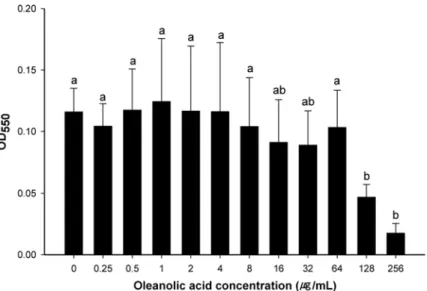
been extensively used for assessing toxicity of antimicrobials in specific organs [29]. The cyto-toxicity of oleanolic acid against HEp-2 cells was examined to determine if oleanolic acid can be used to inhibit the growth of pathogenic bacteria without having a cytotoxic effect on eu-karyotic cells. Oleanolic acid caused no decrease in the viability of HEp-2 cells at concentra-tions up to 64μg mL-1, but cell viability decreased at concentrations over 128μg mL-1(Fig. 1).
This indicates that oleanolic acid is not cytotoxic at levels up to 64μg mL-1. Although oleanolic
acid can be consumed directly or indirectly by humans, it is important to know its effect on karyotic cells. Many other studies investigated the effect of oleanolic acid on the viability of eu-karyotic cells, including leukemic cell line [30], normal liver cell, liver cancer cell line [31,32] and kidney cell line [29]. The results in these studies indicated that oleanolic acid had no cyto-toxicity on eukaryotic cells upto 45μg mL-1. Fontanay et al. [4] also suggested that IC50(50%
inhibitory concentration) of oleanolic acid on HaCaT cell line were 52.4μg mL-1. Taken
to-gether, the application of oleanolic acid for human may be appropriate.
The antimicrobial effects of oleanolic acid onL.monocytogenes,E.faecium, andE.faecalis
were assessed by MIC and MBC. MIC is defined as the minimum concentration that allows no visible bacterial growth; MBC is the lowest concentration that kills bacteria by 99.9% [33]. Oleanolic acid inhibited the growth ofL.monocytogenesstrains at 16μg mL-1other than
L.monocytogenesNCCP10808 that demonstrated no visible growth at 32μg mL-1. Growth of
E.faecium(NCCP11193 and NCCP10887) andE.faecalis(NCCP10874 and KACC11304) was
inhibited at 32μg mL-1, and MIC forE.faecalisNCCP10886 was 64μg mL-1(data not shown).
As a result, the oleanolic acid had antimicrobial activity over the strains used in this study with strain variation. These MIC values are higher as observed in previous studies by Szakiel et al. [34] (15μg mL-1forL.monocytogenes) and Fontanay et al. [4] (8μg mL-1forE.faecalis). For
vancomycin-resistant Enterococci, oleanolic acid had different antimicrobial activity according to strains, 8μg mL-1[2] or 256μg mL-1[4], and it was also concluded that this difference may
be caused by strain variation of bacteria. The results for MBC showed that all strains tested
Fig 1. Cytotoxicity of oleanolic acid on HEp-2 cells.Cells were exposed to twofold-dilluted oleanolic acid (0–256μg mL-1) for 48 hours. After exposure, viability of the cell was measured by MTT assay. a-b: means with different letters are significantly different (P<0.05).
were completely killed (99.9%) by oleanolic acid at>256μg mL-1. It is usually regarded as
bac-tericidal if the MBC of antimicrobial agents is not exceeding four times of the MIC [35]. Thus, this shows that oleanolic acid has bacteriostatic but not bactericidal effects onL.
monocyto-genes,E.faeciumandE.faecalis. Moreover, since 64μg mL-1is the maximum concentration of
oleanolic acid showing no cytotoxicity, its use at less than 64μg mL-1would be expected to be
bacteriostatic toL.monocytogenes,E.faecium, andE.faecalis.
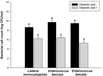
Fig 2. Bacterial cell viability.Listeria monocytogenes,Enterococcus faecium, andEnterococcus faecalis
exposed to oleanolic acid at 2 × MIC. Viabilities of oleanolic acid-treated cells were compared with one of oleanolic acid-untreated cells. a-b: means with different letters are significantly different (P<0.05).
doi:10.1371/journal.pone.0118800.g002
Fig 3. Leakage of material absorbing at 280 nm.AfterListeria monocytogenes,Enterococcus faecium, andEnterococcus faecaliswere exposed to oleanolic acid at 2 × MIC, leakage of material absorbing at 280nm was measured using spectrophotometer. The result was expressed as a relative ratio of OD280of oleanolic acid-treated cells to the one of oleanolic acid-untreated cells.
A bacterial cell viability assay was conducted to measure the fraction of bacterial cells that were able to survive exposure to oleanolic acid. The results of bacterial cell viability assay showed short-time effect of oleanolic acid on the bacteria compared to MIC and MBC. After the bacterial cells were exposed to oleanolic acid for 60 min,L.monocytogenesviability de-creased from 8.6 to 6.1 log CFU mL-1, (p<0.05) (Fig. 2), and the viabilities ofE.faeciumand
E.faecaliswere reduced from 9.5 to 6.5 log CFU mL-1, and from 9.5 to 5.3 log CFU mL-1,
re-spectively (p<0.05) (Fig. 2). These results confirm the antibacterial effects of oleanolic acid on
L.monocytogenes,E.faeciumandE.faecalis.
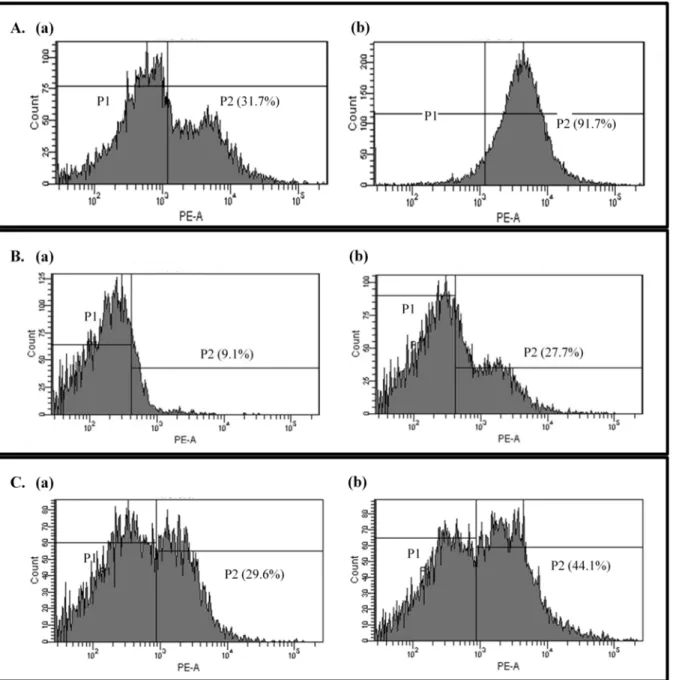
Fig 4. Propidium iodide uptake.Uptake of propidium iodide by untreated (a) or oleanolic acid-treated (2 × MIC) bacterial cells (b):Listeria monocytogenes
(A),Enterococcus faecium(B), andEnterococcus faecalis(C). The area (P2) indicates the ratio of propidium iodide uptake cells.
We investigated potential membrane damage by measuring the leakage of bacterial cell con-tents caused by oleanolic acid. The ratios of compounds absorbing at 280 nm released by bacte-rial cells treated with oleanolic acid at 2 × MIC relative to those released by untreated cells were 1.38, 1.36 and 1.47 forL.monocytogenes,E.faecium, andE.faecalis, respectively (Fig. 3).
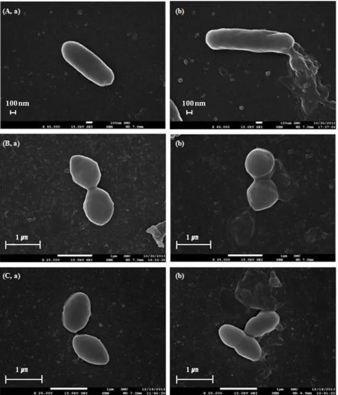
Fig 5. Scanning electron microscope images.After exposed to oleanolic acid at 2 × MIC, untreated (a) and oleanolic acid-treated bacterial cells (b):
Listeria monocytogenes(A),Enterococcus faecium(B), andEnterococcus faecalis(C) were examined by SEM.
Leakage of UV absorbing material is one of indicator presenting cell lysis [36]. This leakage of compounds absorbing at 280 nm is related to the loss of cell proteins that are normally retained by the cell membrane. These results indicate that the bacterial cell membrane is damaged by oleanolic acid.
To further investigate bacterial cell membrane damage, propidium iodide uptake was mea-sured. Propidium iodide binds non-specifically to DNA and emits fluorescence, but due to its large size and negative charge, it cannot pass through intact membranes, fluorescence, there-fore, indicates that the bacterial cell membrane is damaged and has become permeable. X-axis of the graph indicates level of emission. The more propidium iodide binds to DNA, the peaks are placed in the more right side of X-axis. The area inFig. 4indicates the ratio of propidium iodide uptake cells. After oleanolic acid treatment, fluorescentL.monocytogenescells increased from 31.7% to 91.7%. Similarly, forE.faecium, the fraction of membrane-damaged cells went from 9.1% to 27.7%, and forE.faecalis, the percentage increased from 29.6% to 44.1% (Fig. 4). These results further support the idea that oleanolic acid destroys the cell membranes of Gram-positive bacteria.
Bacterial cell damage by oleanolic acid was also visually confirmed by SEM.Fig. 5shows SEM images ofL.monocytogenes,E.faecium, andE.faecalisafter oleanolic acid-treatment at 2 × MIC. Bacterial cells treated with oleanolic acid at 2 × MIC level showed changes in membrane shape in comparison to the control (DMSO-treated). The membranes of oleanolic acid-treated bacterial cells burst, whereas untreated cells had intact cell membranes (Fig. 5). This result directly confirms the implications of the cell leakage and propidium iodide uptake experiments that the antimicrobial activity of oleanolic acid againstL.monocytogenes,E.
fae-ciumandE.faecalisresults from damage to the cell membrane. Kurek et al. [28] found that
oleanolic acid inhibited peptidoglycan turnover and influenced the muropeptides profiles, in-creasing autolysis of the cell wall. A study by Xia et al. [37] showed that oleanolic acid bound to peptidoglycan and teichoic acids, long anionic polymer threading through peptidoglycan. Re-cently, Kurek et al. [38] demonstrated that oleanolic acid bound to the peptidoglycan of the pathogen, which was influenced by teichoic acids. Hence, in our study oleanolic acid may bind to peptidoglycan ofL.monocytogenes,E.faeciumandE.faecalis, and this interaction was affect-ed by teichoic acids, inhibiting peptidoglycan turnover.
In conclusion, oleanolic acid has antimicrobial effects on food-related bacterial pathogens, such asL.monocytogenes,E.faeciumandE.faecalisby destroying the bacterial cell membrane, and the compound has low cytotoxicity on HEp-2 cells.
Acknowledgments
This paper was supported by Wonkwang University in 2013.
Author Contributions
Conceived and designed the experiments: KHC YHY. Performed the experiments: SJK. Ana-lyzed the data: SJK SML HYL YHY KHC. Contributed reagents/materials/analysis tools: SJK LSM LHY YHY KHC. Wrote the paper: SJK YHY KHC.
References
1. Kozai K, Suzuki J, Okada M, Nagasaka N. Effect of oleanolic-acid-cyclodextrin inclusion compounds on carries byin vitroexperiment and rat-carries model. Microbios. 1999; 97: 179–188. PMID:10413873
3. Jiménez AA, Meckes M, Torres JK, Luna HJ. Antimycobacterial triterpenoids fromLantana hispida
(Verbenaceae). J Ethnopharmacol. 2007; 111: 202–205. PMID:17236730
4. Fontanay S, Grare M, Mayer J, Finance C, Duval RE. Ursolic, oleanolic and betulinic acids: antibacteri-al spectra and selectivity indexes. J Ethnopharmacol. 2008; 120: 272–276. doi:10.1016/j.jep.2008.09. 001PMID:18835348
5. Hung CY, Yen GC. Extraction and identification of antioxidative components of Hsian-tsao (Mesona procumbensHemsl.). Food Sci Technol. 2001; 34: 306–311.
6. Kinjo J, Okawa M, Udayama M, Sohno Y, Hirakawa T, Shii Y, et al. Hepatoprotective and hepatotoxic actions of oleanolic acid-type triterpenoidal glucuronoids on rat primary hepatocyte cultures. Chem Pharm Bull. 1999; 47: 290–292. PMID:10071859
7. Wolska KI, Grudniak AM, Fiecek B, Kraczkiewicz-Dowjat A, Kurek A. Antibacterial activity of oleanolic and ursolic acids and their derivatives. Cent Eur J Biol. 2010; 5: 543–553.
8. Yoon Y, Choi KH. Antimicrobial activity of oleanolic acid onListeria monocytogenesunder sublethal stresses of NaCl and pH. Korean J Food Sci Ani Resour. 2010; 30: 717–721.
9. Yoon Y, Choi KH. Identification of inhibitory effect onStreptococcus mutansby oleanolic acid. J Life Sci. 2010; 20: 321–325.
10. Gelsomino R, Vancanneyt M, Cogan TM, Condon S, Swings J. Source of enterococci in a farmhouse raw-milk cheese. Appl Environ Microbiol. 2002; 68: 3560–3565. PMID:12089042
11. Saavedra L, Taranto MP, Sesma F, de Valdez GF. Homemade traditional cheeses for the isolation of probioticEnterococcus faeciumstrains. Int J Food Microbiol. 2003; 88: 241–245. PMID:14596996
12. Canzek MA, Rogelj I, Perko B. Enterococci from Tolminc cheese: population structure, antibiotic sus-ceptibility and incidence of virulence determinants. Int J Food Microbiol. 2005; 102: 239–244. PMID: 15992623
13. Jett BD, Huycke MM, Gilmore MS. Virulence of enterococci. Clin Microbiol Rev. 1994; 7: 462–478. PMID:7834601
14. Hunt CP. The emergence of enterococci as a cause of nosocomial infection. Br J Biomed Sci. 1998; 55: 149–156. PMID:10198473
15. Templer SP, Rohner P, Baumgartner A. Relation ofEnterococcus faecalis and Enterococcus faecium
isolates from foods and clinical specimens. J Food Prot. 2008; 71: 2100–2104. PMID:18939760
16. Swaminathan B, Gerner-Smidt P. The epidemiology of human listeriosis. Microbes Infect. 2007; 9: 1236–1243. PMID:17720602
17. Nuñez de Gonzalez MT, Keeton JT, Acuff GR, Ringer LJ, Lucia LM. Effectiveness of acidic calcium
sul-fate with propionic and lactic acid and lactates as postprocessing dipping solutions to controlListeria monocytogeneson frankfurters with or without potassium lactate and stored vacuum packaged at 4.5°C. J Food Prot. 2004; 67: 915–921. PMID:15151227
18. Mbandi E, Shelef LA. Enhanced antimicrobial effects of combination of lactate and diacetate onListeria monocytogenesandSalmonellaspp. in beef bologna. Int J Food Microbiol. 2002; 76: 191–198. PMID: 12051475
19. Ramaswamy V, Cresence VM, Rejitha JS, Lekshmi MU, Dharsana KS, Prasad SP, et al.Listeria -re-view of epidemiology and pathogenesis. J Microbiol Immunol Infect. 2007; 40: 4–13. PMID:17332901
20. Ennaji H, Timinouni M, Ennaji MM, Hassar M, Cohen N. Characterization and antibiotic susceptibility of
Listeria monocytogenesisolated from poultry and red meat in Morocco. Infect Drug Resist. 2008; 1: 45–50. PMID:21694879
21. Rahimi E, Yazdi F, Farzinezhadizadeh H. Prevalence and antimicrobial resistance ofListeriaspecies isolated from different types of raw meat in Iran. J Food Prot. 2012; 75: 2223–2227. doi: 10.4315/0362-028X.JFP-11-565PMID:23212021
22. Adzitey F, Ali G, Huda N, Cogan T, Corry J. Prevalence, antibiotic resistance and genetic diversity of
Listeria monocytogenesisolated from ducks, their rearing and processing environments in Penang, Malaysia. Food Control. 2013; 32: 607–614.
23. Mellegard H, Strand SP, Christense BE, Granum PE, Hardy SP. Antibacterial activity of chemically de-fined chitosans: Influence of molecular weight, degree of acetylation and test organism. Int J Food Microbiol. 2011; 148: 48–54. doi:10.1016/j.ijfoodmicro.2011.04.023PMID:21605923
24. Reddy MV, Thota N, Sangwan PL, Malhotra P, Ali F, Khan IA, et al. Novel bisstyryl derivatives of baku-chiol: Targeting oral cavity pathogens. Eur J Med Chem. 2010; 45: 3125–3134. doi:10.1016/j.ejmech. 2010.03.049PMID:20427099
26. Cox SD, Mann CM, Markham JL, Bell HC, Gustafson JE, Warmington JR. et al. The mode of antimicro-bial action of the essential oil ofMelaleuca alternifolia(tea tree oil). J Appl Microbiol. 2000; 88: 170–175. PMID:10735256
27. Tyagi AK, Bukvichki D, Gottardi D, Veljic M, Guerzoni ME, Malik A, et al. Antimicrobial potential and chemical characterization of serbian liverwort (Porella arboris-vitae): SEM and TEM observations. Evid Based Complement Alternat Med. 2013, Article ID 382927, 7 pages.
28. Kurek A, Grudniak AM, Szwed M, Klicka A, Samluk L, Wolska KI, et al. Oleanolic acid and ursolic acid affect peptidoglycan metabolism inListeria monocytogenes. Anton Leeuw. 2010; 97: 61–68.
29. Madlala HP, MAsola B, Singh M, Musabayane CT. The effect ofSyzygium aromaticum-derived oleano-lic acid on kidney function of male Sprague-Dawley rats and on kidney and liver cell lines. Renal Fail-ure. 2012; 34: 767–776. doi:10.3109/0886022X.2012.678172PMID:22512664
30. Ovesna Z, Vachalkova A, Horvathova K, Tothova D. Pentacyclic triterpenoic acids: new chemoprotec-tive compounds. Minireview. Neoplasma. 2004; 51: 327–333. PMID:15640935
31. Wang X, Ye X, Chen HL, Bai H, Liang X, Zhang XD, et al. Antioxidant activities of oleanolic acidin vitro: Possible role of Nrf2 and MAP kinases. Chem-Biol Interact. 2010; 184: 328–337. doi:10.1016/j.cbi. 2010.01.034PMID:20100471
32. Yan S, Huang C, Wu S, Yin M. Oleanolic acid and ursolic acid induce apoptois in four human liver can-cer cell lines. Toxicol In Vitro. 2010; 24: 842–848. doi:10.1016/j.tiv.2009.12.008PMID:20005942
33. Lee BL, Sachdeva M, Chambers HF. Effect of protein binding of daptomycin on MIC and antibacterial activity. Antimicrob Agents Chemother. 1991; 35: 2505–2508. PMID:1667253
34. Szakiel A, Ruszkowski D, Grudniak A, Kurek A, Wolsk KI, Doligalska M, et al. Antibacterial and antipar-asitic activity of oleanolic acid and its glycosides isolated from marigold (Calendula officinalis). Planta Med. 2008; 74: 1709–1715. doi:10.1055/s-0028-1088315PMID:18951335
35. French GL. Bactericidal agents in the treatment of MRSA infections—the potential role of daptomycin. J Antimicrob Chemother. 2006; 58: 1107–17. PMID:17040922
36. Zhou K, Zhou W, Li P, Liu G, Zhang J, Dai Y. Mode of action of pentocin 31–1: an antilisteria bacteriocin produced byLactobacillus pentosusfrom chinese traditional ham. Food Control. 2008; 19: 817–822.
37. Xia G, Kohler T, Peschel A. The wall teichoic acid and lipoteichoic acid polymers ofStaphylococcus au-reus. Int J Med Microbiol. 2010; 300: 148–154. doi:10.1016/j.ijmm.2009.10.001PMID:19896895