Juliana de Oliveira da Luz Fontes*, Guilherme Thiesen**
Prospective cephalometric study of the effects
of maxillary protraction therapy associated with
intermaxillary mechanics
Objective: The early diagnosis and treatment of skeletal Class III (Pattern III) is still a much debated topic in orthodontic literature. Maxillary protraction associated with rapid maxillary expansion is the most popular and widely researched approach, producing the best results in the shortest period of time. This study aimed to evaluate the gradual changes that occur in the dentofacial complex in children with Pattern III growth treated with maxillary protrac-tion associated with intermaxillary mechanics. Methods: The sample consisted of 10 patients with Pattern III, whose mean age was 8 years and 2 months at the beginning of treatment, consecutively treated with a modified Haas expander, modified lingual arch, intermaxillary elastics and Petit facemask for maxillary protraction during a 9-month period. Four lateral cephalograms were taken of each patient, one at the beginning of treatment and the other three at regular 3-month intervals (T1, T2, T3 and T4). Cephalometric measurements at each of the four times were compared using ANOVA variance for repeated measures and supplemented by Tukey’s multiple comparisons test. Results: It was observed that the most significant skeletal changes occurred in the first 3 months of treatment. After that period the changes remained constant until the end of treatment. There were few dental compensations and the vertical changes which occurred showed reduced clinical significance. Conclusions:
The therapy used in this study accomplished not only the correction of overjet but also im-provements in the sagittal relationship of the basal bones and in soft tissue esthetics.
Abstract
Keywords: Prognathism. Palatal expansion technique. Extraoral traction appliances. Malocclusion.
* Specialist in Orthodontics, UNISUL.
** MSc in Orthodontics and Dentofacial Orthopedics, PUC/RS. Professor of Orthodontics, UNISUL. How to cite this article: Fontes JOL, Thiesen G. Prospective
cepha-lometric study of the effects of maxillary protraction therapy associ-ated with intermaxillary mechanics. Dental Press J Orthod. 2011 Nov-Dec;16(6):38.e1-9.
» The authors report no commercial, proprietary, or inancial interest in the
INTRODUCTION AND LITERATURE REVIEW
Class III malocclusion, originally defined by Angle as a mesial relationship between man-dibular and maxillary molars, is actually linked to several skeletal and dental changes, which of-ten result in a disharmonious facial appearance. Thus, Pattern III can be defined as a conspicuous anteroposterior imbalance expressed in the soft tissues of the face resulting from a sagittal skele-tal discrepancy between basal bones (mandibular prognathism, maxillary deficiency, or a combina-tion of both), accompanied or not by changes in the vertical and transverse directions. These dis-crepancies promote skeletal changes in the posi-tion of the teeth both inter and intra-arch.4
Despite its low prevalence when compared to other malocclusions,6,21 it’s prominent facial characteristics determine the need for therapy in the early development stages of individuals with this condition. However, early treatment of Pattern III can prove a major challenge to orthodontists in their clinical practice because of its complex control and difficulty in pre-dicting the patient’s morphogenetic growth pattern until adulthood.
Conversely, many professionals prefer to wait for skeletal maturity to treat this deformi-ty through orthognathic surgery given uncer-tainties regarding the success and stability of early treatment. However, recent studies with long-term follow-up of patients who received early treatment with orthopedic mechanics have shown that good results can be achieved in a considerable number of patients.26,28
Among the early treatment modalities of Pattern III, maxillary protraction is the most popular and widely researched approach in the orthodontic literature, yielding the best results in the shortest period of time, especially in in-dividuals who are in the late deciduous or ear-ly mixed dentition stage. Among the changes that occur during treatment, one could high-light maxillary displacement in the anterior
direction, clockwise rotation of the mandibu-lar plane, antero-superior displacement of the upper arch, lingual inclination of mandibular incisors, increased antero-inferior facial height and increased facial convexity.1,6,10,16,18,19,23 Recently published studies report the use of different maxillary expanders, which may be associated with other intra- or intermaxillary appliances designed to enhance the skeletal ef-fects of maxillary protraction.3,12,14,22
Given the undeniable importance of this topic as well as the many controversies that still surround it, this study aimed to evalu-ate how to maximize the effects of a proven method, i.e., maxillary protraction, through the concurrent use of this approach in com-bination with Petit facemask and intermaxil-lary mechanics with the continued use of Class III intraoral elastics. Moreover, there are no orthodontic studies in the literature assessing the gradual effects of this therapy throughout treatment, although some authors4,7 have spec-ulated that most skeletal effects occur during the first months of therapy.
MATERIAL AND METhODs sample description
To perform a prospective cephalometric study of the dental, skeletal and soft tissue changes induced during treatment, 10 patients (6 females and 4 males) were consecutively treated with a modified Haas expansion ap-pliance, modified lingual arch and Petit face-mask for maxillary protraction for a period of 9 months. Mean subject age was 8 years and 2 months at the beginning of treatment (ages ranging from 5 to 11 years). The sample was provided by the Department of Orthodontics of UNISUL – Florianópolis, Brazil.
marked deficiency in the malar region; 2) Pattern III skeletal growth, as determined by Wits≤ 0 mm; 3) deciduous dentition or mixed dentition, 4) an-terior crossbite or end-to-end relationship; 5) no previous orthodontic treatment, 6) absence of congenital and/or other facial deformities.
The skeletal age of the patients was as-sessed using the method of cervical vertebrae maturation proposed by Bacceti, Franchi and McNamara.2 This method consists of six stages of increasing development, but all the patients selected for this study were in the third stage, i.e., before the peak of pubertal growth spurt.
Anchorage system
Upper arch
The anchorage system used in the maxillary arch of all patients (Fig 1) was a modified fixed Haas type expansion appliance made of 1.2 mm steel wire (Dentaurum®) with buccal and lingual connecting bars. The buccal bar was extended distally in order to allow the insertion of Class III intraoral elastics. Deciduous second molars were banded whenever possible. When banding these teeth was not possible the first permanent pre-molars and pre-molars were banded instead (Univer-sal Band Kit – Morelli®). All other posterior teeth were often bonded with composite (Z100 – 3M). An 11-mm expansion screw was used (Dentau-rum) and the activation protocol involved 2/4
turns per day, and the screw was opened to its maximum amplitude in all cases. During the ac-tive expansion phase (14 days after screw acti-vation), Petit facemask for maxillary protraction was placed using an mean force of 400-600 g on each side. Patients were instructed to wear the facemask for an mean time of 12 hours per day.
Lower arch
The anchorage system used in the lower arch (Fig 2) was a modified Nance lingual arch made of 1.0-mm steel wire (Dentaurum®) with a buccal connecting bar and hooks in the canine region for the use of Class III intraoral elas-tics. Either the second deciduous molars or the first permanent molars were banded (universal band kit, Morelli®). The other posterior teeth were bonded with composite (Z100, 3M).
Class III elastics were extended (Fig 3) be-tween the hooks on the posterior region of the modified Haas expander and the hooks in the anterior region of the modified Nance lingual arch. Patients were instructed to wear them 24 hours a day (removing them only for feeding and hygiene). The average force applied by the Class III intraoral elastic was 200-350 g.
All subjects in the experimental group wore the modified Haas appliance, the modified lingual arch and the facemask for a period of 9 months after which a positive overjet was attained.
FIGURE 1 - Modified fixed Haas type expan-sion appliance.
Measuring the changes
The dental, skeletal and soft tissues changes were evaluated through lateral cephalograms ob-tained at four different times: T1) beginning of treatment, before appliance placement; T2) af-ter 3 months of treatment; T3) afaf-ter 6 months of treatment; and T4) after 9 months of treatment, immediately before the appliance removal.
All radiographs were performed in the Radi-ology Department of Nivaldo Nuernberg Den-tal Institute (IONN, Florianópolis/SC, Brazil) using a lead protection and without charging the patients. It was always used the same X-ray machine (Siemens®, Germador-type, with a Margolis cephalostat).
The radiographs were processed by an em-ployee of the IONN radiology department in a Al-pró Imagcorp processor (model A/T 2000M) in a dark chamber, using a total pro-cessing time of 3.5 minutes.
The cephalometric tracings were performed manually by the same previously calibrated ex-aminer using black pencil. Comparative analy-sis between the two groups was conducted by measuring angular and linear profile cepha-lometric radiographs with scales of 0.5º and 0.5 mm, pairing up the four times in the ex-perimental group (T1, T2, T3 and T4 ). No correction was made for linear magnification of the radiographic images (approximately 7% relative to the sagittal plane).
To calculate intexaminer error, 20% of ra-diographs of experimental group were random-ly selected and once again traced and measured with a two-week interval between the first and second evaluation. Error was calculated using Student’s t-test for paired samples, comparing the values obtained in the first measurement with the values of the second measurement at a significance level of 5%. To calculate random error, Dahlberg’s formula was employed.
After collecting the data, a database was struc-tured to enable application of statistical tests
using the software Statistica for Windows, ver-sion 6 (StatSoft). The cephalometric measure-ments were compared between the four times with ANOVA for repeated measures at a 5% significance level, since the same individual was measured at different times. To comple-ment the analysis of variance, Tukey’s multiple comparisons test was applied also at a 5% sig-nificance level, indicating when there were dif-ferences between the means (T1, T2, T3 and T4). Dental, skeletal and soft tissue changes were measured using angular (SNA, SNB, ANB, SN.Ocl, SN.PP, SN.GoMe, 1.NA, 1.NB, FMA, IMPA, 1./PP) and linear (Wits, ANS-Me, Co-A, Co-Gn, NPerp-A, NPerp-Pog, UL, LL, 1/-NA, /1-NB) cephalometric measures.
REsULTs
Calculation of intra-examiner error was performed by applying Student’s t-test for paired samples at a 5% significance level. No statistically significant difference was found for any of the measures assessed. Likewise, no significant random error value was found for the angular and linear measures. The largest measurement differences were found to be 0.5° and 0.7 mm respectively.
Table 2 shows that measures SNB and NPerp-Pog remained virtually stable through-out treatment, exhibiting a slight reduction in the first 3 months of therapy, although not statistically significant. Effective mandibular length increased during treatment, confirming that patients’ mandibles had grown during the evaluation period. However, this mandibular growth did not mean a mandibular protrusion probably due to the fact that the mandible ro-tated during that period.
Lower incisors showed mild retroclination during treatment as demonstrated by measures 1-NB, 1.NB and IMPA.
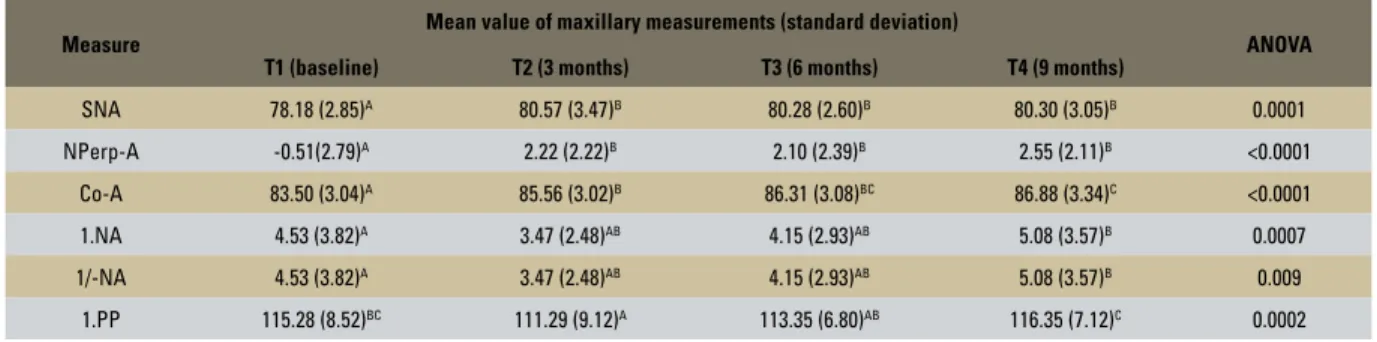
As shown in Table 3, treatment induced significant changes in the maxillomandibular relationship (ANB and Wits). These changes were significant in the first 3 months of treat-ment and remained almost stable until its end.
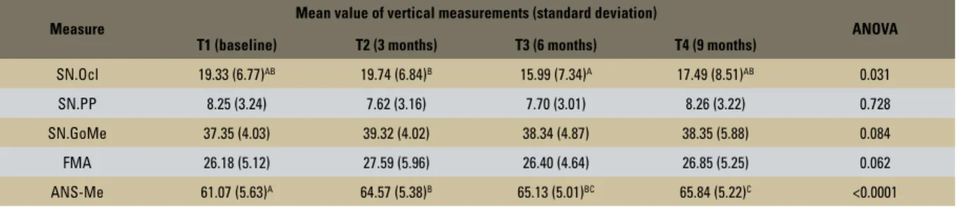
Table 4 allows assessment of vertical changes occurring during the treatment pe-riod. Although some mandibular rotation was noted during therapy (increases in FMA and SN.GoMe), this change was not statistically significant for any of the evaluated times. By the same reason, the palatal plane did not change significantly during treatment.
However, there were statistically signifi-cant changes in the anteroinferior facial height (ANS-Me) and in the occlusal plane (SN.Ocl). It is noteworthy that although there were changes in the occlusal plane during therapy, these changes were considered minor and clin-ically negligible after treatment.
Table 5 shows the measures that assess up-per and lower lip protrusion relative to Steiner’s S line (S-UL and S-LL). Upper lip protrusion was observed, with a significant increase in the
Measure
Mean value of maxillary measurements (standard deviation)
ANOVA
T1 (baseline) T2 (3 months) T3 (6 months) T4 (9 months)
SNA 78.18 (2.85)A 80.57 (3.47)B 80.28 (2.60)B 80.30 (3.05)B 0.0001
NPerp-A -0.51(2.79)A 2.22 (2.22)B 2.10 (2.39)B 2.55 (2.11)B <0.0001
Co-A 83.50 (3.04)A 85.56 (3.02)B 86.31 (3.08)BC 86.88 (3.34)C <0.0001
1.NA 4.53 (3.82)A 3.47 (2.48)AB 4.15 (2.93)AB 5.08 (3.57)B 0.0007
1/-NA 4.53 (3.82)A 3.47 (2.48)AB 4.15 (2.93)AB 5.08 (3.57)B 0.009
1.PP 115.28 (8.52)BC 111.29 (9.12)A 113.35 (6.80)AB 116.35 (7.12)C 0.0002
TABLE 1 - Means, standard deviations and variance analysis for measures used to evaluate maxillary effects.
TABLE 2 - Means, standard deviations and variance analysis for measures used to evaluate mandibular effects.
Measure Mean value of mandibular measurements (standard deviation) ANOVA
T1 (baseline) T2 (3 months) T3 (6 months) T4 (9 months)
SNB 78.20 (3.09) 77.13 (2.88) 77.27 (2.85) 77.10 (3.67) 0. 082
NPerp-Pog -1.19 (6.78) -1.69 (6.40) -1.64 (4.79) -0.67 (4.97) 0.725
Co-Gn 109.79 (5.91)A 110.89 (6.31)AB 111.94 (7.40)AB 112.95 (8.17)B 0.006
1.NB 24.61 (5.87) 22.35 (4.64) 21.43 (6.04) 21.38 (6.57) 0.096
1/-NB 3.75 (2.18) 3.08 (2.14) 3.01 (2.14) 3.10 (2.40) 0.1
IMPA 89.26 (7.15)B 85.50 (6.03)A 85.83 (7.35)A 85.10 (7.01)A 0.0002
S-UL measure during treatment. Conversely, lower lip protrusion gradually decreased until the end of treatment.
DIsCUssION
Maxillary protraction in nowadays orthodon-tics has become the most widely used technique to correct the development of Pattern III maxil-lomandibular growth pattern. The popularity of maxillary protraction has increased due to aware-ness that maxillary deficiency plays a partial or key role in the structural etiology of Pattern III. Moreover, numerous reports have demonstrated that this appliance accomplishes a higher success
rate in the long term when compared to other techniques, such as chin cups, functional appli-ances or camouflage therapy.8,23,25
Regardless of posttreatment stability, the major purpose of performing early treatment of Pattern III is to induce maximal skeletal changes with minimal dental compensation. Thus, a variety of extra and intraoral devices have been developed to enhance desirable or-thopedic effects during oror-thopedic treatment of these patients.12,14
Furthermore, the results achieved by Hol-berg, Mahani and Rudziki9 also should be tak-en into account. They reported that the forces
TABLE 3 - Means, standard deviations and variance analysis for measures used to evaluate intermaxillary effects.
TABLE 4 - Means, standard deviations and variance analysis for measures used to evaluate effects in the vertical direction.
TABLE 5 - Means, standard deviations and variance analysis for measures used to evaluate soft tissues effects.
Measure Periods ANOVA
T1 (baseline) T2 (3 months) T3 (6 months) T4 (9 months)
ANB -0.02 (3.26)A 3.34 (2.31)B 3.02 (2.41)B 3.20 (2.46)B <0.0001
Wits -5.56 (3.29)A -1.78 (4.31)B 0.48 (4.14)B -0.17 (3.46)B <0.0001
* Means followed by different letters differ significantly.
* Means followed by different letters differ significantly; where there are no letters, no significant difference was found.
* Means followed by different letters differ significantly. Measure
Mean value of vertical measurements (standard deviation)
ANOVA T1 (baseline) T2 (3 months) T3 (6 months) T4 (9 months)
SN.Ocl 19.33 (6.77)AB 19.74 (6.84)B 15.99 (7.34)A 17.49 (8.51)AB 0.031
SN.PP 8.25 (3.24) 7.62 (3.16) 7.70 (3.01) 8.26 (3.22) 0.728
SN.GoMe 37.35 (4.03) 39.32 (4.02) 38.34 (4.87) 38.35 (5.88) 0.084
FMA 26.18 (5.12) 27.59 (5.96) 26.40 (4.64) 26.85 (5.25) 0.062
ANS-Me 61.07 (5.63)A 64.57 (5.38)B 65.13 (5.01)BC 65.84 (5.22)C <0.0001
Measure Mean value of soft tissue measurements (standard deviation) ANOVA
T1 (baseline) T2 (3 months) T3 (6 months) T4 (9 months)
S-UL -0.19 (3.32)A 0.15 (3.08)AB 1.03 (3.18)B 1.36 (2.92)C 0.0003
commonly employed to promote maxillary protraction by using facemasks are apparently insufficient to significantly stimulate bone for-mation in circum-maxillary sutures. According to the authors, who analyzed maxillary pro-traction by means of the finite element meth-od, it seems unlikely that the magnitude of the stresses induced in the sutural areas of the mid-face during therapy is sufficient to gener-ate significant skeletal effects.
Therefore, the aim of this study was to eval-uate the use of maxillary protraction associat-ed with intermaxillary mechanics through the analysis of the gradual skeletal, dental and soft tissue effects induced by the treatment. Thus, the major goal of the therapy postulated in this study, which involves the concurrent use of in-termaxillary mechanics in combination with a facemask, would be to maximize the orthope-dic effect of early treatment of Pattern III.
It was noted that the mechanotherapy em-ployed in this study proved superior—in terms of skeletal advancement of the maxilla—com-pared to most findings in the literature.6,17,19,21,23,29 However, its performance was similar or inferior to others.12,13,15,22 The same applies to the maxil-lomandibular relationship, which shows that the mechanotherapy applied in this study proved effective for the treatment of Pattern III, but similar to some studies that employed facemask alone.10,11,15,22 The measures used to assess ante-rior mandibular protrusion exhibited a mild re-duction, statistically insignificant and in line with other studies.10,13,15,16,18,22,23
At the end of the treatment proposed in this study, mild dentoalveolar compensations appeared in the upper arch (slight incisors buccal inclina-tion assessed by measures 1.Na, 1-Na and 1.PP). In the lower arch, mandibular incisors experi-enced a slightly bigger retroclination but similar to what has been reported by other authors.3,8,15
Finally, these results clearly show that the si-multaneous use of facemask and intermaxillary
therapy, as suggested in this article, may not unreasonably potentiate the desired orthopedic effects in the interceptive treatment of Pattern III, but it neither maximize the role of the den-toalveolar component in the treatment.
Analysis of the four different assessment times revealed that most of the skeletal changes caused by therapy occurred within the first 3 months of treatment. After that period they remained almost constant until the end of treatment (as evidenced by the SNA, NPerp-A, Co-A, ANB and Wits mea-sures presented in Tables 1 and 3).
Although most of the skeletal changes oc-curred in the first 3 months, it should be stressed that stopping treatment at this time could lead to relapse. Further investigation is necessary to clarify these issues.
Dental compensations were more evident in the last months of maxillary protraction (T3 and T4), and the maxillary incisors flared and man-dibular incisors experienced progressive retro-clination with treatment. However, the dental changes that occurred in this study can be con-sidered minor. This leads to the conclusion that during treatment there is a tendency toward gradual dentoalveolar compensation with main-tenance of the orthopedic effects achieved in the first months of maxillary protraction therapy.
CONCLUsIONs
In observing the gradual skeletal changes that occurred during treatment one finds that virtually all significant skeletal changes took place in the first three months of treatment and remained constant until its end. From the dental point of view, there were few den-tal compensations. Vertical changes were also
reduced and had little clinical significance. After treatment, it was found that maxillary protraction combined with intermaxillary me-chanics was able to not only correct overjet but also improve sagittal relationship between basal bones and soft tissue esthetics, although these changes were not significantly enhanced when compared to others findings in the literature.
1. Baccetti T, McGill JS, Franchi L, McNamara JA Jr, Tollaro I. Skeletal effects of early treatment of Class III malocclusion with maxillary expansion and face-mask therapy. Am J Orthod Dentofacial Orthop. 1998;113(3):333-43. 2. Baccetti T, Franchi L, McNamara Jr. The cervical vertebral
maturation (CVM) method for the assessment of optimal treatment timing in dentofacial orthopedics. Semin Orthod. 2005;11:119-29.
3. Battagel JM, Orton HS. A comparative study of the effects of customized facemask therapy or headgear to the lower arch on the developing Class III face. Eur J Orthod. 1995;17(6):467-82.
4. Capelozza Filho L. Diagnóstico em Ortodontia. Maringá: Dental Press; 2004.
5. Chung CH, Font B. Skeletal and dental changes in the sagittal, vertical, and transverse dimensions after rapid palatal expansion. Am J Orthod Dentofacial Orthop. 2004;126(5):569-75.
REfERENCEs
6. Gallagher RW, Miranda F, Buschang PH. Maxillary protraction: treatment and posttreatment effects. Am J Orthod Dentofacial Orthop. 1998;113(6):612-9.
7. Haas AJ. Palatal expansion: just the beginning of dentofacial orthopedics. Am J Orthod. 1970;57(3):219-55.
8. Hägg U, Tse A, Bendeus M, Rabie BM. Long-term follow-up of early treatment with reverse headgear. Eur J Orthod. 2003;25:95-102.
9. Holberg C, Mahaini L, Rudzki I. Analysis of sutural strain in maxillary protraction therapy. Angle Orthod. 2007;77(4):586-94.
10. Kapust AJ, Sinclair PM, Turley PK. Cephalometric effects of face mask/expansion therapy in Class III children: a comparison of three age groups. Am J Orthod Dentofacial Orthop. 1998;113(2):204-12.
12. Kircelli BH, Pektas ZO, Uçkan S. Orthopedic protraction with skeletal anchorage in a patient with maxillary hypoplasia and hypodontia. Angle Orthod. 2006;76(1):156-63.
13. Cha KS. Skeletal changes of maxillary protraction in patients exhibiting skeletal class III malocclusion: a comparison of three skeletal maturation groups. Angle Orthod. 2003;73(1):26-35.
14. Liou EJ, Tsai WC. A new protocol for maxillary protraction in cleft patients: repetitive weekly protocol of alternate rapid maxillary expansions and constrictions. Cleft Palate Craniofac J. 2005;42(2):121-7.
15. McDonald KE, Kapust AJ, Turley PK. Cephalometric changes after the correction of Class III malocclusion with maxillary expansion/facemask therapy. Am J Orthod Dentofacial Orthop. 1999;116(1):13-24.
16. Nartallo-Turley PE, Turley PK. Cephalometric effects of combined palatal expansion and facemask therapy on Class III malocclusion. Angle Orthod. 1998;68(3):217-24. 17. Ngan P, Yiu C, Hu A, Hägg U, Wei SH, Gunel E.
Cephalometric and occlusal changes following maxillary expansion and protraction. Eur J Orthod. 1998;20(3):237-54. 18. Ngan PW, Hagg U, Yiu C, Wei SH. Treatment response and
long-term dentofacial adaptations to maxillary expansion and protraction. Semin Orthod. 1997;3(4):255-64. 19. Pangrazio-Kulbersh V, Berger J, Kersten G. Effects of
protraction mechanics on the midface. Am J Orthod Dentofacial Orthop. 1998;114(5):484-91.
20. Saadia M, Torres E. Sagittal changes after maxillary protraction with expansion in class III patients in the primary, mixed, and late mixed dentitions: a longitudinal retrospective study. Am J Orthod Dentofacial Orthop. 2000;117(6):669-80.
21. Silva Filho OG, Magro AC, Capelozza Filho L. Early treatment of the Class III malocclusion with rapid maxillary expansion and maxillary protraction. Am J Orthod Dentofacial Orthop. 1998;113(2):196-203.
22. Silva Filho O, Ozawa TO, Okada CH, Okada HY, Dahmen L. Anquilose intensional dos caninos decíduos como reforço de ancoragem para a tração reversa da maxila. Estudo cefalométrico prospectivo. Rev Dental Press Ortod Ortop Facial. 2006;11(6):35-44.
23. Sung SJ, Baik HS. Assessment of skeletal and dental changes by maxillary protraction. Am J Orthod Dentofacial Orthop. 1998;114(5):492-502.
24. Thiesen G, Hoffelder LB, Rego MVNN, Berthold TB, Marchioro EM. Tratamento precoce do Padrão III por meio de tração reversa da maxila. Rev Odonto Ciênc. 2004;19(45):281-6.
25. Turley PK. Orthopedic correction of Class III malocclusion with palatal expansion and custom protraction headgear. J Clin Orthod. 1988;22(5):314-25.
26. Wells AP, Sarver DM, Profit WR. Long-term eficacy
of reverse pull headgear therapy. Angle Orthod. 2006;76(6):915-22.
27. Wertz RA. Skeletal and dental changes accompanying rapid midpalatal suture opening. Am J Orthod. 1970;58(1):41-66. 28. Westwood PV, McNamara JA Jr, Baccetti T, Franchi L,
Sarver DM. Long-term effects of Class III treatment with rapid maxillary expansion and facemask therapy followed
by ixed appliances. Am J Orthod Dentofacial Orthop.
2003;123(3):306-20.
29. Williams MD, Sarver DM, Sadowsky PL, Bradley E. Combined rapid maxillary expansion and protraction facemask in the treatment of Class III malocclusions in growing children: a prospective long-term study. Semin Orthod. 1997;3(4):265-74.
Contact address
Juliana de Oliveira da Luz Fontes R. Hermínio Millis, 66 – Bom Abrigo
Zip code: 88.085-320 – Florianópolis/SC, Brazil E-mail: [email protected]