Alexandre Magno de Negreiros Diógenes*, Rildo Medeiros Matoso**, Emmanuelle Medeiros de Araújo***, Kenio Costa Lima****, Raniere Luiz dos Santos Sousa*****
Cephalometric evaluation of the effects of
a mandibular protraction appliance (MPA)
combined with fixed orthodontic appliance on
dentoalveolar and soft tissue structures of
Class II, division 1 patients
Objective: To perform a cephalometric evaluation of dentoalveolar and soft tissue changes in Brazilian youths with Class II, division 1 malocclusion, treated with a mandibular protraction appliance (MPA) combined with fixed corrective orthodontics. Methods: The sample consisted of 28 patients (16 females and 12 males) with a mean age of 13.06 years, treated for a mean period of 14.43 months. The changes were measured on 56 specific cephalometric analysis obtained from lateral cephalograms taken before and after treatment by two calibrated examiners in order to identify soft tissue and dentoalveolar changes using linear and angular cephalometric measures. The independent variables sex, age, facial pattern, MPA model, archwire, technique and treatment time were registered and analyzed using linear and angular cephalometric measures. Treatment responses were analyzed and compared by the Wilcoxon Signed Ranks and Mann-Whitney tests at a significance level of 5%. Results: The results showed dentoalveolar changes of great magnitude, which caused positive changes in soft tissue. It was also noted that the variables age, MPA model and technique influenced the treatment. Conclusions: MPA proved to be an effective alternative in the treatment of Class II, division 1 malocclusion, inducing dentoalveolar and soft tissue changes with satisfactory clinical results.
Abstract
Keywords: Cephalometry. Mandibular protraction appliance. Class II, Division 1 malocclusion. Dento-alveolar and soft tissue changes.
* Specialist in Orthodontics, in private practice.
** MSc in Orthodontics, USP. Professor and Coordinator of Orthodontics, Dentistry Department, Federal University of Rio Grande do Norte (UFRN). Professor, Specialization Program in Orthodontics, ABO-EAP/RN.
*** Specialist in Orthodontics, ABO-EAP/RN, in private practice.
**** PhD in Sciences (Medical Microbiology), Federal University of Rio de Janeiro (UFRJ). Adjunct Professor, Federal University of Rio Grande do Norte (UFRN) and Postgraduate Program in Dentistry, UFRN.
***** Specialist in Orthodontics, ABO-EAP/RN, in private practice.
How to cite this article: Diógenes AMN, Matoso RM, Araújo EM, Lima
KC, Sousa RLS. Cephalometric evaluation of the effects of a mandibular protraction appliance (MPA) combined with ixed orthodontic appliance on dentoalveolar and soft tissue structures of Class II, division 1 patients. Den-tal Press J Orthod. 2011 Nov-Dec;16(6):52-62.
intROduCtiOn
Angle Class II, Division 1 malocclusion is highly prevalent among Brazilian children, af-fecting 55% of patients with malocclusion.1 It is
characterized by lack of sagittal harmony between the basal bones due to maxillary prognathism and mandibular retrognathia, either in isolation or in combination, being the mandibular retrognathism one of the major causes of this malocclusion.23,26
In recent decades, many researchers began to develop fixed intraoral orthopedic appli-ances to correct Class II malocclusion caused by mandibular retrognathism. These devices promote changes in mandibular posture by positioning it forward and generating forces that are delivered to the teeth and basal bone, thereby correcting the problem. One such ex-ample is the Herbst appliance.15-21
Due to difficulties on importing, a lack of specialized laboratories, high costs and chal-lenging installation procedure regarding most of these appliances, Coelho Filho3,4 was driven
to devise the Mandibular Protraction Appli-ance 1(MPA 1) as an alternative to the Herbst appliance.18,19 In his articles on this subject,3,4
the author mentions as advantages of the MPA over the Herbst appliance its easy confection and installation, as well as its low cost and less bulky design, ensuring greater patient comfort.
MPA 1 was made with 0.032-in (0.9 mm) wire, which received two small loops between the headgear tube and the distal bend of the mandibular canine.
Although effective, it did not allow brackets to be bonded to premolars, mouth opening was limited and it broke frequently, leading the au-thor to develop a second version.3,4,9 This new
version consisted of two 0.032-in archwire segments with loops in the ends and an open coil intending to maintain a proper relation-ship between the archwire segments, allowing greater mouth opening range and the bonding of brackets to premolars.
Coelho Filho3 described 4 clinical cases of
Class II patients with mandibular retrogna-thism treated during the growth period with fixed appliance and MPA. Noteworthy among the changes were those of a dentoalveolar na-ture, such as lingual inclination of maxillary incisors. However, some skeletal changes were also observed such as increases in the length of the mandible and its ascending ramus, result-ing in reduced facial convexity, improved soft tissue profile and decreased overjet.
The author went on to develop the MPA 3,5,6 with quite different characteristics, with
the purpose of overcoming some limitations of previous versions.
MPA 3 used 0.9 mm wire rods that ran through telescopic stainless steel tubes. This imparted greater stability to the appliance during mouth opening and closing and was easier to install. However, it required a more careful fabrication procedure and, thereby, more complex.
Between the years 2001 and 2002, Coelho Filho9,10,11,13 made changes on MPA 3 with a
new design for the upper arch intermaxillary telescopic tube fitting, enhancing stability dur-ing mouth opendur-ing and closdur-ing. This new ver-sion, named MPA 4,9,10,11,13 surpassed all
previ-ous appliances in terms of breaking strength, ease of installation and fabrication. The
au-thor11,13 pointed out that the current version
has shown clinical efficacy and final results similar to those of previous models since the mechanical principles are the same.
MEtHOdS
This is an uncontrolled, non-randomized clinical trial conducted on a sample comprising 56 lateral cephalograms (28 initial and 28 final cephalograms) of 28 Brazilian patients with An-gle Class II, Division 1 malocclusion, 12 males and 16 females, mean age of 13.06 years at the beginning of treatment, who were treated for a mean period of 14.43 months with mandibular protraction appliances (MPA) associated with fixed orthodontic appliance. All patients were treated at the private practice of Professor Car-los Martins Coelho Filho in São Luís, Maranhão State, Brazil. The following inclusion criteria were adopted: Angle Class II, Division 1 maloc-clusion with mandibular retrognathism, as as-sessed by photographs, study models, in addition to cephalograms that allowed clear visualization of the structures of interest. Sample exclusion criteria were: Patients who had agenesis, extrac-tion or missing permanent teeth, cases of Angle Class II, Division 1 treated only with MPA, pa-tients with pronounced overjet.
A clinical form was used for data collection in-cluding seven variables: Patient age, sex, facial pat-tern (dolichofacial, mesofacial and brachyfacial, although the latter was excluded during sample selection as only one case displayed this facial type, which might lead to statistical results with a higher margin of error), MPA model (types 1, 2, 3 and 4), total time of appliance usage, archwires used during treatment with MPA (0.019 x 0.025-in, 0.021 x 0.025-in and 0.018 x 0.025-in stainless steel wires) as well as the orthodontic technique (standard Edgewise and Straight-Wire).
Research instruments consisted of 56 lateral cephalometric radiographs obtained at two stag-es: before (T1) and after treatment (T2). All ra-diographs used in this study were obtained with a Funk Orbital X15 X-ray device, with a magnifi-cation factor of 9%, by the same operator.
A sheet of acetate paper was fixed over each radiograph with adhesive tape and the
anatomi-cal structures of interest for the construction of the cephalometric analysis were traced. This tracing was performed in a darkened room by two previously calibrated examiners on a view-box to facilitate the visualization of structures.
Examiners calibration lasted approximately three months, during which 30 randomly select-ed radiographs were tracselect-ed and comparselect-ed until minimum error was attained.
The materials used by examiners to build the cephalometric tracing consisted of a viewbox, sheet of transparent acetate paper (Cephalo-metric Tracing Paper® - GAC), 0.07 mm thick,
size 17.5 x 17.5 cm, Pentel mechanical pencil with 0.3 mm fine point, template (Tracing Tem-plate®, Unitek Corp.), millimeter ruler,
adhe-sive tape, soft eraser and black cardboard mask. When double images of the anatomical bony structures were visualized both images were traced and a mean position between them was found for determining the cephalometric points.
In a following stage, the images were import-ed via a scanner into a microcomputer containing the Radiocef Studio®cephalometry program (No.
020576, version 4.0, release 3 - Belo Horizonte, Brazil), which was used to obtain the values for T1 and T2 as well as their respective repetitions. The results were stored and subsequently sub-jected to statistical analysis.
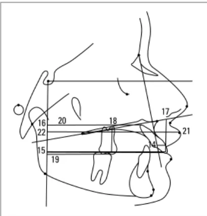
Changes in the dental and soft tissue struc-tures were measured by means of the following angular and linear cephalometric measures. Angu-lar: 1.NA (1), 1.NB (2), IMPA (3), ANL (4); Lin-ear: 1-NB (5), 1-FHp (6), 1a-FHp (7), 1-MP (8), 6-MP (9), 6-FHp (10), 6a-FHp (11), LL-Pog’Sn (12), LL-FHp (13), 1-NA (14), 1-FHp (15), 1a-FHp (16), 1-PP (17), 6-PP (18), 6-1a-FHp (19), 6a-FHp (20), UL-Pog’Sn (21), UL-6a-FHp (22).
1
2 3
4
5 6
7
8 9 10
11
12
14 15
16
17 18
19 20
21 22
13
6-PM (9), 6-FHp (10) and 6a-FHp (11). For the soft tissue profile: ANL (4), UL-Pog’Sn (21), LL-Pog’Sn (12), UL-FHp (22) and LL-FHp (13).
RESuLtS
The dentoalveolar and soft tissue changes in-duced by the combined use of MPA and a fixed orthodontic appliance were evaluated in 28 patients with Class II, Division 1 malocclusion with mean age of 13.06 years, treated for a mean period of 14.43 months (53.6%). These changes were measured in 56 lateral radiographs in two stages: Before (T1) and after treatment (T2).
Regarding the MPA model, the sample ini-tially consisted of four groups of patients that corresponded to the four types of MPA. How-ever, the frequency of the group that used MPA models 1 and 3 was low and statistically discrep-ant in relation to models 2 and 4. Therefore, the four were gathered into two groups — labeled 0 and 1 respectively — the first resulting from the sum of the number of patients with MPA 1 and 2 (46.4%) and the second, the sum of those using MPA models 3 and 4 (53.6%).
For the variable archwire, the following types were noted: 0.019 x 0.025-in stainless steel (57.1% or 16 patients), 0.021 x 0.025-in stainless steel (10.7% or 3 patients) and 0.018 x 0.025-in stainless steel (32.1% or 9 patients). The two latter were also grouped into a total of 12 cases.
The sample consisted of 57.1% (16 patients) of female subjects and 42.9% (12 patients) male. Only two of the three facial patterns (me-sofacial and dolichofacial) were analyzed. The first type accounted for 64.3% (18 patients) of patients and the second, 35.7% (10 patients).
The Straight-Wire technique was used in 42.9% (12 patients) of individuals and stan-dard Edgewise in 57.1% (16 patients). Measures 1-NB, ILi-FHp and 6-PP showed statistically significant differences compared to the group using the Edgewise technique.
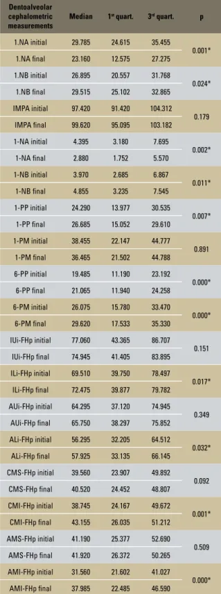
Tables 1 and 2 show the initial (T1) and fi-nal (T2) dentoalveolar and soft tissue cephalo-metric measurements of patients of both sexes, their medians, 1st and 3rd quartiles, and
statis-tical significance (p-value), obtained with the Wilcoxon Signed Ranks Test. Results were
con-FIGURE 1 - Angular cephalometric measure-ments.
FIGURE 2 - Linear cephalometric measure-ments. Lower dentoalveolar and soft tissue components.
Dentoalveolar cephalometric measurements
Median 1st quart. 3rd quart. p
1.NA initial 29.785 24.615 35.455
0.001*
1.NA final 23.160 12.575 27.275
1.NB initial 26.895 20.557 31.768
0.024*
1.NB final 29.515 25.102 32.865
IMPA initial 97.420 91.420 104.312
0.179
IMPA final 99.620 95.095 103.182
1-NA initial 4.395 3.180 7.695
0.002*
1-NA final 2.880 1.752 5.570
1-NB initial 3.970 2.685 6.867
0.011*
1-NB final 4.855 3.235 7.545
1-PP initial 24.290 13.977 30.535
0.007*
1-PP final 26.685 15.052 29.610
1-PM initial 38.455 22.147 44.777
0.891
1-PM final 36.465 21.502 44.788
6-PP initial 19.485 11.190 23.192
0.000*
6-PP final 21.065 11.940 24.258
6-PM initial 26.075 15.780 33.470
0.000*
6-PM final 29.620 17.533 35.330
IUi-FHp initial 77.060 43.365 86.707
0.151
IUi-FHp final 74.945 41.405 83.895
ILi-FHp initial 69.510 39.750 78.497
0.017*
ILi-FHp final 72.475 39.877 79.782
AUi-FHp initial 64.295 37.120 74.945
0.349
AUi-FHp final 65.750 38.297 75.852
ALi-FHp initial 56.295 32.205 64.512
0.032*
ALi-FHp final 57.925 33.135 66.145
CMS-FHp initial 39.560 23.907 49.892
0.092
CMS-FHp final 40.520 24.452 48.807
CMI-FHp initial 38.745 24.167 49.672
0.001*
CMI-FHp final 43.155 26.035 51.212
AMS-FHp initial 41.190 25.377 52.690
0.509
AMS-FHp final 41.920 26.372 50.265
AMI-FHp initial 31.560 21.602 41.027
0.000*
AMI-FHp final 37.985 22.485 46.590
TABLE 1 - Median, 1st and 3rd quartiles for differences between initial and final
dentoalveolar cephalometric measurements and significance value.
TABLE 2 - Median, 1st and 3rd quartiles for differences between initial and final
soft tissue cephalometric measurements and significance value.
*Significant difference (p<0.05) based on the Wilcoxon test.
*Significant difference (p<0.05) based on the Wilcoxon test.
Soft tissue cephalometric measurements
Median 1st quart. 3rd quart. p
ANL initial 111.170 103.657 116.367
0.145
ANL final 112.790 106.480 118.387
UL-Pog’Sn initial 5.030 2.815 7.295
0.001*
UL-Pog’Sn final 3.195 1.945 6.265
LL-Pog’Sn initial 2.155 1.037 3.445
0.838
LL-Pog’Sn final 1.795 1.032 3.435
Pog’-FHp initial 76.170 44.387 87.030
0.010*
Pog’-FHp final 79.970 45.685 89.912
UL-FHp initial 88.905 50.940 101.325
0.48
UL-FHp final 91.690 50.435 101.605
LL-FHp initial 69.510 39.750 78.490
0.016*
LL-FHp final 87.190 48.200 95.390
sidered significant at 5% significance (p<0.05), indicating at least 95% confidence in the con-clusions. In Tables 1 and 2 are shown the fol-lowing statistically significant dentoalveolar and soft tissue cephalometric measures: 1.NA, 1.NB, 1-NA, 1-NB, 1-PP, 6-PP, 6-PM, ILi-FHp, ALi-FHp, CMI-FHp, AMI-FHp (Table 1), UL-Pog´Sn, Pog´-FHp and LL-FHp (Table 2).
Table 3 shows that sex was the only vari-able with a statistically significant influence on cephalometric measures 6-PM and AMI-FHp before treatment.
*Significant difference (p<0.05) based on the Mann-Whitney test.
*Significant difference (p<0.05).
TABLE 3 - Median, 1st and 3rd quartiles and p-value for cephalometric measurements related to the independent variable sex at T1.
TABLE 4 - Median, 1st and 3rd quartiles and p-value for cephalometric measurements related to the independent variables.
Independent variable Initial 6-PM Initial AMI-FHp
Sex n median 1st quart. 3rd quart. p median 1st quart. 3rd quart. p
Female 16 16.325 11.252 30.937
0.016*
22.295 12.995 38.367
0.014*
Male 12 29.760 26.590 34.347 38.230 32.315 44.500
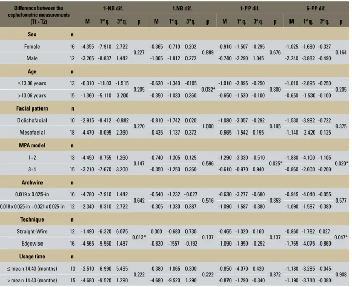
Difference between the cephalometric measurements
(T1 - T2)
1-NB dif. 1.NB dif. 1-PP dif. 6-PP dif.
M 1st q. 3rd q. p M 1st q. 3rd q. p M 1st q. 3rd q. p M 1st q. 3rd q. p
Sex n
Female 16 -4.355 -7.910 2.722
0.227 -0.365 -0.710 0.202 0.889 -0.910 -1.507 -0.295 0.676 -1.025 -1.680 -0.327 0.164
Male 12 -3.265 -8.837 1.442 -1.065 -1.812 0.272 -0.740 -2.290 1.045 -2.240 -3.882 -0.490
Age n
≤13.06 years 13 -6.310 -11.03 -1.515 0.205
-0.620 -1.340 -0105 0.032*
-1.010 -2.895 -0.250 0.300
-1.010 -2.895 -0.250 0.205 >13.06 years 15 -1.360 -5.110 3.200 -0.350 -1.030 0.360 -0.650 -1.530 -0.100 -0.650 -1.530 -0.100
Facial pattern n
Dolichofacial 10 -2.915 -8.412 -0.982 0.270
-0.810 -1.742 0.020 1.000
-1.080 -3.057 -0.292 0.195
-1.530 -3.992 -0.722 0.375
Mesofacial 18 -4.470 -8.095 2.360 -0.435 -1.137 0.372 -0.665 -1.542 0.195 -1.140 -2.420 -0.125
MPA model n
1+2 13 -4.450 -8.755 1.260
0.147
-0.740 -1.305 0.125 0.596
-1.290 -3.330 -0.510 0.025*
-1.880 -4.100 -1.105 0.020*
3+4 15 -3.210 -7.670 3.200 -0.350 -1.250 0.360 -0.610 -0.970 0.940 -0.860 -2.600 -0.200
Archwire n
0.019 x 0.025-in 16 -4.780 -7.910 1.442
0.642 -0.540 -1.232 -0.027 0.516 -0.630 -3.277 -0.680 0.353 -0.945 -4.040 -0.055 0.577 0.018 x 0.025-in + 0.021 x 0.025-in 12 -2.340 -8.310 2.722 -0.305 -1.330 0.387 -1.090 -1.587 -0.380 -1.090 -1.587 -0.380
Technique n
Straight-Wire 12 -1.490 -6.320 6.075 0.013*
0.300 -0.680 0.730 0.137
-0.465 -1.020 0.160 0.137
-0.860 -1.782 0.027 0.047*
Edgewise 16 -4.565 -9.560 1.487 -0.830 -1557 -0.192 -1.090 -1.950 -0.292 -1.765 -4.075 -0.860
Usage time n
≤ mean 14.43 (months) 13 -2.510 -6.990 5.495 0.222
-0.380 -1.065 0.300 0.222
-0.850 -4.070 0.420 0.872
greater extrusion of incisors and molars found in the group using the MPA 1+2. The group us-ing the Edgewise technique showed statistically significant measurements in relation to the la-bial inclination of lower incisors and increased extrusion of maxillary molars.
diSCuSSiOn
The use of MPA in Angle Class II treatment is aimed at correcting the sagittal relationship between the maxilla and the mandible, main-ly through dentoalveolar changes.3-13 In order
to evaluate the influence of the independent
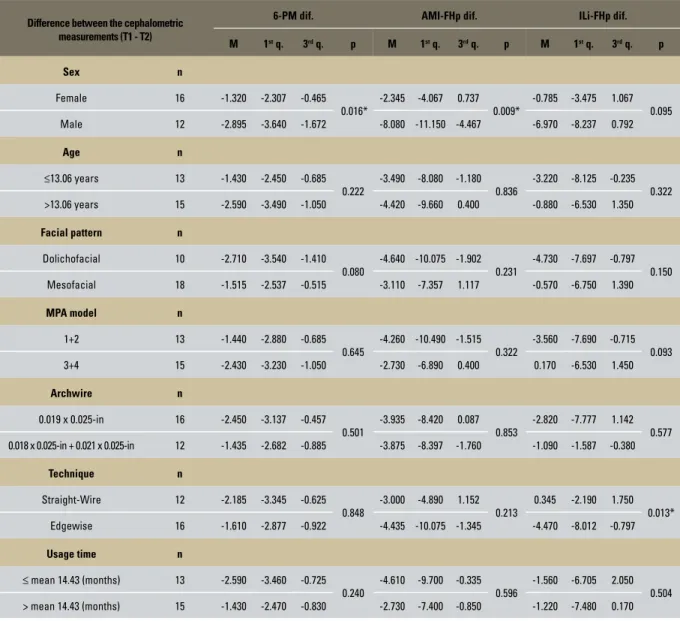
Difference between the cephalometric measurements (T1 - T2)
6-PM dif. AMI-FHp dif. ILi-FHp dif.
M 1st q. 3rd q. p M 1st q. 3rd q. p M 1st q. 3rd q. p
Sex n
Female 16 -1.320 -2.307 -0.465
0.016*
-2.345 -4.067 0.737 0.009*
-0.785 -3.475 1.067 0.095
Male 12 -2.895 -3.640 -1.672 -8.080 -11.150 -4.467 -6.970 -8.237 0.792
Age n
≤13.06 years 13 -1.430 -2.450 -0.685
0.222
-3.490 -8.080 -1.180 0.836
-3.220 -8.125 -0.235 0.322
>13.06 years 15 -2.590 -3.490 -1.050 -4.420 -9.660 0.400 -0.880 -6.530 1.350
Facial pattern n
Dolichofacial 10 -2.710 -3.540 -1.410
0.080
-4.640 -10.075 -1.902 0.231
-4.730 -7.697 -0.797 0.150
Mesofacial 18 -1.515 -2.537 -0.515 -3.110 -7.357 1.117 -0.570 -6.750 1.390
MPA model n
1+2 13 -1.440 -2.880 -0.685
0.645
-4.260 -10.490 -1.515 0.322
-3.560 -7.690 -0.715 0.093
3+4 15 -2.430 -3.230 -1.050 -2.730 -6.890 0.400 0.170 -6.530 1.450
Archwire n
0.019 x 0.025-in 16 -2.450 -3.137 -0.457
0.501
-3.935 -8.420 0.087 0.853
-2.820 -7.777 1.142 0.577
0.018 x 0.025-in + 0.021 x 0.025-in 12 -1.435 -2.682 -0.885 -3.875 -8.397 -1.760 -1.090 -1.587 -0.380
Technique n
Straight-Wire 12 -2.185 -3.345 -0.625
0.848
-3.000 -4.890 1.152 0.213
0.345 -2.190 1.750 0.013*
Edgewise 16 -1.610 -2.877 -0.922 -4.435 -10.075 -1.345 -4.470 -8.012 -0.797
Usage time n
≤ mean 14.43 (months) 13 -2.590 -3.460 -0.725
0.240
-4.610 -9.700 -0.335 0.596
-1.560 -6.705 2.050 0.504
> mean 14.43 (months) 15 -1.430 -2.470 -0.830 -2.730 -7.400 -0.850 -1.220 -7.480 0.170
TABLE 5 - Median, 1st and 3rd quartiles and p-value for cephalometric measurements related to the independent variables.
variables sex, age, facial pattern, MPA model, archwire, technique and MPA treatment time in the sample described before, the means of cephalometric differences between T1 and T2 were compared and yielded statistically signifi-cant results.
The results displayed in these tables show that, for the variable sex, measures 6-PM and AMI-FHp were significant, and higher for fe-males than fe-males. Based on the results of the significance test (p-value), that show which in-dependent variables affected treatment (Table 5), cephalometric measures 6-PM and AMI-FHp were statistically significant at the beginning of treatment, with measurements equal to the me-dians, which shows that the variable sex did not influence treatment outcome, since measure-ments were already significant prior to treatment. The only measure that showed a statisti-cally significant median value with respect to the variable age was 1.NB. The mean found for patients aged below 13.06 years was asso-ciated with greater mandibular incisor inclina-tion. This result is probably related to a failure in banding the lower second molars, which re-duced anchorage. Moreover, another factor that probably contributed to a smaller inclination of the lower incisors in the group older than 13.06 years was that in this group there was a more pronounced mandibular growth component, thereby moving point B to a more anterior posi-tion by correcting the skeletal discrepancy and consequently causing less dental compensation (mandibular incisor inclination).
Regarding variable MPA model, Group 1 (MPA 3+4) showed statistically significant means between T1 and T2 for measures 1-PP and 6-PP. These results were probably due to the aligning and leveling extrusive mechanics applied prior to MPA use and growth. Group 1 (MPA 3+4) showed lower extrusion of incisors and molars relative to the palatal plane, prob-ably due to increased uprighting of the incisors
and limited extrusion of upper molars related to a lower breakage rate in this group.
Measures 1-NB, ILi-FHp and 6-PP showed statistically significant differences in relation to Group 1 (Edgewise technique) for the inde-pendent variable technique. This result prob-ably occurred because the group did not use pre-adjusted brackets, resulting in greater incli-nation of mandibular incisors and consequent lower lip protrusion. In Group 2 (Straight-Wire technique), there was significantly less extru-sion of upper molars, probably due to palatal torque of molar crowns, which positioned the roots of these teeth on the buccal cortex, there-by strengthening anchorage.22,25 The other
inde-pendent variables (facial pattern, archwire and MPA use time) showed no statistically signifi-cant differences.
In order to facilitate the interpretation of re-sults a separate discussion was conducted on the changes that took place in the maxillary dento-alveolar component, mandibular dentodento-alveolar component and soft tissue component.
Maxillary dentoalveolar component
The MPA forces used in this investigation were delivered by means of dental structures and, thus, significant dentoalveolar effects were expected. The maxillary alveolar component (Table 1) was assessed by means of measures 1.NA, 1-NA, IUi-FHp, AUi-IUi-FHp, 6-PP, CMS-IUi-FHp, 1-PP, AMS-FHp. Assessment of the position and inclination of up-per incisors (1.NA, 1-NA, IUi-FHp) showed a marked lingual inclination that was statistically significant for measures 1.NA and 1-NA, and with no statistical significance for measures IUi-FHp and AUi-IUi-FHp, corroborating Coelho Filho,3-13
Siqueira24 and White.27 The results showed a slight
These results were probably due to alveolar bone growth2,14 and the extrusive aligning and leveling
mechanics utilized prior to MPA use. MPA use is probably not related to the extrusion of molars and incisors, since according to Coelho Filho,13
MPA generates intrusive and distal forces and is therefore indicated for use in Class II patients with a high mandibular plane angle.
Mandibular dentoalveolar component
It was assessed trough measures 1.NB, 1-NB, IMPA, ILi-FHp, 1-PM, ALi-FHp, 6-PM, CMI-FHp and AMI-CMI-FHp (Table 1). However, most measures that evaluated lower incisor position (1.NB, 1-NB, ILi-FHp, ALi-FHp) demonstrated significant proclination and protrusion of these teeth, with the exception of IMPA which, al-though increased, was not statistically significant. This result was probably due to the occurrence of bone apposition in the mandibular plane dur-ing treatment.2,14 The measures related to the
as-sessment of lower molar positioning (CMI-FHp, AMI-FHp, 6-PM) exhibited significant mesial movement and extrusion of these teeth. In agree-ment with these results, Siqueira24 hinted that
these effects probably occur due to the direction of forces delivered by the device owing to a lim-ited vertical development of upper molars. It is also believed that the mechanical alignment and leveling occurring prior to MPA use contributes to the extrusion of lower molars. Assessment of the degree of lower incisor intrusion, as revealed by measure 1-PM, showed little intrusion of these teeth, although not statistically significant. This decreased value may be due to lower inci-sor protrusion, which reduces the distance from their incisal edge to the mandibular plane.
Soft tissue component
This component was analyzed by means of cephalometric measures ANL, UL-Pog´Sn, LL-Pog´Sn, Pog´-FHp, UL-FHp, LL-FHp (Table 2). In assessing the upper lip, however, two measures
(UL-Pog´Sn and UL-FHp) disclosed that the up-per lip was retracted, following the retrusion and lingual inclination of the upper incisors, while only UL-Pog’Sn exhibited a statistically signifi-cant difference. This fact is probably related to bone apposition in the pogonion2,14 and the more
anterior position of the mandible at the end of treatment. According to Coelho Filho3-13 and
White27, the dentoskeletal changes induced by
MPA use caused favorable changes in soft tissue, such as upper lip retraction, which improves the soft tissue profile. The lower lip was examined by cephalometric measures LL-Pog´Sn, LL-FHp and Pog´-FHp by comparing T1 with T2. Measures LL-Pog´Sn and LL-FHp showed protrusion of the lower lip due to a marked proclination and pro-trusion of the mandibular incisors at the end of orthodontic treatment, although only measures LL-FHp and Pog’-FHp were statistically signifi-cant. It is worth noting that measure Pog’-FHP is related to bone apposition of the pogonion2,14
and a more anterior position of the mandible at the end of treatment.
These findings, however, cannot be considered fully conclusive due to some limitations in this study, among which are a small sample size, ab-sence of a control group and the fact that patients were not randomly assigned. Thus, further stud-ies with larger samples are warranted in order to assess the influence of independent variables and compare results with the control group.
COnCLuSiOnS
Based on the methods employed and results achieved in this study, the authors concluded that treatment with MPA combined with a fixed orthodontic appliance for correction of Class II, Division 1 malocclusion produces considerable dentoalveolar changes, which can be summa-rized as follows:
2. Mandibular dentoalveolar component: There was protrusion and proclination of the lower incisors, in addition to mesial drift and extrusion of the lower molars. 3. Soft tissue component: The dentoalveolar
changes exerted a positive, significant influ-ence on the soft tissue profile of the patients. 4. Age: A greater lower incisor inclination
was noted in the group of patients
young-er than 13.06 years.
5. MPA model: Less extrusion was observed in the upper incisors and molars in the group that used MPA 3 and 4 due to the greater effectiveness of these appliances. 6. Technique: A higher labial inclination of
lower incisors and greater extrusion of up-per molars were noted in the group using standard Edgewise brackets.
1. Almeida-Pedrin RR, Pinzan A, Almeida RR, Almeida MR, Henriques JFC. Efeitos do AEB conjugado e do Bionator no tratamento da Classe II, 1ª divisão. Rev Dental Press Ortod Ortop Facial. 2005;10(5):37-54.
2. Björk A. Facial growth in man, studied with the aid of metallic implants. Acta Odontol Scand. 1955;13(1):9-34.
3. Coelho Filho CM. Mandibular protraction appliance for Class II treatment. J Clin Orthod. 1995;29(5):319-36.
4. Coelho Filho CM. Clinical application of the Mandibular Protraction Appliance. J Clin Orthod. 1997;31(2):92-102. 5. Coelho Filho CM. Emprego do aparelho de protração
mandibular no tratamento das maloclusões das Classes I e II. 9º Livro Anual do Grupo Brasileiro de Professores de Ortodontia e Odontopediatria. São Paulo; GBPOO; 1997. p. 122-9. 6. Coelho Filho CM. The Mandibular Protraction Appliance n. 3. J
Clin Orthod. 1998;32(6):379-84.
7. Coelho Filho CM. Emprego clínico do aparelho de projeção de mandíbula. Rev Dental Press Ortod Ortop Facial. 1998;3(5):69-130.
REfEREnCES
8. Coelho Filho CM. Crônica de uma vida na Ortodontia. In: Feres MAL, Teodoro L. Ortodontia: algumas histórias de sucesso. 1ª ed. Curitiba: Editek; 1999. p. 23-30. 9. Coelho Filho CM. O aparelho de Protração Mandibular.
In: Baptista JM. E-book de Ortopedia Facial e Ortodontia. 1ª ed. Curitiba: Editek; 2000.
10. Coelho Filho CM. Mandibular Protraction Appliances IV. J Clin Orthod. 2001;35(1):18-24.
11. Coelho Filho CM. O Aparelho de Protração Mandibular IV. Rev Dental Press Ortod Ortop Facial. 2002;7(2):49-60. 12. Coelho Filho CM. O Aparelho de Protração Mandibular
(APM) no tratamento de pacientes adultos. In: Sakai E, Martins NS, Fiúza SC, Barbosa RLL, Grimberg JC, Pereira CCB, et al. Nova visão em Ortodontia e Ortopedia Facial. 1ª ed. São Paulo: Ed. Santos; 2002. p. 457-63.
13. Coelho Filho CM. Entrevista com Carlos Martins Coelho Filho. Rev Dental Press Ortod Ortop Facial. 2003;2(5):5-11. 14. Enlow DH. Crescimento Facial. 3ª ed. São Paulo: Artes
Contact address
Alexandre Magno de Negreiros Diógenes R. Duodécimo Rosado, 322 – Nova Betânia Zip code: 59.607-020 – Mossoró / RN, Brazil E-mail: [email protected]
Submitted: August 26, 2008 Revised and accepted: April 21, 2009
15. Konik M, Pancherz H, Hansen K. The mechanism of Class II correction in late Herbst treatment. Amer J Orthod. 1997;112(1):87-91.
16. Lai M. Molar distalization with the Herbst appliance. Semin Orthod 2000;6(5):119-28.
17. Manfredi C, Cimino R, Trani A, Pancherz H. Skeletal changes of Herbst appliance therapy investigated with more conventional cephalometrics and European norms. Angle Orthod. 2001;71(3):170-6.
18. Pancherz H. Treatment of Class II malocclusions by jumping the bite with the Herbst appliance: a cephalometric investigation. Am J Orthod. 1979;76(4):423-42.
19. Pancherz H. The Herbst appliance: its biologic effects and clinical use. Am J Orthod. 1985;87(1):1-20.
20. Pancherz H. The effects, limitations and long-term dentofacial adaptations to treatment with the Herbst appliance. Semin Orthod. 1997;3(4):232-43.
21. Pancherz H, Ruf S, Kohlhas P. Effective condylar growth and chin position changes in Herbst treatment: a cephalometric long-term study. Am J Orthod. 1998;114(4):437-46.
22. Ricketts RM. Bioprogressive therapy. Denver: Rocky Mountain Orthodontics; 1979.
23. Rothstein TL. Facial morphology and growth from 10 to 14 years of age in children presenting Class II, division 1 malocclusion: a comparative roentgenographic cephalometric study. Am J Orthod. 1971;60(6):619-20.
24. Siqueira DF. Estudo comparativo, por meio de análise cefalométrica em norma lateral, dos efeitos dentoesqueléticos e tegumentares
produzidos pelo aparelho extrabucal cervical e pelo aparelho de protração mandibular, associados ao aparelho ixo, no tratamento
da Classe II, 1ª divisão de Angle [tese]. Bauru (SP): Universidade de São Paulo; 2004.
25. Tweed CH. Clinical orthodontics. St. Louis: CV Mosby; 1966. 2v. 26. Vale DMV. Avaliação cefalométrica das estruturas dentoesqueléticas
em jovens portadores de Classe II, divisão 1, brasileiros, leucodermas e de origem mediterrânea [dissertação]. Bauru (SP): Universidade de São Paulo; 1985.