Eduardo Jacomino Franco*, Arnaldo Pinzan**, Guilherme Janson***,
José Fernando Castanha Henriques****, Célia Regina Maio Pinzan-Vercelino*****
Cephalometric study of tooth position in
young Afro-Caucasian Brazilian individuals
with normal occlusion
Objective: The purpose of this study was to achieve a specific cephalometric pattern for young Afro-Caucasian Brazilian individuals and verify the presence of dimorphism between sexes. Methods: The sample was composed of 40 lateral cephalograms of young Afro-Cauca-sian Brazilian individuals (mulattos), 20 males (mean age 13.25 years) and 20 females (mean age 13.10 years), with normal occlusion and no previous orthodontic treatment. The cephalo-metric variables were determined according to the analyses of Downs, Steiner, Riedel, Tweed, McNamara, Ricketts and Interlandi. Independent t test was applied to compare the variables between sexes. Results: The maxillary and mandibular incisors were protruded and buccally tipped. There was no statistically significant difference between sexes in all variables. Con-clusions: It was observed that young Afro-Caucasian Brazilian individuals without skeletal alterations and with normal occlusion showed specific tooth position and facial features in relation to the other Brazilian ethnic groups.
Abstract
Keywords: Ethnic groups. Cephalometrics. Incisors.
* MSc and Specialist in Orthodontics, Bauru Dental School, University of São Paulo (FOB-USP). Professor of the Specialization Course in Orthodontics, Funorte/ IBPG and Uninga/Bauru.
** Associate Professor of Orthodontics, FOB-USP.
*** Head Professor of Orthodontics, FOB-USP. Coordinator of the Post Graduation Course in Orthodontics, FOB-USP.
**** Head Professor of Orthodontics, FOB-USP. Coordinator of the Post Graduation Course in Orthodontics and Specialization, FOB-USP. ***** PhD in Orthodontics, FOB/USP. Assistant Professor, MSc Program in Dentistry, area of concentration Orthodontics, Uniceuma, São Luís, Brazil.
How to cite this article: Franco EJ, Pinzan A, Janson G, Henriques JFC,
Pinzan-Vercelino CRM. Cephalometric study of tooth position in young Afro-Caucasian Brazilian individuals with normal occlusion. Dental Press J Orthod. 2011 Nov-Dec;16(6):41-51.
» The authors report no commercial, proprietary, or inancial interest in the
intROduCtiOn
Population context determines an intense variation in ethnic groups, especially in large urban centers, evidencing the need to acknowl-edge that a single pattern of facial esthetics may not be appropriate for diagnostic decisions and treatment planning for individuals of different ethnic backgrounds, who migrated to distinct geographic regions.12,16
In Brazil, the admixture between Portuguese settlers, Brazilian Amerindians and Africans led to the formation of a diversified population, and a significant part of the Brazilian population was originated from relationships between White-Black (Mulatto), White-Amerindian (Caboclo) and Black-Amerindian (Zambo). Each one of the three basic groups (Amerindian, White and Black) does not represent a pure ethnicity. It is important to identify the characteristics of the Brazilian population and to analyze the respec-tive somatic aspects.36
The importance of investigating the skeletal and dentofacial components and relate them to normal and individual characteristics in the ethnic groups, with different cultural and so-cial influences, is justified by orthodontic treat-ment limitations and its clinical implications, especially when the mechanics impairs the facial esthetics.7,8,13-16,20,22,27,28,42 The normative values of cephalometric measurements, which are specific for different ethnic groups, should complement the diagnosis and treatment plan-ning according to the individual needs and expectations of the patient.1,39 The literature highlights the lack of cephalometric studies related to differences in facial morphology between ethnic groups.1,7,14,15,26,29 The applica-bility of characteristics of facial, skeletal and soft tissue patterns should also be determined to establish the diagnosis and treatment plan-ning of malocclusions, aiming to achieve occlu-sal, functional and skeletal ideal relationships. Therefore, the cephalometric and occlusal
studies aim to promote stable orthodontic cor-rections, with ideal tooth position.
Therefore, considering the lack of specific studies in Afro-Caucasian individuals, the pres-ent study aimed to determine the existing ceph-alometric variables for the ideal maxillary and mandibular incisors position in Mulatto indi-viduals with balanced faces.
EtHniCitY
Since the terms “racial” and “ethnic” may induce doubtful interpretations, it was used the classification of Cuvier (cited by Ávila5), which highlights the three main racial groups according to differentiation criteria related to the color of human skin, i.e.:
» White race; » Yellow race;
» Black race.
The meaning of each ethnic group is closely related to culture conditions and sociopoliti-cal integration of members of each population, regarding the language, habits and way of life. Therefore, an ethnic group is a population among others that constitutes a racial group when collectively grouped, but maintain their physical and cultural differences by limiting mechanisms as geographic or social barriers.6,37
The skin color classification for Mulatto in-dividuals is due to the miscegenation between Whites and Blacks.
In 1999, IBGE (Brazilian Institute for Ge-ography and Statistics) published data reveal-ing that Brazilian Black population was only 5.4%. However, with the increase of 39.9% of Mulattos, Brazil was mainly defined as a Black country, as it is currently considered by many Americans and Europeans.
FIGURE 2 - Intraoral photographs. Deter-mination of occlusal characteristics in a young Afro-Caucasian individual, with nor-mal occlusion.
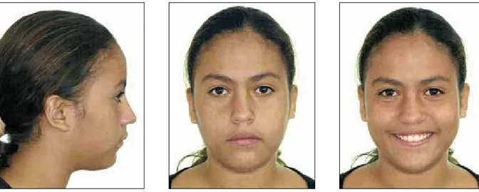
FIGURE 1 - Extraoral photographs. Young Afro-Caucasian male individual. since the normal relationship between skel-etal and tooth position is different according to ethnic variations. Thus, studies should be conducted to support the diagnosis, especially in Afro-Caucasian individuals (Mulattos), be-cause they constitute a large community with-in the Brazilian population.
PROPOSitiOn
This study aimed to present an individual cephalometric pattern for tooth position in young Afro-Caucasian Brazilian individuals with
age ranging from 12 to 14 years, by means of: » Achieving mean values of normality for
dental cephalometric measurements. » Identifying the presence or absence of
di-morphism between sexes.
MAtERiAL And MEtHOdS
FIGURE 3 - Extraoral photographs. Young Afro-Caucasian female individual.
FIGURE 4 - Intraoral photographs. Determi-nation of occlusal characteristics in young female individual with normal occlusion. The ethnic and racial characteristics were
accurately evaluated by a questionnaire, which provided information to classify the skin color of the parents. Therefore, only young individu-als of African descent were included in the sample. Adolescent clinical examinations were performed using a disposable spatula as an ef-fective instrument for intraoral examination.
Additional inclusion criteria were: Presence of permanent teeth in occlusion, but third
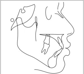
FIGURE 5 - Tracing of anatomical structures. The sample of Afro-Caucasian Brazilian
sub-jects with normal occlusion was composed of 40 young individuals, being 20 males and 20 females, with mean age of 13.15 years (range, 12.0 – 14.30 years).
Radiographs
All lateral cephalograms were obtained in maximum intercuspation with lips at rest, us-ing a Broadbent11 cephalostat to standardize the head positioning.
The magnification factor of the radiographic image was calculated and corrected to achieve greater accuracy. The X-ray machine showed a magnification factor of 9.8%.
tracing of cephalograms
Cephalograms were hand-traced and the landmarks were analyzed in a digitizing tab-let (Numonics Corporation, Montgomeryville, PA, USA), connected to a microcomputer with processor AMD K6-2 500MHz, for achieve-ment of cephalometric measureachieve-ments.
The tracings and digitization of points were performed by the examiner using a customized analysis in the software Dentofacial Planner 7.02 (Dentofacial Planner Software Inc., To-ronto, Ontario, Canada) for the measurements. Correction of the magnification factor (9.8%) was performed by the software (Fig 5).
dental cephalometric variables
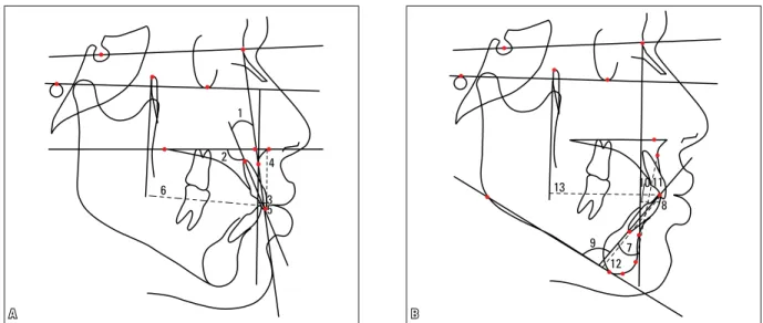
Maxillary dentoalveolar component (Fig 6A) 1. Mx1.NA: Angle between the long axis of the maxillary central incisor and line NA, rep-resenting the degree of inclination of the max-illary central incisor in relation to the maxilla and nasion point.
2. Mx1.PP: Angle between the long axis of the maxillary central incisor and the palatal plane, representing the degree of inclination of the maxillary central incisor in relation to the maxilla.
3. Mx1-NA: Distance between the most an-terior point on the crown of the maxillary cen-tral incisor and line NA, representing the an-teroposterior position of the maxillary incisor in relation to the maxilla and nasion.
4. Mx1-PP: Perpendicular distance between the incisal edge of the maxillary central incisor and the palatal plane, representing the vertical position of the permanent maxillary central in-cisor in the maxilla.
5. Mx1-Aperp: Distance between the EMxI point (incisal edge of maxillary central incisor) and line Aperp.
6. Mx1-PTV: Distance between the EMxI point to the pterygomaxillary vertical plane.
Mandibular dentoalveolar component (Fig 6B)
6
1
13
9 7
8
12 10 11 3
5
2 4
A B
8. Md1-NB: Perpendicular distance be-tween the most anterior point on the crown of the mandibular central incisor and line NB, representing the anteroposterior position of the mandibular incisor in relation to the man-dible and nasion.
9. IMPA: Angle between the long axis of the mandibular central incisor and the mandibular plane (GoMe), representing the tooth inclina-tion in relainclina-tion to the mandible.
10. “I” line: Union of points P’ and “E”, traced in an extent of only 1 cm, at the crossing point with the occlusal plane, determining the degree of retrusion or protrusion of the mandibular central incisor.
11. Md1-APog: Distance between the EMdI point (incisal edge of mandibular incisor) and the line A-Pog.
12. Md1-PM: Perpendicular distance be-tween the incisal edge of the mandibular cen-tral incisor and the mandibular plane, repre-senting the vertical position of the permanent mandibular central incisor in the mandibular symphysis.
13.Md1-PTV: distance between the EMdI point to the pterygomaxillary vertical plane (PTV).
Statistical analysis Method error
Cephalograms were retraced by the same ex-aminer. To determine the reliability of results, fifteen randomly selected radiographs were traced and digitized by the same investigator, after a 20-day interval.
For each cephalometric measurement ana-lyzed, the systematic and casual errors were in-dependently evaluated. The systematic error was calculated by the paired t test, as suggested by Houston.25 Application of the Dahlberg17 formula (Se2=∑d2/2n) allowed to estimate the result of ca-sual errors, considering as significant errors greater than 1 millimeter for linear measurements and 1.5 degree for angular measurements.17
Descriptive and comparative analysis
in Table 1. The descriptive evaluation of val-ues achieved in relation to the dental cephalo-metric variables in the Brazilian Afro-Cauca-sian adolescents is shown in Table 2. Table 3 presents the results of the independent t test for comparison of cephalometric variables be-tween males and females.
For statistical analysis of data, the indepen-dent t test was applied at a significance level of p<0.05, for comparison of values of cepha-lometric variables between sexes. These tests were conducted on the software Statistica for
TABLE 1 - Statistical analysis of ages means for both sexes.
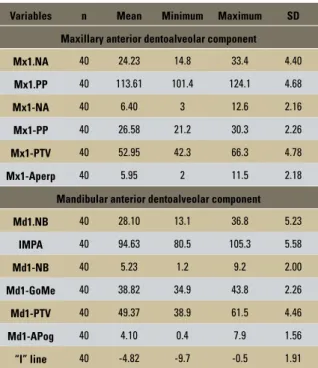
TABLE 2 - Descriptive analysis of the sample, with mean values and standard deviations of young Afro-Caucasian Brazilian individuals.
TABLE 3 - Statistical analysis of independent t test for the dimorphism between sexes, in young Afro-Caucasian Brazilian individuals Significant at p<0.05.
Significant at p<0.05.
Total (n=40) Female (n=20) Male (n=20)
X SD X SD X SD p
Age 13.15 0.61 13.10 0.78 13.25 1.04 0.560
Minimum
age 12.00 12.00 12.20
Maximum
age 14.30 14.21 14.30
Variables n Mean Minimum Maximum SD
Maxillary anterior dentoalveolar component
Mx1.NA 40 24.23 14.8 33.4 4.40
Mx1.PP 40 113.61 101.4 124.1 4.68
Mx1-NA 40 6.40 3 12.6 2.16
Mx1-PP 40 26.58 21.2 30.3 2.26
Mx1-PTV 40 52.95 42.3 66.3 4.78
Mx1-Aperp 40 5.95 2 11.5 2.18
Mandibular anterior dentoalveolar component
Md1.NB 40 28.10 13.1 36.8 5.23
IMPA 40 94.63 80.5 105.3 5.58
Md1-NB 40 5.23 1.2 9.2 2.00
Md1-GoMe 40 38.82 34.9 43.8 2.26
Md1-PTV 40 49.37 38.9 61.5 4.46
Md1-APog 40 4.10 0.4 7.9 1.56
“I” line 40 -4.82 -9.7 -0.5 1.91
Variables
Male Female
p
Mean SD Mean SD
Maxillary anterior dentoalveolar component
Mx1.NA 24.44 4.46 24.01 4.43 0.762
Mx1.PP 113.81 5.09 113.42 4.37 0.796
Mx1-NA 6.53 2.28 6.26 2.08 0.698
Mx1-PP 26.70 2.51 26.46 2.04 0.743
Mx1-PTV 53.44 4.95 52.47 4.69 0.529
Mx1-Aperp 5.82 2.29 6.09 2.13 0.697
Mandibular anterior dentoalveolar component
Md1.NB 27.65 5.70 28.55 4.81 0.591
IMPA 94.63 5.30 94.64 5.98 0.993
Md1-PM 39.51 2.29 38.14 2.07 0.056
Md1-PTV 49.90 4.62 48.84 4.34 0.462
Md1-NB 4.96 2.15 5.49 1.85 0.410
Md1-APog 4.00 1.73 4.20 1.42 0.700
“I” line -4.69 2.19 -4.95 1.62 0.667 Windows 5.0 (Statistica for Windows. StatSoft, Inc., 1995, Tulsa, OK, USA).
RESuLtS
The mean age of subjects (n=40) was 13.15 years, according to the t test. A descriptive eval-uation of the dental cephalometric variables in Afro-Caucasian Brazilian adolescents is present-ed in Table 2.
Table 3 shows the absence of dimorphism be-tween sexes. According to the results, no significant difference was observed for all analyzed variables.
diSCuSSiOn
were used to accomplish the present study, yet in-cluding young Afro-Caucasian individuals.8,36,38,51
Considering the face of young Afro-Cau-casian Brazilian individuals as harmonic and pleasant, different classifications were found in the literature regarding the facial types.12,21 The literature shows that the craniofacial features of the evaluated subjects may suffer alterations during the growth period due to heritage.9,12,52
The characteristic of Pattern I for Afro-Cau-casian individuals slightly varied in relation to the classification proposed by Capelozza Filho.12 The facial phenotype of male and female Mulattos pre-sented facial convexity and a closed nasolabial, due to a slightly maxillary protrusion or incisors flaring. However, the thickness of the upper lip significant-ly influenced the nasolabial angle in most sample subjects, considering that the facial characteristics of these individuals exhibited peculiarities that were intermediate between Blacks and Whites.
The oblique line of nose insertion was sig-nificant in most sample subjects.
It was also observe that the mandible pre-sented greater clockwise rotation, with com-pensation of mandibular incisors (buccal incli-nation),9,21 significant increase of facial convex-ity and slight increase in the LAFH.
To certify the incisor positioning, 13 dental cephalometric variables were implemented due to the structure diversity of the craniofacial com-plex. Many cephalometric measurements are ob-tained considering the cranial base or maxillary and mandibular bone bases. However, there is large variation of the structural morphology in-volving the cranial base.7,21 A short SN line may induce the orthodontist to a less accurate diagno-sis, leading to a erroneously evidence of the pro-trusion or repro-trusion of bone and dental bases.41,53 Deflection of the cranial base may also lead to erroneous interpretations during the comple-mentary diagnosis. However, many variations may be analyzed as typical characteristics of a certain ethnic group.21,41
Some variables suggested by Downs19 and Steiner44 are obtained from the cranial base. Con-versely, Ricketts,43 McNamara35 and Tweed47,48 did not use the cranial base as reference to evalu-ate the dental variables.
Therefore, analysis of the skeletal morphology of young Afro-Caucasian individuals on the cepha-logram evidenced the great potential of tooth com-pensation because of the skeletal characteristics of the jaws. The maxilla exhibited slight protrusion in relation to the forehead, which therefore should be considered normal for this ethnic group. However, the mandible exhibited slight clockwise rotation with marked mandibular incisors flaring. This skel-etal and dental characteristic may induce a false di-agnosis of bimaxillary protrusion of the bone bases for Afro-Caucasian and Black individuals.21
In addition to the facial and morpho-differen-tial diagnosis of the cephalogram, the main ref-erence points for orthodontic treatment include the curve of Spee, anterior and posterior tooth crowding, inclination of maxillary and especially of mandibular incisors.19,27,32,44,47,48 Therefore, when establishing these analyses, the specific-ity and applicabilspecific-ity in different racial groups demonstrate evident craniofacial and dental dif-ferences. Thus, there should be a customized therapy to avoid interference on the phenotypic characteristics of the craniofacial complex.
Maxillary and mandibular incisor position for the different ethnic groups
When comparing some linear and angular vari-ables of the present study with the report of Me-deiros36 on Black individuals, it may be inferred that the variables 1.NB, 1-NB and “I” line demonstrated significant differences. There was a greater protru-sion and inclination, especially in mandibular inci-sors of young Afro-Brazilian individuals, which cor-roborates with some other studies.2,4,9,14,16,20,22,30
protrusion of mandibular incisors in Afro-Cau-casians and Black individuals,8,36 compared to studies that analyzed this variable in Cauca-sians27,28 and individuals of Asian descent.42
Another study with young Caucasian individ-uals assessed the craniofacial growth and devel-opment in young individuals aged 6 to 18 years.34 The comparative analysis of values evidences that most dental variables exhibited great prox-imity or similarity with the present results. How-ever, it was observed that the dental variables in young Afro-Caucasian Brazilian individuals re-vealed slight protrusion and inclination of inci-sors in relation to the Caucasians, except for the variable Mx1-PTV. For the variables Md1-GoMe and Mx1-PP, which quantified the dental extru-sion and stages of eruption of mandibular and maxillary molars, the value was slightly greater for young Caucasian individuals.
Quantitative evaluation, in relation to mandibular incisor position, according to the tweed variable (iMPA)
The Afro-Caucasian individuals showed a mean value of 94.6º for the variable IMPA. In comparison, it was observed a mean of 92.3º for the IMPA in Brazilians with Japanese de-scent,46 which was greater than the value of 90º observed by Margolis32 and Tweed48 in North American individuals, and also greater than the 91º observed by Martins et al,34 in Caucasian in-dividuals aged 12 to 14 years. Conversely, Harris, Kowalski and Walker24 found a value of 95.6º for Black individuals.
Comparison between male and female sexes
Several studies14,18,23,30,40,42,46,51 established pat-terns for different ethnic groups with standard-ized samples, correlating the presence or absence of dimorphism between sexes. However, other studies8,10,22,36 individually evaluated the ideal values for young and adult Black individuals.
The comparative evaluation between sex-es is important to identify differencsex-es in the growth and development process between young male and female individuals.41 Martins et al34 observed tendencies of earlier craniofacial development for young females compared to the males, at different ages.
The observation of numeric and statistical similarity in the analysis between male and fe-male sexes indicates the absence of sexual dimor-phism between young Afro-Caucasian Brazilian individuals (Tab. 3), assuming that the dentoal-veolar variables may be applied for both sexes.
Clinical application
It should be highlighted to the clinician that, since this is a cephalometric study, measure-ments of other studies should also be evaluated and correlated to the current study. However, the variables are obtained by mean values that pres-ent large variation (standard deviation). The or-thodontist should understand these variations to properly apply this knowledge, considering that facial analysis should be the main instrument to determine the final diagnosis with the secondary aid of cephalometric measurements and lateral cephalograms. The orthodontist should also con-sider the variations and limitation of the facial, skeletal and dental context in the different ethnic groups when selecting the best treatment plan.
COnCLuSiOnS
The results showed the mean values of dental cephalometric variables in young Afro-Caucasian Brazilian individuals (Pattern I) with normal occlusion, which leads to the following conclusions:
» There was no dimorphism between sexes. » Further studies should be conducted in Afro-Caucasian individuals with skeletal dis-crepancy (Pattern II, Pattern III, Short Face and Long Face).
1. Alcalde RE, Jinno T, Pogrel MA, Matsumura T.
Cephalometric norms in Japanese adults. J Oral Maxillofac Surg. 1998;56(2):129-34.
2. Alexander TL, Hitcock HP. Cephalometric standards for American Negro children. Am J Orthod. 1978;74(3):298-304. 3. Altemus LA. Cephalofacial relationships. Angle Orthod.
1968;38(3):175-84.
4. Altemus LA. A comparison of cephalofacial relationships. Angle Orthod. 1960;30(4):223-40.
5. Avila JB. Antropologia Física. Rio de Janeiro: Agir; 1958. 6. Azevedo T. Cultura e situação racial no Brasil. Rio de
Janeiro: Civilização Brasileira; 1966.
7. Bacon W, Girardin P, Turlot JC. A comparison of
cephalometric norms for the African Bantu and a Caucasoid population. Eur J Orthod. 1983;5(3):233-40.
8. Bertoz FA. Determinação da linha “I” em melanodermas brasileiros, masculinos de 12 a 17 anos, com oclusão normal [dissertação]. Bauru (SP): Universidade de São Paulo; 1981.
9. Bjork A. Some biological aspects of prognathism and occlusion of the teeth. Acta Odontol Scand. 1950;9(1):1-40. 10. Briedenhann SJ, Roos EC. A cephalometric appraisal of
the Herero-speaking Negro male. J Dent Assoc S Afr. 1988 Dec;43(12):569-75.
11. Broadbent BH. A new X-ray technique and its application to orthodontia. Angle Orthod. 1931;1(2):45-66.
12. Capelozza Filho LF. Diagnóstico em Ortodontia. Maringá: Dental Press; 2004.
13. Coben SE. The integration of facial skeletal variants. Am J Orthod. 1955;41(6):407-34.
14. Connor AM, Moshiri F. Orthognathic surgery norms for American black patients. Am J Orthod. 1985;87(2):119-34. 15. Cooke MS, Wei SH. A comparative study of southern
Chinese and British Caucasian cephalometric standards. Angle Orthod. 1989;59(2):131-8.
16. Cotton WN, Takano WS, Wong WM. The Downs analysis applied to three other ethnic groups. Angle Orthod. 1951;21(4):213-20.
17. Dahlberg G. Statistical methods for medical and biological students. New York: Interscience; 1940.
18. Dandajena TC, Nanda RS. Bialveolar protrusion in a Zimbabwean sample. Am J Orthod Dentofacial Orthop. 2003;123(2):133-7.
19. Downs WB. Analysis of the dentofacial profile. Angle Orthod. 1956;26(4):191-212.
20. Drummond RA. A determination of cephalometric norms for the Negro race. Am J Orthod. 1968;54(9):670-82. 21. Enlow DH, Hans MG. Essentials of facial growth.
Philadelphia: W.B. Saunders; 1996.
22. Fonseca RJ, Klein WD. A cephalometric evaluation of American Negro women. Am J Orthod. 1978;73(2):152-60. 23. Gormley MB, Carlo JM, Reardon J. Cranio-skeletal
morphology for a segment of the black urban population using sella nasion as a cranial base line. Quintessence Int Dent Dig. 1975;6(3):67-70.
24. Harris JE, Kowalski CJ, Walker SJ. Distribution of the mandibular incisor-mandibular plane Angle in nubian schoolchildren. J Dent Res. 1975;54(3):699. 25. Houston WJB. The analysis of errors in orthodontic
measurements. Am J Orthod. 1983;83(5):382-90.
26. Hwang HS, Kim WS, McNamara JA Jr. Ethnic differences in the soft tissue profile of Korean and European-American adults with normal occlusions and well-balanced faces. Angle Orthod. 2002;72(1):72-80.
27. Interlandi S. Linha “I” na análise morfodiferencial para diagnóstico ortodôntico. Rev Fac Odontol Univ São Paulo. 1971;9(2):289-310.
28. Interlandi S. Ortodontia: bases para a iniciação. São Paulo: Artes Médicas; 1977.
Contact address
Eduardo Jacomino Franco
Av. Araucárias- Lote 1835, Qualis Odontologia, Sala 412 Zip code: 71.936-250 – Brasília/DF, Brazil
E-mail: [email protected]
42. Raddi IMG, Henriques JFC, Martins DR. Determinação da linha “I” em xantodermas nipo-brasileiros, dos 12 anos aos 18 anos e 6 meses, com “oclusão normal”. Ortodontia. 1989;22(3):24-32.
43. Ricketts RMA. A foundation for cephalometric communication. Am J Orthod. 1960;46(5):330-57. 44. Steiner CC. Cephalometrics for you and me. Am J Orthod.
1953;39(10):729-55.
45. Takahashi R. Determinação cefalométrica das alturas faciais anterior e posterior, em jovens brasileiros, descendentes de xandodermas e leucodermas, com oclusão normal [tese]. Universidade de São Paulo; 2002. 46. Takahashi R. Padrão cefalométrico FOB-USP para jovens nipo-brasileiros com oclusão normal [dissertação]. Bauru (SP): Universidade de São Paulo; 1998.
47. Tweed CH. The Frankfort mandibular plane Angle in orthodontic diagnosis, classification, treatment planning and prognosis. Am J Orthod. 1946;32(4):175-221. 48. Tweed CH. Frankfort mandibular incisor angle (FMIA)
in diagnosis treatment planning and prognosis. Angle Orthod. 1954;24(3):121-69.
49. Tweed CH. Indications for the extraction of teeth in orthodontic procedure. Am J Orthod. 1944;30(8):405-28. 50. Tweed CH. A philosophy of orthodontic treatment. Am J
Orthod. 1945;31(2):74-103.
51. Uchiyama LMAF. Estudo cefalométrico das alturas faciais anterior e posterior, em jovens brasileiros melanodermas, com “oclusão normal” [dissertação]. Bauru (SP): Universidade de São Paulo; 2005.
52. Van der Linden FPGM. Crescimento e Ortopedia Facial. São Paulo: Quintessence; 1990.
53. Vargas Neto J, Pinzan A, Henriques JFC, Freitas MR, Janson GRP, Almeida RR. Avaliação comparativa entre a linha sela-násio e o plano horizontal de Francfort como parâmetros para o diagnóstico das posições ântero-posterior e vertical das bases ósseas, em jovens brasileiros leucodermas com más oclusões de Classe I e II de Angle. Rev Dental Press Ortod Ortop Facial. 1999;4(2):13-22.
Submitted: April 3, 2007
Revised and accepted: February 4, 2009
29. Jacobson A. The craniofacial skeletal pattern of the South African Negro. Am J Orthod. 1978;73(6):681-91.
30. Jacobson A, Oosthuizen L. The craniofacial skeletal pattern of the South African Bantu. J Dent Assoc S Afr. 1970;25(10):361-5. 31. Kowalski CJ, Nasjleti CE, Walker GF. Differential diagnosis
of adult male black and white populations. Angle Orthod. 1974;44(4):346-50.
32. Margolis HI. The axial inclination of the mandibular incisors. Am J Orthod. 1943;29(10):571-94.
33. Martins DR. Estudo comparativo dos valores cefalométricos das análises de Downs, Tweed, Steiner e Alabama, com os adolescentes brasileiros, leucodermas, de origem mediterrânea [tese]. Bauru (SP): Universidade de São Paulo; 1979.
34. Martins DR. Atlas de crescimento craniofacial. São Paulo: Ed. Santos; 1998.
35. McNamara JA Jr. A method of cephalometric evaluation. Am J Orthod. 1984;86(6):449-69.
36. Medeiros MAQB. Estudo cefalométrico do padrão dentário de jovens brasileiros melanodermas do sexo feminino com “oclusão normal”. Ortodontia. 1988;21(1):18-33. 37. Montagu A. Introdução à antropologia física. São Paulo:
Cultrix; 1969.
38. Moraes C, Freitas MR, Henriques JFC. Cefalometria: determinação do padrão esquelético das adolescentes melanodermas brasileiras, com “oclusão normal”. Ortodontia. 1988;22(4):4-14.
39. Okuyama CC. Preferência do peril tegumentar em jovens melanodermas, leucodermas e xantodermas de ambos os sexos, avaliados por ortodontistas, leigos e artistas plásticos [dissertação]. Bauru (SP): Universidade de São Paulo; 1995. 40. Oliveira JN. Estudo longitudinal e comparativo da variação
do pogônio com os incisivos inferiores, em relação à linha NB, em adolescentes brasileiros, leucodermas, de 12 aos 18 anos de idade, com “oclusão normal” [dissertação]. Bauru (SP): Universidade de São Paulo; 1977.