Alberto Consolaro*, Renata B. Consolaro**
Orthodontic movement does not induce
external cervical resorption (ECR)
or
Orthodontic movement does not change gingival color
and volume, and does not induce gingival inlammation
HOW THE CERVICAL REGION OF THE TOOTH IS STRUCTURED AND ORGANIZED
On the surface of cervical region of human teeth, in the area where the enamel borders the cementum, there is a line known as the cementoenamel junction (CEJ) (Fig 1). On the circumference of the neck of all human teeth the CEJ line alternates three types
of relationship between enamel and cementum.3,7 In some areas of CEJ the cementum covers the enamel (Fig 4), in others the enamel and cementum meet edge to edge, but in other regions the enamel and cementum remain distant from each other, thereby exposing microscopic dentin gaps or “windows” fac-ing the gfac-ingival connective tissue.
* Head Professor of Pathology, FOB-USP, and postgraduation courses, FORP-USP. ** Professor, FOA-Unesp and Faculdades Adamantinenses Integradas (FAI).
This study sought to explain, both anatomically and functionally, how the cervical region of human teeth is structured and organized in order to address the following questions: 1) Why does External Cervical Resorption (ECR) occur in human dentition? 2) Why is there no ECR in gingivitis and periodontitis? 3) Why ECR can occur after dental trauma and internal bleaching? 4) Why does orthodontic movement not change the gingival color and volume during treatment? 5) Why does orthodontic movement not induce ECR al-though it is common knowledge that the cervical region can undergo much stress? The existence of sequestered antigens in the dentin, the presence of dentin gaps in the cervical region of all teeth, the reaction of the junctional epithelium and the gingival distribution of blood vessels may explain why ECR does not occur, nor do gingival color and volume change when teeth are orthodontically moved.
Abstract
Keywords: Tooth resorption. External cervical resorption. Orthodontic treatment. Gingiva.
How to cite this article: Consolaro A, Consolaro RB. Orthodontic movement does not induce external cervical resorption (ECR). Dental Press J Orthod. 2011 Nov-Dec;16(6):22-7.
» The authors report no commercial, proprietary, or inancial interest in the
Having completed its key function of produc-ing enamel, this organ — formed by the outer epi-thelium, stellate reticulum, stratum intermedium and ameloblasts — undergoes a sandwich-like re-stratification and gives rise to the reduced epithe-lium of the enamel organ. This epitheepithe-lium is firm-ly attached to the enamel and receives nutrients from the peripheral connective tissue. Together, they form the dental follicle, which generates the image of the pericoronal space in unerupted teeth.
As a tooth gradually begins to appear in the mouth, the reduced epithelium of the enamel or-gan fuses with the oral mucosa and together form the junctional epithelium. Initially this epithe-lium has three to four layers, reaching as many 10 to 20 cells of thickness with age. Close to 30 years of age, the junctional epithelium lines the enamel in its cervical-most portion, but eventu-ally progresses gradueventu-ally towards the cementum. The length of the junctional epithelium4 ranges from 0.25 to 1.35 mm.
In the cervical root region, next to the enamel, it is the connective tissue of the dental follicle, which gives rise to the connective attachment in the erupted tooth (Fig 4). Conceptually, the con-nective attachment in the erupted tooth comprises the space between the final portion of the enamel and the initial portion of the alveolar bone crest.
The cementum extends from the final edge of the cervical enamel and its junctional epi-thelium. On the cementum surface are the cementoblasts (Fig 4) nourished by the con-nective tissue of the dental follicle tissue and connective attachment. Progressing toward the apex, the connective tissue and cementoblasts on the root become part of the periodontal liga-ment. The average thickness of the cementum in the cervical third ranges from 16 to 60 µm, which is equivalent to the thickness of a human hair. In the apical third, bifurcations and trifur-cations range from 150 to 200 µm. The average thickness of the cementum in a youth aged 20 is 95 µm and at age 60, 215 µm.9
Whatever the relationship between enamel and cementum in the cervical region, the CEJ and its components are in direct contact with the fibrous gingival connective tissue or, more spe-cifically, with its collagen fibers and extracellular matrix gel interspersed with fibroblasts (Fig 4). In the CEJ segments, where the dentin gaps are located, it is "concealed" or protected from expo-sure to macrophages by the extracellular matrix gel, which plays this role with great competence.
1) WHy DOES ExTERNAL CERVICAL RESORp-TION (ECR) OCCUR IN HUmAN DENTIRESORp-TION?
Some proteins in the dentin are recognized as antigenic by the immune system because they are deposited during odontogenesis without direct contact with the protein memory cells which de-velop during intrauterine life.1 Thus, the dentin should be protected from this contact throughout its life, including in the dentin gaps of the CEJ present in all teeth.
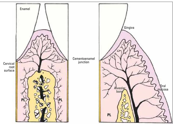
FIGURE 1 - The dentogingival plexus features anastomosis with ves-sels extending from the periodontal ligament (1st), oral mucosa (2nd) and alveolar bone (3rd) in the region of the connective attachment next to the cervical root surface (scheme modified from Glickman4).
Enamel
Gingiva
Vascular dento-gingival plexus
Alveolar bone
Oral mucosa
Alveolar bone
PL
1st 3
rd
2nd Cervical
root surface
Before the dentin gaps are exposed to macro-phages at the CEJ, with consequent immunologi-cal recognition, a direct exposure is required to occur as a result of the dissolution of the extra-cellular matrix of connective tissue where these cells circulate.
The destruction and/or dissolution of the ex-tracellular matrix occur mainly in inflammation: A defense mechanism typical of connective tis-sues and dependent on vessels. The mediators and enzymes that make up the inflammatory exudate arising from vessels and leukocytes dissolve the extracellular matrix gel and, at the gingival con-nective attachment can provide the dentin with access to the macrophages.
Once the macrophages identify the dentin as
a foreign body, they initiate a process through which foreign proteins are eliminated and, when these macrophages are present in hard tissues, bone resorption is the mechanism of choice.1
2) WHy IS THERE NO ECR IN GINGIVITIS AND pERIODONTITIS?
When gingival inflammation is induced by dental bacterial plaque, it promotes epithelial hy-perplasia while the junctional epithelium migrates toward the apical region, placing the CEJ and its gaps in the gingival sulcus and oral environment, thereby "protecting" the ECR region. In the oral environment, macrophages are not capable of rec-ognizing foreign proteins in the dentin and there-fore fail to trigger an immune response.
Enamel
Gingiva
Cervical root surface
Cementoenamel junction
Oral mucosa Alveolar
bone
PL PL
PL
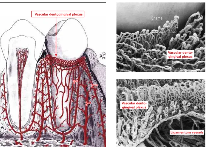
FIGURE 3 - A) The lace-shaped or net-shaped dentogingival plexus consists of vessels originating from the periodontium (1st), mucosa (2nd) and bone (3rd) (scheme introduced by Lascala and Moussalli5). B and C highlight the numerous loop-shaped capillaries of dentogingival plexus in rodents, revealed by scanning electron microscopy performed by Selliseth and Selvig (Source: Newman et al,8 2006).
3) WHy ECR CAN OCCUR AFTER DENTAL TRAUmA AND INTERNAL bLEACHING?
In dental trauma, inflammation of gingival connective tissue may occur, but without junc-tional epithelial hyperplasia. Such is the case in internal dental bleaching, where hydrogen perox-ide leaks into the dentinal tubules that open out at the junction, causing a local gingival inflam-mation in the connective tissue, without epithe-lial hyperplasia. In these two situations, gingival inflammation dissolves the extracellular matrix, exposing the dentin in the CEJ gaps. However, hyperplasia of the junctional epithelium does not occur, allowing the occurrence of ECR.2
4) WHy DOES ORTHODONTIC mOVEmENT NOT CHANGE GINGIVAL COLOR AND VOLUmE DURING TREATmENT?
One way to induce inflammation is by ob-structing blood vessels, which results in local cell death. Proteins released by necrotic cells induce inflammation, as in the gingival connec-tive tissue. Compression of the teeth against the periodontal ligament and alveolar bone of-ten causes cells death and hyalinization of a ligament segment. Not only the arterioles and capillaries, but the veinlets would also be ob-structed, along with areas of necrosis, followed by inflammation.
Vascular dentogingival plexus
1st
2nd
3rd
Vascular dento-gingival plexus
Vascular dento-gingival plexus
Ligamentum vessels
Enamel
A C
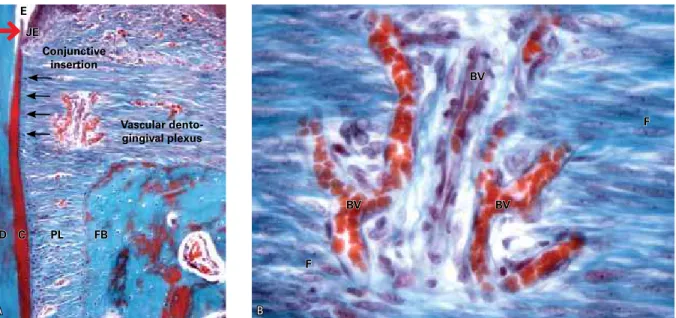
FIGURE 4 - Net-shaped dentogingival plexus in the cervical dental region of monkeys, more specifically at the site of the cementoenamel junction (larger arrow). On the root one can see the cementoblasts (small arrows) (E= enamel, JE= junctional epithelium, D= dentin, C= cementum, PL= periodontal liga-ment, FB= fasciculate bone, BV= blood vessel, F= fibroblast ).
In orthodontic movement, compression of the vessels occurs in the periodontal ligament and could potentially influence gingival blood flow, but this does not occur. It would be natural to ex-pect ischemia would make the gingiva turn whit-ish, or passive hyperemia would make it violet-red due to veinlet compression.
In the gingival tissue, more specifically at the site of the gingival connective attachment, blood vessels form a lace-like dentogingival vascular network or plexus with connections arising from periodontal vascular components. These connections also originate in the marrow spaces of the alveolar bone crest and its perios-teum, as well as from the vessels of the alveo-lar mucous membrane via attached gingiva.5,6,8 There are 50 capillaries within every square millimeter of gingiva5 (Figs 1-4).
The effects of vascular compression of the cer-vical periodontal tissues and gingival tissues are offset by anastomosis or by connections from oth-er sources (Fig 2). Blood supply to gingival tissues
and their venous and lymphatic drainage remain at normal levels. Gingival color and volume are not changed during orthodontic movement by this mechanism or anatomical feature. Neither an edema develops — due to venous obstruction — nor does any inflammatory process emerge.
5) WHy DOES ORTHODONTIC
mOVEmENT NOT INDUCE ExTERNAL CERVICAL RESORpTION?
In a similar compensatory manner, compres-sion of the periodontal and gingival vessels does not promote focus areas of necrosis and/or gingi-val inflammation in the region of the connective attachment (Fig 2). In the absence of necrosis, characterized by cell rupture and protein spill, orthodontic movement does not induce inflam-mation and dissolution of extracellular matrix in the gingival connective tissue. The dentin gaps will not be exposed to macrophages, which in turn will not perform antigen recognition, nor present with foreign proteins that might trigger E
JE
D C PL FB
BV BV
BV
F
F Conjunctive
insertion
Vascular dento-gingival plexus
external cervical immune and resorptive respons-es. This mechanism and anatomical feature does not induce ECR during orthodontic movement.
On the other hand, damage to the gingiva caused by dental trauma and internal dental bleaching cannot be offset by extensive anas-tomosis: Cellular injury occurs mechanically, directly and independently of blood supply. During trauma, cells and vessels are ruptured by sudden shifts while in tooth bleaching the toxic effect of hydrogen peroxide occurs inde-pendently of local blood supply.
FINAL CONSIDERATIONS
1) Dentin comprises proteins, which are con-sidered sequestered antigens and when it is ex-posed to connective tissues it tends to be resorbed as a form of elimination by the body.
2) At the cementoenamel junction (CEJ), the dentin gaps found in all permanent teeth may expose the dentin whenever inflammation is in-duced at the connective attachment.
3) Before dentin is exposed at the CEJ, gingi-vitis and periodontitis reveal a hyperplasia of the junctional epithelium that covers the gaps, placing the dentin in the oral environment.
4) In dental trauma and internal bleaching, in-flammation of connective gingival tissue does not promote junctional epithelial hyperplasia and di-rect exposure of the dentin allows antigen recog-nition by macrophages.
5) The gingiva receives vessels from the peri-odontium, bone, periosteum and mucous mem-brane so that its nutrition and drainage are not affected, even when the vessels of the periodon-tal ligament are compressed during orthodontic movement.
As a result, there is no inflammation at the connective attachment, nor there is cell death in the gingiva during orthodontic movement, with gingival color and volume remaining unchanged, even if the dentin is exposed at the CEJ gaps, which is necessary to initiate ECR.
1. Consolaro A. Reabsorções dentárias nas especialidades clínicas. Maringá: Dental Press; 2005.
2. Esberard R, Esberard RR, Esberard RM, Consolaro A, Pameijer CH. Effect of bleaching on the cemento-enamel junction. Am J Dent. 2007;20(4):245-9.
3. Francischone LA, Consolaro A. Morphology of the cementoenamel junction of primary teeth. J Dent Child (Chic). 2008;75(3):252-9. 4. Glickman I. Periodontia clínica de Glickman. Rio de Janeiro:
Interamericana; 1983.
5. Lascala NT, Moussalli NH. Periodontia clínica e especialidades
ains. São Paulo: Artes Médicas; 1980.
6. Lindhe J, Karring T, Lang NP. Tratado de Periodontia clínica e Implantodontia oral. 3ª ed. Rio de Janeiro: Guanabara Koogan; 1999.
7. Neuvald L, Consolaro A. Cementoenamel junction:
microscopic analysis and external cervical resorption. J Endod. 2000;26(9):503-8.
8. Newman MG, Takei HH, Klokkevold, PR, Carranza, FA. Carranza’s clinical periodontology. 10ª ed. St. Louis: Saunders Elsevier; 2006. 9. Zander HA, Hurzeler B. Continuous cementum apposition J Dent
Res. 1958;37(6):1035-44. REFERENCES
Contact address
Alberto Consolaro
E-mail: [email protected]
Submitted: October 7, 2011