Sources of controversies over analgesics
prescribed after activation of orthodontic appliances
Acetylsalicylic acid or acetaminophen?
Alberto Consolaro*, Vanessa Bernardini Maldonado**, Milton Santamaria Júnior***, Maria Fernanda M-O. Consolaro****
The wide range of available drugs raises questions about the desirable and undesirable effects on orthodontic movement. Could medi-cations such as non-steroidal anti-inflammatory drugs (NSAIDs)—ASA, acetaminophen, di-clofenac, ibuprofen, indomethacin and celecox-ib—, corticosteroids and bisphosphonates have any impact on bone metabolism to the extent
of interfering with orthodontic movement?3,18
When administered to control pain and dis-comfort after activation of the orthodontic ap-pliance, what would be the influence of ASA and acetaminophen on tooth movement and the root resorption associated with it? To help in clarifying this question, Maldonado34 and his team set out to quantitatively determine the influence of ASA and acetaminophen on induced tooth movement and on root resorp-tion by means of optical microscopy. In anoth-er study they described the history and action
mechanisms of these two medications.14
However, when analyzing the literature on the subject, it appears that no work repeated the same experimental model of any other work. The appliance used to move a tooth, the
force applied, the dosage and duration of drug use, route of administration, the experimental animal, the teeth that were moved, the types of tissue section and tooth movement assess-ment as well as many other details differ from study to study, thus precluding comparisons. Without comparisons and without relevant discussions grounded and based on relevant literature it becomes difficult to draw infer-ences for clinical applications.
Fundamentals and assumptions
Orthodontic movements occur by the appli-cation of continued and controlled mechanical forces on the tooth pushing it against the bone, resulting in areas of pressure and tension in the periodontal ligament. The sort of bone remodel-ing that promotes tooth movement is the result of cellular stress and occasionally inflammation involving clast cells, osteoblasts, osteocytes, ce-mentoblasts and fibroblasts. Mediators—such as various cytokines, growth factors and arachi-donic acid products such as prostaglandins and leukotrienes—participate in the process as in-teraction molecules between cells.11,13,16
* Full Professor of Pathology at FOB-USP and at FORP-USP Postgraduate courses. ** Master’s degree from FORP-USP and private clinic in Ribeirão Preto.
Kinins primarily induce pain in free nerve endings and prostaglandins potentiate the ef-fect of kinins, which after 5 to 6 hours no longer produce desirable effects. This poten-tiation of kinin effects by prostaglandins also occurs in microcirculation vessels increasing vascular permeability. The action mechanism of NSAIDs and acetaminophen has been prov-en by specific studies conducted on the sub-ject.1,4,6,7,8,10,15,19,20,21,26,30,31,35,39,42,45,46,53
Apparently, drug prescription after activa-tion of the orthodontic appliance poses a para-dox: If, on the one hand, they suppress the pa-tient’s pain and discomfort with analgesics that reduce the level of prostaglandins, could it be that, on the other hand, they reduce the effec-tiveness of cellular stress and inflammation as a mechanism of bone resorption and induced tooth movement?
Based on reports found in the literature and the behavior of orthodontic clinical cases
with-in the framework advanced by Maldonado34 the
authors developed the following hypothesis: Could the prescription of ASA or acet-aminophen have no effect on the biologi-cal reactions related to effective orthodontic movement and the frequency and severity of root resorption associated with it? Could the occasional and random administration of such drugs or longer periods of administration pro-duce different effects?
Studies that focused on shorter periods of
use of these drugs have been criticized3,56 for
ignoring the option of longer administration
periods. As proposed by Maldonado,34 could
the results of the experimental group under-going the long and short-term administration of acetaminophen and acetylsalicylic acid help in answering how the short and long-term administration of these drugs influence orth-odontic movement? In the view of Jerome et
al,25 anti-inflammatory medication might even
offer protection against root resorption during
orthodontic movement.
It is a fact that prostaglandins induce cellu-lar, tissue and vascular alterations but there is no evidence of inhibition of orthodontic tooth
movement by painkillers and NSAIDs.56 Bone
resorption induced by tooth movement is not
mediated solely by prostaglandins9,58 but by a
pool of mediators such as leukotrienes, cyclic
adenosine monophosphate (cAMP),16,58
colla-genase and many others,29 which are generated
by forces applied to periodontal tissues. The molecular life of prostaglandins con-sists of only a few seconds. Occasional, ran-domly administered doses over short periods of time lead to a transient reduction of prosta-glandins but are enough to reduce the pain and discomfort after the activation of orthodontic appliances. It is only the long-term reduction of prostaglandins after the administration of regular and repeated doses that would be able to reduce the edema effect and the efficiency of the inflammatory cells, known as anti-in-flammatory effect, widely used in postopera-tive surgical periods.12,13
Inconsistencies in the literature and possible explanations
The literature on the effects of drugs dur-ing induced tooth movement is still lackdur-ing standardized methodology in all its aspects. Results are often very diverse and inconsistent and cause confusing interpretation. The litera-ture on the subject often demonstrates inex-perience with the experimental model and the animal used. Thus, veritable “literature trends” arise whereby one study cites the other with-out checking basic methodological details. The main variables involved in the lack of consis-tency between the results reported in the lit-erature are described below.
1. Medication inconsistencies
administration periods used in the various stud-ies about induced tooth movement differ widely: • Wongetal57 (1992): 65mg of
acetylsali-cylic acid for 28 days.
• Kehoe et al27 (1996): 200mg of acet-aminophen for 11 days.
• Roche et al47 (1997): 1,000mg de acet-aminophen for 21 days.
• Sarietal50 (2004): 500mg of acetylsali-cylic acid for 2 days.
• Ariasetal2 (2006): 100mg of acetylsali-cylic acid and 200mg of acetaminophen for 10 days.
• Salmassianetal48 (2009): 600mg of acet-aminophen for 7 days.
• Stabile et al52 (2009): 200mg de acet-aminophen for 2 days.
These differences led two groups of re-searchers to review the matter: Walker and Bu-ring56 and Bartzela et al.3 In a systematic review
of Bartzela et al3 on induced tooth movement
and the use of NSAIDs, including acetamino-phen, the authors found 49 articles. The selec-tion criteria were experimental design, mag-nitude of force and medication. The authors criticized the results of studies using NSAIDs in induced tooth movement concluding that most of these studies evaluated short-term experi-ments underestimating the effects of prolonged drug administration.
A similar review was conducted in 2001 by
Walker and Buring56 with conclusions similar to
those reached by Bartzela et al3 in 2009. The authors emphasized that there is no evidence nor any reasonable grounds to account for a complete inhibition of induced tooth move-ment either with short or long-term drug ad-ministration. They justified their conclusions based on the concept that prostaglandins are not the only modulators of bone resorption. Furthermore, based on previous studies, they claim that the exact amount of force neces-sary to create tooth movement has not been
standardized, and conclude that animal studies have been limited and short-term.
2. Inconsistencies regarding animals used and teeth moved
Methods using induced tooth movement ac-companied by drug administration are numerous in the literature, but the choice of experimental model insofar as the orthodontic appliance and the animals are concerned is fraught with incon-sistencies. Rabbits,47 cats,9 pigs27,57 and rats2,17,32,3 3,36,37,38,40,41,49,52,54,55,59 have been used.
The teeth used for induced tooth move-ment are yet another variable that influences the wide variety of results achieved by such studies. Compared with human incisors rodent incisors are quite different in their morphology. Moreover, their odontogenesis is continuous. For example:
• Wongetal57 (1992) used guinea pig inci-sors.
• Kehoeetal27 (1996) studied guinea pig incisors.
• Roche et al47 (1997) investigated rabbit molars.
• Sari et al50 (2004) experimented using human gingival fluid.
• Ariasetal2 (2006) used the upper inci-sors of rats.
• Salmassian et al48 (2009) measured the painful effects of fixed orthodontic appli-ances in humans.
• Stabileetal52 (2009) investigated rat in-cisors.
Research in animals is justified because it is numerically more representative than evalu-ation in humans. The microscopic analysis of induced tooth movement in humans is limit-ed since it requires extraction, which ruptures periodontal ligament fibers. As a result, part of the tissue that should be examined remains at-tached to the alveolar bone thereby precluding
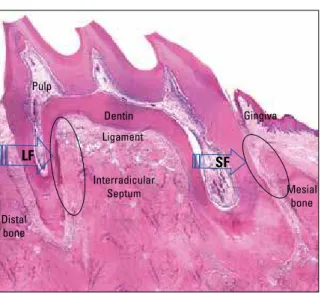
FIGURE 1 - By observing the longitudinal section of a murine first molar one can compare the similarities between a murine tooth and a human molar. In the experimental model proposed by Heller and Nanda22 the
forces applied to the mesial root are considered light (LF) due to its vol-ume and size. The small dimensions of the distobuccal root imply strong forces (SF) with greater action exerted on the periodontal ligament, which is consequently extensively hyalinized (red area in the larger circle).
FIGURE 2 - By observing the cross-section of a murine first molar one can compare the similarities between this murine tooth and a human molar and its reactions with greater histomorphometric accuracy—in the face of light forces (small arrow) and intense forces (large arrow)—
such as the hyaline areas in the periodontal ligament (circles) and the attendant dental resorption. In these cross-sections one can observe all five roots of the murine first molar and adjacent bone.
The model of induced tooth movement in rats is universally accepted due to the fact that it is more ethical and can be extrapolated to hu-mans, considering the favorable root morphol-ogy of rodents, the size of their molars (Figs 1, 2 and 3) and their anatomical peculiarities, al-though their metabolism is twice as high as that of humans.12,43,51
3. Inconsistencies regarding the design of the experimental orthodontic appliance
Many different appliance designs are em-ployed to obtain experimental tooth movement and these designs influence the results, regard-less of whether or not associated with drug use. • V-shaped appliances are intended to
separate the incisors,2,52 which in rodents undergo continuous root formation. • Whenplacedbetweentheirstandsec
-ond molars elastomers exert a not so effi-cient or continuous force, which tends to dissipate swiftly, as it acts on two teeth,
instead of just one. Moreover, the force is further absorbed by a considerable de-flection of the bony ridges and septa of two teeth, and not just one.40,55
• When expansion screws are placed be -tween the first molars they cause upper first molar proclination.23,24 Transverse coil springs for molar proclination28 work both on the teeth and on the palatal su-tures, and it is not possible to determine with any degree of reliability the extent
Dentin Dentin
Ligament
Gingiva
Pulp
Ligament
Interradicular Septum
Alveolar mucosa
Buccal surface of the Maxilla Mesial
bone Distal
bone Pulp
Interradicular Septum
incisorswithaV-shapedapplianceusing a force of 8cN.
• Kehoe et al27 (1996) moved guinea pig incisors using aV-shaped appliance and applied forces up to 25cN.
• Rocheetal47 (1997) moved rabbit molars mesially using springs interposed to the incisors by applying forces up to 100cN. • Sarietal50 (2004) moved human canines
distally applying forces up to 120cN. • Arias et al2 (2006) separated the upper
incisorsofrodentsusingaV-shapedap -pliance and a force of 35cN.
• Salmassian et al48 (2009) did not stan-dardize force intensity.
• Stabile et al52 (2009) moved the upper incisorsofratswithaV-shapedappliance using a force of 30cN.
These significant discrepancies could also be found in the data reported in 2004 after a
sys-tematic review of Ren.44 In 20% of the studies
on induced tooth movement the forces applied were lower than 20cN; 27% of the studies used elastomers of unknown forces; 37% applied forces ranging from 20 to 50cN and 12% used forces ranging from 50 to 100cN.
5. Inconsistencies regarding result interpreta-tion and measurement
The criteria for interpreting and measur-ing the effects of applied forces on cells and tissues, of associated root resorption and the influence of drugs on periodontal tissues rep-resent another variable limiting the extrapola-tion of the clinical results previously reported in the literature.
For example, in research on induced tooth movement areas of hyalinization in the peri-odontal ligament mean that the forces applied were effective in light of a microscopic analysis of experimental results. The results found by Bohl et al5 about the extent of hyalinization areas after experimental tooth movement are
FIGURE 3 - By magnifying a cross-section of a murine first molar root one can measure its reactions with histomorphometric accuracy in the face of the forces (arrow) applied, such as the hyaline areas (H) in the periodontal ligament (circle) and related bone and tooth resorption when these are present.
of such occurrences. The bone plate of the rat is very thin and does not allow a standardized use of these springs.
• The use of coil springs interposed be -tween incisors and molars in order to move molars mesially is currently wide-spread enabling the quantification and standardization of forces deployed in the experimental model, thereby imparting greater credibility to the extrapolation of results.17,36,37,38,41,49,54,59 This model was
de-signed by Heller and Nanda22 and
perfect-ed by Martins-Ortiz36 (Figs 1, 2 and 3).
4. Inconsistencies regarding the intensity and distribution of forces
Perhaps the most decisive factors influenc-ing the differences and diversities of the results regarding this matter do not concern only the experimental appliance but especially the in-tensity and distribution of forces applied to achieve tooth movement:
• Wong et al57 (1992) moved guinea pig
Interradicular Septum
Ligament Lingual
Surface of Maxilla
Buccal Surface of
Maxilla
Dentin
H
H H
rather inadequate in the literature due to: • Widevariationamongthemethods. • Variabilityinsizeandtimeasregardsthe
emergence of hyaline areas.
• Considerable type and intensity differ -ences in terms of force application. In 2007, Fracalossi17 revealed that, under mi-croscopic analysis, the differences may also occur in the cross-section plane of the moved teeth. Cross-sections allowed a better assessment of tis-sue effects making it easy to visualize the roots in a single, parallel plane17 (Figs 1, 2 and 3).
Acetylsalicylic acid or acetaminophen?
In the work of Maldonado,34 the
compara-tive results between the short and long-term administration of acetylsalicylic acid and ac-etaminophen were obtained using the model
created by Heller and Nanda22 and later
modi-fied by Martin-Ortiz36 since it allows for force
and movement type standardization. The first molars of young rats are very similar in mor-phology to that of humans and their forma-tion is not continuous as is the case with rat incisors.
Result reproducibility was based on the measurement of two important parameters, namely, the occurrence of hyaline areas (Figs 1, 2 and 3) and the presence of root resorption. Hyaline areas indicate that there was vascu-lar compression and areas of hypoxia that de-termine cell migration and necrosis. In other words, this means that the force was active in the first hours and days of the experiment. The areas of root resorption in turn indicate cell mobilization, especially of clast cells, indicating that the forces continued to operate even sev-eral days after the force had been applied and the teeth moved.
The dosage administered in the Maldonado34
investigation was based on the dosage recom-mended for humans taking into account that the rat’s metabolism is twice that of humans.
For the relief of human pain, these drugs—ace-tylsalicylic acid and acetaminophen—are pre-scribed and patients recommended to swallow them whenever the need arises, i.e., administra-tion follows a random pattern in time. Thus, the administration of these drugs for short periods attempted to simulate this situation of random-ness and contingency. However, certain patients endowed with higher sensitivity to pain can take them several times over a period of just a few days. Long-term administration was meant to simulate this situation.
In this study, a histomorphometric analysis was performed of the effects of induced tooth movement in rats (200g) with springs inter-posed between the upper molars and the inci-sors and a force of 75cN associated with 25mg of ASA and 25mg of acetaminophen (short-term administration/2 days, and long-(short-term/9 days). When compared with the control, the authors found no statistically significant differ-ences in percentage means of hyaline areas in the periodontal ligament. Tissue reactions in the experimental groups were similar to those in the control group.
Root resorption was also assessed by the quantitative histomorphometric method and no statistically significant differences were found in the percentage means of the areas of root resorption between the acetylsalicylic acid group and the acetaminophen group compared with the control group, in the time periods used for evaluation.
The same findings were observed when comparing the experimental acetylsalicylic acid group with the acetaminophen group in terms of the percentage of hyaline areas found. In the authors’ opinion, no statistically significant dif-ference was found in the means of the hyaline areas between the experimental groups at 5, 7 and 9 days. The microscopic effects were simi-lar in the experimental groups.
acid group and the acetaminophen group (short and long term), in light of the percentage means of root resorption areas found no statistical dif-ference in the time periods under evaluation.
The findings were difficult to be compared directly with those of other authors as the ex-perimental model in almost all respects differed significantly.
In this experimental model results can be more safely and directly extrapolated to hu-mans using the measurements and analysis conducted by the authors. In particular, this is due to the fact that this experimental model allows the observation and measurement—on the same tooth—of the tissue and cellular ef-fects brought about by light and heavy forces, both without drugs and under their influence. Such precautions were suggested by Bartzela et al,3 Walker and Buring.56
Final considerations
The results of the study carried out by
Mal-donado34 indicated that acetylsalicylic acid and
acetaminophen did not interfere in the cell and tissue phenomena caused by induced tooth movement or in the degree and frequency of
the attendant root resorption. This corrobo-rates the finding of Walker and Buring56 that prostaglandins—the main targets of acetyl-salicylic acid and acetaminophen—are not the only local mediators of bone resorption. These drugs act as modulators of prostaglandin pro-duction to control pain and discomfort rather than inhibiting total prostaglandin synthesis in the cellular stress and inflammation region, while other mediators of bone resorption con-tinued to operate normally in the periodontal tissues.
1. Abramson SB, Weissmann G. The mechanism of action of nonsteroidal anti-inflammatory drugs. Arthritis Rheum. 1989; 32:1-9.
2. Arias OR, Marquez-Orozco MC. Aspirin, acetaminophen, and ibuprofen: Their effects on orthodontic tooth movement. Am J Orthod Dentofacial Orthop. 2006 Sep;130(3):364-70. 3. Bartzela T, Türp JC, Motschall E, Maltha JC. Medication effects
on the rate of orthodontic tooth movement: a systematic literature review. Am J Orthod Dentofacial Orthop. 2009 Jan;135(1):16-26.
4. Bianchi M, Panerai AE. The dose-related effects of paracetamol on hyperalgesia and nociception in the rat. British Journal of Pharmac. 1996;117:130-32.
5. von Böhl M, Kuijpers-Jagtman AM. Hyalinization during orthodontic tooth movement: a systematic review on tissue reactions. Eur J Orthod. 2009 Feb;31(1):30-6.
6. de Carlos F, Cobo J, Díaz-Esnal B, Arguelles J, Vijande M, Costales M. Orthodontic tooth movement after inhibition of cyclooxygenase-2. Am J Orthod Dentofacial Orthop. 2006 Mar;129(3):402-6.
7. de Carlos F, Cobo J, Perillan C, Garcia MA, Arguelles J, Vijande M, Costales M. Orthodontic tooth movement after different coxib therapies. Eur J Orthod. 2007 Dec;29(6):596-9.
8. Chandrasekharan NV, Dai H, Roos KL, Evanson NK, Tomsik J, Elton TS, Simmons DL. COX-3, a cyclooxygenase 1 variant inhibited by acetaminophen and other analgesic/antipyretic drugs: cloning, structure, and expression. Proc Natl Acad Sci USA. 2002 Oct;99(21):13926-31.
9. Chumbley AB, Tuncay OC. The effect of indomethacin (an aspirin-like drug) on the rate of orthodontic tooth movement. Am J Orthod. 1986 Apr;89(4):312-4.
10. Clissold SP. Acetaminophen and phenacetin. Drugs. 1986; 32:46-59.
11. Consolaro A. Reabsorções dentárias nas especialidades clínicas. 2ª ed. Maringá: Dental Press; 2005.
12. Consolaro A. Analgésicos e antiinflamatórios na movimentação dentária induzida: metodologia e interpretação. Você sabe o que é extrapolação cefalométrica? Rev Dental Press Ortod Ortop Facial. 2007 maio/jun;12(3):19-23.
13. Consolaro A. Inflamação e reparo. 1ª ed. Maringá: Dental Press; 2009.
14. Consolaro A, Maldonado VB, Santamaria Jr, M Consolaro MFMO. Ácido acetilsalicílico ou acetaminofeno? Qual o medicamento para aliviar a dor e o desconforto após a ativação dos aparelhos ortodônticos? Rev Clín Ortodon Dental Press. 2009 dez;2010 jan;8(6):106-10.
15. Courade JP, Caussade F, Martin K, Besse D, Delchambre C, Hanoun N, Hamon M, Eschalier A, Cloarec A. Effects of acetaminophen on monoaminergic systems in the rat central nervous system. Naunyn Schmiedebergs Arch Pharmacol. 2001 Dec;364(6):534-7.
16. Davidovitch Z, Shanfeld J. Cyclic AMP level in alveolar bone of orthodontically treated cats. Arch Oral Biol. 1975 Sep; 20(9):567-74.
17. Fracalossi ACC. Análise da movimentação dentária induzida em ratos. Influências do alendronato nas reabsorções dentárias, estudo comparativo em cortes transversais e longitudinais e avaliação microscópica em diferentes períodos de observação. [Dissertação]. Bauru (SP): Universidade de São Paulo; 2007. 18. Gameiro GH, Pereira-Neto JS, Magnani MB, Nouer DF. The
influence of drugs and systemic factors on orthodontic tooth movement. J Clin Orthod. 2007 Feb;41(2):73-8.
19. Giunta D, Keller J, Nielsen FF, Melsen B. Influence of indomethacin on bone turnover related to orthodontic tooth movement in miniature pigs. Am J Orthod Dentofacial Orthop. 1995 Oct;108(4):361-6.
20. Goodman A. As bases farmacológicas da terapêutica. 10ª ed. New York: Mc Graw-Hill; 2003. p. 517-50.
21. Graf P, Glatt M, Brune K. Acidic nonsteroid anti-inflammatory drugs accumulating in inflamed tissue. Experientia. 1975 Aug
REFERENCES
15;31(8):951-3.
22. Heller IJ, Nanda R. Effect of metabolic of periodontal fibers on orthodontic tooth movement. Am J Orthod. 1979 Mar;75(3):239-58.
23. Igarashi K, Mitani H, Adachi H, Shinoda H. Anchorage and retentive effects of a bisphosphonate (AHBuBP) on tooth movements in rats. Am J Orthod Dentofacial Orthop. 1994 Sep;106(3):279-89.
24. Igarashi K, Adachi H, Mitani H, Shinoda H. Inhibitory effect of the topical administration of a bisphosphonate (risedronate) on root resorption incident to orthodontic tooth movement in rats. J Dent Res. 1996 Sep;75(9):1644-9.
25. Jerome J, Brunson T, Takeoka G, Foster C, Moon HB, Grageda E, Zeichner-David M. Celebrex offers a small protection from root resorption associated with orthodontic movement. J Calif Dent Assoc. 2005 Dec;33(12):951-9.
26. Jones M, Chan C. The pain and discomfort experienced during orthodontic treatment: a randomized controlled clinical trial of two aligning archwires. Am J Orthod Dentofacial Orthop. 1992;102:373-81.
27. Kehoe MJ, Cohen SM, Zarrinnia K, Cowan A. The effect of acetaminophen, ibuprofen and misoprostol on prostaglandin E2 synthesis and the degree and rate of orthodontic tooth movement. Angle Orthod. 1996;66(5):339-49.
28. Iwase M, Kim KJ, Kobayashi Y, Itoh M, Itoh T. A novel bisphosphonate inhibits inflammatory bone resorption in rat osteolysis model with continuous infusion of polyethylene particles. J Orthop Res. 2002 May;20(3):499-505. 29. King GJ, Collier J. A bone resorptive agent extracted from
orthodontically-treated tissues of the rat. Angle Orthod. 1986 Oct;56(4):299-308.
30. Krishnan V. Orthodontic pain: from causes to management - a review. Eur J Orthod. 2007 Apr;29(2):170-9.
31. Law SLS, Southard KA, Law AS, Logan HL, Jakobsen JR. An evolution of preoperative ibuprofen for treatment of pain associated with orthodontic separator placement. Am J Orthod Dentofacial Orthop. 2000 Dec;118(6):629-35.
32. Leiker BJ, Nanda RS, Currier GF, Howes RI, Sinha PK. The effects of exogenous prostaglandins on orthodontic tooth movement in rats. Am J Orthod Dentofacial Orthop. 1995 Oct;108(4):380-8. 33. Macapanpan LC et al. Early tissue changes following tooth
movement in rats. Angle Orthod. 1954;24(2):79-95. 34. Maldonado VB. Efeitos microscópicos do ácido acetilsalicílico
(aspirina) e do acetaminofeno (tylenol) na movimentação dentária induzida e nas reabsorções radiculares associadas. [Dissertação] Ribeirão Preto (SP): Universidade de São Paulo; 2009. 35. Marshall PJ, Kulmacz RJ, Lands WE. Constraints on
prostaglandins biosynthesis in tissues. J Biol Chem. 1987 Mar 15;262(8):3510-7.
36. Martins-Ortiz MF. Influência dos bisfosfonatos na movimentação dentária induzida, na freqüência e nas dimensões das reabsorções radiculares associadas. [tese]. Bauru (SP): Universidade de São Paulo; 2004.
37. Mazzieiro ET. Bisfosfonato e movimentação dentária induzida: avaliação microscópica de seus efeitos. [Tese]. Bauru (SP): Universidade de São Paulo; 1999.
38. Mohammed AH, Tatakis DN, Dziak R. Leukotrienes in orthodontic tooth movement. Am J Orthod. 1989 Mar;95(3):232-37. 39. Ngan P, Kess B, Wilson S. Perception of discomfort by patients
undergoing orthodontic treatment. Am J Orthod Dentofacial Orthop. 1989 Jul;96(1):47-53.
40. Ohkawa S. Effects of orthodontic forces and anti-inflammatory drugs on the mechanical strength of the periodontium in the rat mandibular first molar. Am J Orthod. 1982 Jun;81(6):498-502. 41. Pereira AAC. Avaliação microscópica da influência de
anticoncepcional e gravidez na movimentação dentária induzida, em especial nos fenômenos da reabsorção dentária. [dissertação]. Bauru (SP): Universidade de São Paulo; 1995. 42. Polat O, Karaman AI. Pain control during fixed appliance therapy.
Contact address
Alberto Consolaro
E-mail: [email protected]
43. Porter G. The norway rat (Rattus norvegicus). In: Lanne-Petter, W. et al. The UFAW handbook on the care and management of laboratory animals. 3rd ed. London: Livingstone; 1967. p. 353-90. 44. Ren Y, Maltha JC, Jagtman-Kuijpers AM. The rat as a model for
orthodontic tooth movement - a critical review and a proposed solution. Eur J Orthod. 2004;26:483-490.
45. Rodan GA, Martin TJ. Role of osteoblasts in hormonal control of bone resorption: a hypothesis. Calcif Tissue Int. 1981;33(4):349-51.
46. Rodan GA, Yeh CK, Thompson DT. Prostanglandin and bone. In: Norton LA, Burstone CJ. The biology of tooth movement. Boca Raton: CRC Press; 1989. Chapter 16:263-7.
47. Roche JJ, Cisneros GJ, Acs G. The effect of acetaminophen on tooth movement in rabbits. Angle Orthod. 1997;67(3):231-6. 48. Salmassian R, Oesterle LJ, Shellhart WC, Newman SM.
Comparison of the efficacy of ibuprofen and acetaminophen in controlling pain after orthodontic tooth movement. Am J Orthod Dentofacial Orthop. 2009 Apr;135(4):516-21.
49. Santamaria MJR. Biologia da movimentação dentária induzida e das reabsorções radiculares associadas. Influência do gênero e dos bisfosfonatos. [tese].Bauru (SP): Universidade de São Paulo; 2009.
50. Sari E, Olmez H, Gürton AU. Comparison of some effects of acetylsalicylic acid and rofecoxib during orthodontic tooth movement. Am J Orthod Dentofacial Orthop. 2004 Mar;125(3):310-5.
51. Schour I, Massler M. The teeth. In: Farris EJ, Griffith JQ. The rat in laboratoty investigation. 2nd ed. New York: Hafner; 1963.6:104-65.
52. Stabile AC, Stuani MB, Leite-Panissi CR, Rocha MJ. Effects of short-term acetaminophen and celecoxib treatment on orthodontic tooth movement and neuronal activation in rat. Brain Res Bull. 2009 Aug 14;79(6):396-401.
53. Swierkosz TA, Jordan L, McBride M, McGough K, Devlin J, Botting RM. Actions of paracetamol on cyclooxygenases in tissue and cell homogenates of mouse and rabbit. Med Sci Monit. 2002 Dec;8(12):BR496-503.
54. Vasconcelos MHF et al. A histological study of tooth movement in rats under contraceptive use. In: Davidovitch Z, Mah J. Biological mechanisms of tooth eruption, resorption and replacement by implants. Alabama: Harvard Society for the Advancement of Orthodontics; 1998.
55. Waldo CM, Rothblatt JM. Histologic response to tooth movement in the laboratory rat: procedure and preliminary observations. J Dent Res. 1954 Aug;33(4):481-6.
56. Walker JB, Buring SM. NSAID impairment of orthodontic tooth movement. Ann Pharmacother. 2001 Jan;35(1):113-5. 57. Wong A, Reynolds EC, West VC. The effect of acetylsalicylic acid
on orthodontic tooth movement in the guinea pig. Am J Orthod Dentofacial Orthop. 1992 Oct;102(4):360-5.
58. Yamasaki K. The role of cyclic AMP, calcium and prostaglandins in the induction of osteoclastic bone resorption associated with experimental tooth movement. J Dent Res. 1983 Aug;62(8):877-81.