The influence of bilateral lower first
permanent molar loss on dentofacial
morfology – a cephalometric study
David Normando*, Cristina Cavacami**
Objective: To evaluate cephalometric changes in patients with bilateral loss of lower first permanent molar teeth. Methods: Sixty-eight lateral radiographs of patients from private practices were analyzed. The sample was divided into two groups matched for age and gender: 34 individuals without loss (control group) and 34 presenting with bilateral loss of lower first permanent molar teeth (loss group). Patients who had lost teeth other than lower first molars, cases of agenesis and patients under 16 years of age were excluded from the sample. Only individuals who reported losing teeth at least 5 years earlier were evaluated. Results: It was found that bilateral loss of lower first permanent molars leads to smooth closure of GnSN angle (P = 0.05), counterclockwise rotation of the occlusal plane (P = 0.0001), mild decrease in lower anterior face height (P = 0.05), pronounced lingual tipping (P = 0.04) and retrusion of mandibular incisors (P = 0.03). Moreover, bilateral loss of lower first permanent molars did not affect the maxillomandibular relationship in the anteroposterior direction (P = 0.21), amount of chin (P = 0.45), inclination of upper incisors (P = 0.12) and anteroposterior position of maxillary incisors (P = 0.46). Conclusion: Bilateral loss of lower first molars can pro-duce marked changes in lower incisor positioning and in the occlusal plane as well as a mild vertical reduction of the face.
Abstract
Keywords: First permanent molar. Cephalometry.
* Specialist in Orthodontics, PROFIS-USP/Bauru. Professor of Orthodontics, UFPA. Coordinator, Specialization Program in Orthodontics, EAP / ABO-PA. M.Sc. in Clinical Dentistry, FOUSP, Doctoral in Dentistry, UERJ.
intROduCtiOn
Despite the vast scientific knowledge avail-able concerning effective methods to prevent dental caries disease, epidemiological data on tooth loss show alarming rates in Brazil, espe-cially in the low-income population.2,8,9,15 Loss of lower first permanent molars not only con-tributes to these statistical data but has been identified as the most prevalent.8,9
Over the years literature has highlighted the importance of first permanent molars in occlu-sion. Their loss can lead to serious problems with remarkable clinical changes in the position of neighboring and antagonist teeth,5,10,11 which may require orthodontic and rehabilitation treatment due to the complexity of the result-ing malocclusion.
Several occlusal changes caused by missing first molars have been described in the litera-ture. Second molars have been shown to mi-grate mesially into the posterior region of the dental arch,5,11 while second premolars5,6,11 and canines10,11 drift distally. However, it is clear that the effects of lower first molar loss are not restricted to the posterior region as they seem to significantly influence anterior teeth, increasing the occurrence of diastemas10 and midline shifts.10,11 Few studies have sought to examine the effects of missing first permanent molars on the cephalometric pattern. These studies1,12 showed spontaneous cephalometric changes in overbite and overjet and in incisor inclination after extraction of lower first per-manent molars. A tendency was observed to-ward increased overjet and overbite in associa-tion with retroclinaassocia-tion of lower incisors and protrusion of upper incisors, with relatively significant variation in these changes.12 In most cases where overjet and overbite were normal, the overbite remained stable after extraction.12 However, no evidence has been found to sup-port the occurrence of changes in the vertical relationships of the face.1
MAtERiAL And MEtHOdS
This study was developed through the analysis of 68 lateral cephalometric X-rays from routine orth-odontic records. The sample was divided into two groups matched for gender and age: a control group (no loss), consisting of 34 radiographs, 8 men and 26 women, whose mean age was 19.5 years (16-26.2), and another group with bilateral loss of first perma-nent molars, consisting of 34 radiographs, 8 men and 26 women with a mean age of 24.6 years (16-36). Patients who had lost teeth other than the first mo-lar, cases of agenesis and patients under 16 years of age were excluded from the sample.
Information regarding age and gender was col-lected directly from the patients’ dental records. Despite the authors’ efforts, it was not possible to accurately determine the time at which the molars were lost. The patients who were able to determine it reported having lost them at least 5 years earlier. Patients who reported a recent loss were excluded from the sample.
The radiographs were traced manually by one investigator and checked by another. The cepha-lometric measurements were made using the pro-gram Measurement and Cephalometric Tracing System (SMTC). Cephalometric landmarks and linear and angular measures were performed as outlined by Silva Filho et al.13
Random error was defined by Dahlberg’s for-mula and systematic error was examined by the intraclass correlation test, duplicating measures in 20 randomly selected radiographs, 10 from each group. Student’s t-test at 95% confidence was employed for statistical analysis of differences be-tween groups.
RESuLtS
direction of facial growth and facial height
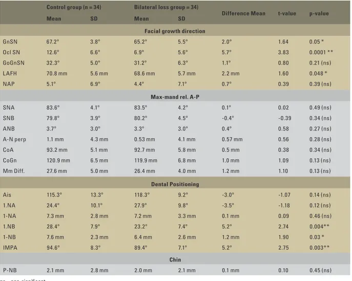
A comparative analysis of the GnSN angle, which defines the resultant vector of the antero-inferior growth of the mandible, showed a more smoothly closed GnSN angle (P=0.05) in the loss group (mean= 65.2°, SD=5.5°) compared to the control group (mean = 67.2°, SD = 3.8°).
The OclSN angle, which defines the occlu-sal plane from the cranial base, showed a mean of 12.6° (SD=6.6) in the control group, and 5.6º (SD=5.7°) in the loss group, demonstrating that bilateral loss of first molars causes a nearly 6º (P=0.0001) counterclockwise rotation of the oc-clusal plane.
The GoGnSN angle, which provides insight into the behavior of the mandibular base relative to the cranial base, showed a mean of 32.3º (SD= 5.0°) in the control group and 31.2° in the loss group (SD= 6.3), with no statistically significant difference (P = 0.21). However, LAFH, which is the linear expression of lower face height, where the mean value obtained for the control group was 70.8 mm (SD = 5.6 mm), and for the loss group, 68.6 mm (SD = 5.7 mm), revealed that bilateral loss of first molars causes a mild, statistically sig-nificant (P = 0.048) decrease in LAFH.
Anteroposterior maxillomandibular relationship
Comparative analysis between the control group and the group with bilateral loss of first molars revealed that the anteroposterior maxillo-mandibular relationship did not undergo signifi-cant change due to the loss.
Regarding NAP measure, which aids in quali-fying maxillary protrusion in relation to total fa-cial profile, its mean value in the control group was 5.1° (SD= 3.8°), and in the loss group, 4.4° (SD= 7.1º). This difference was not statistically significant (P = 0.39).
The SNA angle, which defines the position of the maxilla in relation to the cranial base, yielded a value of 83.6° (SD= 4.1°) in the control group,
and 83.5º (SD= 4.2) in the loss group, with no sig-nificant difference (P = 0.49). A similar behavior was noted in analyzing the anteroposterior posi-tion of the mandible in relaposi-tion to the skull base, which is obtained by means of the SNB angle. The mean value obtained in the control group was 79.8 ° (SD= 3.9°), and in the loss group, 80.2º (SD= 4.5°). This difference was not statistically significant (P= 0.34). As a result, there was no sig-nificant difference (P = 0.27) in ANB angle. Con-trol group mean was 3.7° (SD = 3.0°) and loss group mean, 3.3° (SD = 3.0°).
When linear distances were analyzed for the A-N Perp line, which relates the maxilla to the cranial base, the control group achieved a mean value of 1.1 mm (SD= 4.3 mm), and the loss group, 0.53 mm (SD = 4.1 mm), this difference was not statistically significant (P = 0.28).
As regards the numerical expression of the size of the maxilla, obtained through the Co-A distance, the control group’s mean value was 93.2 mm (SD = 5.1 mm) and the loss group’s, 92.7 mm (SD = 5.8 mm), P = 0.34. The size of the mandible given by the Co-Gn line was found to be 120.9 mm (SD = 6.5 mm) in the control group, and 119.9 mm (SD = 6.8 mm) in the loss group, with no statistically significant difference (P = 0.13). Consequently, the maxillomandibular differential (Mm Diff), which is the difference between the CoGn and CoA measures, was sta-tistically similar (P = 0.13) between the control group (mean = 27.6 mm, SD = 5.0 mm) and the group with bilateral loss of lower first molars (mean = 26.4 mm, SD= 4mm).
dental pattern
Dental pattern results showed that the AIs an-gle, which reflects upper incisor inclination in the basal bone, exhibited no statistically significant difference between the control group (mean= 115.3°, SD= 13.3°) and the loss group (mean= 118.3°, D.P = 9.2°).
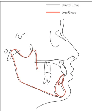
up-FIGURE 1 - Mean differences observed between the control group (black tracing) and the group with bilateral loss of lower first molar (red tracing).
per incisors in the alveolar bone with the aid of 1.NA angle, the mean found in the control group was 24.4º (SD= 10.1), and in the loss group, 27.9º (SD= 9.8°), once again, this difference was not statistically significant (P = 0.12).
As regards the anteroposterior position of maxillary incisors in relation to their apical base, obtained by measuring 1-NA, the control group’s mean was 7.3 mm (SD= 2.8 mm), and the loss group’s, 7.2 mm (SD= 3.3 mm), indicating no sta-tistically significant difference (P = 0.46).
Concerning the axial inclination of lower inci-sors in the alveolar bone, obtained with the 1.NB angle, the average found in the control group was 28.4° (SD = 7.9°), and the loss group, 23.2º (SD = 7.4º). This result indicates that the lower incisors are tipped lingually due to bilateral loss of lower first permanent molars (P = 0.004). This finding is confirmed by IMPA, where there was a marked lingual inclination of lower incisors in patients with missing first molars (P = 0.003), with control group mean equal to 94.6º (SD = 8.3°) and loss group mean of 89.4º (SD = 7.1°).
Regarding the anteroposterior position of low-er incisors in relation to their apical base, mea-sured by 1-NB, the control group’s mean was 7.6 mm (SD= 2.3 mm) and the loss group’s, 6.4 mm (SD= 2.6 mm). Moreover, a mild retrusion was found in the mandibular incisors of patients with missing first molars (P = 0.03).
Chin
Analysis of amount of chin through P-NB highlights a similarity between the control group (mean = 2.1 mm, SD = 2.8 mm) and the group with bilateral loss of first molars (mean = 2.0 mm, SD = 2.1 mm).
diSCuSSiOn
The literature has long discussed the key role played by first permanent molars in maintaining the morphology of the dental arches. The 50’s and 60’s saw the emergence of two orthodontic
schools of thought. One of these argued that first molars were instrumental in determining a nor-mal occlusion and therefore of paramount impor-tance in maintaining incisal relationships. For this group of researchers4,16 the loss of first permanent molars would lead to lingual collapse of lower in-cisors and increased overjet and overbite, as was indeed later confirmed by cephalometric stud-ies.12 Conversely, the other group contended that the loss of first molars produced no detrimental effect on incisal relationships.3,7,14
While little has been assessed regarding mor-phological changes in the dental arches arising from missing lower first permanent molars, almost nothing seems to have been reported regarding dentoskeletal changes resulting from these losses in facial morphology. Studies1,12 have reported a tendency toward increased overjet and overbite associated with retroclination of the lower incisors
Control Group
TABLE 1 - Mean, standard deviation (SD), mean differences “t” and “P” values used to analyze differences between the control group and the group with bilateral loss of lower first molars.
ns = non-significant. * P<0.05.
** P<0.01.
12 to 18 months after the loss of lower first per-manent molars.
The findings of this study corroborate the re-sults of previous studies,1,11,12 which showed a marked influence of bilateral loss of lower first permanent molars on the positioning of lower in-cisors (Table 1, Fig 1). Cephalometric evaluation comparing the two groups—control and loss— shows that the bilateral loss of lower first perma-nent molars causes an approximate 5º retroclina-tion of lower incisors both in terms of 1.NB, which assesses the angulation of lower incisors through a
craniomandibular reference, and IMPA, which as-sesses the positioning of mandibular incisors rela-tive to the mandibular plane. However, the group cross-sectional analysis used in this study did not disclose any changes in the positioning of the up-per incisors, which confirms the clinical data of Normando et al10 and diverges from the longitudi-nal cephalometric data12 that point to an increase in the protrusion of upper incisors one year after the loss of lower first permanent molars.
It seems reasonable to believe, however, that the influence of bilateral loss of lower first
Control group (n = 34) Bilateral loss group = 34)
Difference Mean t-value p-value
Mean SD Mean SD
Facial growth direction
GnSN 67.2º 3.8º 65.2º 5.5º 2.0º 1.64 0.05 *
Ocl SN 12.6º 6.6º 6.9º 5.6º 5.7º 3.83 0.0001 **
GoGnSN 32.3º 5.0º 31.2º 6.3º 1.1º 0.80 0.21 (ns)
L AFH 70.8 mm 5.6 mm 68.6 mm 5.7 mm 2.2 mm 1.60 0.048 *
NAP 5.1º 6.9º 4.4º 7.1º 0.7º 0.39 0.39 (ns)
Max-mand rel. A-P
SNA 83.6º 4.1º 83.5º 4.2º 0.1º 0.02 0.49 (ns)
SNB 79.8º 3.9º 80.2º 4.5º -0.4º -0.39 0.34 (ns)
ANB 3.7º 3.0º 3.3º 3.0º 0.4º 0.58 0.27 (ns)
A-N perp 1.1 mm 4.3 mm 0.53 mm 4.1 mm 0.57 mm 0.56 0.28 (ns)
CoA 93.2 mm 5.1 mm 92.7 mm 5.8 mm 0.5 mm 0.38 0.34 (ns)
CoGn 120.9 mm 6.5 mm 119.9 mm 6.8 mm 1.0 mm 1.09 0.13 (ns)
Mm Diff. 27.6 mm 5.0 mm 26.4 mm 4.0 mm 1.2 mm 1.10 0.13 (ns)
Dental Positioning
Ais 115.3º 13.3º 118.3º 9.2º -3.0º -1.07 0.14 (ns)
1.NA 24.4º 10.1º 27.9º 9.8º -3.5º -1.18 0.12 (ns)
1-NA 7.3 mm 2.8 mm 7.2 mm 3.3 mm 0.1 mm 0.09 0.46 (ns)
1.NB 28.4º 7.9º 23.2º 7.4º 5.2º 2.74 0.004**
1-NB 7.6 mm 2.3 mm 6.4 mm 2.6 mm 1.2 mm 1.90 0.03 *
IMPA 94.6º 8.3º 89.4º 7.1º 5.2º 2.75 0.003**
Chin
permanent molars, although on a smaller scale, is not confined only to the anteroposterior position of lower incisors (Fig 1). The group with bilateral loss also displayed changes in several measures that make up the vertical analysis of the face. Table 1 portrays a slightly decreased lower ante-rior face height (LAFH) in the loss group, sub-stantiated by a decrease in the GnSN angle and a counterclockwise rotation of the occlusal plane. Although these cephalometric data do not lend support to previous studies,1 they reinforce the common clinical evidence regarding the loss of vertical dimension that results from bilateral loss of first permanent molars.
Evidently, from a scientific point of view, a confident study of the influence of tooth loss on dentoskeletal development should be conducted by means of a longitudinal evaluation. However, ethical requirements render the adoption of this methodology impossible, leaving to investiga-tors only those evaluations of a cross-sectional nature, along with the obvious disadvantages of working with two samples composed of differ-ent individuals. In the presdiffer-ent investigation sev-eral measures were adopted in order to make it as reliable as possible, among which one should highlight the use of patients with no potential for growth and matched for age and gender.
Another point to be discussed focuses on the fact that cephalometric studies generally use as
control group subjects with normal occlusion. In this study, the sample that comprised the group with missing first permanent molars was not ob-tained through an epidemiological survey in a ran-dom population, but rather from a dental office sample. It is reasonable to believe that if a patient seeks orthodontic treatment, they probably pres-ent with an occlusal problem. Therefore, in order to obtain a control group that could be different from the experimental group in terms of the vari-able of interest, i.e., bilateral loss of lower first per-manent molars, a control sample was used which consisted of individuals who sought orthodontic treatment without, however, having lost any teeth.
COnCLuSiOnS
The following conclusions can be drawn based on the results of this study:
1. Bilateral loss of lower first permanent molars did not affect the anteroposterior maxillo-mandibular relationship, the dental pattern of the upper dental arch or the chin.
1. Abu Aihaija ES, McSheny PF, Richardson A. A cephalometric
study of the effect of extraction of lower irst permanent
molars. J Clin Pediatr Dent. 2000 Spring;24(3):195-8. 2. Ferlin LHM, Daruge AD, Daruge RJ, Rancan SV. Prevalência da
perda de primeiros molares permanentes, em escolares de 6 a 12 anos, de ambos os sexos, da cidade de Ribeirão Preto (SP). Rev Odontol Univ São Paulo. 1989 jan-mar;3(1):239-45.
3. Hallet GEM, Burke PH. Symmetrical extraction of irst
permanent molars. Trans Eur Orthod Soc. 1961;7:238-55. 4. Hovell JH. Malocclusion: diagnosis and treatment. In: Wather
DP, editor. Current orthodontics. Bristol: John Wright; 1966. 5. Jälevik B, Möller M. Evaluation of spontaneous space closure
and development of permanent dentition after extraction of
hypomineralized permanent irst molars. Int J Paediatr Dent.
2007 Sep;17(5):328-35.
6. Matteson SR, Kantor ML, Profit WR. Extreme distal migration
of the mandibular second bicuspid. A variant of eruption. Angle Orthod. 1982 Jan;52(1):11-8.
7. McEwen JD, McHugh WD. An epidemiological investigation
into the effects of the loss of irst permanent molar teeth. Rep
Congr Eur Orthod Soc. 1970:337-48.
8. Modesto A, Miranda DKB, Bastos EPS, Asturian C, Garcia Eliane S. Prevalência da perda do primeiro molar permanente. Rev Bras Odontol. 1993 maio-jun;50(3):52-4.
9. Normando ADC, Brandão AM, Matos JN, Cunha AV, Mohry O, Jorge ST. Má oclusão e oclusão normal na dentição permanente: um estudo epidemiológico em escolares do município de Belém-PA. Rev Paraense Odontol. 1999 jan-jun; 4(1):21-36.
REfEREnCES
10. Normando ADC, Silva MC, Le Bihan R, Simone JL. Alterações oclusais espontâneas decorrentes da perda dos primeiros molares permanentes inferiores. Rev Dental Press Ortod Ortop Facial. 2003 maio-jun;8(3):15-23.
11. Normando ADC, Maia FA, Ursi WJ, Simone L. Dento-alveolar
changes after unilateral loss of the lower irst permanent molar and their inluence on third molar position and development.
World J Orthod. 2010;11(1):55-60.
12. Richardson A. Spontaneous changes in the incisor relationship
following extraction of lower irst permanent molars. Br J
Orthod. 1979 Apr;6(2):85-90.
13. Silva Filho OG. Cefalometria radiográica. Bauru: Universidade
de São Paulo. Hospital de Pesquisa e Reabilitação de Lesões Lábio-Palatais; 1984.
14. Thunold K. Early loss of the irst molars 25 years after. Rep
Congr Eur Orthod Soc. 1970:349-65.
15. Vieira RS, Ammon ION, Silva HC. Prevalência da perda de primeiros molares permanentes de crianças de 06 a 12 anos matriculadas no serviço de triagem do curso de graduação em Odontologia da Universidade Federal de Santa Catarina. Rev Ciênc Saúde. 1988-89;7/8(1/2):112-21.
16. White TC, Gardiner JH, Leighton BC. Orthodontics for Dental Students. Missouri: Macmillan; 1954.
Contact address
David Normando
Rua Boaventura da Silva, 567- Apt. 1201 CEP: 66.635-540 - Belém / PA, Brazil E-mail: davidnor@amazon.com.br