Microcirculation improvement after short-term infusion
of vasopressin in septic shock is dependent on
noradrenaline
Ana Paula Metran Nascente,IFla´vio Geraldo Rezende Freitas,IJan Bakker,II,IIIAntoˆnio Tonete Bafi,IRenata Teixeira Ladeira,ILuciano Cesar Pontes Azevedo,IAlexandre Lima,IIFlavia Ribeiro MachadoI,*
IDepartamento de Anestesiologia, Dor e Terapia Intensiva, Universidade Federal de Sao Paulo, Sao Paulo, SP, BR.IIDepartment of Intensive Care Adults,
Erasmus MC - University Hospital Rotterdam, the Netherlands.IIIDivision of Pulmonary, Allergy and Critical Care, Department of Medicine. College of Physicians & Surgeons of Columbia, University of New York, USA.
OBJECTIVES:To assess the impact of vasopressin on the microcirculation and to develop a predictive model to estimate the probability of microcirculatory recruitment in patients with septic shock.
METHODS:This prospective interventional study included patients with septic shock receiving noradrenaline for less than 48 hours. We infused vasopressin at 0.04 U/min for one hour. Hemodynamic measurements, including sidestream dark-field imaging, were obtained immediately before vasopressin infusion, 1 hour after vasopressin infusion and 1 hour after vasopressin withdrawal. We defined patients with more than a 10% increase in total vascular density and perfused vascular density as responders. ClinicalTrials.gov: NCT02053675.
RESULTS:Eighteen patients were included, and nine (50%) showed improved microcirculation after infusion of vasopressin. The noradrenaline dose was significantly reduced after vasopressin (p=0.001) and was higher both at baseline and during vasopressin infusion in the responders than in the non-responders. The strongest predictor for a favorable microcirculatory response was the dose of noradrenaline at baseline (OR=4.5; 95% CI: 1.2-17.0;p=0.027). For patients using a noradrenaline dose higher than 0.38 mcg/kg/min, the probability that microcirculatory perfusion would be improved with vasopressin was 53% (sensitivity 78%, specificity 77%). CONCLUSIONS:In patients with septic shock for no longer than 48 h, administration of vasopressin is likely to result in an improvement in microcirculation when the baseline noradrenaline dose is higher than 0.38 mcg/kg/min.
KEYWORDS: Septic Shock; Vasopressin; Microcirculation; Vasopressors; Hemodynamic.
Nascente AP, Freitas FG, Bakker J, Bafi AT, Ladeira RT, Azevedo LC, et al. Microcirculation improvement after short-term infusion of vasopressin in septic shock is dependent on noradrenaline. Clinics. 2017;72(12):750-757
Received for publication onMay 31, 2017;First review completed onJuly 17, 2017;Accepted for publication onOctober 11, 2017 *Corresponding author. E-mail: [email protected]
’ INTRODUCTION
In patients with septic shock, noradrenaline is usually administered to achieve an adequate mean arterial pressure (MAP) to maintain sufficient organ perfusion. However, adrenergic receptors are hyposensitive during the advanced stages of septic shock (1,2), and the use of a high nor-adrenaline dose in these circumstances is associated with adverse events (3-6). Excessive use of adrenergic drugs is associated with not only undesirable hemodynamic effects but also enhanced coagulation, reduced innate and adap-tive immunity and increased bacterial replication and virulence (7). Thus, the rationale for developing strategies
aiming to sparingly use catecholamines in critically ill patients is strong.
Vasopressin has been used as an adjunct to noradrenaline for severe hypotension. A previous meta-analysis of rando-mized trials suggested improved survival in patients with septic shock who received vasopressin (8-10), although a recent study failed to show improvement in renal dysfunc-tion (11). In addidysfunc-tion, several studies have demonstrated that adding vasopressin to a noradrenaline infusion decreases catecholamine requirements (12-17).
Microcirculatory alterations are a hallmark of sepsis, are associated with outcomes (18-20) and are stronger predictors than global hemodynamic variables (21). The effects of vaso-pressin on the microcirculation have not been adequately studied. The strong vasoconstrictive action, which might hamper microcirculatory flow, of vasopressin is a focus of concern (22). However, the effects of vasopressin on V1 receptors in combination with the reduced dose of adrenergic vasopressors could potentially lead to improved perfusion at the microcirculatory level despite potential negative effects on macrocirculatory parameters such as cardiac output (23-26).
DOI:10.6061/clinics/2017(12)06
Copyright&2017CLINICS–This is an Open Access article distributed under the terms of the Creative Commons License (http://creativecommons.org/licenses/by/ 4.0/) which permits unrestricted use, distribution, and reproduction in any medium or format, provided the original work is properly cited.
Based on these observations, we carried out a prospective study to evaluate the effects of vasopressin on microcircu-latory parameters. In addition, we assessed potential predic-tive factors related to microcirculation recruitment by vasopressin in septic patients using noradrenaline to sustain MAP.
’ MATERIALS AND METHODS
Study population
This prospective study was conducted in a 35-bed inten-sive care unit of a teaching hospital. Between June 2010 and August 2012, we included patients with septic shock who received adrenergic vasopressors for less than 48 hours and were monitored with an arterial catheter and a pulmonary artery catheter because we required the monitoring of the cardiac index (CI). According to the unit protocol, in the absence of contraindications, patients with septic shock using noradre-naline above 0.3 mcg/kg/min during the first 24 hours of shock were monitored with a pulmonary artery catheter. Sepsis was defined according to the Society of Critical Care Medicine-American College of Chest Physicians Consensus Conference (27). Septic shock was defined as fluid-refractory hypotension requiring vasopressors with no requirement of elevated lactate levels because the study was conducted before the new definition of septic shock (28). Exclusion criteria included the following: use of vasopressin; acute coronary disease; suspected or confirmed acute mesenteric ischemia; severe hyponatremia (Na+o130 mmol/L); Raynaud’s phenomenon; systemic sclerosis; pregnancy; or a technical difficulty preventing sublingual video microscopy. The study was conducted according to the Helsinki Declaration, which was revised in 1983, and accord-ing to the Resolution 196/96 of the Conselho Nacional de Saude. The Research and Ethics Committee of the institution approved study number 2081/08, and all patients or their legal representatives provided informed consent. Clinical-Trials.gov: NCT02053675.
Measurements
The demographic and sepsis characteristics and the severity scores from the APACHE II and SOFA were recorded. Hemo-dynamic measurements included semi-continuous thermo-dilution CI (Vigilance, Edwards LifeSciences, Irvine, CA, USA), heart rate (HR), MAP, central venous pressure (CVP), pulmonary arterial pressure (PAP), and pulmonary arterial occlusion pressure (PAOP). Ventilator settings were recorded and arterial and mixed venous blood were collected for blood gases analysis, oxygen venous saturation (SvO2) and serum lactate.
We assessed sublingual microcirculation using sidestream dark-field (SDF) imaging (Microscan, MicroVision Medical, Amsterdam, Netherlands). To ensure image quality, a skilled physician using the recommended techniques obtained all of the images (29). Three high-quality steady images of at least 20 seconds on both sides of the tongue were obtained while avoiding pressure artifacts. All images were captured using a portable computer and an analog/digital video converter. Microcirculatory parameters, including the microcirculatory flow index (MFI), total vascular density (TVD), proportion of perfused vessels (PPV), perfused vascular density (PVD) and heterogeneity index (HI) (29), were analyzed using AVA 3.0s
software (MicroVision Medical, Amsterdam, Netherlands). We obtained only images that were related to vessels with diameters less than 20mm. We assigned a random number to each image, and the investigator (A.P.M.N.) who analyzed
the images was blinded to the patients and details associated with the images.
Study protocol
Immediately before vasopressin infusion, each patient was evaluated for adequate intravascular volume as evidenced by pulse pressure variation assessment (DX 2020, Dixtal, São Paulo, Brazil) after adequate continuous sedation to control spontaneous ventilatory efforts. Patients with a pulse pressure variation413% received repeated Ringer’s lactate challenges until the pulse pressure variation was below this value. Patients with pulse pressure variations that could not be measured received fluid challenges until no increase in cardiac output greater than 10% was evident. Vasopressors were used to maintain MAP above 65 mmHg. The oxygen inspiratory fraction was adjusted to maintain peripheral oxygen saturation above 92%.
Thirty minutes after the initial stabilization, we obtained the baseline hemodynamic, respiratory, and sublingual micro-circulatory measurements (T0). After the baseline measure-ments, vasopressin was administered at a fixed dose of 0.04 U/min. One hour after vasopressin infusion, we collected the data from the same variables (T1). Vasopressin was stop-ped, and new measurements were recorded after 1 hour (T2). If clinically required, the vasopressor infusion was adjusted during the study period to maintain a target MAP level 465 mmHg. If the patients received dobutamine, the doses were kept constant during the study procedure. Patients were excluded from the study if there was a clinical indication of tapering of the ventilator parameters or additional sedation during the intended study period.
Statistical analysis
Data are expressed as the mean±standard deviation,
Discriminative ability was determined with the c-statistic, which is equivalent to the area under the receiver operator character-istic (ROC) curve. The results were then summarized in a graphical assessment of the expected probability for a micro-circulatory response based on the final model.
The statistical comparisons at each time point of the study were performed using a generalized mixed-model analysis to estimate the mean response differences and significance of the covariates (global hemodynamic variables and sub-lingual microcirculation parameters) within the time points (factor) between responders and non-responders (dependent variable). The interaction was tested for each time point to investigate the relationship between changes in hemo-dynamic variables (MAP, CVP and CI) and microcirculatory parameters. SPSS (version 23.0, SPSS, Chicago, IL, USA) was used for statistical analyses. Ap-valueo0.05 was considered to be statistically significant.
’ RESULTS
We screened 116 patients with septic shock who were admit-ted to the intensive care unit. Fifty-one patients were not included because SDF or the study team were not available. Other reasons for non-eligibility included the following: absence of a Swan-Ganz catheter (n=27), reversal of shock before the baseline assessment (n=10), lack of informed consent (n=3), acute coronary disease (n=2), use of norepinephrine longer than 48 hours (n=2), previous use of vasopressin (n=1), pregnancy (n=1) and death before inclusion (n=1). Eighteen consecutive patients with septic shock were included in the
study. Their characteristics at baseline are listed in Table 1. The patients received adrenergic vasopressors for a mean of 27.2±12.2 hours, and the hospital mortality rate was 72%. All
patients received 0.04 U/min of vasopressin for one hour, except for one patient who received a dose of 0.02 U/min. At baseline, only four patients received epinephrine (mean dose, 0.28±0.08 mcg/kg/min), and five patients received
dobuta-mine (mean dose, 5.6±2.9 mcg/kg/min). For most patients,
we did not change these medication doses; however, for one patient we reduced the epinephrine dose from 0.26mg/kg/min to 0.13mg/kg/min instead of reducing the noradrenaline dose because an increase in MAP and excessive tachycardia were evident. In 3 patients, an assessment of the T2 measurement was not possible because there was a clinical indication of tapering of the ventilator parameters or additional sedation.
After vasopressin infusion, the absolute mean reduc-tion in the norepinephrine infusion rate was 32.2±27.4%;
p=0.001. Nine patients showed improvements in microvascular
density after 1 hour of vasopressin infusion. Tables 2 and 3 show the hemodynamic and sublingual microcirculation parameters, respectively, which are stratified by responders and non-responders. Both responders and non-responders had a significant decrease in the noradrenaline dose during the infusion of vasopressin, whereas the noradrenaline dose was significantly higher in the responders than the non-responders both at baseline and during vasopressin infusion (Table 2). Only 5 patients were using dobutamine at baseline; 2 were responders, and 3 were non-responders. After dis-continuation of the vasopressin infusion, the noradrenaline dose increased significantly in the non-responder group only, Table 1-Demographic data and sepsis characteristics.
Variable All patients (n=18) Responders (n=9) Non-responders (n=9) pvalue
Age (years) 62.3±17.8 63.9±17.4 60.2±19.8 0.601
Males 11 (61.1) 6 (66.7) 5 (55.6) 0.629
APACHE II 13.4±4.6 14.1±6.1 12.9±3.1 0.630
Admission SOFA 11 (8, 13) 10 (7, 12.5) 11 (9.5,13)
Admission category 0.881
Ward 7 (38.9) 4 (44.4) 3 (33.3)
Surgery 9 (50) 4 (44.4) 5 (55.6)
Emergency department 2 (11.1) 1 (11.1) 1 (11.1)
Patient category 0.774
Medical 9 (50) 5 (55.6) 4 (44.4)
Elective surgical 3 (16.7) 2 (22.2) 1 (11.1)
Emergency surgical 6 (33.4) 2 (22.2) 4 (44.4)
Type of infection 0.287
Community 5 (27.8) 1 (11.1) 4 (44.4)
Nosocomial - ward 8 (44.4) 5 (55.6) 3 (33.3)
Nosocomial - ICU 5 (27.8) 3 (33.3) 2 (22.2)
Site of infection 0.164
Lung 5 (27.8) 2 (22.2) 3 (33.3)
Abdominal 6 (33.3) 2 (22.2) 4 (44.4)
Urinary 2 (11.1) 0 (0) 2 (22.2)
Catheter 1 (5.6) 1 (11.1) 0 (0)
Bloodstream 2 (11.1) 2 (22.2) 0 (0)
Unknown 2 (11.1) 2 (22.2) 0 (0)
Organ dysfunctions (number) 4.17±1.61 3.78±1.4 4.56±1.8 0.322
Duration of organ dysfunction (hours) 36.54 (30.0, 51.8) 39.9 (29.7, 56.5) 34.7 (27.7, 45.4) 0.606 Duration of vasopressor use (hours) 27.2±12.2 24.7±13.8 29.3±10.9 0.457
ICU mortality 11 (61.1) 6 (66.7) 5 (55.6) 0.629
Hospital mortality 13 (72.2) 6 (66.7) 7 (77.8) 0.599
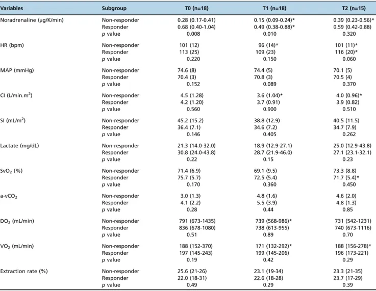
Table 2 -Global hemodynamic parameters and noradrenaline dose recorded at the three different time points during the study protocol and categorized according to responders and non-responders.
Variables Subgroup T0 (n=18) T1 (n=18) T2 (n=15)
Noradrenaline (mg/K/min) Non-responder 0.28 (0.17-0.41) 0.15 (0.09-0.24)* 0.39 (0.23-0.56)* Responder 0.68 (0.40-1.04) 0.49 (0.38-0.88)* 0.59 (0.42-0.88)
pvalue 0.008 0.010 0.320
HR (bpm) Non-responder 101 (12) 96 (14)* 101 (11)*
Responder 113 (25) 109 (23) 116 (20)*
pvalue 0.220 0.150 0.060
MAP (mmHg) Non-responder 74.6 (8) 74.4 (5) 70.1 (5)
Responder 70.4 (3) 70.8 (3) 70.5 (4)
pvalue 0.152 0.089 0.370
CI (L/min.m2) Non-responder 4.5 (1.28) 3.6 (1.04)* 4.0 (0.96)*
Responder 4.2 (1.20) 3.7 (0.91) 3.9 (0.82)
pvalue 0.560 0.900 0.510
SI (mL/m2) Non-responder 45.2 (15.2) 38.8 (12.9) 40.5 (11.5)
Responder 36.4 (7.1) 34.6 (7.2) 34.7 (7.9)
pvalue 0.146 0.405 0.262
Lactate (mg/dL) Non-responder 21.3 (14.0-32.0) 18.9 (12.9-27.1) 25.0 (12.9-43.8) Responder 30.8 (24.0-43.8) 28.7 (21.9-46.0) 27.1 (23.1-32.1)
pvalue 0.22 0.15 0.23
SvO2(%) Non-responder 71.4 (6.9) 69.1 (9.5) 73.3 (8.8)
Responder 75.7 (5.7) 72.5 (5.4) 71.7 (5.4)*
pvalue 0.170 0.360 0.450
a-vCO2 Non-responder 3.0 (1.3) 4.8 (1.6) 4.6 (2.0)
Responder 4.1 (2.2) 5.5 (3.9) 4.8 (1.3)
pvalue 0.28 0.44 0.85
DO2(mL/min) Non-responder 791 (673-1435) 739 (568-986)* 731 (542-1231)
Responder 836 (678-1080) 738 (613-955) 740 (673-1116)
pvalue 0.51 0.89 0.70
VO2(mL/min) Non-responder 188 (152-370) 171 (132-292)* 188 (156-278)*
Responder 197 (145-243) 199 (145-206) 196 (173-221)
pvalue 0.19 0.42 0.29
Extraction rate (%) Non-responder 25.6 (21-26) 23.1 (19-34) 23.3 (21-35)
Responder 22.0 (18-31) 22.6 (18-28) 23.7 (17-29)
pvalue 0.49 0.29 0.39
Time points are defined as before (T0), during (T1) and after (T2) vasopressin infusion. There were 9 responders and 9 non-responders. HR, heart rate; MAP, mean arterial pressure; CI, cardiac index; SI, systolic index; SvO2, oxygen mixed venous saturation. Data are expressed as the mean (standard deviation) or median (25%, 75%).
*po0.05vs. previous time point within the same group.
Table 3 -Sublingual microcirculation parameters recorded at the three different time points during the study protocol and categorized according to responders and non-responders.
Variable Subgroup T0 T1 T2
TVD (mm/mm2) Non-responder 15.9 (2.5) 14.4 (1.8)* 14.8 (0.58)
Responder 14.3 (1.7) 16.3 (1.6)* 15.2 (1.57)*
pvalue 0.13 0.03 0.35
PVD (mm/mm2) Non-responder 13.9 (2.7) 12.4 (1.8)* 12.6 (1.3)
Responder 12.1 (1.9) 14.4 (1.4)* 13.0 (1.9)*
pvalue 0.11 0.02 0.47
PPV (%) Non-responder 87.3 (7.6) 86.5 (6.4) 85.0 (7.8)
Responder 83.5 (6.0) 89.0 (4.8)* 86.6 (7.0)
pvalue 0.26 0.36 0.62
MFI Non-responder 2.7 (0.2) 2.8 (0.2) 2.7 (0.2)
Responder 2.7 (0.1) 2.6 (0.3) 2.7 (0.2)
pvalue 0.69 0.15 0.89
Time points are defined as before (T0), during (T1) and after (T2) vasopressin infusion. There were 9 responders and 9 non-responders. TVD, total vascular density; PPV, proportion of perfused vessels; PVD, perfused vascular density; MFI, microcirculatory flow index; Data are expressed as the mean (standard deviation) or median (25%, 75%).
and both responders and non-responders received similar noradrenaline doses at that time. According to our interac-tion analysis in the generalized mixed-model, the changes in CI, MAP and CVP had no significant effects on TVD or PVD. Interestingly, the decrease in noradrenaline dosing in the non-responder group was associated with a decrease in CI, oxygen delivery (DO2) and oxygen consumption (VO2), whereas CI was restored to baseline levels after discontinua-tion of the vasopressin and the subsequent increase in noradrenaline dosing to maintain MAP (Table 2). The hemo-dynamic parameters, laboratory variables and microcircula-tory measurements in the whole population obtained before and after the vasopressin infusion are listed in the electronic supplementary material (Table S1 and S2).
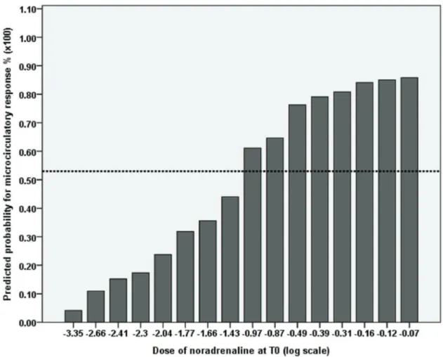
In a multivariate regression model (Table S3), the strongest predictor for an improvement in the microcirculation was the baseline dose of noradrenaline (OR=4.5; 95% CI: 1.2-17.0; p=0.027). Figure 1 shows the probability of a
microcirculatory response based on the noradrenaline dose. The model showed adequate calibration, good discrimina-tion and an area under the ROC curve of 0.85 (95% CI: 0.66-0.99). The probability for 78% sensitivity is indicated, which corresponds to a predicted probability of more than 50% for patients receiving a noradrenaline dose higher than 0.38 mcg/kg/min (log scale4-0.97 in the graph). Table 4 shows the final model with predicted probabilities for a microcirculatory response to vasopressin according to different
infusion doses of noradrenaline to facilitate practical application.
’ DISCUSSION
The primary finding of this study was that the improve-ment in the microcirculatory parameters after vasopressin infusion in the septic shock patients was highly associated with the level of noradrenaline dependency and was inde-pendent of systemic hemodynamic parameters (CI, MAP, and CVP). This finding suggests that the patient’s noradrena-line dose is directly proportional to the likelihood that the patient will respond to vasopressin infusion. For clinical decision making, patients receiving a noradrenaline dose above 0.38 mcg/kg/min (independent of hemodynamic conditions)
Figure 1 -Probability of microcirculatory response based on a dose of noradrenaline at T0 (logarithmic scale). The dotted line shows the threshold value of the predicted probability of 53% for patients receiving a noradrenaline dose higher than 0.38 mcg/kg/min (78% sensitivity and 77% specificity).
Table 4-Predicted probability of the different doses of noradrenaline for microcirculatory responses with their corresponding sensitivity and specificity.
Noradrenaline dose (mcg/kg/min)
Predicted probability for the microcirculatory response to vasopressin
Sensitivity* Specificity*
40.17 27.8% 89% 56%
40.38 53.0% 78% 77%
40.68 77.6% 67% 99%
are candidates for vasopressin use with a 53% probability of recruiting the microcirculation. These findings, which were not previously reported, may help with selecting patients who may benefit from the association of vasopressin according to their noradrenaline dependency status.
The effects of vasopressin in the microcirculation have already been demonstrated by Morelli’s group, which showed that continuous infusions of low-dose vasopressin and terli-pressin improved the MFI after 6 hours. However, the authors found similar results in the control group, which suggests that these changes may not have been related to vasopressin use (31). In our study, we assessed only the short-term effects of vasopressin. Although a subgroup analysis of the VASST study has suggested that patients with less severe septic shock, who require 5 to 14 mcg of noradrenaline per minute, would have better clinical outcomes with the use of vasopressin (32) at the microcirculatory level; however, our results point in the oppo-site direction. Our multivariate model showed that the most clinically useful predictor for a microcirculatory response was the noradrenaline dose required to maintain a MAP above 65 mmHg. Vasopressin infusion also reduced norepinephrine requirements without any significant adverse effect during this short period of infusion.
Microcirculatory alterations can be observed even when systemic hemodynamics are within satisfactory goals. The independence of the microcirculation parameters has been previously reported (18) (33) (34,35). However, microcircu-latory perfusion can be affected by cardiac output and arterial pressure when these variables are critically altered. We found that non-responders had a significant reduction in their cardiac output, DO2 and VO2 during vasopressin infusion, which suggests that changes in the systemic hemodynamics may lead to microcirculatory alterations. However, the reduction in DO2was not followed by an increase in arterial lactate or differences in other tissue perfusion parameters, suggesting that these macrohemodynamic alterations were not clinically rele-vant. However, changes in the CI were not associated with improvement in the microcirculation in our multivariate model. This result highlights the relevance of measuring CI in studies aiming to assess microcirculation even if the enrollment rate is compromised. In fact, one of the major reasons for not enrolling patients in our study was the absence of CI measurements.
Additionally, the reduction in CI may have been related to the reduction in the noradrenaline dose and, consequently, to its inotropic and chronotropic effects or its effect on venous return. A non-significant reduction in the SI was evident during vasopressin infusion. The reduction in noradrenaline dose might be considered a beneficial effect as increasing evidence indicates that the excessive use of catecholamines is associated with potential iatrogenic complications. These potential harmful effects are not exclusively related to well-known hemodynamic effects, namely, increased energy expendi-ture; excessive vasoconstriction; and splanchnic hypoperfusion with altered gut motility, function and potential bacterial trans-location. Convincing evidence exists showing that the phago-cytic capacity of macrophages and neutrophils is inhibited (36), together with lymphopenia and a shift toward a Th2 pattern (37). Bacterial virulence and proliferation is also enhanced (7). Metabolic changes include hyperglycemia, hypertriglyceride-mia and thyroid hormone alterations. Coagulation disorders with enhanced clot formation and a reduction in fibrinolysis have also been reported (7). Thus, the use of vasopressin is part of the decatecholaminization strategy that has gained increased support in critical care.
Although our study was not designed to assess causality, we can provide several hypotheses. Vasopressin led to a reduction in the noradrenaline dose in all patients. This result suggests that the use of receptors other than adrenergic receptors will lead to a better response in vasopressor tone and, consequently, to a reduction in the need for vasopres-sors. Reducing the excessive vasopressor effect of noradrena-line may have led to an increase in microcirculatory density and perfusion in the responders. Non-responders were using a smaller dose of noradrenaline, which suggests less severe disease and vasodilation. In this context, vasopressin might have caused excessive vasoconstriction, as suggested by the finding of a decrease in PVD. Moreover, the potential negative effects of vasopressin in cardiac output may have played a role. Patients who had a smaller reduction in CI after vasopressin infusion would be likely to show improve-ments in microcirculation. The differences between the respon-ders and non-responrespon-ders regarding the impact of vasopressin on CI, DO2and VO2were interesting. For the non-responders, the CI reduction did not compromise the microcirculation but was associated with a decrease in VO2 that was reversed when vasopressin was suspended. This finding hypothetically suggests that if vasopressin is used, a decrease in noradrena-line requirements may occur if the microcirculation does not improve the overall effect of vasopressin, which might be harmful. However, these notions are speculations because distinguishing the effects of vasopressin on contractility from its vasoconstrictive effects on microcirculation is difficult.
Although this study provides novel observations, a fixed dose of vasopressin was used, which could be considered insufficient to show any beneficial or harmful effects. A pre-vious study suggested that a higher dose of vasopressin (0.067 U/min) would restore hemodynamics more effectively and that the dose was not associated with a high incidence of adverse effects (38). Another limitation of the present study is the absence of a control group. However, this study was designed to allow every patient to serve as his or her own control, which minimizes bias. Therefore, the significant improvement or decrease in the sublingual microcirculation parameters during and after vasopressin infusion strengthens our findings. An additional limitation is the short observation period of our study as a longer AVP infusion period might have a different impact on the microcirculatory response. Notably, the high severity of illness in our population is another limitation worth considering. These patients had a mean organ dysfunction incidence of 4.2 and a high mortality rate. Thus, the effects of vasopressin may be different in patients with a less severe shock.
In conclusion, the clinical monitoring of the sublingual microcirculation can help identify patients with septic shock that might benefit from the association of vasopressin. Our results suggest that a noradrenaline dose above 0.38 mcg/ kg/min might be a good predictor for the microcirculatory response to the vasopressin infusion. Additional research that explores different microcirculatory beds and uses dif-ferent measurement tools for assessing microcirculation will improve our knowledge concerning the role of vasopressin in septic shock resuscitation.
’ ACKNOWLEDGMENTS
Dr. Ivan Koh for assisting us in using the SDF device. Financial support: Fundac¸ão de Amparo a Pesquisa do Estado de São Paulo (FAPESP), Grant 2009/50096-6. The funding organization played no role in the design of the study, data collection, analysis, interpretation of data or writing of the manuscript.
’ AUTHOR CONTRIBUTIONS
Nascente AP and Machado FR are the guarantors of the entire manuscript. Nascente AP, Machado FR and Freitas FG designed the study. Nascente AP collected all of the data. All authors helped with the data interpretation and drafting of the manuscript. All authors revised and approved thefinal version of the manuscript.
’ REFERENCES
1. Jones SB, Romano FD. Myocardial beta adrenergic receptor coupling to adenylate cyclase during developing septic shock. Circ Shock. 1990; 30(1):51-61.
2. Chernow B, Roth BL. Pharmacologic manipulation of the peripheral vasculature in shock: clinical and experimental approaches. Circ Shock. 1986;18(2):141-55.
3. Cronin RE, Erickson AM, de Torrente A, McDonald KM, Schrier RW. Norepinephrine-induced acute renal failure: a reversible ischemic model of acute renal failure. Kidney Int. 1978;14(2):187-90, http://dx.doi.org/ 10.1038/ki.1978.106.
4. De Backer D, Creteur J, Silva E, Vincent JL. Effects of dopamine, nor-epinephrine, and epinephrine on the splanchnic circulation in septic shock: which is best? Crit Care Med. 2003;31(6):1659-67, http://dx.doi. org/10.1097/01.CCM.0000063045.77339.B6.
5. Meier-Hellmann A, Reinhart K, Bredle DL, Specht M, Spies CD, Hanne-mann L. Epinephrine impairs splanchnic perfusion in septic shock. Crit Care Med. 1997;25(3):399-404, http://dx.doi.org/10.1097/00003246-19970 3000-00005.
6. Meier-Hellmann A, Bredle DL, Specht M, Spies C, Hannemann L, Reinhart K. The effects of low-dose dopamine on splanchnic blood flow and oxygen uptake in patients with septic shock. Intensive Care Med. 1997;23(1):31-7, http://dx.doi.org/10.1007/s001340050287.
7. Andreis DT, Singer M. Catecholamines for inflammatory shock: a Jekyll-and-Hyde conundrum. Intensive Care Med. 2016;42(9):1387-97, http:// dx.doi.org/10.1007/s00134-016-4249-z.
8. Belletti A, Castro ML, Silvetti S, Greco T, Biondi-Zoccai G, Pasin L, et al. The Effect of inotropes and vasopressors on mortality: a meta-analysis of randomized clinical trials. Br J Anaesth. 2015;115(5):656-75, http://dx.doi. org/10.1093/bja/aev284.
9. Belletti A, Musu M, Silvetti S, Saleh O, Pasin L, Monaco F, et al. Non-Adrenergic Vasopressors in Patients with or at Risk for Vasodilatory Shock. A Systematic Review and Meta-Analysis of Randomized Trials. PLoS One. 2015;10(11):e0142605, http://dx.doi.org/10.1371/journal.pone.0142605. 10. Oba Y, Lone NA. Mortality benefit of vasopressor and inotropic agents in septic shock: a Bayesian network meta-analysis of randomized controlled trials. J Crit Care. 2014;29(5):706-10, http://dx.doi.org/10.1016/j.jcrc.2014.04.011. 11. Gordon AC, Mason AJ, Thirunavukkarasu N, Perkins GD, Cecconi M,
Cepkova M, et al. Effect of Early Vasopressin vs Norepinephrine on Kidney Failure in Patients With Septic Shock: The VANISH Randomized Clinical Trial. JAMA. 2016;316(5):509-18, http://dx.doi.org/10.1001/jama.2016.10485. 12. Tsuneyoshi I, Yamada H, Kakihana Y, Nakamura M, Nakano Y, Boyle WA, 3rd. Hemodynamic and metabolic effects of low-dose vasopressin infusions in vasodilatory septic shock. Crit Care Med. 2001;29(3):487-93, http://dx.doi.org/10.1097/00003246-200103000-00004.
13. Landry DW, Levin HR, Gallant EM, Ashton RC Jr, Seo S, D’Alessandro D,
et al. Vasopressin deficiency contributes to the vasodilation of septic shock. Circulation. 1997;95(5):1122-5, http://dx.doi.org/10.1161/01.CIR.95.5.1122. 14. Malay MB, Ashton RC Jr, Landry DW, Townsend RN. Low-dose
vaso-pressin in the treatment of vasodilatory septic shock. J Trauma. 1999;47(4): 699-703; discussion 703-5.
15. Holmes CL, Walley KR, Chittock DR, Lehman T, Russell JA. The effects of vasopressin on hemodynamics and renal function in severe septic shock: a case series. Intensive Care Med. 2001;27(8):1416-21, http://dx.doi.org/ 10.1007/s001340101014.
16. Patel BM, Chittock DR, Russell JA, Walley KR. Beneficial effects of short-term vasopressin infusion during severe septic shock. Anesthesiology. 2002;96(3):576-82, http://dx.doi.org/10.1097/00000542-200203000-00011. 17. Argenziano M, Chen JM, Choudhri AF, Cullinane S, Garfein E, Weinberg AD, et al. Management of vasodilatory shock after cardiac surgery: identification of predisposing factors and use of a novel pressor agent. J Thorac Cardiovasc Surg. 1998;116(6):973-80, http://dx.doi.org/10.1016/ S0022-5223(98)70049-2.
18. De Backer D, Ortiz JA, Salgado D. Coupling microcirculation to systemic hemodynamics. Curr Opin Crit Care. 2010;16(3):250-4, http://dx.doi.org/ 10.1097/MCC.0b013e3283383621.
19. De Backer D, Creteur J, Preiser JC, Dubois MJ, Vincent JL. Microvascular blood flow is altered in patients with sepsis. Am J Respir Crit Care Med. 2002;166(1):98-104, http://dx.doi.org/10.1164/rccm.200109-016OC. 20. Sakr Y, Dubois MJ, De Backer D, Creteur J, Vincent JL. Persistent
micro-circulatory alterations are associated with organ failure and death in patients with septic shock. Crit Care Med. 2004;32(9):1825-31, http://dx. doi.org/10.1097/01.CCM.0000138558.16257.3F.
21. De Backer D, Donadello K, Sakr Y, Ospina-Tascon G, Salgado D, Scolletta S, et al. Microcirculatory Alterations in Patients With Severe Sepsis: Impact of Time of Assessment and Relationship With Outcome. Crit Care Med. 2013;41(3):791-9, http://dx.doi.org/10.1097/CCM.0b013e3182742e8b. 22. Dunser MW, Mayr AJ, Tur A, Pajk W, Barbara F, Knotzer H, et al.
Ischemic skin lesions as a complication of continuous vasopressin infusion in catecholamine-resistant vasodilatory shock: incidence and risk factors. Crit Care Med. 2003;31(5):1394-8, http://dx.doi.org/10.1097/01.CCM.0000059722. 94182.79.
23. Luckner G, Dunser MW, Stadlbauer KH, Mayr VD, Jochberger S, Wenzel V, et al. Cutaneous vascular reactivity and flow motion response to vasopressin in advanced vasodilatory shock and severe postoperative multiple organ dysfunction syndrome. Crit Care. 2006;10(2):R40, http:// dx.doi.org/10.1186/cc4845.
24. Dunser MW, Mayr AJ, Stallinger A, Ulmer H, Ritsch N, Knotzer H, et al. Cardiac performance during vasopressin infusion in postcardiotomy shock. Intensive Care Med. 2002;28(6):746-51, http://dx.doi.org/10.1007/ s00134-002-1265-y.
25. Gordon AC, Russell JA, Walley KR, Singer J, Ayers D, Storms MM, et al. The effects of vasopressin on acute kidney injury in septic shock. Intensive Care Med. 2010;36(1):83-91, http://dx.doi.org/10.1007/s00134-009-1687-x. 26. Ertmer C, Rehberg S, Westphal M. Vasopressin analogues in the treatment of shock states: potential pitfalls. Best Pract Res Clin Anaesthesiol. 2008; 22(2):393-406, http://dx.doi.org/10.1016/j.bpa.2008.02.007.
27. Bone RC, Balk RA, Cerra FB, Dellinger RP, Fein AM, Knaus WA, et al. Definitions for sepsis and organ failure and guidelines for the use of innova-tive therapies in sepsis. The ACCP/SCCM Consensus Conference Committee. American College of Chest Physicians/Society of Critical Care Medicine. Chest. 1992;101(6):1644-55, http://dx.doi.org/10.1378/chest.101.6.1644. 28. Singer M, Deutschman CS, Seymour CW, Shankar-Hari M, Annane D,
Bauer M, et al. The Third International Consensus Definitions for Sepsis and Septic Shock (Sepsis-3). JAMA. 2016;315(8):801-10, http://dx.doi.org/ 10.1001/jama.2016.0287.
29. De Backer D, Hollenberg S, Boerma C, Goedhart P, Buchele G, Ospina-Tascon G, et al. How to evaluate the microcirculation: report of a round table conference. Crit Care. 2007;11(5):R101, http://dx.doi.org/10.1186/cc6118. 30. Trzeciak S, McCoy JV, Phillip Dellinger R, Arnold RC, Rizzuto M, Abate
NL, et al. Early increases in microcirculatory perfusion during protocol-directed resuscitation are associated with reduced multi-organ failure at 24 h in patients with sepsis. Intensive Care Med. 2008;34(12):2210-7, http://dx.doi.org/10.1007/s00134-008-1193-6.
31. Morelli A, Donati A, Ertmer C, Rehberg S, Kampmeier T, Orecchioni A, et al. Effects of vasopressinergic receptor agonists on sublingual micro-circulation in norepinephrine-dependent septic shock. Crit Care. 2011;15 (5):R217, http://dx.doi.org/10.1186/cc10453.
32. Russell JA, Walley KR, Singer J, Gordon AC, Hebert PC, Cooper DJ, et al. Vasopressin versus norepinephrine infusion in patients with septic shock. N Engl J Med. 2008;358(9):877-87, http://dx.doi.org/10.1056/NEJMoa067373. 33. De Backer D, Creteur J, Dubois MJ, Sakr Y, Koch M, Verdant C, et al. The effects of dobutamine on microcirculatory alterations in patients with septic shock are independent of its systemic effects. Crit Care Med. 2006;34(2):403-8, http://dx.doi.org/10.1097/01.CCM.0000198107. 61493.5A.
34. Jhanji S, Stirling S, Patel N, Hinds CJ, Pearse RM. The effect of increasing doses of norepinephrine on tissue oxygenation and microvascular flow in patients with septic shock. Crit Care Med. 2009;37(6):1961-6, http://dx. doi.org/10.1097/CCM.0b013e3181a00a1c.
35. Dubin A, Pozo MO, Casabella CA, Palizas F Jr, Murias G, Moseinco MC, et al. Increasing arterial blood pressure with norepinephrine does not improve microcirculatory blood flow: a prospective study. Crit Care. 2009;13(3):R92, http://dx.doi.org/10.1186/cc7922.
36. Wenisch C, Parschalk B, Weiss A, Zedwitz-Liebenstein K, Hahsler B, Wenisch H, et al. High-dose catecholamine treatment decreases poly-morphonuclear leukocyte phagocytic capacity and reactive oxygen pro-duction. Clin Diagn Lab Immunol. 1996;3(4):423-8.
37. Kohm AP, Sanders VM. Norepinephrine and beta 2-adrenergic receptor
stimulation regulate CD4+T and B lymphocyte function in vitro and in
vivo. Pharmacol Rev. 2001;53(4):487-525.
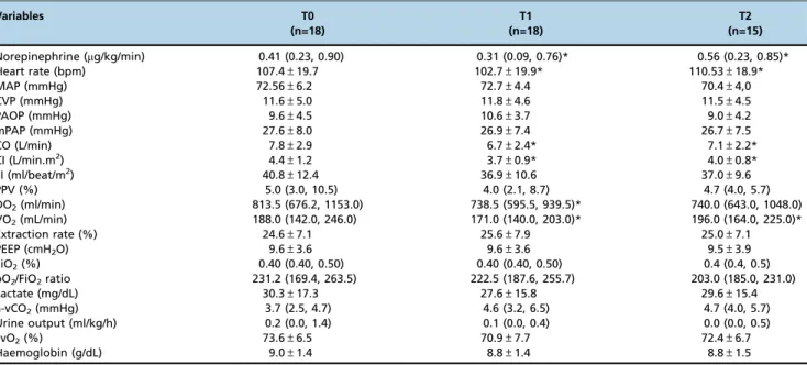
’ APPENDIX
Table S1-Changes in hemodynamic, respiratory and metabolic variables after vasopressin infusion – global analysis.
Variables T0
(n=18)
T1 (n=18)
T2 (n=15)
Norepinephrine (mg/kg/min) 0.41 (0.23, 0.90) 0.31 (0.09, 0.76)* 0.56 (0.23, 0.85)*
Heart rate (bpm) 107.4±19.7 102.7±19.9* 110.53±18.9*
MAP (mmHg) 72.56±6.2 72.7±4.4 70.4±4,0
CVP (mmHg) 11.6±5.0 11.8±4.6 11.5±4.5
PAOP (mmHg) 9.6±4.5 10.6±3.7 9.0±4.2
mPAP (mmHg) 27.6±8.0 26.9±7.4 26.7±7.5
CO (L/min) 7.8±2.9 6.7±2.4* 7.1±2.2*
CI (L/min.m2) 4.4±1.2 3.7±0.9* 4.0±0.8*
SI (ml/beat/m2) 40.8±12.4 36.9±10.6 37.0±9.6
PPV (%) 5.0 (3.0, 10.5) 4.0 (2.1, 8.7) 4.7 (4.0, 5.7)
DO2(ml/min) 813.5 (676.2, 1153.0) 738.5 (595.5, 939.5)* 740.0 (643.0, 1048.0) VO2(mL/min) 188.0 (142.0, 246.0) 171.0 (140.0, 203.0)* 196.0 (164.0, 225.0)*
Extraction rate (%) 24.6±7.1 25.6±7.9 25.0±7.1
PEEP (cmH2O) 9.6±3.6 9.6±3.6 9.5±3.9
FiO2(%) 0.40 (0.40, 0.50) 0.40 (0.40, 0.50) 0.4 (0.4, 0.5)
pO2/FiO2ratio 231.2 (169.4, 263.5) 222.5 (187.6, 255.7) 203.0 (185.0, 231.0)
Lactate (mg/dL) 30.3±17.3 27.6±15.8 29.6±15.4
a-vCO2(mmHg) 3.7 (2.5, 4.7) 4.6 (3.2, 6.5) 4.7 (4.0, 5.7)
Urine output (ml/kg/h) 0.2 (0.0, 1.4) 0.1 (0.0, 0.4) 0.0 (0.0, 0.5)
SvO2(%) 73.6±6.5 70.9±7.7 72.4±6.7
Haemoglobin (g/dL) 9.0±1.4 8.8±1.4 8.8±1.5
Time points are defined as before (T0), during (T1) and after (T2) vasopressin infusion. MAP, mean arterial pressure; CVP, central venous pressure; PAOP, pulmonary artery occluded pressure; PAP, pulmonary artery pressure; CO, cardiac output; CI, cardiac index; SI, systolic index; PPV, pulse pressure variation; DO2, oxygen delivery; PEEP, positive end expiratory pressure; FiO2, fraction of inspired oxygen; pO2, oxygen partial pressure;DCO2, venous-arterial CO2 gradient; SvO2, oxygen mixed venous saturation. Data are expressed as number (%), mean±standard deviation or median (25%, 75%).
*po0.05vs. previous time point.
Table S2-Changes in the microcirculatory variables after vasopressin infusion – global analysis.
Variable T0 T1 T2
TVD (mm/mm2) 15.1±2.2 15.2±1.8 15.2±1.5
PVD (mm/mm2) 13.0±2.4 13.4±1.8 12.9±1.7
PPV (%) 87.1 (83.0, 90.5) 88.6 (84.1, 90.5) 85.8 (83.4, 90.5)
MFI 2.7 (2.6, 2.9) 2.9 (2.5, 3.0) 2.7 (2.5, 3.0)
Time points are defined as before (T0), during (T1) and after (T2) vasopressin infusion. TVD, total vascular density; PPV, proportion of perfused vessels; PVD, perfused vascular density; MFI, microcirculatory flow index; HI, heterogeneity index. Data are expressed as mean±standard deviation or median (25%, 75%).
*po0.05vs. previous time point.
Table S3-Multivariable regression model to changes in microcirculatory response.
Independent variable bcoefficient±SE OR 95% CI p-value
TVD -0.73±0.4 0.479 (0.180 – 1.272) 0.14
HR 0.013±0.07 1.013 (0.877 – 1.172) 0.85
MAP -0.15±0.31 0.855 (0.463 – 1.579) 0.61
CVP 0.09±0.34 1.104 (0.566 – 2.154) 0.77
Lactate -0.07±0.068 0.925 (0.811 – 1.056) 0.25
CI -0.83±0.81 0.433 (0.088 – 2.128) 0.30
Noradrenaline dose* 4.8±2.3 5.611 (1.130 – 27.861) 0.03
* Noradrenaline dose was used as a categorized variable.