REVISTA
BRASILEIRA
DE
ANESTESIOLOGIA
PublicaçãoOficialdaSociedadeBrasileiradeAnestesiologiawww.sba.com.br
SCIENTIFIC
ARTICLE
Effects
of
the
positive
end-expiratory
pressure
increase
on
sublingual
microcirculation
in
patients
with
acute
respiratory
distress
syndrome
Nathaly
Fonseca
Nunes,
Antônio
Tonete
Bafi
∗,
Eduardo
Souza
Pacheco,
Luciano
Cesar
Pontes
de
Azevedo,
Flavia
Ribeiro
Machado,
Flávio
Geraldo
Rezende
Freitas
UniversidadeFederaldeSãoPaulo(UNIFESP),HospitaldeSãoPaulo,DisciplinadeAnestesiologia,DoreTerapiaIntensiva, SãoPaulo,SP,Brazil
Received21May2015;accepted5October2015 Availableonline31May2016
KEYWORDS
Adultrespiratory distresssyndrome; Positive
end-expiratory pressure; Microcirculation; Hemodynamics; Shock;
Mechanical ventilators
Abstract
Objective:Theaimofthisstudywastoevaluatetheimpactofincreasedpositiveend-expiratory
pressureonthesublingualmicrocirculation.
Methods:Adultpatientswhoweresedated,undermechanicalventilation,andhadadiagnosis
ofcirculatoryshockandacuterespiratorydistresssyndromewereincluded.Thepositive end-expiratorypressurelevelwassettledtoobtainaplateaupressureof30cmH2Oandthen main-tainedatthislevelfor20minutes.Microcirculatory(obtainedbyvideomicroscopy)and hemo-dynamicvariableswerecollectedatbaselineandcomparedwiththoseattheendof20min.
Results:Twelvepatientswereenrolled.Overall,themicrocirculationparametersdidnot
sig-nificantlychange after increasing thepositive end-expiratorypressure. However, therewas considerableinterindividualvariability.Therewasanegative,moderatecorrelationbetween the changes in the De Backer score (r=−0.58, p=0.048), total vessel density (r=−0.60,
p=0.039)andbaselinevalues.Thechangesintotalvesseldensity(r=0.54,p=0.07)and per-fusedvesseldensity(r=0.52,p=0.08)trendedtowardcorrelatingwiththechangesinthemean arterialpressure.
Conclusion:Overall,themicrocirculationparametersdidnotsignificantlychangeafter
increas-ing the positive end-expiratory pressure. However, at individual level, such response was heterogeneous.Thechangesinthemicrocirculationparameterscouldbecorrelatedwiththe baselinevaluesandchangesinthemeanarterialpressure.
©2016SociedadeBrasileiradeAnestesiologia.PublishedbyElsevierEditoraLtda.Thisisan openaccessarticleundertheCCBY-NC-NDlicense( http://creativecommons.org/licenses/by-nc-nd/4.0/).
∗Correspondingauthor.
E-mail:[email protected](A.T.Bafi).
http://dx.doi.org/10.1016/j.bjane.2015.10.002
PALAVRAS-CHAVE
Síndromedo
desconforto respiratóriodo adulto;
Pressãopositiva expiratóriafinal; Microcirculac¸ão; Hemodinâmica; Choque; Ventiladores mecânicos
Efeitosdoaumentodepressãopositivaaofinaldaexpirac¸ãosobreamicrocirculac¸ão sublingualempacientescomsíndromedodesconfortorespiratórioagudo
Resumo
Objetivo: Oobjetivodesteestudofoiavaliaroimpactodoaumentodepressãopositiva
expi-ratóriafinal(PEEP)sobreamicrocirculac¸ãosublingual.
Métodos: Ospacientesadultosqueforamsedados,sobventilac¸ãomecânica,comdiagnóstico
dechoquecirculatórioesíndromedodesconfortorespiratórioagudoforamincluídos.Onívelda PEEPestabelecidoparaobterumapressãodeplatôde30cmH2Oedepoismantidanessenível por20minutos.Asvariáveismicrocirculatória(obtidapormicroscopiadevídeo)ehemodinâmica foramregistradasnafasebasalecomparadascomaquelasaofinalde20min.
Resultados: Dozepacientesforamincluídos.Emgeral,osparâmetrosdamicrocirculac¸ãonão
apresentaram alterac¸ões significativasapós oaumento daPEEP. Porém, houveconsiderável variabilidadeinterindividual.Houveumacorrelac¸ãonegativa,moderada,entreasalterac¸ões noescoredeDeBacker(r=-0,58,p=0,048),nadensidadetotaldovaso(r=-0,60,p=0,039)e nosvaloresbasais.Asalterac¸õesnadensidadetotaldovaso(r=0,54,p=0,07)enadensidade dovasoperfundido(r=0,52,p=0,08)apresentaramtendênciadecorrelac¸ãocomasalterac¸ões napressãoarterialmédia.
Conclusão:Emgeral,osparâmetrosdamicrocirculac¸ãonãoapresentaramalterac¸ões
significa-tivasapósoaumentodaPEEP.Noentanto,individualmente,essarespostafoiheterogênea. Asalterac¸õesnosparâmetrosdamicrocirculac¸ãopuderamsercorrelacionadascomosvalores basaisealterac¸õesnapressãoarterialmédia.
©2016SociedadeBrasileiradeAnestesiologia.PublicadoporElsevierEditoraLtda.Este ´eum artigo OpenAccess sobumalicenc¸aCCBY-NC-ND( http://creativecommons.org/licenses/by-nc-nd/4.0/).
Introduction
Inpatientswithmoderateor severeacuterespiratory dis-tress syndrome (ARDS), a ventilator strategy based on higherrather thanlower levelsof positiveend-expiratory pressure(PEEP)isrecommended.1,2ThePEEPresultsin
alve-olar recruitment, reducedshunting, andincreased partial pressure ofoxygen (PaO2).2 However,theextrapulmonary
effects of high PEEP can limit this approach. The effects onthehemodynamicsandregionalbloodflowarethemain concerns.3
Theultimategoalofrespiratoryandhemodynamic inter-ventionsistorestoreeffectivetissueperfusionandoxygen delivery to maintain cellular metabolism. Therefore, the assessment of the microcirculation might improve our understandingoftheeffectsoftherapies beyondrestoring systemichemodynamics.4Alterationsinmicrovascularblood
flowareunderlyingmechanismsthatareimplicatedinthe developmentofmultipleorgandysfunctionand,ultimately, death.5Severalstudieshaveshownthatsevereand
persis-tentmicrocirculatoryalterationsarestrongpredictorsofthe outcome.6---9Microcirculatoryalterationscanstillbepresent
evenwhentheglobalhemodynamicsareoptimized.10These
findingssuggestthattargetingthemicrocirculationisa log-icalapproachfor interventionsthataimtoimprovetissue perfusion.4
Clinical studies exploring the regional perfusion alter-ations induced by the PEEP using different tools have focused on the splanchnic area and shown conflicting results.11---13AsthemicrocirculatoryeffectsofthePEEPhave
notbeenestablished,theaimofthisstudywastoevaluate theimpactof increasingthePEEP levelsin thesublingual microcirculationparametersusingvideomicroscopy.
Methods
Thisstudywasconductedina35bedmixedIntensiveCare Unit(ICU)in auniversityhospitalfromJuly 2011to Octo-ber2012.The localethicscommitteeapprovedthe study, andthepatients’closestrelativessignedinformedconsent formstoallowfordatacollection.
WeincludedadultpatientswithARDSwhowere mechan-icallyventilated witha plateau pressure≤25cm H2O and
PEEP≤10cm H2O as well asan indication of an increase
inthe PEEP by theattending physician.Allpatients were receivingsedation witha Ramsay scale of 6, had circula-toryshockwiththeneedforvasopressorandhemodynamic monitoringwithapulmonaryarterycatheterandanarterial catheter. ARDS was defined according to the Consen-sus conference.14 The exclusion criteria were pregnancy,
intracranial hypertension, abdominal compartment syn-dromeandoralinjuries.Wealsoexcludedpatientsinwhom theprimarycauseofcirculatoryshockwasactivebleeding (suspectedor confirmed)or cardiogenic shock, which was definedasacardiacindex(CI)<1.8L·min−1·m−2without
sup-portandpulmonaryarteryocclusionpressure≥18mmHg.
Interventions
Theselectedpatientsweremechanicallyventilated(Vela, Viasys,PalmSprings,CA,USA)usingthevolume-controlled mode.The tidalvolumewasadjustedto6mL·kg−1(based
werenosignificantvariationsinthehemodynamicand ven-tilatorparameters. The PEEP level wasthen increased to obtainaplateaupressureof30cmH2O(measuredafteran
end-inspiratorypauseof 2s).The PEEPwasmaintainedat theselevelsfor 20min. Throughoutthe study period,the doses of the sedative, inotropic and vasopressor medica-tionsremainedconstant.Ifthemeanarterialpressure(MAP) decreasedbelow65mmHg,theCIdecreasedmorethan50% orpulseoximetry decreased below90% duringthis period ofobservation,theinterventionwasinterrupted.Afterthe protocol,theattendingphysicianadjustedthePEEPlevel.
Wemeasuredthehemodynamic,ventilatoryand micro-circulatory parameters at baseline (T0) and immediately afterthe20minperiod(T1).TheCIwasmeasured usinga semi-continuousthermodilution techniquethatconsidered themeanvalueoffourconsecutivemeasurementsfromthe STATmodescreenoftheVigilance® monitor(Edwards
Life-sciences,Irvine,CA,USA).Allpressuresweredeterminedat theend-expirationwiththezeroreferencelevelsettledat the4thto5thintercostalspacealongthemid-axillaryline. Weassessedthesublingualmicrovascularnetworkusing SidestreamDarkField(SDF)imaging(Microscan;MicroVision Medical, Amsterdam, Netherlands). Briefly, the Microscan is a hand-held video microscope system that illuminates atissueof interestwithstroboscopic green(530nm) light emittingdiodes.Hemoglobinabsorbsthe530nmwavelength light,whichinturniscapturedviatheimagingprobe’slight guideand a charge-coupled device camera. Clear images of flowing RBCs are depicted as dark moving globules in the lumen of blood vesselsagainst a white/grayish back-ground. The recommended techniques for ensuring high image quality were adopted.15 After removing saliva and
oralsecretions,theprobewasappliedoverthemucosa.At each timepoint, three videos wererecorded in different sitesatthebaseofthetongue,atleast10s persite. Spe-cialcarewastakentoavoid pressureartifacts,whichwas verifiedbycheckingongoingflowinlargermicrovessels.All ofthesevideoswereobtainedusingtheAVA3.0® software
(Microvision Medical, Amsterdam, Netherlands) consider-ing for analyses vessels with a diameter less than 20m
(smallvessels).Theentiresequencewasusedto character-izethe semi-quantitative characteristics of microvascular bloodflow,particularlythepresenceofstoppedor intermit-tentflow.Itdistinguishesbetweennoflow(0),intermittent flow(1),sluggishflow(2),andcontinuousflow(3).Avalue wasassignedtoeachindividualvessel.Afterstabilizationof theimagesusingtheAVA3.0software,wedeterminedthe microcirculatoryflowindex(MFI),totalvesseldensity(TVD), proportionofperfusedvessels(PPV),DeBackerscore,and perfused vessel density (PVD) as previously described.15
Blindedinvestigators(ATBandNFN)analyzedallimagesin arandomorder.
Statisticalanalysis
We hypothesized a mean decrease of 0.5 and a standard deviation of the difference of 0.5 in the MFI after an increasedinthePEEPtocalculatethesamplesizerequired for comparing two paired samples (significance level of 0.05 and power of 80%). The required sample size was 10 patients; to correct for the potential non-parametric
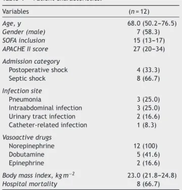
Table1 Patientcharacteristics.
Variables (n=12)
Age,y 68.0(50.2---76.5)
Gender(male) 7(58.3)
SOFAinclusion 15(13---17)
APACHEIIscore 27(20---34)
Admissioncategory
Postoperativeshock 4(33.3)
Septicshock 8(66.7)
Infectionsite
Pneumonia 3(25.0)
Intraabdominalinfection 3(25.0)
Urinarytractinfection 2(16.6)
Catheter-relatedinfection 1(8.3)
Vasoactivedrugs
Norepinephrine 12(100)
Dobutamine 5(41.6)
Epinephrine 2(16.6)
Bodymassindex,kgm−2 23.0(21.8---24.8)
Hospitalmortality 8(66.7)
SOFA, Sequential OrganFailure Assessment;APACHE II,Acute PhysiologicalChronicHealthEvaluation.Results areexpressed asthenumber(%)ormedian(25%---75%).
distributionofthevariable,weadjustedthisrequired sam-plesizeto12patients.
Data are expressed as numbers (%) or medians and
interquartileranges(25thto75thpercentile). Nonparamet-rictestswereusedbecauseofthesmallsamplesize.The hemodynamic, respiratory, and microcirculatory variables werecomparedatT0andT1usingtheWilcoxonpairedtest. Additionalanalyseswereconductedtotestthelinear corre-lationbetweenthebaselinemicrocirculatoryvariablesand theirchangesafterthePEEPincrease(MFI,TVD,PPV, PVDandDeBackerscore)usingtheSpearmancorrelation test.
We used SPSS version 17.0 for Windows (SPSS Inc.,
Chicago, IL, USA). The results with p-values < 0.05 were consideredsignificant.Forthesample sizecalculation,we
used MedCalc software 14.12.0 (MedCalc Software bvba,
Belgium).
Results
We enrolled 12 patients with ARDS and circulatory shock
withamedianageof68.0(50.25---76.50)years.Septicshock
wasthemostcommonreasonforICU admission.Themain
clinicaldataareshowninTable1.
The median increase in the PEEP levels to achieve a plateaupressureof 30cmH2Owas7.5(6.0---10.0)cmH2O.
AfteranincreaseinthePEEP,tenpatientshadadecreased CIandninehadadecreasedMAP.IncreasingthePEEPlevels led to a significant increase in the PaO2 (p=0.05);
Table2 Changesinthehemodynamic,respiratoryandmetabolicvariablesafterachangeinthePEEP.
Variables Baseline AfterPEEP p-value
HR,bpm 100.50(92.50---111.25) 98.50(91.50---114.50) 0.710
CI,L·min−1·m−2 3.71(3.45---5.00) 3.45(2.65---4.52) 0.005
MAP,mmHg 78.50(75.25---86.00) 77.00(68.25---85.00) 0.100
CVP,mmHg 12.00(7.25---13.75) 13.00(9.75---16.00) 0.004
mPAP,mmHg 24.00(18.50---37.75) 26.50(23.25---35.50) 0.110
PAOP,mmHg 9.90(8.15---13.50) 13.90(12.92---16.25) 0.008
PP,% 3.75(2.37---7.35) 6.50(3.20---13.00) 0.009
PaO2/FiO2ratio 163.50(126.42---228.66) 205.00(154.13---238.57) 0.070
PaO2,mmHg 88.00(80.50---108.50) 101.50(89.05---119.00) 0.050
Tidalvolume,mL 467.50(438.75---557.50) 467.50(438.75---557.50) 1.000
PEEP,cmH2O 7.00(5.00---9.50) 15.00(14.25---19.00) 0.002
SvO2,% 75.50(64.40---81.22) 75.60(62.62---83.12) 0.630
Lactate,mg·dL−1 30.00(26.25---49.25) 28.00(17.50---44.25) 0.640
DO2,mL·min−1 855.53(532.81---1044.77) 781.39(504.55---970.11) 0.010
VO2,mL·min−1 192.83(161.60---231.00) 180.65(151.73---202.72) 0.230
Compliance,mL·cm−1H
2O 35.89(24.31---44.16) 35.85(24.31---44.64) 0.480
HR,hearthrate;CI,cardiacindex;MAP,meanarterialpressure;CVP,centralvenouspressure;mPAP,pulmonaryarterypressure;PAOP, pulmonaryarteryoccludedpressure;PP,pulsepressurevariation;PaO2,oxygenpartialpressure;FiO2,fractionofinspiredoxygen; PEEP,positiveend-expiratorypressure;SvO2,oxygenmixedvenoussaturation;DO2,oxygendelivery;andVO2,oxygenconsumption. Dataareexpressedasthemedian(25%---75%).Wilcoxonpairedtest.
Table3 ChangesinthemicrocirculatoryvariablesafterachangeinthePEEP.
Variable Baseline AfterPEEP p-value
TVD,mm·mm−2 13.51(12.64---15.24) 14.95(11.80---15.63) 0.875
PVD,mm·mm−2 11.25(10.06---14.24) 11.87(10.26---13.09) 0.583
PPV,% 83.14(77.72---91.48) 81.02(77.25---87.26) 0.695
DeBacker,n·mm−2 9.50(8.73---10.24) 9.60(8.09---10.40) 0.875
MFI 2.62(2.28---2.75) 2.55(2.28---2.75) 0.799
PEEP,positiveend-expiratorypressure;TVD,totalvesseldensity;PPV,proportionofperfusedvessels;PVD,perfusedvesseldensity;and MFI,microcirculatoryflowindex.Dataareexpressedasthemedian(25%---75%).Wilcoxonpairedtest.
25
20
15
10
T0 T1
T0 T1 T0 T1
T0 T1 T0 T1
TVD m
m.
mm
-2
PVD m
m.
mm
-2
PPV
%
De Backer
n.
mm
-1
MF
I
100
90
80
70
60
50
20
15
10
15
10
3
2
1
Overall, the microcirculation parameters did not vary significantlyafteranincrease inthePEEP (Table3). How-ever,therewasconsiderableinterindividualvariability.The individualchangesin the microcirculatoryparameters are showninFig.1.Intwopatients,thereweredramaticfalls inthePPV.ThesepatientshadimportantdecreaseintheCI andMAP.
There was a negative, moderate correlation between theDeBacker(r=−0.58,p=0.048)andTVD(r=−0.60,
p=0.039)andtheirbaselinevalues.Thiswasnotthecasefor theMFI(r=−0.29,p=0.36)orPPV(r=−0.48,p=0.12).
There was a trend negative correlation between PVD (r=−0.57,p=0.05)andbaselinevalue.Interestingly,TVD (r=0.54,p=0.07)andPVD(r=0.52,p=0.08)hadatrend ofcorrelatingwiththechangesintheMAP.Noother corre-lationwasfound betweenthechanges inmicrocirculatory parametersandchanges inthesystemichemodynamicsor changesinthePEEPlevels.
Discussion
Wefoundthattherewasconsiderablevariationinthe indi-vidual sublingual microcirculatory responses to increases inthe PEEP,although therewerenooverall changes. The PEEP-induced alterations in the microcirculatory parame-terscorrelatedwiththebaselinevaluesintheTVDandDe Backerscores.Moreover,therewasatrendofacorrelation betweenchanges in theMAP andchanges in thePVDand TVD.
Overall, the microcirculation parameters did not sig-nificantly change after increasing the PEEP. However,the considerable interindividual variability suggests the need for further studies aiming at understanding the factors that influence the individual variations of response. The mechanismsinvolvedinmicrocirculationalterationsaftera PEEPincreaseprobablyincludedfactorsotherthansystemic hemodynamics.Theroleofintra-abdominalpressure,16
neu-rohumoralactivity,17oxygen-dependentmetabolicsignals,18
the potential effect of increased PEEP levels on central venous pressure,19 and changes in the organ blow flow
induced by sepsis cannotbe neglected.20 Microcirculation
bloodflowcontrolis averycomplexphenomenonandthe highly heterogeneous responses at a patient-level in our study could be explained by the interactions of multiple factors.21
Our study was the first to evaluate the sublingual microcirculatoryresponses toincreases inthe PEEP. How-ever, previous studies have examined the effects of the PEEP on regional perfusion using other tools. Bruhn et al. showed that a PEEP of 10---20cm H2O did not
affect the gastric mucosal perfusion measured by gas-tric tonometry, and it was hemodynamically tolerated in most of the ARDS patients included in the study.12
Kiefer et al. reported that a PEEP increase of 5cm H2O did not have a consistent effect on the
splanch-nic blood flow and metabolism when the cardiac index is stable.13 By contrary, in another study, increasing the
PEEP levels from 5 to 15cm H2O induced a decrease
in the CO with a concomitant drop in the hepatic vein O2 saturation and hepatic glucose production.11 Data
fromexperimental studies suggest that the effect of the
PEEP on splanchnic blood is dose-dependent and can usually be reversed with the maintenance of systemic hemodynamics.3,22
Unfortunately,oursmallsamplesizeprecludesadvanced statistical analyses to determine the microcirculatory behavior in the subgroup of patients with hemodynamic impairment. However, we observed a trend in the cor-relation between the changes in the MAP and sublingual microcirculation;patientswithadecreaseintheMAPafter a PEEP increase had decreased microvascular perfusion. The vast majority of the studies withvideomicroscopy to evaluatetherapeuticinterventionshaveindicatedthatthe sublingualmicrocirculatoryeffectswererelatively indepen-dent ofthesystemiceffects.8,23,24 However,thereis some
evidence suggesting thatthe microcirculationis not com-pletely dissociated fromthesystemic hemodynamics,and changes in the microcirculation perfusion could parallel changes in the MAP.23,25---27 Of note, we did not find any
correlation betweenchanges inthe CIandchangesin the microcirculatoryvariables.
We cannot rule out the possibility that the changes between measurements could be random variations asso-ciated with SDF technique or statistical phenomenon.28
However, the negative correlation between the microcir-culatoryvariablesandtheirbaselinevalueswassignificant for TVD and De Backer score and trended to be signifi-cant for PVD. Interestingly, some studies have indicated that these changes could be correlated with the base-linevalues.Insepsis,theresponseofthemicrocirculation tonoradrenalinedependsonthebaselinemicrocirculatory state; the perfusedcapillarydensity improved in patients whohadanalteredsublingualperfusionatbaseline.23 The
increase in the microcirculatory blood flow was inversely correlatedwiththebaselinelevelsinsepticshockpatients after12hoursofhighvolumehemofiltration.29Thechange
in the capillary perfusion after red blood cell transfusion was correlated withthe baseline capillary perfusion, and it improvedin patientswithalteredcapillaryperfusionat baseline.30Ourresultsareinagreementwiththesestudies
andsuggestthatthemicrocirculatorybloodflowimproved in patients with a lowersublingual perfusion at baseline, while it decreased in patients with higher microvascular bloodflow.Theseresultsneedtobeconfirmedbyadditional studies.
Ourstudy has several limitations other than the small samplesize.Thestudyperiodwasshort,andweonly evalu-atedthesublingualmicrocirculationatonetimepointafter thePEEPincreases.Therefore,theresultscannotbe extrap-olated toprolonged changes in the PEEP. We also lacked a control group. Finally, we did not evaluate the impact of stepwise PEEP elevation, and we useda variablePEEP value.
Conclusion
Funding
This study was fully supported by Fundac¸ão de Amparo à PesquisadoEstadodeSãoPaulo---FAPESP---2010/50096-6.
Conflicts
of
interest
Theauthorsdeclarenoconflictsofinterests.
References
1.DellingerRP,LevyMM,RhodesA,etal.SurvivingSepsis Cam-paign:internationalguidelinesformanagementofseveresepsis andsepticshock,2012.IntensiveCareMed.2013;39:165---228. 2.BrielM,MeadeM,MercatA,etal.Highervslowerpositive end-expiratorypressureinpatientswithacutelunginjuryandacute respiratory distress syndrome: systematic review and meta-analysis.JAMA.2010;303:865---73.
3.DeBackerD.Theeffectsofpositiveend-expiratorypressureon thesplanchniccirculation.IntensiveCareMed.2000;26:361---3. 4.HarroisA,DupicL,DuranteauJ.Targetingthemicrocirculation inresuscitationofacutelyunwellpatients.CurrOpinCritCare. 2011;17:303---7.
5.DeBackerD,OrbegozoCortesD,DonadelloK,etal. Pathophy-siologyofmicrocirculatorydysfunctionandthepathogenesisof septicshock.Virulence.2014;5:73---9.
6.De Backer D, Creteur J, Dubois MJ, et al. Microvascular alterationsinpatientswithacutesevereheartfailureand car-diogenicshock.AmHeartJ.2004;147:91---9.
7.TrzeciakS,DellingerRP,ParrilloJE,etal.Early microcircula-toryperfusionderangementsinpatientswithseveresepsisand septicshock:relationshiptohemodynamics,oxygentransport, andsurvival.AnnEmergMed.2007;49:88---98,98.e81---2. 8.Jhanji S, Lee C, Watson D, et al. Microvascular flow and
tissue oxygenation after major abdominal surgery: associa-tion with post-operative complications. Intensive Care Med. 2009;35:671---7.
9.DeBackerD,CreteurJ,PreiserJC,etal.Microvascularblood flowisalteredinpatientswithsepsis. AmJRespirCritCare Med.2002;166:98---104.
10.SakrY,DuboisMJ,DeBackerD,etal.Persistent microcircula-toryalterationsareassociatedwithorganfailureanddeathin patientswithsepticshock.CritCareMed.2004;32:1825---31. 11.Trager K, Radermacher P, Georgieff M. PEEP and hepatic
metabolic performance in septicshock. Intensive Care Med. 1996;22:1274---5.
12.BruhnA, Hernandez G, Bugedo G, et al. Effects of positive end-expiratorypressureongastricmucosalperfusioninacute respiratorydistresssyndrome.CritCare.2004;8:R306---11. 13.Kiefer P,Nunes S, Kosonen P,et al. Effect of positive
end-expiratorypressureonsplanchnicperfusioninacutelunginjury. IntensiveCareMed.2000;26:376---83.
14.Ranieri VM, Rubenfeld GD, Thompson BT, et al. Acute respiratory distress syndrome: the Berlin Definition. JAMA. 2012;307:2526---33.
15.DeBackerD,HollenbergS,BoermaC,etal.Howtoevaluate themicrocirculation:reportofaroundtableconference.Crit Care.2007;11:R101.
16.Maddison L,Karjagin J,BuldakovM, etal.Sublingual micro-circulation in patients withintra-abdominal hypertension: a pilotstudyin 15criticallyill patients. JCritCare. 2014;29, 183.e181---6.
17.NanasS,MagderS.Adaptationsoftheperipheralcirculationto PEEP.AmRevRespirDis.1992;146:688---93.
18.Orbegozo CortesD,Puflea F,Donadello K,et al.Normobaric hyperoxia alters the microcirculation in healthy volunteers. MicrovascRes.2014;98c:23---8.
19.VellingaNA,InceC,BoermaEC.Elevatedcentralvenous pres-sureisassociatedwithimpairmentofmicrocirculatoryblood flowinsepsis:ahypothesisgeneratingposthocanalysis.BMC Anesthesiol.2013;13:17.
20.BerstenAD,GnidecAA,RutledgeFS,etal.Hyperdynamicsepsis modifiesaPEEP-mediatedredistributioninorganbloodflows. AmRevRespirDis.1990;141Pt1:1198---208.
21.FryBC, RoyTK, SecombTW. Capillary recruitmentina the-oretical model for blood flow regulation in heterogeneous microvesselnetworks.PhysiolRep.2013;1:e00050.
22.Putensen C,Wrigge H, Hering R. The effects ofmechanical ventilation on the gut and abdomen. Curr Opin Crit Care. 2006;12:160---5.
23.DubinA,PozoMO,CasabellaCA,etal.Increasingarterialblood pressurewithnorepinephrinedoesnotimprove microcircula-torybloodflow:aprospectivestudy.CritCare.2009;13:R92. 24.DeBackerD,CreteurJ,DuboisMJ,etal.Theeffectsof
dobut-amineonmicrocirculatoryalterationsin patientswithseptic shockareindependentofitssystemiceffects.CritCareMed. 2006;34:403---8.
25.SilvaS,TeboulJL.Definingtheadequatearterialpressuretarget duringsepticshock:nota‘micro’issuebutthemicrocirculation canhelp.CritCare.2011;15:1004.
26.Thooft A,Favory R, SalgadoDR,etal. Effectsofchangesin arterialpressureonorganperfusionduringsepticshock.Crit Care.2011;15:R222.
27.Pottecher J, Deruddre S, Teboul JL, et al. Both passive leg raisingandintravascularvolumeexpansionimprovesublingual microcirculatory perfusion in severe sepsis and septic shock patients.IntensiveCareMed.2010;36:1867---74.
28.LindenA. Assessingregressionto themeaneffects inhealth careinitiatives.BMCMedResMethodol.2013;13:119. 29.Ruiz C, Hernandez G, Godoy C, et al. Sublingual
micro-circulatory changes during high-volume hemofiltration in hyperdynamicsepticshockpatients.CritCare.2010;14:R170. 30.SakrY,ChieregoM,PiagnerelliM,etal.Microvascularresponse