CT-guided biopsy of lung lesions: defining the best
needle option for a specific diagnosis
Marcos Duarte Guimara˜es,I Edson Marchiori,IIBruno Hochhegger,IIIRubens Chojniak,I Jefferson Luiz GrossIV
IAC Camargo Cancer Center, Department of Imaging, Sa˜o Paulo/SP, Brazil.IIUniversidade Federal do Rio de Janeiro, Department of Radiology, Petro´polis/
RJ, Brazil.IIIUniversidade Federal de Cieˆncias da Sau´de de Porto Alegre, Department of Radiology, Porto Alegre/RS, Brazil.IVAC Camargo Cancer Center, Department of Thoracic Surgery, Sa˜o Paulo/SP, Brazil.
OBJECTIVES:To evaluate the performance of fine and cutting needles in computed tomography guided-biopsy of lung lesions suspicious for malignancy and to determine which technique is the best option for a specific diagnosis.
METHODS:This retrospective study reviewed the data from 362 (71.6%) patients who underwent fine-needle aspiration biopsy and from 97 (19.7%) patients who underwent cutting-needle biopsy between January 2006 and December 2011. The data concerning demographic and lesion characteristics, procedures, biopsy sample adequacy, specific diagnoses, and complications were collected. The success and complication rates of both biopsy techniques were calculated.
RESULTS: Cutting-needle biopsy yielded significantly higher percentages of adequate biopsy samples and specific diagnoses than did fine-needle aspiration biopsy (p,0.05). The sensitivity, specificity, and accuracy of cutting-needle biopsy were 93.8%, 97.3%, and 95.2%, respectively; those of fine-needle aspiration biopsy were 82.6%, 81.3%, and 81.8%, respectively (allp,0.05). The incidence of pneumothorax was higher for fine-needle aspiration biopsy, and that of hematoma was higher for cutting-needle biopsy (bothp,0.05).
CONCLUSIONS:Our experience using these two techniques for computed tomography-guided percutaneous biopsy showed that cutting-needle biopsy yielded better results than did fine-needle aspiration biopsy and that there was no significant increase in complication rates to indicate the best option for specific diagnoses.
KEYWORDS: Computed Tomography-Guided Biopsy; Lung Lesion; Neoplasm; Diagnosis; Malignancy; Tomography.
Guimara˜es MD, Marchiori E, Hochhdegger B, Chojniak R, Gross JL. CT-guided biopsy of lung lesions: defining the best needle option for a specific diagnosis. Clinics. 2014;69(5):335-340.
Received for publication onSeptember 2, 2013;First review publication onOctober 1, 2013;Accepted for publication onOctober 23, 2013
E-mail: [email protected]
Tel.: 55 11 3208-5327
& INTRODUCTION
Computed tomography (CT)-guided percutaneous needle biopsy (PNB) is a reliable technique for diagnosing thoracic diseases, particularly for assessing lung lesions. Since the first reported use of CT-guided biopsy, fine- and cutting-needle biopsy techniques have improved diagnostic yields and have reduced the risk of complications (1). Both techniques yield satisfactory results with acceptable com-plication rates; thus, the selection of needle type should be
based on factors related to the procedure, the biopsy purpose, and the radiological lesion characteristics (2-4).
The type of needle is the most important source of variation in biopsy techniques (5); fine-needle aspiration biopsy (FNAB) uses cytological techniques for analysis, whereas cutting-needle biopsy provides larger tissue sam-ples that are suitable for histological evaluation (6,7). Nearly all comparisons of FNAB and cutting-needle biopsy have been based only on the capacity to differentiate between benign and malignant lesions. In the contemporary oncolo-gical context of personalized medicine, such preliminary diagnoses are used only for staging purposes. Because up to 75% of the patients do not undergo surgical resection, tissue specimens collected for therapeutic planning purposes should allow for more complete diagnoses, including the determination of biomarker profiles (8,9). Macrobiopsies provide more adequate and high-quality tissue samples for such purposes than do microbiopsies (i.e., FNABs) (10). The objectives of this study were to compare the performance of cutting and fine needles in CT-guided biopsies of lung Copyrightß2014CLINICS– This is an Open Access article distributed under
the terms of the Creative Commons Attribution Non-Commercial License (http:// creativecommons.org/licenses/by-nc/3.0/) which permits unrestricted non-commercial use, distribution, and reproduction in any medium, provided the original work is properly cited.
No potential conflict of interest was reported.
lesions suspicious for malignancy with respect to the capacity to obtain a specific diagnosis and to determine which technique is optimal for cancer patient management.
& PATIENTS AND METHODS
This retrospective analysis included 434 consecutive outpatients admitted to an oncology center between 2006 and 2011. These patients underwent 459 lung biopsies employing either the cutting-needle (n = 97, 19.7%) or the fine-needle (n = 362, 71.6%) technique.
Two inclusion criteria were considered: 1) the presence of pulmonary lesions with CT characteristics suggestive of malignancy; and 2) patients who were regularly admitted to the hospital and followed in the institution. Because this analysis was retrospective, no chest CT scan standardization was performed. No patient had previously undergone CT-guided biopsy that yielded inconclusive results; screening coagulation tests were routinely ordered. We conducted this study in compliance with the Declaration of Helsinki. The Institutional Review Board approved this study, and the requirement for informed patient consent was waived.
The data concerning the patients’ demographic character-istics, adequacy of biopsy material, specific diagnoses, and complications were collected from medical charts using the hospital’s medical archive service. Information concerning procedures and lesion characteristics (i.e., location, size, number, contour, and depth) was collected from percuta-neous biopsy forms. The radiological findings were extracted from the reports of CT examinations performed before biopsy.
The biopsies were performed using helical CT (HiSpeed, General Electric Medical Systems, Milwaukee, WI, USA) and standard techniques. In all of the patients, panoramic scout images were acquired using 5- to 10-mm cuts to specifically locate lesions and to compare their character-istics with those found in previous examinations.
The patients’ charts and percutaneous biopsy forms did not contain specific information concerning the reasons underlying needle selection; the data were generally based on lesion characteristics (e.g., size, depth, relationship to vascular structures, and necrosis) and the patients’ clinical condition. According to hospital protocol, fine needles are typically used in critical patients and for smaller, deeper, and potentially malignant lesions, and cutting needles are used in stable patients and for larger, superficial lesions with unknown malignancy status.
Patient preparation and lesion localization procedures were similar for both needle types. The patients were admitted after 6 hours of fasting in preparation for anesthetics and, if needed, intravenous contrast adminis-tration. All of the biopsies were performed using a helical CT unit (HiSpeed, General Electric Medical Systems, Milwaukee, WI, USA). CT was used to localize the lesions and to guide needle insertion. The majority of the procedures were performed under local anesthesia; how-ever, several younger and uncooperative patients required general anesthesia. The patients were positioned to allow the most direct access to the lesions; when the lesions were located in the thorax, passage through the aerated lung was minimized to reduce the risk of pneumothorax.
For all of the procedures, local anesthesia using 1% lidocaine was applied from the skin to the pleura following the planned biopsy pathway. The needle was introduced
when the patient was in expiratory apnea, and new CT scans were obtained to check (and modify, if necessary) the needle’s position in relation to the lesion. The computer’s cursor was used to measure the lesion’s dimensions and distance to a metallic marker placed on the skin surface. All of the procedures were repeated until the physician determined that the sample obtained was macroscopically appropriate.
FNABs were performed using 22-gauge Chiba-type needles (Meditech, Gainesville, FL, USA). Biopsy sample smears were prepared and immersed in 90% alcohol and submitted to cytological evaluation for malignancy detec-tion and, whenever possible, for a specific diagnosis.
Cutting-needle biopsies were performed using an auto-mated 20-gauge coaxial system (Angiotech, Vancouver, British Columbia, Canada). The needles were 10 cm or 15 cm in length, depending on the distance between the skin and the lesion. The biopsy specimens were preserved in 10% formalin and submitted to histological evaluation for a specific diagnosis.
A pathologist routinely classified biopsy samples as adequate (indicating procedural success) or inadequate for analysis and as positive, negative, or suspicious for malignancy. For technical reasons, this study did not evaluate molecular profiles. Surgical biopsy or radiological and clinical follow-up was considered to be the gold standard for evaluating the sensitivity and specificity of FNAB, and cutting-needle biopsy was used to determine the presence of malignancy.
We defined a procedure-related complication as a new radiological occurrence, such as pulmonary hematoma or pneumothorax, on a post-biopsy CT image or a new (post-biopsy) clinical symptom or sign, such as shortness of breath, intense dyspnea, thoracic pain, or hemoptysis. Success and complication rates were described according to demographic, radiological, and procedure characteristics. Descriptive analyses of demographic, clinical, radiologi-cal, and pathological characteristics were performed. Microsoft Excel 2000 software was used for collecting data and for calculating correlations. All of the statistical analyses were performed using SPSS software (version 17.0, SPSS) for
Microsoft Windows; p,0.05 was considered to indicate a
significant difference.
& RESULTS
Of 459 biopsies, 263 (57.3%) biopsies were performed in male patients, and 196 (42.7%) biopsies were performed in
female patients. The patients’ mean age was 61¡16
(median, 63) years. The biopsies were performed either due to the primary diagnosis of a focal lung lesion that was suspicious for malignancy (FNAB, n = 215 [59.4%]; cutting needle, n = 74 [76.3%]) or for the confirmation of metastasis (FNAB, n = 147 [40.6%]; cutting needle, n = 23 [23.7%]).
In 180 (39.2%) cases, the patients had single lung lesions; 49 (10.7%) patients had two lung lesions, 34 (7.4%) patients had three lung lesions, and 53 (11.5%) patients had four or more lung lesions. Information on lesion number was not available in 143 (31.2%) cases. Lung lesion diameters ranged from 9 mm to 140 mm, with a mean diameter of
42.6¡24.3 mm and a median diameter of 40 mm. The
lesion depth ranged from 5 mm to 130 mm, with a mean
depth of 51.9¡20.9 mm and a median depth of 52 mm. For
irregular in 96 (26.5%) cases and 32 (33%) cases, speculated in 38 (10.5%) cases and 14 (14.4%) cases, smooth in 37 (10.2%) cases and 12 (12.4%) cases, and lobulated in 22 (6.0%) cases and 8 (8.2%) cases, respectively. Information on lesion contour was not available in 188 (41%) cases.
Adequate biopsy samples were obtained in 398 (86.7%) cases, including 94 (96.9%) cutting-needle biopsies and 304
(84%) FNABs (p= 0.045). The incidence of biopsy sample
adequacy was significantly higher among patients under-going FNAB for primary diagnosis (n = 187, 89.0%) than among those patients undergoing this procedure to docu-ment possible secondary malignancies (n = 117, 80.1%,
p= 0.03); no similar difference was observed in patients
undergoing cutting-needle biopsy. The adequacy of biopsy samples did not differ according to patient sex or age.
Significantly more specific diagnoses were obtained based on specimens collected by cutting-needle biopsy (n = 83,
87.7%) than by FNAB (n = 247, 68.2%, p= 0.003) (Table 1).
For cutting-needle biopsy specimens, the diagnoses were confirmed using histological analysis and clinical follow-up in 78 (80.4%) cases and by surgery in 5 (5.2%) cases. These diagnoses were considered to be definitive, and therapeutic regimens were established. The confirmation of specific diagnoses based on cutting-needle biopsy specimens was not possible in 11 (11.3%) cases because the patients were no longer being treated at our institution; in 3 (3.1%) cases, the material was inadequate for analysis. Specific diagnoses based on FNAB specimens were confirmed using cytologi-cal analysis and clinicytologi-cal follow-up (n = 188, 51.9%) and surgery (n = 59, 16.3%). These diagnoses were considered to be definitive, and therapeutic regimens were established. Confirming specific diagnoses based on FNAB specimens was not possible in 57 (15.7%) cases because the patients were no longer being treated at our institution; the
specimens were inadequate for analysis in 53 (15.7%) cases, and no information was available in 5 (1.4%) cases.
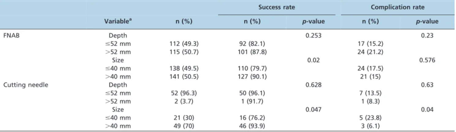
Table 2 shows the success and complication rates of FNAB and cutting-needle biopsy, using the average lesion depth and size as references. The success rates of both biopsy techniques were higher for larger-than-average ($40 mm) than for smaller-than-average (,40 mm) lesions (p,0.05). The complication rate did not differ according to
lesion size among the patients undergoing FNAB; however, the complication rate was significantly higher for smaller-than-average lesions in patients undergoing cutting-needle
biopsy (p,0.05). The success and complication rates did not
differ between biopsy techniques according to lesion depth, using average depth as a reference, or according to the side
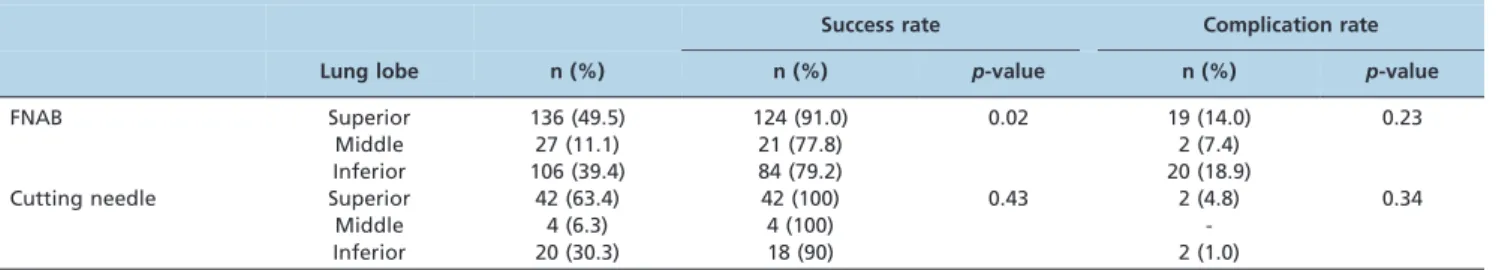
of the affected lung (p.0.05). The success rate of FNAB was
higher in superior lobes than in other lobes; no similar difference was observed for cutting-needle biopsy (Table 3). No significant difference according to the affected lobe was observed between the two techniques.
Complications occurred in 63 (13.8%) cases; pneu-mothorax occurred most frequently (n = 43, 9.4%), followed by hematoma (n = 11, 2.4%) and hemoptysis (n = 9, 2.0%) (Table 4). The overall complication rate did not differ between FNAB (n = 51, 14.1%) and cutting-needle biopsy
(n = 12, 12.4%, p.0.05); however, the incidence of
pneu-mothorax was significantly higher for FNAB (n = 40, 11.1%)
than for cutting-needle biopsy (n = 3, 3.1%,p,0.05), and the
incidence of pulmonary hematoma was significantly higher for cutting-needle biopsy (n = 7, 7.2%) than for FNAB (n = 4,
1.1%, p,0.05). The rate of hemoptysis did not differ
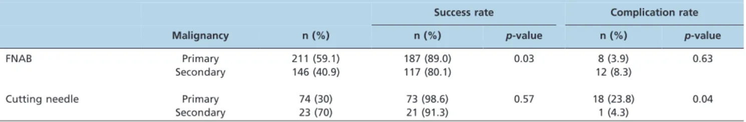
between the two techniques. Of the 43 (9.4%) patients with pneumothorax, only 11 (2.5%) patients, all of whom under-went FNAB, required thoracic drainage. The complication rate did not differ according to FNAB indication; however, the complication rate was significantly higher among the patients undergoing cutting-needle biopsy for primary diagnosis (n = 18, 23.8%) than among those undergoing this procedure for secondary diagnosis (n = 1, 4.3%, p = 0.04) (Table 5). Complications did not differ according to patient sex or age.
The sensitivity, specificity, and accuracy of cutting-needle biopsy (93.8%, 97.3%, and 95.2%, respectively) were significantly higher than those for FNAB (82.6%, 81.3%,
and 81.8%, respectively; all p,0.05). The rate of
false-negative diagnosis of malignancy was significantly higher
Table 1 -Rates of biopsy sample adequacy and specific diagnosis.
Sample adequacy n (%) p-value
Specific diagnosis n (%) p-value
FNAB 304 (84) 0.045 247 (68.2) 0.003
Cutting needle 94 (96.9) 83 (85.6)
FNAB, fine-needle aspiration biopsy; n, number of lung lesions.
Table 2 -Success and complication rates of FNAB and cutting-needle biopsy according to lesion depth and size.
Success rate Complication rate
Variablea n (%) n (%)
p-value n (%) p-value
FNAB Depth 0.253 0.23
#52 mm 112 (49.3) 92 (82.1) 17 (15.2)
.52 mm 115 (50.7) 101 (87.8) 24 (21.2)
Size 0.02 0.576
#40 mm 138 (49.5) 110 (79.7) 24 (17.5)
.40 mm 141 (50.5) 127 (90.1) 21 (15)
Cutting needle Depth 0.628 0.63
#52 mm 52 (96.3) 50 (96.1) 7 (13.5)
.52 mm 2 (3.7) 1 (91.7) 1 (8.3)
Size 0.047 0.04
#40 mm 21 (30) 16 (76.2) 5 (23.8)
.40 mm 49 (70) 46 (93.9) 3 (6.1)
for FNAB (n = 45, 17%) than for cutting-needle biopsy (n = 4,
2.6%, p,0.005); no similar difference was noted for
benignity.
& DISCUSSION
Our results indicated that CT-guided PNB is a reliable and reproducible method for diagnosing lung lesions. Using this method yielded high success rates for collecting adequate material for analysis and acceptable rates for procedure-related complications. However, several impor-tant issues related to needle type are discussed.
Avritscher et al. (11) reported that CT-guided PNB of pulmonary hilar lesions had a sensitivity of 91.4% and an accuracy of 92.8%, despite the procedure’s technical difficulty. These results are similar to those we obtained using cutting-needle biopsy. Avritscher et al. reported higher rates of pneumothorax and chest tube drainage
compared to our findings (48%vs. 13.7% and 32%vs. 2.4%,
respectively), which are likely due to the complexity and difficulty of accessing hilar lesions.
Large lesions usually provide more adequate samples for analysis than do small lesions (12). Layfield et al. (13) demonstrated that lesion location and size affect the diag-nostic accuracy of thoracic FNAB and that the best results are obtained for peripheral and larger lesions. In the present
study, lesions with diameters $40 mm supplied larger
amounts of adequate biopsy material, thus enabling more specific diagnoses compared to smaller lesions using both biopsy techniques. In contrast to our results, which indicated that sample adequacy and success rates were not affected by lesion contour or depth, Miller et al. (14) demonstrated that speculated lesion contour and greater depth were predictive of successful biopsy sample acquisition.
The lesion location in the superior lobes has been described as predictive of FNAB success (15), which is likely due to minimal respiratory lung motion and ease of
access (direct, regardless of the patient’s position) relative to other lobes. Our results support this finding. Superior lesions frequently have pleural bases and thus exhibit close-fitting surfaces or chest wall infiltration; thus, the biopsy needle can be introduced without transposing the pleural space. We found that lesion location had no effect on the success of cutting-needle biopsy.
The high success rate of cutting-needle biopsy could be partially explained by the selection of needle type and size according to the lesion characteristics and the patient’s clinical condition. In the current era of personalized medicine, in which advanced oncological treatment is based on molecular profiling and targeted therapy, collecting adequate tissue specimens is important. Macrobiopsies and tissue quality from cutting needles provide more adequate tissue samples for such purposes than do microbiopsies (i.e., FNABs).
Advances in oncological treatment have been based on targeted therapy and molecular profiling. The characteriza-tion of intratumoral heterogeneity enables the colleccharacteriza-tion of tissue specimens from regions that represent the lesion’s most aggressive behavior. For this reason, functional/ metabolic methods have been sought that enable the identification of the most suspicious areas within a lesion for biopsy. Moreover, functional imaging methods, such as diffusion-weighted imaging (DWI) and positron emission tomography (PET)/CT, are key to achieving effective image-guided percutaneous biopsies, obviating more inva-sive procedures for proper diagnosis and therapeutic planning when using cutting needles (16,17).
Several reports have demonstrated that clinical, radiolo-gical, and procedural factors are related to the risks of complications (18-20). Yeow et al. (21) reported a higher rate
of pneumothorax in lesions ,2 cm and deeper than
0.1-2 cm and for procedures performed by less-experienced
radiologists. The bleeding risk was higher in lesions,2 cm
and deeper than 2.1 cm and in the absence of pleural effusion. In our study, no radiological lesion characteristic
predicted complications in FNAB, and only lesions,40 mm
were predictive of complications in cutting-needle biopsy. Additionally, a skilled radiologist or a resident under the radiologist’s supervision performed all of the biopsies in our study, thus minimizing the effect of operator’s experience on success and complication rates.
In the present study, the incidence of pulmonary hematoma was higher for cutting-needle biopsy than for FNAB; however, this difference had no major clinical implication. This difference may be explained by the greater damage to the net vessels of the tumor caused by cutting-needle biopsy due to the large cutting-needle’s disruption of the tissue sample. In agreement with our results, Khan et al. (22)
Table 3 -Success and complication rates of FNAB and cutting-needle biopsy according to lung lobe affected.
Success rate Complication rate
Lung lobe n (%) n (%) p-value n (%) p-value
FNAB Superior 136 (49.5) 124 (91.0) 0.02 19 (14.0) 0.23
Middle 27 (11.1) 21 (77.8) 2 (7.4)
Inferior 106 (39.4) 84 (79.2) 20 (18.9)
Cutting needle Superior 42 (63.4) 42 (100) 0.43 2 (4.8) 0.34
Middle 4 (6.3) 4 (100)
-Inferior 20 (30.3) 18 (90) 2 (1.0)
FNAB, fine-needle aspiration biopsy; n, number of lung lesions.
Table 4 -Distribution of complication rates for CT-guided biopsies of lung lesions.
Complication n (%)
FNAB n (%)
Cutting needle n (%)
Pneumothorax 43 (9.4) 40 (11.1) 3 (3.1)
Hematoma 11 (2.4) 04 (1.1) 07 (7.2)
Hemoptysis 09 (2.0) 07 (1.9) 02 (2.1)
Total complications 63 (13.8) 51 (14.1) 12 (12.4) No complications 396 (86.2) 311 (85.9) 85 (87.6)
Total sample 459 (100) 362 (100) 97 (100)
demonstrated that the lesion contour had no significant effect on the complication rate.
The incidence of pneumothorax was higher for FNAB than for cutting-needle biopsy, which might be explained by the tendency to select FNAB for lesions with difficult approaches, including small and deep lesions and lesions near large vessels; these situations require more time and transfixation to obtain adequate biopsy material. In contrast to our finding that complication rates did not differ according to the patient’s age or sex, Geraghty et al. (23) demonstrated that the patient’s age had a significant effect on the rate of pneumothorax, which occurred in 153 (60.7%)
patients aged.60 years and 99 (39.3%) patients aged#60
years (p,0.02).
One way to minimize the complication rates when applying cutting needles is to use the coaxial technique, in which the needle passes inside a needle guide positioned toward the tumor. Because this device reduces the number of pleural punctures, the pneumothorax rates are similarly reduced (24). Several study limitations should be emphasized. First, the retrospective selection of patients examined in an oncology center under strong presumption of malignancy clearly introduced selection bias into our series. Second, the reasons for needle type selection could not be evaluated because this information was not available. Third, the nature of procedures performed in our institution has changed over time; several years ago, FNAB was the preferred technique for confirming malignancy in nearly all lung lesions. However, cutting-needle biopsy has become the preferred technique for diagnosis and management because the operator’s confidence with this procedure has increased. Fourth, a pathologist was not available during biopsy procedures, as is typical in many oncological centers in developing countries. Thus, the numbers of thoracic transfixations performed and the smears or specimens collected were related to the radiologist’s experience and the incidence of complications at the time of the procedure. In conclusion, our results indicate that the overall performance of cutting-needle biopsy was stronger than that of FNAB, with no significant increase in complication rates. Considering that biomarker profiling and targeted therapy are important components of oncological patient management, cutting-needle biopsy is currently used in our center for nearly all diagnosed lung lesions. FNAB is reserved for patients with coagulation deficits or when malignancy documentation is required.
& AUTHOR CONTRIBUTIONS
Guimaraes MD, the principal investigator and the guarantor of the entire and final review of the manuscript, contributed to the coordination and
design of the study, statistics, and data interpretation. Marchiori E contributed to the study design, statistics, data interpretation, literature review, and manuscript revision. Hochhegger B contributed to the data collection, literature review, and manuscript preparation. Chojniak R contributed to the data collection, high-resolution CT scan evaluation, literature review, data interpretation, and manuscript preparation. Gross JL contributed to the data collection, high-resolution CT scan evaluation, literature review, and manuscript preparation.
& REFERENCES
1. Haaga JR, Alfidi RJ. Precise biopsy localization by computer tomogra-phy. Radiology. 1976;118(3):603-7.
2. McSweeney SE, O’Regan KN, Mc Laughlin PD, Crush L, Maher MM. Evaluation of the efficacy and safety of percutaneous biopsy of lung. Open Respir Med J. 2012;6:82-8, http://dx.doi.org/10.2174/ 1874306401206010082.
3. Chojniak R, Isberner RK, Viana LM, Yu LS, Aita AA, Soares FA. Computed tomography guided needle biopsy: experience from 1,300 procedures. Sao Paulo Med J. 2006;124(1):10-4, http://dx.doi.org/10. 1590/S1516-31802006000100003.
4. Tsai IC, Tsai WL, Chen MC, Chang GC, Tseng WS, Chan SW, et al. CT-guided core biopsy of lung lesions: a primer. AJR Am J Roentgenol. 2009;193(5):1228-35, http://dx.doi.org/10.2214/AJR.08.2113.
5. Yao X, Gomes MM, Tsao MS, Allen CJ, Geddie W, Sekhon H. Fine-needle aspiration biopsy versus core-needle biopsy in diagnosing lung cancer: a systematic review. Curr Oncol. 2012;19(1):e16-27.
6. Uruga H, Takaya H, Hanada S, Beika Y, Miyamoto A, Morokawa N, et al. Diagnostic efficacy of CT-guided transthoracic needle biopsy and fine needle aspiration in cases of pulmonary infectious disease. Jpn J Radiol. 2012;30(7):589-93.
7. Beslic S, Zukic F, Milisic S. Percutaneous transthoracic CT guided biopsies of lung lesions; fine needle aspiration biopsy versus core biopsy. Radiol Oncol. 2012;46(1):19-22.
8. Kalia M. Personalized oncology: recent advances and future challenges. Metabolism. 2013;62 Suppl 1:S11-4, http://dx.doi.org/10.1016/j.metabol. 2012.08.016.
9. Guimaraes MD, Gross JL, Bitencourt AG, Chojniak R. CT-guided transthoracic biopsies of lung lesions suspected of malignancy. Ir J Med Sci. 2013;182(3):533.
10. Guimaraes MD, de Andrade MQ, da Fonte AC, Chojniak R, Gross JL. CT-guided cutting needle biopsy of lung lesions—an effective procedure for adequate material and specific diagnose. Eur J Radiol. 2011; 80(3):e488-90.
11. Avritscher R, Krishnamurthy S, Ensor J, Gupta S, Tam A, Madoff DC, et al. Accuracy and sensitivity of computed tomography-guided percutaneous needle biopsy of pulmonary hilar lymph nodes. Cancer. 2010;116(8):1974-80, http://dx.doi.org/10.1002/cncr.24968.
12. Yuan DM, Lu¨ YL, Yao YW, Liu HB, Wang Q, Xiao XW, et al. Diagnostic efficiency and complication rate of CT-guided lung biopsy: a single center experience of the procedures conducted over a 10-year period. Chin Med J (Engl). 2011;124(20):3227-31.
13. Layfield LJ, Coogan A, Johnston WW, Patz EF. Transthoracic fine needle aspiration biopsy. Sensitivity in relation to guidance technique and lesion size and location. Acta Cytol. 1996;40(4):687-90, http://dx.doi. org/10.1159/000333940.
14. Miller JA, Pramanik BK, Lavenhar MA. Predicting the rates of success and complications of computed tomography-guided percutaneous core-needle biopsies of the thorax from the findings of the preprocedure chest computed tomography scan. J Thorac Imaging. 1998;13(1):7-13, http:// dx.doi.org/10.1097/00005382-199801000-00003.
15. Guimara˜es MD, Chojniak R, Gross JL, Bitencourt AG. Predictive success factors for CT-guided fine needle aspiration biopsy of pulmonary lesions. Clinics. 2009;64(12):1139-44.
Table 5 -Success and complication rates of FNAB and cutting-needle biopsy according to the reason for the procedure (primaryvs. secondary malignancy)
Success rate Complication rate
Malignancy n (%) n (%) p-value n (%) p-value
FNAB Primary 211 (59.1) 187 (89.0) 0.03 8 (3.9) 0.63
Secondary 146 (40.9) 117 (80.1) 12 (8.3)
Cutting needle Primary 74 (30) 73 (98.6) 0.57 18 (23.8) 0.04
Secondary 23 (70) 21 (91.3) 1 (4.3)
16. Guimaraes MD, Gross JL, Chojniak R, Marchiori E. MRI-Guided Biopsy: A valuable procedure alternative to avoid the risks of ionizing radiation from diagnostic imaging methods. Cardiovasc Intervent Radiol. 2013 Jun 28. [Epub ahead of print].
17. Cerci JJ, Pereira Neto CC, Krauzer C, Sakamoto DG, Vitola JV. The impact of coaxial core biopsy guided by FDG PET/CT in oncological patients. Eur J Nucl Med Mol Imaging. 2013;40(1):98-103, http://dx.doi. org/10.1007/s00259-012-2263-0.
18. Lima CD, Nunes RA, Saito EH, Higa C, Cardona ZJ, Santos DB. Results and complications of CT-guided transthoracic fine-needle aspiration biopsy of pulmonary lesions. J Bras Pneumol. 2011;37(2):209-16, http:// dx.doi.org/10.1590/S1806-37132011000200011.
19. Nakatani M, Tanigawa N, Kariya S, Komemushi A, Yagi R, Sawada S. Analysis of factors influencing accuracy and complications in CT-guided lung biopsy. Minim Invasive Ther Allied Technol. 2012;21(6):415-22, http://dx.doi.org/10.3109/13645706.2012.662155.
20. Guimara˜es MD, Andrade MQ, Fonte AC, Benevides G, Chojniak R, Gross JL. Predictive complication factors for CT-guided fine needle
aspiration biopsy of pulmonary lesions. Clinics. 2010;65(9):847-50, http://dx.doi.org/10.1590/S1807-59322010000900006.
21. Yeow KM, Su IH, Pan KT, Tsay PK, Lui KW, Cheung YC, et al. Risk factors of pneumothorax and bleeding: multivariate analysis of 660 CT-guided coaxial cutting needle lung biopsies. Chest. 2004;126(3):748-54, http://dx.doi.org/10.1378/chest.126.3.748.
22. Khan MF, Straub R, Moghaddam SR, Maataoui A, Gurung J, Wagner TO, et al. Variables affecting the risk of pneumothorax and intrapulmonal hemorrhage in CT-guided transthoracic biopsy. Eur Radiol. 2008;18(7):1356-63, http://dx.doi.org/10.1007/s00330-008-0893-1.
23. Geraghty PR, Kee ST, McFarlane G, Razavi MK, Sze DY, Dake MD. CT-guided transthoracic needle aspiration biopsy of pulmonary nodules: needle size and pneumothorax rate. Eur Radiol. 2008; 18(7):1356-63.